Digital Pathology and Tissue-based Diagnosis. How do they differ?
|
|
|
- Luke Doyle
- 6 years ago
- Views:
Transcription

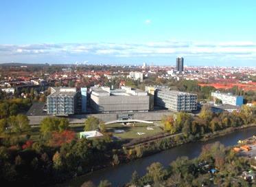
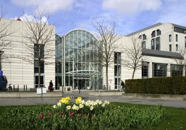
1 Digital Pathology and Tissue-based Diagnosis. How do they differ? P. Hufnagl Institute of Pathology (Rudolf-Virchow-Haus). Humboldt University, Berlin?
2 Structure of the talk Possible workflow routine diagnostic diagnostic support Which scanner? Quantification / image analysis Conclusions
3 Structure of the talk Possible workflow routine diagnostic diagnostic support Which scanner can we trust? Quantification / image analysis Conclusions
4 The Conventional Workflow Tissue Lab: Cutting, Staining, Coverslip Clinical request HIS PLIMS Report Physician: Dictation, Images, Physician: Assignment, diagnosing, Annotation consultation


5 The Digital Workflow Tissue Lab: Cutting, Staining, Coverslip Slide Scanner: Registration, Digitalisation PACS: Storage, Image streaming Clinical request HIS PLIMS Report Physician: Dictation, Images, Physician: Assignment, diagnosing, Image analysis: marker quantification Annotation consultation
6 The Digital Workflow Missing link between cover slipper and scanner Tissue Lab: Cutting, Staining, Coverslip Missing Slide Scanner: link between Registration, scanner Digitalisation and PLIS PACS: Storage, Image streaming Clinical request HIS PLIMS Report Physician: Dictation, Images, Physician: Assignment, diagnosing, Image analysis: marker quantification Annotation consultation


7 Advantages of Virtual Microscopy User Viewer Slide Server No glass transportation, no glass archive (?) Previous slides are always available Microscopic diagnostic - anytime anywhere - Parallel viewing of different stainings, positions Viewing and handling parallel at different locations Facilitated second opinion - online Quantification and image analysis just in time Annotations are simple to be handled much more.. Slide Scanner


8 Problems of the digital workflow Disadvantages Additional process step: Digitalisation Huge and continuous hardware and software investments Training of personnel Diagnosis on the monitor is unfamiliar for pathologists Legal Problems
9 Strategy Start with a less critical application Biobanking Introduce VM to applications with most effects Tumour board Second opinion in-house Solve all problems along the workflow LIMS integration Sure barcode identification Establishing of continuous testing


10 ZEBANC CHARITÉ BIOBANK CVK CCM CBF

 WSI generation")


11 CURRENTLY OFFERED SERVICES Aliquotation Quality documentation (SPREC) WSI generation TMA generation and WSI* based analysis Automatic DNA- extraction DNA Sequencing... *whole slide image 11


12 VALUE OF A SAMPLE Sample Clinical Data Whole Slide Images Quantification of Morphology V A L U E Next Generation Sequencing Data Data generated during experiments...
13 ZEBANC AND VM Digitalisation established for samples from frozen section labs Technical quality control implemented Infrastructure for block-centric navigation established Medical quality control in use (prototype tumor area detection) TMA as sample array in CentraXX integrated Several quantification procedures implemented to generate additional sample features Virtual studies on virtual slides instead of real samples Virtual microscopy has a huge potential for services in the context of a biomaterial bank

14 Clinical Pathology Second Opinion, Studies, Marker Quantification Medical Workstation - All Information in One View


15 Workflow of Tumor Board Meetings Set bookmarks and make annotations Browse slides and move to annotations Before meeting On meeting
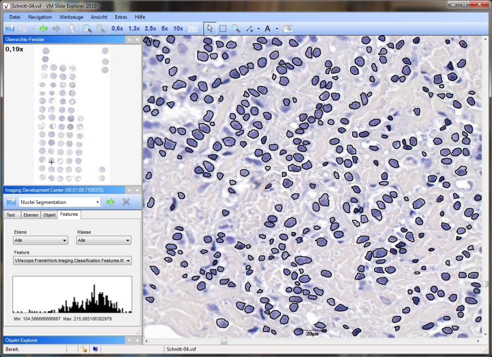
16 Quantification
17 Structure of the talk Possible workflow routine diagnostic diagnostic support Which scanner? Quantification / image analysis Conclusions
18 Requirements on Slide Scanning Correct slide information is present Completeness of tissue Image sharpness Color fidelity


19 VIRTUAL MICROSCOPY SCANNER CONTEST P. Hufnagl 1,2, N.Zerbe 2 1 University of Applied Science Berlin, Berlin, German 2 Institute for Pathology, Charité Berlin, Germany 2 nd International Scanner Contest technology meets pathology
20 MISSION 2 nd International Scanner Contest technology meets pathology Determination of the state of the art in slide scanning Support of pathologists and scientists to find the appropriate scanner for their applications Support of vendors to understand the needs of pathologists Determination of quality of WSI within the context of pathology Development of a set of standard features for the characterization and comparison of scanning devices

21 DISCIPLINES 2 nd International Scanner Contest technology meets pathology High Throughput Quality Fluorescence Image Analysis Technical

22 2 nd International Scanner Contest QUALITY EVALUATION TERMINALS technology meets pathology


23 VENDORS. 2 nd International Scanner Contest technology meets pathology

24 2 nd International Scanner Contest TECHNICAL COLOUR & GEOMETRY AIMS & MATERIAL technology meets pathology Aims: Measurement of colour fidelity and colour resolution of devices Determination of true effective pixel size Detection of image distortions Material: IT8.7/1 Colour Target mounted on glas slide 264 colour & 24 grey value fields known absorption spectra and colourimetric coordinates Grid pattern glas slide overall size: 1 x 3 (25mm x 75mm) image area: 20mm x 50mm clear aperture: 8.5 µm² opaque lines: 1.5 µm² pitch: 10 µm
25 2 nd International Scanner Contest TECHNICAL COLOUR & GEOMETRY TASK technology meets pathology General Conditions: All participants had to scan the same slide Any manual interaction was allowed Rescan of slide was allowed 1h time limit Evaluation: Colour difference calculation to CIEDE2000 average inside middle 50% of each field low-resolution scan Measurements inside whole slide images Inside sensor field (no stitching) 9 sensor fields 18 measurements each


26 2 nd International Scanner Contest technology meets pathology TECHNICAL COLOUR TEST METHOD Fidelity test: average de over 144 fields de avg = Σ de(c * mes, c ref ) /144 mix-colours matrix for fidelity test Colour difference calculation to CIEDE2000 colour step wedges


27 2 nd International Scanner Contest TECHNICAL COLOUR SOFTWARE technology meets pathology

28 COMPLETENESS OF SCAN 2 nd International Scanner Contest technology meets pathology

29 2 nd International Scanner Contest technology meets pathology HIGH THROUGHPUT AIMS & MATERIAL 29

30 2 nd International Scanner Contest technology meets pathology HIGH THROUGHPUT AIMS & MATERIAL 30

 Sharpness assessment +")
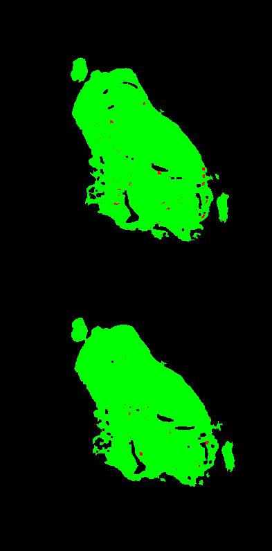
31 2 nd International Scanner Contest DIGITALISATION - SCANMASTER technology meets pathology Sample identifikation (barcode / OCR) Sharpness assessment + Completeness of particles


32 2 nd International Scanner Contest FOCUS QUALITY ASSESSMENT technology meets pathology Green: quality sufficiant Red: not sharp, possible artefacts
33


34 2 nd International Scanner Contest technology meets pathology
35 2 nd International Scanner Contest technology meets pathology VIEWING ON A VIRTUAL MICROSCOPE


36 RESOLUTION 2 nd International Scanner Contest technology meets pathology MAGNIFICATION IS NOT RESOLUTION AND OPTICAL RESOLUTION IS NOT DIGITAL RESOLUTION!


37 Several Positions in Test Leap Motion over the table Leap Motion under acryl glass pane


38 Leap Motion Stereo imaging based on infrared cameras (


39 Handling Possible workflow
40 Gesture Control Next/ previous slide Zoom in/ out
41 Histological Image Registration Goal Inter-Modal Registration (Stain-To-Stain) Applications WSI Navigation Support Virtual Staining 3D Reconstruction Approach Intensity Based Multi-Resolution Similarity Measure Mutual Information Transformation Models Rigid: Rotation + Translation Affine: Linear Transformation + Translation Free Form Deformation: B-Splines Optimization Gradient Descent
42 Registration Of Renal Images: Reference Image (H&E) Template Image (SFOG)
43 Reference Image (H&E)

44 Template Image (SFOG)
45 Coarse to Fine Image Registration: Rigid, Affine, Free Form Model

46 Rigid Registration
47 Affine Registration
48 Free Form Registration
49 Reference Image
50 Structure of the talk Possible workflow routine diagnostic diagnostic support Which scanner? Quantification / image analysis Conclusion

51 The strong reputation of pathology is becoming weaker.. if trust is gone, you almost never get it back

52 Standardized Quantification in Tumor Pathology Nat J Inst., preprint November
53 . At the St. Gallen cutoff of 13.5 % there are 32.3 % high Ki67 by Lab A while Lab B would call the same cases low Ki

54 Optical Illusions Not always funny, sometimes really critical

55 Human brain: Square a is lighter than square b! Reality: Both are identical

56 Simulated Ki67 15%
57 Estimation of variation of Ki67 scoring


58 FEATURE BASED MULTIRESOLUTION CORRESPONDENCE Combined tumor annotations Individual tumor annotations Annotated as tumor by 10 pathologists By 4 pathologists By 2 pathologists tumor non-tumor by no pathologist


59 CLASSIFICATION RESULTS Gastric Cancer (HER2) Transmission to HE Learning Sample 59
60 Structure of the talk Possible workflow routine diagnostic diagnostic support Which scanner? Quantification / image analysis Conclusions
61 Summary on Relevance of VM Workflow routine diagnostic diagnostic support Which scanner? Quantification / image analysis In routine path not yet active on a broad level Excellent instruments exist, but they have to be integrated properly Will become very important in personalized medicine Clinical-pathological tumorconferences (tumor board) Is already very important Biobanking Is important in research institutions
62 Most important requirements on VM Clinical data (LIMS) are correctly connected to WSI Completeness of tissue Image sharpness Color fidelity Compression is adequate Resolution and quality of the monitor is sufficiant Test continously!

63 Let s go virtual 13th European Conference on Digital Pathology Berlin May 25. /


64 Acknowledgement Team Norman Zerbe, Karsten Schlüns, Sebastian Lohmann, Mario Domhardt, Björn Lindequist, Daniel Heim, Stephan Wienert, Kai Saeger, Thorsten Knape, Arend Müller, Wolfram Schädel, Uwe Brunner, Thomas Schrader, Manfred Dietel 2 nd International Scanner Contest technology meets pathology
Second Announcement Call for Participation. (Evaluation Criteria added)
") Second Announcement Call for Participation 2 nd International Scanner Contest (ISC) (Evaluation Criteria added) P. Hufnagl 1, T. Schrader 1, 2, M.G. Rojo 3, A. Laurinavicius 4, G. Kayser 5, Y. Yagi 6 1
Second Announcement Call for Participation 2 nd International Scanner Contest (ISC) (Evaluation Criteria added) P. Hufnagl 1, T. Schrader 1, 2, M.G. Rojo 3, A. Laurinavicius 4, G. Kayser 5, Y. Yagi 6 1
technology meets pathology Institute of Pathology, Charité Universitätsmedizin Berlin, Berlin, Germany 3 Overview
 ASSESSMENT OF TECHNICAL PARAMETERS A. Alekseychuk 1, N. Zerbe 2, Y. Yagi 3 1 Computer Vision and Remote Sensing, TU Berlin, Berlin, Germany 2 Institute of Pathology, Charité Universitätsmedizin Berlin,
FRAUNHOFER INSTITUTE FOR INTEGRATED CIRCUITS IIS. MANUAL PANORAMIC MICROSCOPY WITH istix
 FRAUNHOFER INSTITUTE FOR INTEGRATED CIRCUITS IIS MANUAL PANORAMIC MICROSCOPY WITH istix CLINICAL DIAGNOSTICS AND MATERIAL SCIENCES IMPROVED BY DIGITAL MICROSCOPY B A C K G R O U N D Due to a high grade
DICOM-compatible compression of WSI and diagnostic evaluation
 of WSI and diagnostic evaluation R. Zwönitzer, H. Hofmann, A. Roessner, T. Kalinski 2nd European Workshop in Tissue Imaging and Analysis June 25-26, 2010 - Heidelberg 1 GPWL / GP-PPS Introduction Overview
InScape: Making Virtual Pathology a Reality
 InScape: Making Virtual Pathology a Reality Sally S. Agersborg, M.D., Ph.D. Quest Diagnostics, Nichols Institute San Juan Capistrano, CA Company Overview Quest Diagnostics, Nichols Institute the world
MIRAX SCAN The new way of looking at pathology
 Microscopy from Carl Zeiss MIRAX SCAN The new way of looking at pathology Greater reliability. Greater efficiency. Plus points for your diagnostics Better. More efficient. Quality as a factor for success
Technical Aspects in Digital Pathology
 Technical Aspects in Digital Pathology Yukako Yagi, PhD yyagi@mgh.harvard.edu Director of the MGH Pathology Imaging & Communication Technology Center Assistant Professor of Pathology, Harvard Medical School
Digital Pathology at Johns Hopkins Practical Research and Clinical Considerations
 Digital Pathology at Johns Hopkins Practical Research and Clinical Considerations July 10, 2017 Alexander Baras, MD, PhD Assistant Professor of Pathology, Urology, and Oncology Associate Director of Pathology
Stereotopix Research. Precision Pathology. Highthroughput. pathology. powered by newcast. Advantages of Stereotopix : RUO
 Precision Pathology Highthroughput pathology Stereotopix Research powered by newcast RUO Researchers use quantitative microscopy in many ways with the goal of producing high-quality, quantitative results
How to introduce virtual microscopy (VM) in routine diagnostic pathology: constraints, ideas, and solutions
 in routine diagnostic pathology: constraints, ideas, and solutions") How to introduce virtual microscopy (VM) in routine diagnostic pathology: constraints, ideas, and solutions Klaus Kayser, Stephan Borkenfeld, Gian Kayser History Workflow Design of virtual microscopy Constraints
How to introduce virtual microscopy (VM) in routine diagnostic pathology: constraints, ideas, and solutions Klaus Kayser, Stephan Borkenfeld, Gian Kayser History Workflow Design of virtual microscopy Constraints
Digital Pathology and Image Analysis. Queen s University Department of Pathology and Molecular Medicine Shakeel Virk
 Digital Pathology and Image Analysis Queen s University Department of Pathology and Molecular Medicine Shakeel Virk Outline Digital Pathology and Image Analysis capabilities at Queen s Laboratory for Molecular
IHE Anatomic Pathology Redesign. Sardinia, Italy Nov , 2017
 IHE Anatomic Pathology Redesign Sardinia, Italy Nov. 13-15, 2017 Specimen Workflow in a Nutshell (variations likely depending on context, e.g. collection location) Create Encounter Generate Results Order
BRINGING DEEP LEARNING TO ENTERPRISE IMAGING CLINICAL PRACTICE
 BRINGING DEEP LEARNING TO ENTERPRISE IMAGING CLINICAL PRACTICE Esteban Rubens Global Enterprise Imaging Principal Pure Storage @pureesteban AI IN HEALTHCARE What is Artificial Intelligence (AI)? How is
Color aspects and Color Standardization in Digital Microscopy
 Color aspects and Color Standardization in Digital Microscopy Yukako Yagi, PhD yyagi@partners.org Director of the MGH Pathology Imaging & Communication Technology Center Assistant Professor of Pathology,
Digital Pathology Update
 Digital Pathology Update J. Mark Tuthill, MD Division Head, Pathology Informatics Department of Pathology & Laboratory Medicine Henry Ford Hospital Detroit, MI mtuthil1@hfhs.org ASCT Webinar, January 2016
Digital Microscopy. Past, Present, and Future. Cyrus V. Hedvat, MD, PhD
 Digital Microscopy Past, Present, and Future Cyrus V. Hedvat, MD, PhD N Context. Digital viewing of histologic images is moving from presentations and publications to incorporation into the daily work
GALILEO TMA CK 4500 HTS Tissue Microarray Platform
 GALILEO TMA CK 4500 HTS Tissue Microarray Platform Tissue Microarray (TMA) A Block Of Samples From Hundreds Of Blocks (S. M. Hewitt, M.D., Ph.D., Tissue Array Research Program, LP, CCR, NCI, NIH) TMA technology
THEORY AND APPROACHES TO AUTOMATED IMAGE ANALYSIS IN DIGITAL PATHOLOGY
 THEORY AND APPROACHES TO AUTOMATED IMAGE ANALYSIS IN DIGITAL PATHOLOGY Kyle Takayama, MS Charles River Laboratories EVERY STEP OF THE WAY EVERY STEP OF THE WAY MORPHOMETRY Measurements or counts performed
MECOS-C2 microscopy systems
 MECOS-C2 microscopy systems Microscopy systems of the MECOS-C2 family production LLC "Medical computer Systems (MECOS)" belong to a class of scanning microscopes-analyzers and are intended for: Increase
Multilayer scanning enhances sensitivity of artificial intelligence-aided Mycobacterium tuberculosis detection
 Multilayer scanning enhances sensitivity of artificial intelligence-aided Mycobacterium tuberculosis detection Yan Xiong Peking University First Hospital, China. yanxiong1109@163.com Ao Hou ao_sure@foxmail.com
Digital Microscopy: New Paradigm's for Teaching Microscopic Anatomy and Pathology
 Digital Microscopy: New Paradigm's for Teaching Microscopic Anatomy and Pathology Michael Feldman, MD, PhD Assistant Dean IT Assistant Professor Pathology University of Pennsylvania Health System Feldmanm@mail.med.upenn.edu
Overview of Digital Pathology s Current State: Technologies, Systems, Capabilities, Limitations, and Opportunities
 Overview of Digital Pathology s Current State: Technologies, Systems, Capabilities, Limitations, and Opportunities David McClintock, MD Executive War College Post-Conference Workshop Digital Pathology
Teaching Digital Histology
 Teaching Digital Histology Carlos R. Morales Department of Anatomy and Cell Biology, McGill University, Montreal, Quebec, Canada The light microscope is one of the most widely used scientific instruments
DMETRIX S (FUTURE) PERSPECTIVES ON DIGITAL IMAGING & DIGITAL PATHOLOGY SYSTEMS
 PERSPECTIVES ON DIGITAL IMAGING & DIGITAL PATHOLOGY SYSTEMS") Michael R. Descour, Ph.D., DMetrix, Inc., & University of Arizona Lloyd J. LaComb, Jr., Ph.D., DMetrix, Inc. DMETRIX S (FUTURE) PERSPECTIVES ON DIGITAL IMAGING & DIGITAL PATHOLOGY SYSTEMS Outline of presentation
Michael R. Descour, Ph.D., DMetrix, Inc., & University of Arizona Lloyd J. LaComb, Jr., Ph.D., DMetrix, Inc. DMETRIX S (FUTURE) PERSPECTIVES ON DIGITAL IMAGING & DIGITAL PATHOLOGY SYSTEMS Outline of presentation
PACS Fundamentals. By: Eng. Valentino T. Mvanga Ministry of Health and Social Welfare Tanzania
 PACS Fundamentals By: Eng. Valentino T. Mvanga Ministry of Health and Social Welfare Tanzania 1 Learning Goals To Understand the importance of PACS To Understand PACS infrastructure requirement Introduction
Positive Pixel Count Algorithm. User s Guide
 Positive Pixel Count Algorithm User s Guide Copyright 2004, 2006 2008 Aperio Technologies, Inc. Part Number/Revision: MAN 0024, Revision B Date: December 9, 2008 This document applies to software versions
PathStation TM Fits Your Lab
 PathStation TM Fits Your Lab PathStation hood Compact In-hood Imaging system. Supports in-process documentation PathStation Key Features 20 MP Image Capture Great HD Video 8.33 X Optical zoom 4X Digital
Leica DMi8A Quick Guide
 Leica DMi8A Quick Guide 1 Optical Microscope Quick Start Guide The following instructions are provided as a Quick Start Guide for powering up, running measurements, and shutting down Leica s DMi8A Inverted
SPOT PathSuite Solutions
 SPOT PathSuite Solutions The Perfect Fit PathStation TM Imaging system for the gross dissection in hood PathStand TM 40 imaging station for medium to large specimens PathSuite Is Easy To Use A turnkey
Anatomic and Computational Pathology Diagnostic Artificial Intelligence at Scale
 Anatomic and Computational Pathology Diagnostic Artificial Intelligence at Scale John Gilbertson MD Department of Pathology Massachusetts General Hospital Partners Healthcare System Harvard Medical School
Image Viewing. with ImageScope
 Image Viewing with ImageScope ImageScope Components Use ImageScope to View These File Types: ScanScope Virtual Slides.SVS files created when the ScanScope scanner scans glass microscope slides. JPEG files
Multi-resolution Cervical Cell Dataset
 Report 37 Multi-resolution Cervical Cell Dataset Patrik Malm December 2013 Centre for Image Analysis Swedish University of Agricultural Sciences Uppsala University Uppsala 2013 Multi-resolution Cervical
GE Healthcare. Senographe 2000D Full-field digital mammography system
 GE Healthcare Senographe 2000D Full-field digital mammography system Digital has arrived. The Senographe 2000D Full-Field Digital Mammography (FFDM) system gives you a unique competitive advantage. That
Quality and GLP for Histology and Pathology of Drug Safety Studies
 Quality and GLP for Histology and Pathology of Drug Safety Studies Roger Alison BVSc MRCVS DiplECVP Consultant Toxicological Pathologist What is Quality Histology? It depends upon the purpose - Answer
Digital Imaging and Communication in Medicine- A Digital Window for Oral Pathology
 IOSR Journal of Dental and Medical Sciences (IOSR-JDMS) e-issn: 2279-0853, p-issn: 2279-0861.Volume 13, Issue 10 Ver. III (Oct. 2014), PP 67-72 Digital Imaging and Communication in Medicine- A Digital
Report on interoperability of commercial digital pathology software
 Report on interoperability of commercial digital pathology software Deliverable 1.1 Project acronym: BIOPOOL Grant Agreement: 296162 Version: Due date: Month 2 Submission date: 15/12/12 Dissemination level:
Automated Digitization of Gram Stains. Centralized Reading. Decentralized Assessment. Improved Quality Management.
 Automated Digitization of Gram Stains Centralized Reading. Decentralized Assessment. Improved Quality Management. A GROWING DEMAND Gram staining is the rapid, easy, and inexpensive method for the assessment
An Autonomous Vehicle Navigation System using Panoramic Machine Vision Techniques
 An Autonomous Vehicle Navigation System using Panoramic Machine Vision Techniques Kevin Rushant, Department of Computer Science, University of Sheffield, GB. email: krusha@dcs.shef.ac.uk Libor Spacek,
Trust the Colors with Olympus True Color LED
 White Paper Olympus True Color LED Trust the Colors with Olympus True Color LED True Color LED illumination is a durable, bright light source with spectral properties that closely match halogen illumination.
Image Analysis for Fluorescence
 Image Analysis for Fluorescence Terminology Table Image Analysis Macro Colocalization Intensity Dye AFI The extraction of meaningful information from digital images by means of digital image processing
(12) United States Patent (10) Patent No.: US 6,525,828 B1
 United States Patent (10) Patent No.: US 6,525,828 B1") USOO6525828B1 (12) United States Patent (10) Patent No.: US 6,525,828 B1 Grosskopf (45) Date of Patent: *Feb. 25, 2003 (54) CONFOCAL COLOR 5,978,095 A 11/1999 Tanaami... 356/445 6,031,661. A 2/2000 Tanaami...
USOO6525828B1 (12) United States Patent (10) Patent No.: US 6,525,828 B1 Grosskopf (45) Date of Patent: *Feb. 25, 2003 (54) CONFOCAL COLOR 5,978,095 A 11/1999 Tanaami... 356/445 6,031,661. A 2/2000 Tanaami...
Image Processing. COMP 3072 / GV12 Gabriel Brostow. TA: Josias P. Elisee (with help from Dr Wole Oyekoya) Image Processing.
 Image Processing.") Image Processing COMP 3072 / GV12 Gabriel Brostow TA: Josias P. Elisee (with help from Dr Wole Oyekoya) 1 2 Motivation and Goals Grounding in image processing techniques Concentrate on algorithms used
Image Processing COMP 3072 / GV12 Gabriel Brostow TA: Josias P. Elisee (with help from Dr Wole Oyekoya) 1 2 Motivation and Goals Grounding in image processing techniques Concentrate on algorithms used
34X ZOOM. PathStand TM Routine. Superior Image Quality. PathStand Key Features. 1X Zoom. 8.33X Zoom
 PathStand TM Routine Complete stand-alone Imaging system. Supports in-process documentation. PathStand Key Features 20 MP Image Capture 1080p Live Video 34X zoom Metal Construction All-in-one Computer
DICOM Correction Proposal
 Tracking Information - Administration Use Only DICOM Correction Proposal Correction Proposal Number Status CP-1713 Letter Ballot Date of Last Update 2018/01/23 Person Assigned Submitter Name David Clunie
34X ZOOM. PathStand TM Routine. Superior Image Quality. PathStand Key Features. 1X Zoom. 8.33X Zoom
 PathStand TM Routine Complete stand-alone Imaging system. Supports in-process documentation. PathStand Key Features 20 MP Image Capture 720p Live Video 34X zoom Metal Construction All-in-one Computer LED
The First True Color Confocal Scanner on the Market
 The First True Color Confocal Scanner on the Market White color and infrared confocal images: the advantages of white color and confocality together for better fundus images. The infrared to see what our
HTT project: High Throughput Truthing. FY2019 Critical Path Proposal PI: Brandon D. Gallas
 HTT project: High Throughput Truthing FY2019 Critical Path Proposal PI: Brandon D. Gallas Full Project Title High throughput truthing of microscope slides to validate artificial intelligence algorithms
Bowen Hills Histopathology
 Bowen Hills Histopathology Histology s Bowen Hills Challenges Relocate and amalgamate Taringa and Indooroopilly Labs into a single complex histology laboratory Change workflow, staff structure, install
Virtual 3D Microscopy Using Multiplane Whole Slide Images in Diagnostic Pathology
 Anatomic Pathology / Diagnostic Virtual Pathology Virtual 3D Microscopy Using Multiplane Whole Slide Images in Diagnostic Pathology Thomas Kalinski, MD, 1 Ralf Zwönitzer, 2 Saadettin Sel, MD, 1 Matthias
Digital Imaging Outline
 Digital Microscopy and Imaging Update Michael Feldman, MD, PhD Associate Professor of Pathology Assistant Dean for Information Technology University of Pennsylvania School of Medicine (Feldmanm@mail.med.upenn.edu)
Virtual Electron Microscopy update after one year of routine use
 The Hong Kong College of Pathologists, Incorporated in Hong Kong with Limited Liability Volume 7, Issue 2 July 2012 Editorial note: Many of us who have tried operating an electron microscope would share
Malignancy Detection of Candidate for Basal Cell Carcinoma Using Image Processing and Artificial Neural Network
 DLSU Engineering e-journal Vol. 1 No. 1, March 2007, pp.70-79 Malignancy Detection of Candidate for Basal Cell Carcinoma Using Image Processing and Artificial Neural Network Armida R. Bayot Louise Ann
Project: Sudoku solver
 Project: Sudoku solver Write a program that finds the sudoku square in the image, detects the 81 fields, and identifies the number in the fields that have a number. The output should be a 9x9 matrix with
Digital Imaging in Anatomic Pathology
 your lab focus CE update [generalist histology] Digital Imaging in Anatomic Pathology Ted F. Beals, MD From the Department of Veteran Affairs, Veterans Health Administration, and Department of Pathology,
Contents. Telepathology: Revolution or Evolution? Histology Automation and Laboratory Workflow: Is Automation Right for My Lab? November.
 November 2006 No 3 Telepathology: Revolution or Evolution? By Doug Giszczynski, Leica Senior Marketing Manager Histology Automation and Laboratory Workflow: Is Automation Right for My Lab? By Andreas Kaepplein,
MEDICAL & LIFE SCIENCES
 MEDICAL & LIFE SCIENCES Basler cameras - the power of sight for medical and life science technology Broad industrial camera portfolio for digital imaging -year warranty, long-term availability Trust in
4321 FIRST STREET LOREM ET $525,000
 4321 FIRST STREET LOREM ET $525,000 UPMC DIGITAL PATHOLOGY CONSULTATION SERVICES OFFERS EXPERT SECOND-OPINION DIAGNOSTIC As the leader in Pathology Informatics for the last 20 years, the Department of
Impressive Wide Field Image Quality with Small Pupil Size
 Impressive Wide Field Image Quality with Small Pupil Size White color and infrared confocal images: the advantages of white color and confocality together for better fundus images. The infrared to see
1. Editorial. N 9 June Content
 N 9 June 2010 Content 1. Editorial 2. Timelapse: news and updates 3. n vivo rodent imaging setup available in Epalinges 4. 2010 Workshops 5. Spotlight on mage Stitching 1. Editorial We welcome new and
DR _ solutions. We understand that customers don t need just products, they want. solutions
 DR _ solutions We understand that customers don t need just products, they want solutions index company profile 1974-2005 2006-2007 - 2008 ITALRAY Srl was founded in 1974 as the production branch of Marzocchi
Bringing Answers to the Surface
 3D Bringing Answers to the Surface 1 Expanding the Boundaries of Laser Microscopy Measurements and images you can count on. Every time. LEXT OLS4100 Widely used in quality control, research, and development
Image Processing. Gabriel Brostow & Simon Prince. GV12/3072 Image Processing.
 Image Processing Gabriel Brostow & Simon Prince GV12/3072 Image Processing. 1 GV12/3072 Image Processing. 2 Motivation and Goals Grounding in image processing techniques Concentrate on algorithms used
SECTION I - CHAPTER 2 DIGITAL IMAGING PROCESSING CONCEPTS
 RADT 3463 - COMPUTERIZED IMAGING Section I: Chapter 2 RADT 3463 Computerized Imaging 1 SECTION I - CHAPTER 2 DIGITAL IMAGING PROCESSING CONCEPTS RADT 3463 COMPUTERIZED IMAGING Section I: Chapter 2 RADT
ELE 882: Introduction to Digital Image Processing (DIP)
") ELE882 Introduction to Digital Image Processing Course Instructor: Prof. Ling Guan Department of Electrical & Computer Engineering Room 315, ENG Building Tel: (416)979-5000 ext 6072 Email: lguan@ee.ryerson.ca
ELE882 Introduction to Digital Image Processing Course Instructor: Prof. Ling Guan Department of Electrical & Computer Engineering Room 315, ENG Building Tel: (416)979-5000 ext 6072 Email: lguan@ee.ryerson.ca
Innovation and Quality Made in Germany
 EN Headquarters Canfield Scientific, Inc. Parsippany New Jersey Canfield Scientific GmbH Bielefeld Germany Innovation and Quality Made in Germany VISIOMED GmbH has been one of the most innovative companies
BACKGROUND SEGMENTATION IN MICROSCOPY IMAGES
 BACKGROUND SEGMENTATION IN MICROSCOPY IMAGES J.J. Charles, L.I. Kuncheva School of Computer Science, University of Wales, Bangor, LL57 1UT, United Kingdom jjc@informatics.bangor.ac.uk B. Wells Conwy Valley
Computed Radiography
 Computed Radiography High-Capacity, Modular, CR Solutions Performance...Reliability...Ease-of-Use Widely recognized as the optimum combination of quality, performance and value in the industry, Xpress
An Activity in Computed Tomography
 Pre-lab Discussion An Activity in Computed Tomography X-rays X-rays are high energy electromagnetic radiation with wavelengths smaller than those in the visible spectrum (0.01-10nm and 4000-800nm respectively).
Changing the Shape of Nuclear Medicine
 TRUTH IN IMAGING Changing the Shape of Nuclear Medicine Multi-Purpose SPECT Scanner Nothing Gets Closer Introducing 360 Body Contour Scanning With 360 degree detector coverage, and unique proximity sensors
contents TABLE OF The SECOM platform Applications - sections Applications - whole cells Features Integrated workflow Automated overlay
 S E C O M TABLE OF contents The SECOM platform 4 Applications - sections 5 Applications - whole cells 8 Features 9 Integrated workflow 12 Automated overlay ODEMIS - integrated software Specifications 13
Solutions Page Content ImagePilot. Primary keywords: Digital radiography Computed radiography Image viewing and storage
 Solutions Page Content Primary keywords: Digital radiography Computed radiography Image viewing and storage Solution Description For facilities with medium volume imaging, Konica Minolta s original all-in-one
Application Note #548 AcuityXR Technology Significantly Enhances Lateral Resolution of White-Light Optical Profilers
 Application Note #548 AcuityXR Technology Significantly Enhances Lateral Resolution of White-Light Optical Profilers ContourGT with AcuityXR TM capability White light interferometry is firmly established
Dynamic Phase-Shifting Microscopy Tracks Living Cells
 from photonics.com: 04/01/2012 http://www.photonics.com/article.aspx?aid=50654 Dynamic Phase-Shifting Microscopy Tracks Living Cells Dr. Katherine Creath, Goldie Goldstein and Mike Zecchino, 4D Technology
COST EFFECTIVE FLAT PANEL DIGITAL RADIOGRAPHY UPGRADE SOLUTIONS
 COST EFFECTIVE FLAT PANEL DIGITAL RADIOGRAPHY UPGRADE SOLUTIONS DRive is a digital imaging DR hardware & Software solution designed for General Radiography of anatomy. It intended to replace film/screen
The Fastest, Easiest, Most Accurate Way To Compare Parts To Their CAD Data
 210 Brunswick Pointe-Claire (Quebec) Canada H9R 1A6 Web: www.visionxinc.com Email: info@visionxinc.com tel: (514) 694-9290 fax: (514) 694-9488 VISIONx INC. The Fastest, Easiest, Most Accurate Way To Compare
An Introduction to Automatic Optical Inspection (AOI)
") An Introduction to Automatic Optical Inspection (AOI) Process Analysis The following script has been prepared by DCB Automation to give more information to organisations who are considering the use of
An Introduction to Automatic Optical Inspection (AOI) Process Analysis The following script has been prepared by DCB Automation to give more information to organisations who are considering the use of
Building of a composite virtual slide from contiguous tissue samples
 PROCEEDINGS Building of a composite virtual slide from contiguous tissue samples Open Access Benoît Plancoulaine 1,3, Myriam Oger 1,2*, Nicolas Elie 1,3, Philippe Belhomme 1,3, Paulette Herlin 1,2, Abir
Centre for Computational and Numerical Studies, Institute of Advanced Study in Science and Technology 2. Dept. of Statistics, Gauhati University
 Cervix Cancer Diagnosis from Pap Smear Images Using Structure Based Segmentation and Shape Analysis 1 Lipi B. Mahanta, 2 Dilip Ch. Nath, 1 Chandan Kr. Nath 1 Centre for Computational and Numerical Studies,
White Paper Focusing more on the forest, and less on the trees
 White Paper Focusing more on the forest, and less on the trees Why total system image quality is more important than any single component of your next document scanner Contents Evaluating total system
A Parametric Method of Perspective Alignment and Color Correction for Skin Lesion Imaging
 A Parametric Method of Perspective Alignment and Color Correction for Skin Lesion Imaging A Thesis presented to the Faculty of California Polytechnic State University, San Luis Obispo In Partial Fulfillment
Light Microscopy. Upon completion of this lecture, the student should be able to:
 Light Light microscopy is based on the interaction of light and tissue components and can be used to study tissue features. Upon completion of this lecture, the student should be able to: 1- Explain the
Digital Image Processing and Machine Vision Fundamentals
 Digital Image Processing and Machine Vision Fundamentals By Dr. Rajeev Srivastava Associate Professor Dept. of Computer Sc. & Engineering, IIT(BHU), Varanasi Overview In early days of computing, data was
A Practical Guide to Frozen Section Technique
 A Practical Guide to Frozen Section Technique Editor A Practical Guide to Frozen Section Technique Editor University of Medicine and Dentistry of New Jersey New Jersey Medical School Newark, NJ USA petepath@yahoo.com
Image sensor combining the best of different worlds
 Image sensors and vision systems Image sensor combining the best of different worlds First multispectral time-delay-and-integration (TDI) image sensor based on CCD-in-CMOS technology. Introduction Jonathan
2067_C002.fm Page 7 Wednesday, May 11, :02 AM. Section I Technological Advances
 2067_C002.fm Page 7 Wednesday, May 11, 2005 10:02 AM Section I Technological Advances 2067_C002.fm Page 8 Wednesday, May 11, 2005 10:02 AM 2067_C002.fm Page 9 Wednesday, May 11, 2005 10:02 AM 2 Reinvention
CAPTURING IMAGES ON THE HIGH-MAGNIFICATION MICROSCOPE
 University of Virginia ITC Academic Computing Health Sciences CAPTURING IMAGES ON THE HIGH-MAGNIFICATION MICROSCOPE Introduction The Olympus BH-2 microscope in ACHS s microscope lab has objectives from
CLARITY IN ST PERFECTFOCUS
 CLARITY IN ST PERFECTFOCUS Digital Age Capture in the digital age than when the technology was first introduced. Previously, when working with 35 mm film, one would have to project and enlarge the image
Motion Solutions for Digital Pathology. White Paper
 Motion Solutions for Digital Pathology White Paper Design Considerations for Digital Pathology Instruments With an ever increasing demand on throughput, pathology scanning applications are some of the
IBEX MATERIALS DETECTION TECHNOLOGY
 WHITE PAPER: IBEX MATERIALS DETECTION TECHNOLOGY IBEX Innovations Ltd. Registered in England and Wales: 07208355 Address: Discovery 2, NETPark, William Armstrong Way, Sedgefield, TS21 3FH, UK Patents held
FLEXVIEW MICROFILM SCANNER. today s technology simplified
 FLEXVIEW MICROFILM SCANNER today s technology simplified The Solution for Cost Effective Film Conversion with Professional Results Microforms have been the preferred document archival standard since the
DICOM Conformance. DICOM Detailed Specification for Diagnostic Labs and Radiology Center Connectivity
 DICOM Detailed Specification for Diagnostic Labs and Radiology Center Connectivity Authored by Global Engineering Team, Health Gorilla April 10, 2014 Table of Contents About Health Gorilla s Online Healthcare
Weekly sampling drum 14 mm
 Sampling techniques Microscope area computation. Examples of microscope slides analysis The conversion factor counting Sampler VPPS 2000 (Lanzoni) cutting block Weekly sampling drum 14 mm Tape s length
egross Ergonomic, mobile grossing workcenter with digital documentation of surgical specimens MILESTONE
 egross Ergonomic, mobile grossing workcenter with digital documentation of surgical specimens MILESTONE H E L P I N G P A T I E N T S The egross This all-in-one grossing workstation has been specifically
FCR XL-2 and FCR XC-2. High-quality digital x-ray that's perfect for your private practice.
 F U J I F I L M D I G I T A L X - R A Y FCR XL-2 and FCR XC-2. High-quality digital x-ray that's perfect for your private practice. So small, yet so powerful. With a compact footprint of less than 2.5
ADVANCED MEDICAL SYSTEMS PTE LTD Singapore Malaysia India Australia
 Innovative design is combined with cutting-edge technology to yield a definitive diagnosis and never before seen ergonomics GIOTTO CLASS is the result of 25 years of experience in the research and development
Hematoxylin and Eosin Stained Tissue
 A p p l i c a t i o n N o t e Hematoxylin and Eosin Stained Tissue Using Color Brightfield Imaging with the Cytation 5 to Image Fixed and Stained Tissue Paul Held Ph. D., Laboratory Manager, Applications
E90 Project Proposal. 6 December 2006 Paul Azunre Thomas Murray David Wright
 E90 Project Proposal 6 December 2006 Paul Azunre Thomas Murray David Wright Table of Contents Abstract 3 Introduction..4 Technical Discussion...4 Tracking Input..4 Haptic Feedack.6 Project Implementation....7
Chemical Imaging. Whiskbroom Imaging. Staring Imaging. Pushbroom Imaging. Whiskbroom. Staring. Pushbroom
 Chemical Imaging Whiskbroom Chemical Imaging (CI) combines different technologies like optical microscopy, digital imaging and molecular spectroscopy in combination with multivariate data analysis methods.
DIGITAL-MICROSCOPY CAMERA SOLUTIONS USB 3.0
 DIGITAL-MICROSCOPY CAMERA SOLUTIONS USB 3.0 PixeLINK for Microscopy Applications PixeLINK will work with you to choose and integrate the optimal USB 3.0 camera for your microscopy project. Ideal for use
NeuroSim - The Prototype of a Neurosurgical Training Simulator
 NeuroSim - The Prototype of a Neurosurgical Training Simulator Florian BEIER a,1,stephandiederich a,kirstenschmieder b and Reinhard MÄNNER a,c a Institute for Computational Medicine, University of Heidelberg