CLEMEX intelligent microscopy
|
|
|
- Beverly Knight
- 5 years ago
- Views:
Transcription
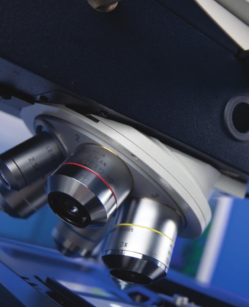
1 CLEMEX intelligent microscopy Vision PE 5.0 Advanced Image Analysis


2 Experience in Image Analysis Research and Quality Control Solutions With Vision PE, Clemex provides a powerful image analysis solution for automated microscopy. From measuring grain size to counting particles, Clemex Vision PE s intuitive software empowers you to easily custom-make image analysis macros without writing a single line of code. Clemex Vision PE also allows you to choose and use existing application packages for frequently run applications or for teaching purposes. Clemex Vision PE is the high level solution that is suitable for research and specialized analysis! Clemex Vision Lite, an abridged version of Clemex Vision PE, has a limited number of built-in imaging functions. When combined with application packages, Clemex Vision Lite offers intermediate-level solutions suitable for repetitive analyses or as an introduction to building customized image analysis routines.


3 Intermediate VS Advanced Solution Features Vision Lite Vision PE Acquisition Manual and Automatic* Multi-Layer Grab x x Reconstruction of Mosaic Image x x Software Controlled Stage Focus x x D Rendering* x Support Hot Stages * x Analysis Application Packages* x x Grain and Cell Sizing* x x Layer Thickness* x x Phase Analysis* x x Particle Sizing* x x Instantaneous Object Tracking x x Flexibility in Results Management x x Save and Retrieve Results x x Histograms with Logarithmic Scale x x Statistics (Percentiles) x x Support of USB.0 High Resolution Cameras x x Control of Input Parameters Settings x x Camera x x Light Intensity x x Image Amendement Functions limited extented Automated Microscope Control* x Image Analysis with Robotic Control* x Results Clemex Report Generator* x x Clemex R'Kive Explorer* x x * Are Optional


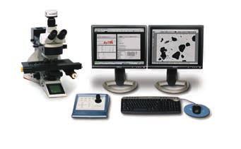
4 Modular Solution From Basic Software to Fully Operational Turnkey System When you need to acquire, quantify, and archive your images, choose from our complete line of digital imaging tools. Available in various configurations, from basic software to fully operational turnkey systems including hardware, software and on-site training and installation, you can choose the solution that's right for you today and add additional modules in the future as required. I


5 Rapid Image Analysis Step by Step Capture Images Large range of Monochrome or Color Digital Cameras can be used to capture images quickly and easily. Analyze your images using a live feed or save high resolution images for further analysis. Quantify Images Writing an analysis routine is just a click away, no programming required. Simply establish a list of actions taken from the Toolbox and your Routine writes itself. Manage Results Validate your results easily within the Data Browser where each measurement is directly linked to its corresponding feature on the image. You can then save them in Excel or binary format. Create Report Customize reports to your specifications quickly and easily. Produce secure printable reports or saved spreadsheet programs. I


6 Image Capture Mosaic step Forget tape and scissors! With the optional mosaic feature, Clemex Vision automatically stitches multiple fields to form a large image of unlimited resolution. This example demonstrates a Silicon Implant magnified at 00X with six stitched fields. + Use with a manual or motorized stage + For uneven surfaces, combine the multi-layer grab function with the mosaic feature (requires optional autofocus kit) + When running in manual mode, positioning arrows allow you to accurately stitch multiple fields + Control the number of fields to stitch, starting points, and more 5 6 Images to 6 captured with a Clemex 80 Color Firewire Camera at a magnification of 00x Perfect stitching x composite image of Silicon Particles in Implant Sample with total resolution of 7.89 MegaPixels I

. Layer 5 (00x).")
7 Multi-layer Grab Focusing on uneven surfaces can be troublesome. With the Automatic Multi-layer Grab function, intelligent software slices your image at varying z-intervals to reconstruct a completely focused image. If image input device systems do not have a motorized focus, the Manual Multi-layer Grab instruction generates a sharp composite image based on multiple planes acquired continuously as the user is turning the microscope focus knob.. Layer (00x). Layer 5 (00x). Reconstructed final image (5 00x) D Rendering Using the focus motor, the stage controller and the new Advanced Multi-Layer Grab, this new function renders D topography of a sample in as many as 56 layers. New measurements for D topographic images include Solid Volume, Void Volume, Z (Depth) Average, Minimum and Maximum values.. D Rendering of chip with Z (depth) measurement along green line. Statistic Results for Depth Measurements. Graph of Depth of chip along green I 5


8 Capture Image Multiple Field Automation step Stage and Auto-Focus Kit For high throughput environments, the optional motorized stage and auto-focus kit is highly recommended. Adaptable to all major microscope brands, you can significantly increase your productivity while taking advantage of some key Clemex Vision software features such as image stitching, D image reconstruction, D rendering, and object tracking. Software Controlled Stage and Focus Simplify the analysis of multiple fields with Clemex Vision's stage and focus automation tools. Highly flexible, you can determine sample origin, set the number of fields to analyze, establish specific autofocus fields or opt for a linear focus for sloping samples. + Set up multiple simultaneous stage patterns + Control field origin spacing + Move stage patterns + Save patterns. Clemex Joystick and Controller. Motorized Stage. Define number of fields needed. See stage moves with this window 6 I


9 Quantify Image Rapid Routine Development step With Clemex Vision's intuitive interface, you can quickly develop image analysis routines in a matter of minutes. Sequentially establish a list of image acquisition, processing, and measurement instructions by selecting them from the Toolbox and see them appear in the Routine window below. No programming required whatsoever! Just point and click. From Blood Cells to Cast Iron Nodules. Image Input Loads image and applies a Delineate filter in order to enhance object edges.. Threshold Performs an Autothreshold according to grey levels to separate phases and assigns a Bitplane color.. Binary Transformations Fills holes, removes small features and bumps from larger objects and separates objects.. Measures Measures length of cast iron nodules. I


10 Manage Results Magnifying Glass Tool step The Magnifying Glass Tool highlights the area at the tip of the mouse to show accurate detail. It is integrated in the image window as a child window, constrained to move with the mouse.. By right clicking, choose magnification. Move arrow where needed Validate Results with Ease Use the Data Browser tool to validate measurements and eliminate oversize particles or artifacts from invalidating results. The Data Browser tool is a spreadsheet of measurements linked to the outlines of each particle. Because the automated stage remembers the positions of all analyzed features, clicking on any given measurement returns the user instantly to the corresponding field of view.. Clicking on any particle highlights the corresponding measurements in the Data Browser in Figure. I


11 More Flexibility in Results Management Save and Retrieve Results Image analysis results can be saved in a raw format in addition to the existing export function that allows data to be saved in Excel format. Saving results in binary format guarantees authenticity of data as required by CFR Part rules of the FDA. Results can also be retrieved in Clemex Vision for further analysis of saved data. Histograms with Logarithmic Scale Histograms can now be displayed in logarithmic scale, in addition to the linear scale and user defined scale.. Results are saved in.cxr format. Results in Linear Scale. Results in Logarithmic Scale. Other Statistic Menu Window 5. Statistical Results with D0 Percentiles selected Percentiles Statistics Specifically for particle size, Clemex Vision automatically calculates the percentiles for any size distribution. This is particularly usefull in particle size analysis. 5 I


12 Customize reports to meet your unique needs. Create Reports step Everyone has special needs that distinguish their business. We know this and we cater to those needs by providing the Clemex Report Generator, a quick and easy way to customize reports and create report templates. Reports can be tailored to user specifications, to produce either a secure printable report for distribution and review, or to save to a spreadsheet program for further analysis and calculation. Once the templates are defined, reports are generated by inserting a single Generate Report instruction into the routine, assuring seamless integration with the run.. Results as they appear on the screen. Final custom report in PDF format. D0, D50 and D90 results 0 I


.")
13 Clemex Vision Options High Throughput The optional Clemex Maestroscope manages multiple samples without operator intervention, providing labs and manufacturing facilities with valuable walk-away time. The system can operate unattended and process up to 80 slides in one run. Advanced slide screening increases throughput, ensuring accuracy of measurements. Bar code identification of samples on the slides allows the Data Browser to link directly to specific particles. "The Clemex Maestroscope allows us to process many samples quickly and efficiently. Its nice to know that our analysis can be performed without supervision and we can return knowing our results are accurate". Robot selects sample holder from hotel Geert Rombaut, INVE (glass slides or microplates). Bar codes are scanned for sample identification. Robot positions sample holder in stage plate. Robot places the samples in analysis position I


14 Hot or Cool Stages Within Clemex Vision, a temperature profile can be created and the temperature can be read as a measurement parameter. The user can acquire time sequences of images showing the evolution of temperature related phenomena. Control of Microscope Parameters Settings This new set of functions allows the user to control the configuration parameters by software. Camera and Light Intensity settings can be modified within a routine as well as microscope parameters such as objective lens change for a motorized nosepiece. Camera and Microscope Configuration Instruction By inserting a Select Configuration instruction into the prolog of a routine, the user tells the system to load a specific set of camera and microscope settings to match the corresponding image analysis application. This is particularly useful when performing analyses requiring special illumination techniques, such as polarization, fluorescence or DIC, this instruction allows parameters such as objective magnification, white balance and shutter speed to be preset in order to avoid user manipulation errors.. Routine Window showing the Select Configuration instruction for Bright Field and the Select Magnification Instruction. Live image of Aluminium Grains as seen in the Image Viewer with Bright Field Illumination and 00X. Routine Window showing the Select Configuration instruction for Cross Polarization.. Automatic Camera adjustments based on Configuration Instruction in Step. I

15 CLEMEX A Commitment to Excellence in Imaging Choosing an imaging system can be a time consuming and often difficult process. Here are some key benefits you can count on when making Clemex your preferred system provider: Highly Qualified Team works closely with users for customized approach. Reputation: As a result of the company's highly focused efforts in building the best digital imaging systems, Clemex acquired a leadership position as provider of innovative solutions in image analysis. Experience: Management along with a highly specialized staff have decades of combined experience in the digital imaging industry. Ease-of-use: The intuitive nature of our systems allows users to get up and running surprisingly quickly. Support: Our dedicated staff of engineers and technicians are ready to provide you with fast, reliable assistance. Commitment: With the majority of employees retaining shares in the company, Clemex is highly committed to its continued development. Our track record of sustainable growth demonstrates that Clemex is a stable company, ready to serve your needs for years to come. Leadership: Since the company's inception, Clemex has succeeded in maintaining a competitive lead based on its superior technology. To remain the leader, Clemex continues to reinvest large portions of its revenues in R&D each year.
16 CLEMEX Printed in Canada 0/07 Some images may not be representative of the exact product. Clemex Vision, Clemex Vision Lite, Clemex Captiva, Clemex R Kive Explorer, Clemex CMT and Clemex CIR are trademarks of Clemex Technologies Inc. All other products mentioned are trademarks of their respective companies Clemex Technologies Inc. All rights reserved. Rev.0/07
Clemex Vision Lite. Capture, Measure, Analyze. the image analysis people
 Clemex Vision Lite Capture, Measure, Analyze the image analysis people Acquire, Measure, Analyze with Clemex Vision Lite Clemex Vision Lite is a flexible yet affordable software solution that enables
8.0. High precision solutions for cleanliness analysis CLEMEX PSFILTER. Accurate measurement of particulates as small as 0.
 High precision solutions for cleanliness analysis Mise à jour des logiciels Clemex depuis la version 6.0 et 7.0 VERSION 8.0 CLEMEX PSFILTER Accurate measurement of particulates as small as 0.5 microns
8.0. High precision solutions for cleanliness analysis CLEMEX PSFILTER. Metallic and non-metallic particles analyzed in one step
 High precision solutions for cleanliness analysis Mise à jour des logiciels Clemex depuis la version 6.0 et 7.0 VERSION 8.0 CLEMEX PSFILTER Metallic and non-metallic particles analyzed in one step Installed
the image analysis people
 Clemex CMT Computerized Microhardness Testing Clemex CMT (automatic) the image analysis people Modular Solutions Clemex CMT (automatic) Clemex CMT (semi-automatic) Clemex CMT Lite When you need a microhardness
DIGITAL-MICROSCOPY CAMERA SOLUTIONS USB 3.0
 DIGITAL-MICROSCOPY CAMERA SOLUTIONS USB 3.0 PixeLINK for Microscopy Applications PixeLINK will work with you to choose and integrate the optimal USB 3.0 camera for your microscopy project. Ideal for use
Zeiss LSM 880 Protocol
 Zeiss LSM 880 Protocol 1) System Startup Please note put sign-up policy. You must inform the facility at least 24 hours beforehand if you can t come; otherwise, you will receive a charge for unused time.
WITec Alpha 300R Quick Operation Summary October 2018
 WITec Alpha 300R Quick Operation Summary October 2018 This document is frequently updated if you feel information should be added, please indicate that to the facility manager (currently Philip Carubia,
Quick Operation Guide
 Quick Operation Guide Power ON Mounting specimens Set the specimen on the sample holder, and install the sample holder to the holder frame. Attach the holder frame to the XY stage. Type of holder Main
μscope Microscopy Software
 μscope Microscopy Software Pixelink μscope Essentials (ES) Software is an easy-to-use robust image capture tool optimized for productivity. Pixelink μscope Standard (SE) Software had added features, making
INTRODUCTION We believe that every laboratory working in the field of nanotechnology needs an SEM, therefore we would like to introduce to you our IEM
 INTRODUCTION We believe that every laboratory working in the field of nanotechnology needs an SEM, therefore we would like to introduce to you our IEM series of SEM. In short space of time, our device
Quick and simple installation and no maintenance needed. 3 Times More affordable Than a normal SEM. Obtaining results in less than 4 minutes
 INTRODUCTION We believe that every laboratory working in the field of nanotechnology needs an SEM, therefore we would like to introduce to you our IEM series of SEM. In short space of time, our device
Automated Imaging Technology to Simplify Your Workflow!
 Automated Imaging Technology to Simplify Your Workflow! BioSpectrum Imaging System Imaging Made Easy for Chemiluminescence Bioluminescence Colorimetric Fluorescence MegaCam 810 Camera OptiChemi 600 Camera
Using Olympus dotslide for polarising microscopy
 Using Olympus dotslide for polarising microscopy Background Because the dotslide system is built using a conventional microscope frame it lends itself to acquiring scans in different imaging modes whose
EXC500p-- PATHOLOGY MICROSCOPE. EXC500hd -- HD DIGITAL PATHOLOGY MICROSCOPE. EXC500r -- RESEARCH MICROSCOPE EXC500-LABORATORY SCOPE
 EXC500p-- PATHOLOGY MICROSCOPE EXC500hd -- HD DIGITAL PATHOLOGY MICROSCOPE EXC500r -- RESEARCH MICROSCOPE EXC500-LABORATORY SCOPE The EXC500 Pathology and Laboratory Microscope is the most optically advanced
Spotlight 150 and 200 FT-IR Microscopy Systems
 S P E C I F I C A T I O N S Spotlight 150 and 200 FT-IR Microscopy Systems FT-IR Microscopy Spotlight 200 with Frontier FT-IR Spectrometer Introduction PerkinElmer Spotlight FT-IR Microscopy Systems are
KEYENCE VKX LASER-SCANNING CONFOCAL MICROSCOPE Standard Operating Procedures (updated Oct 2017)
") KEYENCE VKX LASER-SCANNING CONFOCAL MICROSCOPE Standard Operating Procedures (updated Oct 2017) 1 Introduction You must be trained to operate the Laser-scanning confocal microscope (LSCM) independently.
KEYENCE VKX LASER-SCANNING CONFOCAL MICROSCOPE Standard Operating Procedures (updated Oct 2017) 1 Introduction You must be trained to operate the Laser-scanning confocal microscope (LSCM) independently.
BioSpectrum Imaging System
 BioSpectrum Imaging System Imaging Made Easy for Chemiluminescence Bioluminescence Colorimetric Fluorescence MegaCam 810 Camera OptiChemi 610 Camera BioChemi 510 Camera GelCam 310 Camera 8.1 megapixel
Zeiss Axiovert 135 Fluorescence Microscope Quick Guide / Operations Manual (v. 1.0 February 09)
") University of Chicago Integrated Light Microscopy Core Dr. Vytas Bindokas, Director http://digital.bsd.uchicago.edu By: Christine Labno, Assistant Director Room: AB-129 Phone: 4-9040 Zeiss Axiovert 135
University of Chicago Integrated Light Microscopy Core Dr. Vytas Bindokas, Director http://digital.bsd.uchicago.edu By: Christine Labno, Assistant Director Room: AB-129 Phone: 4-9040 Zeiss Axiovert 135
Nikon SIM-E & A1-R System
 Nikon SIM-E & A1-R System USER GUIDE LSU Health Sciences Center Shreveport Research Core Facility June 01 2017 Chaowei Shang 1 Table of Content 1. Start Up the System... Page 3 Hardware and microscope
Automated Particle Counting Systems Fast, Accurate Measurement Data
 Advanced Image Analysis Software OLYMPUS Inspector Series For Materials Science and Metrology Microscopes Automated Particle Counting Systems Fast, Accurate Measurement Data 1 The OLYMPUS Inspector Series
Turnkey Solution for Technical Cleanliness Inspection
 Technical Cleanliness Inspection System CIX90 OLYMPUS CIX series Turnkey Solution for Technical Cleanliness Inspection Simplify Your Technical Cleanliness Standard process for cleanliness inspection: preparation
Systematic Workflow via Intuitive GUI. Easy operation accomplishes your goals faster than ever.
 Systematic Workflow via Intuitive GUI Easy operation accomplishes your goals faster than ever. 16 With the LEXT OLS4100, observation or measurement begins immediately once the sample is placed on the stage.
Leica SP8 Resonant Confocal. Quick-Start Guide
 Leica SP8 Resonant Confocal Quick-Start Guide Contents Start-up Preparing for Imaging Part 1 On the scope Part 2 Software interface Part 3 Heat & CO2 incubation Part 4 Other hardware options Shut-down
LSM 780 Confocal Microscope Standard Operation Protocol
 LSM 780 Confocal Microscope Standard Operation Protocol Basic Operation Turning on the system 1. Sign on log sheet according to Actual start time 2. Check Compressed Air supply for the air table 3. Switch
Huvitz Digital Microscope HDS-5800
 Huvitz Digital Microscope HDS-5800 Dimensions unit : mm Huvitz Digital Microscope HDS-5800 HDS-MC HDS-SS50 The world s first, convert the magnification from 50x to 5,800x with a zoom lens HDS-TS50 Huvitz
Ordering Information & Specifications. VisionWorksLS Capabilities. Image Analysis Capabilities
 Ordering Information & Specifications VisionWorksLS Capabilities Each system includes: Camera and lens, darkroom with motorized or manual platform, three emission filters, white light illuminator, choice
Zeiss LSM 780 Protocol
 Zeiss LSM 780 Protocol 1) System Startup F Please note the sign-up policy. You must inform the facility at least 24 hours beforehand if you can t come; otherwise, you will receive a charge for unused time.
Contents STARTUP MICROSCOPE CONTROLS CAMERA CONTROLS SOFTWARE CONTROLS EXPOSURE AND CONTRAST MONOCHROME IMAGE HANDLING
 Operations Guide Contents STARTUP MICROSCOPE CONTROLS CAMERA CONTROLS SOFTWARE CONTROLS EXPOSURE AND CONTRAST MONOCHROME IMAGE HANDLING Nikon Eclipse 90i Operations Guide STARTUP Startup Powering Up Fluorescence
Monochrome Print Features: True 600 dpi print resolution Produces 6 D, A1 size prints or copies per minute 100% efficient No waste toner
 KIP 3100 SERIES KIP 3100 Monochrome Print, Copy & Scan The KIP 3100 system accurately reproduces technical documents at true 600 x 600 dpi resolution. Prints and copies may be delivered to the integrated
4 Use the adjustable Focus meter tool to take the subjectivity out of focusing the image, to get the best possible image
 Standard Edition VISIONx INC. www.visionxinc.com Real-Time Full Color Image Acquisition 4 Full support for NTSC and PAL cameras with Composite, Y/C (i.e. S-Video) and RGB video signal formats 4 Image display
Electron Microscopy RADIUS. Control & Imaging Software. RADIUS - The way forward in electron microscopy
 RADIUS - The way forward in electron microscopy Electron Microscopy RADIUS Control & Imaging Software THE ESSENCE OF ELECTRON MICROSCOPY: RADIUS RADIUS is the visionary software for electron microscopy
A DIVISION OF FORENSIC TECHNOLOGY SEE MORE. SOLVE MORE. CLASS-LEADING COMPARISON MICROSCOPES FOR FORENSIC INVESTIGATIONS
 A DIVISION OF FORENSIC TECHNOLOGY SEE MORE. SOLVE MORE. CLASS-LEADING COMPARISON MICROSCOPES FOR FORENSIC INVESTIGATIONS PROJECTINA AND FORENSIC TECHNOLOGY Forensic Technology pioneered automated ballistic
Leading in Desktop SEM Imaging and Analysis
 Leading in Desktop SEM Imaging and Analysis Fast. Outstanding. Reliable SEM imaging and analysis. The Phenom: World s Fastest Scanning Electron Microscope With its market-leading Phenom desktop Scanning
MAKE SURE YOUR SLIDES ARE CLEAN (TOP & BOTTOM) BEFORE LOADING DO NOT LOAD SLIDES DURING SOFTWARE INITIALIZATION
 BEFORE LOADING DO NOT LOAD SLIDES DURING SOFTWARE INITIALIZATION") Olympus VS120-L100 Slide Scanner Standard Operating Procedure Startup 1) Red power bar switch (behind monitor) 2) Computer 3) Login: UserVS120 account (no password) 4) Double click: WAIT FOR INITIALIZATION
Olympus VS120-L100 Slide Scanner Standard Operating Procedure Startup 1) Red power bar switch (behind monitor) 2) Computer 3) Login: UserVS120 account (no password) 4) Double click: WAIT FOR INITIALIZATION
Center for Microscopy and Image Analysis Axio Scan.Z1 Operating Manual
 No index entries found. Center for Microscopy and Image Analysis Axio Scan.Z1 Operating Manual Table of contents 1. Starting procedure... 3 1.1. Turn on hardware... 3 1.2. Starting ZEN blue... 4 2. Load
AxioVision User's Guide. Release 4.1
 AxioVision User's Guide Release 4.1 Number of this manual: B 48-0038 e 10.2003 Date of issue: 10.2003 Carl Zeiss Vision draws the User's attention to the fact that the information and references contained
WIDE. KIP 3100 Series CONNECT_ COMMUNICATE_ CONTROL_. business_by design
 powerful new touch screen based wide format digital system designed to provide a combination of high productivity and superior image quality. To satisfy the diverse requirements of decentralized technical
Quick Guide for Zeiss 710 Laser Scanning Confocal MGH Cancer Center
 Quick Guide for Zeiss 710 Laser Scanning Confocal MGH Cancer Center For any questions or concerns, please contact: Linda Nieman lnieman@mgh.harvard.edu Office: (617) 643-9684 Cell: (512) 565-8076 Chenyue
Before you start, make sure that you have a properly calibrated system to obtain high-quality images.
 CONTENT Step 1: Optimizing your Workspace for Acquisition... 1 Step 2: Tracing the Region of Interest... 2 Step 3: Camera (& Multichannel) Settings... 3 Step 4: Acquiring a Background Image (Brightfield)...
User Manual. Copyright 2010 Lumos. All rights reserved
 User Manual The contents of this document may not be copied nor duplicated in any form, in whole or in part, without prior written consent from Lumos. Lumos makes no warranties as to the accuracy of the
Material analysis by infrared mapping: A case study using a multilayer
 Material analysis by infrared mapping: A case study using a multilayer paint sample Application Note Author Dr. Jonah Kirkwood, Dr. John Wilson and Dr. Mustafa Kansiz Agilent Technologies, Inc. Introduction
RENISHAW INVIA RAMAN SPECTROMETER
 STANDARD OPERATING PROCEDURE: RENISHAW INVIA RAMAN SPECTROMETER Purpose of this Instrument: The Renishaw invia Raman Spectrometer is an instrument used to analyze the Raman scattered light from samples
Zeiss AxioImager.Z2 Brightfield Protocol
 Zeiss AxioImager.Z2 Brightfield Protocol 1) System Startup Please note put sign-up policy. You must inform the facility at least 24 hours beforehand if you can t come; otherwise, you will receive a charge
Using the Microscope for a NANSLO Remote Web-based Science Lab Activity
 Using the Microscope for a NANSLO Remote Web-based Science Lab Activity MICROSCOPE RWSL LAB INTERFACE INSTRUCTIONS The Remote Web-based Science Lab (RWSL) microscope is a high quality digital microscope
Standard Operating Procedure
 Standard Operating Procedure Nanosurf Atomic Force Microscopy Operation Facility NCCRD Nanotechnology Center for Collaborative Research and Development Department of Chemistry and Engineering Physics The
SCIENTIFIC INSTRUMENT NEWS. Introduction. Design of the FlexSEM 1000
 SCIENTIFIC INSTRUMENT NEWS 2017 Vol. 9 SEPTEMBER Technical magazine of Electron Microscope and Analytical Instruments. Technical Explanation The FlexSEM 1000: A Scanning Electron Microscope Specializing
Camera Overview. Digital Microscope Cameras for Material Science: Clear Images, Precise Analysis. Digital Cameras for Microscopy
 Digital Cameras for Microscopy Camera Overview For Materials Science Microscopes Digital Microscope Cameras for Material Science: Clear Images, Precise Analysis Passionate about Imaging: Olympus Digital
ScanArray Overview. Principle of Operation. Instrument Components
 ScanArray Overview The GSI Lumonics ScanArrayÒ Microarray Analysis System is a scanning laser confocal fluorescence microscope that is used to determine the fluorescence intensity of a two-dimensional
Y N C R O S C O P Y A DIVISION OF THE SYNOPTICS GROUP
 S Y N C R O S C O P Y A DIVISION OF THE SYNOPTICS GROUP THE PROBLEM: As a microscopist you often have to work with samples that are difficult to focus. When viewing a 3-D sample using an optical microscope
Camera Overview. Digital Microscope Cameras for Material Science: Clear Images, Precise Analysis. Digital Cameras for Microscopy
 Digital Cameras for Microscopy Camera Overview For Materials Science Microscopes Digital Microscope Cameras for Material Science: Clear Images, Precise Analysis Passionate about Imaging: Olympus Digital
Automated Cellular Imaging and Analysis System
 Automated Cellular Imaging and Analysis System CELLULAR IMAGING AND ANALYSIS FOR SCREENING AUTOMATED ACQUISITION AUTOMATED ANALYSIS HIGH RESOLUTION The ImageXpress TM 5000A automated cellular imaging and
Quick Guide for Zeiss 710 Laser Scanning Confocal MGH Cancer Center
 Quick Guide for Zeiss 710 Laser Scanning Confocal MGH Cancer Center For any questions or concerns, please contact: Linda Nieman lnieman@mgh.harvard.edu Office: (617) 643-9684 Cell: (512) 565-8076 Chenyue
VOLLSTÄDT DIAMANT GmbH. DiaInspect.OSM Automated particle analysis for superabrasives and surface analysis. Operation guide Version 1.2.
 VOLLSTÄDT DIAMANT GmbH DiaInspect.OSM 2010 Automated particle analysis for superabrasives and surface analysis Operation guide Version 1.2.8 Vollstädt-Diamant GmbH DiaInspect.OSM 2010 Microscope manual,
SIEVE CERTIFICATION SYSTEM
 MACHINE VISION SYSTEMS AND TECHNOLOGY Tel: (781) 275-2020 Fax: (781) 275-2028 www.vision-machines.com SIEVE CERTIFICATION SYSTEM Ensure ASTM E11 Compliance! Automated Sieve Certification System (ASCS)
GALILEO TMA CK 4500 HTS Tissue Microarray Platform
 GALILEO TMA CK 4500 HTS Tissue Microarray Platform Tissue Microarray (TMA) A Block Of Samples From Hundreds Of Blocks (S. M. Hewitt, M.D., Ph.D., Tissue Array Research Program, LP, CCR, NCI, NIH) TMA technology
UNIVERSITY OF WATERLOO Physics 360/460 Experiment #2 ATOMIC FORCE MICROSCOPY
 UNIVERSITY OF WATERLOO Physics 360/460 Experiment #2 ATOMIC FORCE MICROSCOPY References: http://virlab.virginia.edu/vl/home.htm (University of Virginia virtual lab. Click on the AFM link) An atomic force
Relationship to theory: This activity involves the motion of bodies under constant velocity.
 UNIFORM MOTION Lab format: this lab is a remote lab activity Relationship to theory: This activity involves the motion of bodies under constant velocity. LEARNING OBJECTIVES Read and understand these instructions
COMPACT MANUAL FOR GI USERS OF THE JEM 1400 FLASH BEGINNERS (For internal use only) Gray means additional information at the end of this mini-manual
 Gray means additional information at the end of this mini-manual") 1 COMPACT MANUAL FOR GI USERS OF THE JEM 1400 FLASH BEGINNERS (For internal use only) Gray means additional information at the end of this mini-manual ABOUT THIS MICROSCOPE (room HG01.240) The JEM-1400Flash
1 COMPACT MANUAL FOR GI USERS OF THE JEM 1400 FLASH BEGINNERS (For internal use only) Gray means additional information at the end of this mini-manual ABOUT THIS MICROSCOPE (room HG01.240) The JEM-1400Flash
Using Autofocus in NIS-Elements
 Using Autofocus in NIS-Elements Overview This technical note provides an overview of the available autofocus routines in NIS-Elements, and describes the necessary steps for using the autofocus functions.
ImageXpress Micro XLS Widefield High Content Screening System. Imaging with a vision.
 ImageXpress Micro XLS Widefield High Content Screening System Imaging with a vision www.moleculardevices.com The ImageXpress Micro Widefield High Content Screening System is the ultimate combination of
BioSpectrum Imaging System
 BioSpectrum Imaging System Imaging Made Easy for Chemiluminescence Bioluminescence Colorimetric Fluorescence MegaCam 800 Camera OptiCam 600 Camera BioChemi 500 Camera ChemiCam 410 Camera GelCam 310 Camera
QAQC LAB 589 Rappahannnock Drive White Stone Va TEL (866)
") OCCHIO Pharma CLICK FOR PRODUCT DEMO 400 OCCHIO Pharma O. O. O. O. OCCHIO Pharma 4 G 00 NANO OCCHIO 500 Occhio 500nano TECHNICAL DATASHEET Reference code: OCC023 Occhio500nano Technical specifications
OCCHIO Pharma CLICK FOR PRODUCT DEMO 400 OCCHIO Pharma O. O. O. O. OCCHIO Pharma 4 G 00 NANO OCCHIO 500 Occhio 500nano TECHNICAL DATASHEET Reference code: OCC023 Occhio500nano Technical specifications
Renishaw InVia Raman microscope
 Laser Spectroscopy Labs Renishaw InVia Raman microscope Operation instructions 1. Turn On the power switch, system power switch is located towards the back of the system on the right hand side. Wait ~10
Stereotopix Research. Precision Pathology. Highthroughput. pathology. powered by newcast. Advantages of Stereotopix : RUO
 Precision Pathology Highthroughput pathology Stereotopix Research powered by newcast RUO Researchers use quantitative microscopy in many ways with the goal of producing high-quality, quantitative results
LEICA TCS SP5 AOBS TANDEM USER MANUAL
 LEICA TCS SP5 AOBS TANDEM USER MANUAL STARTING THE SYSTEM...2 THE LAS AF SOFTWARE...3 THE «ACQUIRE» MENU...5 CHOOSE AND CREATE A SETTING...6 THE CONTROL PANEL...8 THE DMI6000B MICROSCOPE...10 ACQUIRE ONE
XLS Electronic Pipettes
 XLS Electronic Pipettes E4 XLS Maximum reproducibility Application versatility Intuitive operation The App Master Accelerate your Workflow Electronic XLS Pipettes Optimize your Applications The Options
Leica DMi8A Quick Guide
 Leica DMi8A Quick Guide 1 Optical Microscope Quick Start Guide The following instructions are provided as a Quick Start Guide for powering up, running measurements, and shutting down Leica s DMi8A Inverted
Histograms& Light Meters HOW THEY WORK TOGETHER
 Histograms& Light Meters HOW THEY WORK TOGETHER WHAT IS A HISTOGRAM? Frequency* 0 Darker to Lighter Steps 255 Shadow Midtones Highlights Figure 1 Anatomy of a Photographic Histogram *Frequency indicates
Using the Nikon TE2000 Inverted Microscope
 Wellcome Trust Centre for Human Genetics Molecular Cytogenetics and Microscopy Core Using the Nikon TE2000 Inverted Microscope Fluorescence image acquisition using Scanalytic s IPLab software and the B&W
OLYMPUS Digital Cameras for Materials Science Applications: Get the Best out of Your Microscope
 Digital Cameras for Microscopy Camera Overview For Materials Science Microscopes OLYMPUS Digital Cameras for Materials Science Applications: Get the Best out of Your Microscope Passionate About Imaging
MIF ZEISS LSM510 CONFOCAL USER PROTOCOL
 MIF ZEISS LSM510 CONFOCAL USER PROTOCOL START-UP Turn on the Mercury Bulb Power Supply (if needed). Power-on the Control Box. Turn on the computer. Open the LSM 510 software. Choose Scan New Images and
Cast Iron Analysis. Nodular cast iron analysis
 Cast Iron Analysis This dhs Image Data Base module analyzes the microstructure of cast iron. The graphite particles actual make-up is crucial for material properties such as elasticity and fracture behaviour.
FT-IR.
 FT-IR varian, inc. 610/620-IR ft-ir MICROSCOPY AND IMAGING SoLUTIONS www.varianinc.com VARIAN, INC. Setting the Standard Again When Only the Best Will Do The world leader in molecular spectroscopy innovation
Morphologi. Advanced image analysis for high sensitivity particle characterization. Particle size. Particle shape
 Particle size Particle shape Morphologi detailed specification sheets from www.malvern.co.uk Introducing a new concept in image analysis The Morphologi high sensitivity particle analyzer is more than just
Camera Overview. Olympus Digital Cameras for Materials Science Applications: For Clear and Precise Image Analysis. Digital Cameras for Microscopy
 Digital Cameras for Microscopy Camera Overview For Materials Science Microscopes Olympus Digital Cameras for Materials Science Applications: For Clear and Precise Image Analysis Passionate about Imaging
Figure 1. Oil-immersion objectives available for use with the Lionheart FX.
 Tech Note Oil Objective Introduction The Lionheart FX automated imager is compatible with high numerical aperture oil immersion objectives. These objectives offer magnification up to 100X and significantly
THE ULTIMATE DOCUMENT EXAMINATION SYSTEM STATE-OF-THE-ART SPECTRAL ANALYSIS FORENSIC LABS SECURITY PRINTERS IMMIGRATION AUTHORITIES
 THE ULTIMATE DOCUMENT EXAMINATION SYSTEM STATE-OF-THE-ART SPECTRAL ANALYSIS FORENSIC LABS SECURITY PRINTERS IMMIGRATION AUTHORITIES WHEN DETAILS MATTER PROJECTINA SPECTRA PRO The Ultimate Document Examination
Nikon AZ100. Laser Scanning Macro Confocal Microscope. Jordan Briscoe Adam Fries Kyle Marchuk Kaitlin Corbin. May 2017.
 Nikon AZ100 Laser Scanning Macro Confocal Microscope Jordan Briscoe Adam Fries Kyle Marchuk Kaitlin Corbin May 2017 Contents 1 Introduction 2 2 Hardware - Startup 2 3 Software/Operation 4 3.1 Multidimensional
Simplified Instructions: Olympus Widefield Microscope S1230
 Contents General Microscope Operation Simple Image Capture Multi-Wavelength Capture Z-Series Timelapse Combining Capture Modes Synopsis of Other Functions Pages 2-23 24-40 41-47 48-56 57-59 60-68 69-83
Automated Digitization of Gram Stains. Centralized Reading. Decentralized Assessment. Improved Quality Management.
 Automated Digitization of Gram Stains Centralized Reading. Decentralized Assessment. Improved Quality Management. A GROWING DEMAND Gram staining is the rapid, easy, and inexpensive method for the assessment
CARAT 930/950. Hardness Testing & Analysis CARAT 930 / 950
 STABLE CONSTRUCTION The vibration-damped cast aluminum body comprises a robust basis for the high load-bearing Carat table with automatic X/Y axis and automatic Z axis with 8-times objective revolver (LED
Microscopic Structures
 Microscopic Structures Image Analysis Metal, 3D Image (Red-Green) The microscopic methods range from dark field / bright field microscopy through polarisation- and inverse microscopy to techniques like
TEM Cameras. Digital Cameras for Electron Microscopy
 Digital Imaging Solutions TEM Cameras Side- and bottom-mounted TEM cameras Digital Cameras for Electron Microscopy IMAGING SOLUTIONS FOR ELECTRON MICROSCOPY. BASED ON OPTO-DIGITAL KNOW-HOW. DESIGNED BY
R I T. Title: Wyko RST Plus. Semiconductor & Microsystems Fabrication Laboratory Revision: A Rev Date: 05/23/06 1 SCOPE 2 REFERENCE DOCUMENTS
 Approved by: Process Engineer / / / / Equipment Engineer 1 SCOPE The purpose of this document is to detail the use of the Wyko RST Plus. All users are expected to have read and understood this document.
Nikon COOLSCAN V ED Major Features
 Nikon COOLSCAN V ED Major Features 4,000-dpi true optical-resolution scanning, 14-bit A/D converter featuring 16-/8-bit output for clear, colorful images Exclusive Scanner Nikkor ED high-performance lens
Camera Overview. Digital Microscope Cameras for Material Science: Clear Images, Precise Analysis. Digital Cameras for Microscopy
 Digital Cameras for Microscopy Camera Overview For Materials Science Microscopes Digital Microscope Cameras for Material Science: Clear Images, Precise Analysis Passionate about Imaging: Olympus Digital
ChemiDoc-It Imaging System
 ChemiDoc-It Imaging System Ultra dark chamber and highly sensitive, scientific-grade CCD camera for chemiluminescence imaging ChemiDoc-It darkroom is light tight creating optimum imaging conditions for
System NMI. Accuracy is the Key. Classifying the Content of Non-metallic Inclusions in Steel in Accordance with Current Industrial Standards
 Microscopy from Carl Zeiss System NMI Accuracy is the Key Classifying the Content of Non-metallic Inclusions in Steel in Accordance with Current Industrial Standards New Guidelines Require New Priorities:
Olympus LEXT OLS 4000 Confocal Laser Microscope
 Olympus LEXT OLS 4000 Confocal Laser Microscope The Olympus LEXT OLS4000 is a confocal microscope capable of taking high-resolution 3D images. The magnification (Optical and Digital) of this microscope
Comparators. Contour Projector Technologies. Compare the Quality. Precision for People
 Precision for People Comparators Contour Projector Technologies Compare the Quality Certified Comparator Products Focused on Quality Certified Comparator Products offers a complete line of optical comparators
nanovea.com PROFILOMETERS 3D Non Contact Metrology
 PROFILOMETERS 3D Non Contact Metrology nanovea.com PROFILOMETER INTRO Nanovea 3D Non-Contact Profilometers are designed with leading edge optical pens using superior white light axial chromatism. Nano
ASM Webinar Digital Microscopy for Materials Science
 Digital Microscopy Defined The term Digital Microscopy applies to any optical platform that integrates a digital camera and software to acquire images; macroscopes, stereomicroscopes, compound microscopes
User Manual. cellsens 1.16 LIFE SCIENCE IMAGING SOFTWARE
 User Manual cellsens 1.16 LIFE SCIENCE IMAGING SOFTWARE Any copyrights relating to this manual shall belong to OLYMPUS CORPORATION. We at OLYMPUS CORPORATION have tried to make the information contained
Indian Institute of technology Madras Presents NPTEL NATIONAL PROGRAMME ON TECHNOLOGY ENHANCED LEARNING
 Indian Institute of technology Madras Presents NPTEL NATIONAL PROGRAMME ON TECHNOLOGY ENHANCED LEARNING Lecture - 5 Materials Characterization Fundamentals of Optical microscopy Dr. S. Sankaran Associate
BLIPS perfectly aligned cleaned by any impurity Gently press the lens
 BLIPS - TIPS & TRICKS BLIPS, the thinnest Macro and Micro lenses for smartphones in the world, can guarantee an excellent picture quality for photos and videos, but they need some simple tricks to get
Axioscan - Startup. 1. Turn on the Axioscan on (button to the left) and turn on the computer
 and turn on the computer") Axioscan - Startup 1. Turn on the Axioscan on (button to the left) and turn on the computer 2. Log on and start the ZEN Blue software from the desktop 3. Press ZEN slidescan and Start System 4. Start by
Axioscan - Startup 1. Turn on the Axioscan on (button to the left) and turn on the computer 2. Log on and start the ZEN Blue software from the desktop 3. Press ZEN slidescan and Start System 4. Start by
Very short introduction to light microscopy and digital imaging
 Very short introduction to light microscopy and digital imaging Hernan G. Garcia August 1, 2005 1 Light Microscopy Basics In this section we will briefly describe the basic principles of operation and
Stereo Viewing Systems
 Includes NEW image capture option Stereo Viewing Systems Superior imaging for a wide range of inspection & rework tasks Patented optical technology for fatigue-free viewing and superb image quality Wide
Highest Resolution: 5400 dpi (optical) Finest Image Quality due to employing sophisticated Grain Dissolver Comfortability: Digital ICE Manual Focus
 Finest Image Quality due to employing sophisticated Grain Dissolver Comfortability: Digital ICE Manual Focus") Highest Resolution: 5400 dpi (optical) Finest Image Quality due to employing sophisticated Grain Dissolver Comfortability: Digital ICE Manual Focus Button Quick Scan Button Refined Design Dual Interfaces:
Highest Resolution: 5400 dpi (optical) Finest Image Quality due to employing sophisticated Grain Dissolver Comfortability: Digital ICE Manual Focus Button Quick Scan Button Refined Design Dual Interfaces:
Microscopy from Carl Zeiss
 Microscopy from Carl Zeiss Contents Page Contents... 1 Introduction... 1 Starting the System... 2 Introduction to ZEN Efficient Navigation... 5 Setting up the microscope... 10 Configuring the beam path