Introduction. INSTRUCTION MANUAL CAT XL, 6500-XLCORE, 6500-FL Evos-XL, Evos-XL/Core, Evos-FL
|
|
|
- Chloe Warner
- 5 years ago
- Views:
Transcription


1 1 INSTRUCTION MANUAL CAT XL, 6500-XLCORE, 6500-FL Evos-XL, Evos-XL/Core, Evos-FL Introduction Experience faster results and easier cell imaging with an EVOS imaging system! An EVOS system is the ideal item to have in the laboratory. These imaging systems allow researchers to focus on their data without the hassle of operating a microscope. EVOS imaging systems will help you to perform a wide range of applications such as complex protein analysis and multi-channel fluorescent imaging, to name a few. The proprietary LED light cube technology minimizes photo bleaching, offers more than 50,000 hours of illumination, and allows adjustable intensity all with no need for a darkroom or consumable costs. All of the EVOS systems are designed to work together from the initial cell culture check to more complex analyses such as time lapse and image tilting and stitching. EVOS systems allow you to dedicate more time analyzing images and less time capturing them.
2 2 Contents Introduction..1 Compact and Portable Systems.. 3 The power of LED illumination EVOS-FL Auto Imaging System EVOS-FL Auto Imaging System and Onstage Incubator....8 EVOS-FL Auto Imaging System Additional applications.10 EVOS-FL Imaging System..11 EVOS-XL Imaging System..13 EVOS-XLCORE Imaging System..15 Objectives..17 Proprietary LED light cubes 18 Vessel Holders and Stage Plates.. 19


3 3 Compact and Portable Systems With an EVOS Imaging System, you can now have convenient cell imaging where and when you want it. Simply place your EVOS Imaging System at your desired location, flip the switch, and you re ready to go in just under two minutes. Whether dealing with a large or small audience, the EVOS Imaging System is perfect for teaching in classrooms or lecture halls. Publication-quality imaging In today s competitive scientific environment, generating publication-quality images is critical to your success. To help guarantee that you get the images you need, EVOS systems give you top-of-the-line imaging components, including: - High-quality camera and optics to capture high resolution images - LED illumination to produce superior signal-to- noise ratios Easy-to-use image capture and processing software for ready-to-publish images Bovine pulmonary artery endothelial cells, 60x oil objective. Light cubes: DApi, gfp, texas Red. Moss antheridial head polytrichum. 40x objective. Technology that is better for the environment Traditional fluorescence microscopy light sources use mercury, a toxic carcinogen requiring special handling and disposal. By using LED light sources, EVOS systems do not require these special steps and are thereby more environmentally-friendly and energy-efficient. Osteoblasts in bone, 40x coverslip corrected objective. Light cubes: Cy 7, texas Red.


4 4 The Power of LED Illumination All EVOS fluorescent cell imaging systems utilize LED light sources, which means that you are given high-intensity output over a short light path for the most efficient fluorophore excitation. - Shorter light path provides better detection of fluorescent signals - Continuous illumination gives consistent results - More than 50,000-hour bulb lifetime avoids high laboratory costs - Adjustable light intensity reduces photo bleaching Revolutionary light path By placing the LED light cube as close as possible to the objective turret, the number of optical elements in the light path is minimized. High-intensity illumination over a short light path increases the efficiency of fluorophore excitation, providing better detection of weak fluorescent signals. Stability comparison Mercury and metal halide vs. LED Continuous light intensity Mercury arc lamps can decrease in intensity by as much as 50% in the first 100 hours of operation. In addition, images acquired in different sessions cannot be quantitatively compared using mercury illumination without complicated calibrations. Because EVOS systems have continuous light cube intensity, users can rely on its consistent illumination and can compare quantitative results from images acquired on different days!
, compared to 300 hours for a typical mercury bulb (1,500 hours for a metal halide bulb). This is equivalent to 70-75% in savings in the overall maintenance of your instrument!")

5 5 Illumination costs over time Less expensive to own and maintain The LED bulbs on the EVOS systems are rated for more than 50,000 hours (about 17 years!), compared to 300 hours for a typical mercury bulb (1,500 hours for a metal halide bulb). This is equivalent to 70-75% in savings in the overall maintenance of your instrument! EVOS hard-coated filter sets for higher transmission efficiencies Hard-coated filter sets are more expensive, but they have sharper edges and significantly higher transmission efficiencies that typically result in more than 25% more light transmission than traditional soft-coated filters. With the EVOS system s hard-coated filter sets, the cost of your light cubes decreases over time. Furthermore, you will have significantly brighter fluorescence, higher transmission efficiencies, the ability to detect faint fluorescent signals, and better signal-to-noise ratios. Take a look at these transmission efficiency comparisons

 10.")
6 6 EVOS-FL Auto Imaging System Experience an intuitive, fully-automated and affordable imaging system! FL Auto footprint 1. Power input jack 2. Power switch 3. Computer port 4. Lifting handholds 5. Condenser (contains automatic phase annulus selector) 6. Condenser slider slot 7. Automatic X-y axis stage high-resolution touch screen monitor 9. Onstage incubator (optional) 10. Stagetop environmental chamber (optional)

7 7 System highlights Software Integrated software is a key component of the all-in-one system. The EVOS-FL system, accessed by a touch screen monitor, features standard functions such as a scale bar and image review tool as well as a variety of advanced imaging and analysis tools. All images acquired can be saved in.jpeg,.bmp,.tiff, and.png formats. Key software features: - Time-lapse imaging - Image tilting and stitching - Automated cell counting - Auto-focus and automated multi-well plate scanning - Z-stacking - Environmental control with EVOS Onstage incubator - Reuse function for easy duplication of previously acquired images

8 8 Applications The EVOS-FL system was designed to be used for a range of applications including, but not limited to, multi-channel fluorescence imaging, image tilting and stitching, cell density assays, multiple-position vessel scanning, and time-lapse imaging. Hela cells, 100x oil objective light cubes: DApi, gfp, RFp Reagents: nucblue tm live (blue), Celllight mito-gfp (green), Celllight H2B-RFp EVOS-FL Auto Imaging System and Onstage Incubator Time-lapse imaging When combined with the new onstage incubation system, the EVOS-FL Imaging System is ideal for longterm monitoring of cell cultures and time-lapse imaging at high resolution. The EVOS Onstage Incubator is an environmental chamber enabling precise control of temperature, humidity, and three gases for timelapse imaging of live cells under both physiological and non-physiological conditions. Environmental settings and image acquisition parameters are all seamlessly integrated into the EVOS-FL Imaging System interface, creating a high performance inverted imaging system with superb flexibility, ease of use, and optical performance for demanding time-lapse imaging experiments. With the integrated environmental chamber, you can: - Intuitively set environmental and image acquisition parameters - Easily maintain physiological or non-physiological conditions with precise control - Adjust environmental parameters while the experiment is running - Choose from a range of vessel holders - Save lab space with a small footprint and sleek design


 and Celllight mitochondria-rfp (red), and stained with")
9 9 - Once captured, you can seamlessly create and export fluorescence or bright-field images as movies: o o o o Create time-lapse images or every well or 96-well plate, simultaneously Acquire time-lapse images in a single place or z-stacks Autofocus in each channel and region of interest Metadata and time stamps are included with each image frame of time-lapse movies Time-lapse imaging of dividing HeLa cells, using the EVOS FL Auto Imaging System with Onstage Incubator. images were captured every 12 minutes over a period of 24 hours. Cells were transduced with Celllight Histone 2BgFp (green) and Celllight mitochondria-rfp (red), and stained with nucblue live Readyprobes Reagent (blue)


10 10 EVOS-FL Auto Imaging System Additional applications Image stitching The EVOS-FL Imaging System allows you to capture multiple images and mosaic tiling to stitch a high-resolution image of a large area. This is ideal for analyzing tissue sections or stem cell colonies, or for viewing ever cell in the well of a 96-well plate. In addition: - Acquire images at high magnifications and stitch for high-resolution mapping - Batch export plate scans of large wells in one step - Scan in bright-field, phase-contrast, or fluorescent mode - Save individual images as well as composite images Automated cell counting The EVOS-FL Imaging System contains advanced software algorithms that allow extremely accurate cell counting. Following labeling of nuclei using a fluorescent dye such as nucblue live cell stain, the EVOS-FL Imaging System will calculate the number of cells in a field of view, making it easy to determine the number of cells in a well or dish. - Accurate cell counting even at 4x magnification - Adjust intensity levels with a convenient slider bar - Easily visualize GFP expression, determine live/dead cell ratio, and count total cell numbers Stitched image of one well from a 96- well plate, taken using a 10x objective (A). CAKi cells were labeled with anti- Oxphos subunit V primary antibody and goat anti-mouse Alexa Fluor 488 secondary antibody (green), ActinRed 555 reagent (red), and nucblue fixed cell stain (blue). Subsequent images are shown at 200% (B), 400% (C), and 800% (D) magnification. Screen shot from the automated cell counting feature of the EVOS-FL Imaging System. Cells were stained with nucblue live cell stain prior to analysis.


11 11 Z-stacking The EVOS-FL Imaging System has the option to produce flat-focus z-stack images. The z-stack flat focus feature collects a series of images, extracts the most in focus pixels from each image, and then returns a single, focused image even if the sample is very thick. - Images can be made into a video, montage, 3D reconstruction, or maximum projection image - Z-stack range can be performed automatically in fluorescence imaging mode - Z-stack can uncover changes in cellular morphology not seen in standard widefield microscopy EVOS-FL Imaging System Experience form, function and flexibility, in one! FL footprint 1. Power input jack 2. Power switch 3. USB and DVI ports 4. Coarse stage positioning knobs 5. Stage x-axis knob 6. X-axis stage brake Stage y-axis knob 8. Y-axis stage brake 9. Focusing knobs 10. Objective selection wheel 11. Light cube selection lever 12. Phase annulus selector 13. Condenser slider slot 10


12 12 System highlights Software Integrated software is a key component of the all-in-one system. The EVOS-FL software features standard functions including a scale bar and image review tool along with a wide range of advanced imaging and analysis tools. All images acquired can be saved in.jpeg,.bmp,.tiff,.png, and.avi formats. Key software includes: - 1-click, multi-channel overlay - Time-lapse capability - Cell counting capability - Transfection capability Applications The EVOS-FL Imaging System was designed for a broad range of applications including, but not limited to, multiple-channel fluorescence imaging, protein analysis, pathology, cell culture, and in situ imaging. With positions for 5 objectives and 4 fluorescent light cubes, the EVOS-FL Imaging System provides the flexibility to meet most imaging research applications.


13 13 EVOS-XL Imaging System Experience an advanced transmitted-light system that delivers high-definition results with the same form, functions, and features that are standard on all EVOS systems! XL footprint 1. Power input jack 2. Power switch 3. USB and DVI ports 4. Coarse stage positioning knobs 5. Stage x-axis knob 6. X-axis stage brake 7. Stage y-axis knob 8. Y-axis stage brake 9. Focusing knobs 10. Objective selection wheel 11. Phase annulus selector 12. Condenser slider slot


14 14 System highlights Software Integrated software is a key component of the all-in-one system. Our software features standard functions such as a scale bar and image review tool as well as a variety of advanced imaging and analysis tools. All images acquired can be saved in.jpeg,.bmp,.tiff,.png, and.avi formats. Key software features: - Time-lapse imaging - Cell counting Applications The EVOS-XL Imaging System was designed for a broad range of applications including, but not limited to, cell viability assays, stem cell growth and differentiation, stem cell passaging, hematoxylin and eosin imaging, and diaminobenzidine (DAB) imaging. The EVOS-XL Imaging System is ideal for routine cell and tissue culture, cell confluence determination, stem cell passaging, stem cell growth and differentiation, and developmental biology and tissue slice analysis. Mitosis in onion root tip, 40x objective.


15 15 EVOS-XLCORE Imaging System Experience a compact, simple transmitted-light system perfect for use in the cell culture hood or tissue culture facility! XLCORE footprint 1. Power input jack 2. Power switch 3. USB ports 4. Objective turret 5. Coaxial focusing knob 6. Phase turret 7. Illumination wheel 8. Freeze button 9. Save button age knob knob


 W Applications The EVOS-XLCORE Imaging")

16 16 System highlights Software Integrated software is a key component of the all-in-one system. Our software includes a variety of features such as color temperature control. All images can be saved in.jpeg,.bmp, and.tiff formats. Key software features: - Adjustable saturation and contrast - Color temperature controls (warm and cool) W Applications The EVOS-XLCORE Imaging System was designed for a broad range of applications including, but not limited to, routine cell and tissue culture visualization and imaging, stem cell applications, and sample staining differentiation (such as gram staining).

17 17 Objectives Bright-field vs. phase contrast The most basic form of light microscopy, bright-field contrast, is mediated by the absorption on light by the sample. A higher-density area in a sample will absorb more light, thus increasing contrast in those areas. Phase contrast, on the other hand, is most useful for hard-to-see translucent specimens. It is accomplished by converting phase shifts, caused by light passing through a translucent specimen, into brightness changes. Long working distance vs. coverslip corrected Long working distance is optimized for use through vessels with nominal wall thickness of mm (slides, flasks, microtiter dishes, etc.). Coverslip corrected is optimized for use through #1.5 coverslips (approx mm thick). These have a higher magnification-to-ratio and provide higher resolution compared to long working distance.


18 18 Proprietary LED light cubes At the heart of EVOS fluorescence technology is the proprietary LED light cubes. Each cube contains an LED, collimating optics, and filters. Light cubes are user-interchangeable, auto-configured by the system, with plug-and-play capability. The wide range of light cubes available provides flexibility for multiplefluorescence research applications. Custom light cubes Do you need a customized light cube to accommodate your fluorescent needs? Contact Electron Microscopy Sciences at to obtain your specialty light cube. Common light cubes CHO cells transfected with eukaryotic expression plasmid, 40x objective. light cubes: Cy 7, DApi Gold, 10x objective. Light cube: white.

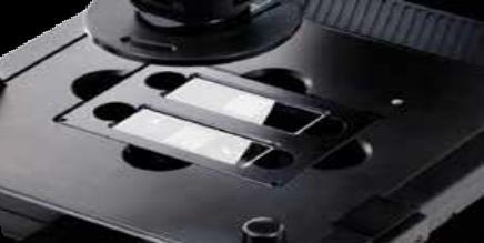
19 19 Vessel holders and stage plates


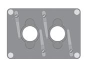
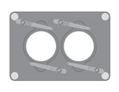
20 20 or andar 56 Holds
Using the Nikon TE2000 Inverted Microscope
 Wellcome Trust Centre for Human Genetics Molecular Cytogenetics and Microscopy Core Using the Nikon TE2000 Inverted Microscope Fluorescence image acquisition using Scanalytic s IPLab software and the B&W
LSM 710 Confocal Microscope Standard Operation Protocol
 LSM 710 Confocal Microscope Standard Operation Protocol Basic Operation Turning on the system 1. Switch on Main power switch 2. Switch on System / PC power button 3. Switch on Components power button 4.
Leica SP8 TCS Users Manual
 Leica SP8 TCS Users Manual Follow the procedure for start up and log on as posted in the lab. Please log on with your account only and do not share your password with anyone. We track and confirm usage
Things to check before start-up.
 Byeong Cha Page 1 11/24/2009 Manual for Leica SP2 Confocal Microscope Enter you name, the date, the time, and the account number in the user log book. Things to check before start-up. Make sure that your
Leica SP8 TCS Users Manual
 Version : 07/08/0 Leica SP8 TCS Users Manual Start up:. Turn the PC Microscope, Scanner Power, Laser Power, and the Laser Emission key to on (bottom right of desk).. Turn on the fluorescent lamp (top left
LSM 780 Confocal Microscope Standard Operation Protocol
 LSM 780 Confocal Microscope Standard Operation Protocol Basic Operation Turning on the system 1. Sign on log sheet according to Actual start time 2. Check Compressed Air supply for the air table 3. Switch
Last updated: May 2014 Y.DeGraaf
 FLINDERS MICROSCOPY BIOMEDICAL SERVICES AVAILABLE MICROSCOPES AND SPECIFICATIONS & INFORMATION REGARDING TRAINING FOR NEW USERS Last updated: May 2014 Y.DeGraaf If you have new staff or students (Honours/Masters
ImageXpress Micro XLS Widefield High Content Screening System. Imaging with a vision.
 ImageXpress Micro XLS Widefield High Content Screening System Imaging with a vision www.moleculardevices.com The ImageXpress Micro Widefield High Content Screening System is the ultimate combination of
ZEISS LSM510META confocal manual
 ZEISS LSM510META confocal manual Switching on the system 1) Switch on the Remote Control button located on the table to the right of the microscope. This is the main switch for the whole system including
ANSWER KEY Lab 2 (IGB): Bright Field and Fluorescence Optical Microscopy and Sectioning
: Bright Field and Fluorescence Optical Microscopy and Sectioning") Phys598BP Spring 2016 University of Illinois at Urbana-Champaign ANSWER KEY Lab 2 (IGB): Bright Field and Fluorescence Optical Microscopy and Sectioning Location: IGB Core Microscopy Facility Microscope:
Phys598BP Spring 2016 University of Illinois at Urbana-Champaign ANSWER KEY Lab 2 (IGB): Bright Field and Fluorescence Optical Microscopy and Sectioning Location: IGB Core Microscopy Facility Microscope:
Nikon Eclipse Ti2-E Widefield/Spinning Disk Confocal Microscope Standard Operation Protocol
 Nikon Eclipse Ti-E Widefield/Spinning Disk Confocal Microscope Standard Operation Protocol Please sign on the log sheet before switching on system. Turn on system Turn on A only if confocal mode or laser
Operating Instructions for Zeiss LSM 510
 Operating Instructions for Zeiss LSM 510 Location: GNL 6.312q (BSL3) Questions? Contact: Maxim Ivannikov, maivanni@utmb.edu 1 Attend A Complementary Training Before Using The Microscope All future users
Nikon SIM-E & A1-R System
 Nikon SIM-E & A1-R System USER GUIDE LSU Health Sciences Center Shreveport Research Core Facility June 01 2017 Chaowei Shang 1 Table of Content 1. Start Up the System... Page 3 Hardware and microscope
Opterra II Multipoint Scanning Confocal Microscope. Innovation with Integrity
 Opterra II Multipoint Scanning Confocal Microscope Enabling 4D Live-Cell Fluorescence Imaging through Speed, Sensitivity, Viability and Simplicity Innovation with Integrity Fluorescence Microscopy The
Practical work no. 3: Confocal Live Cell Microscopy
 Practical work no. 3: Confocal Live Cell Microscopy Course Instructor: Mikko Liljeström (MIU) 1 Background Confocal microscopy: The main idea behind confocality is that it suppresses the signal outside
Confocal Laser Scanning Microscopy
 Name of the Core Facility: Confocal Laser Scanning Microscopy CORE Forschungszentrum Immunologie Mainz Welcome to the CSLM Core Facility: The CLSM Core Facility enables working groups to incorporate high
Zeiss Axiovert 135 Fluorescence Microscope Quick Guide / Operations Manual (v. 1.0 February 09)
") University of Chicago Integrated Light Microscopy Core Dr. Vytas Bindokas, Director http://digital.bsd.uchicago.edu By: Christine Labno, Assistant Director Room: AB-129 Phone: 4-9040 Zeiss Axiovert 135
University of Chicago Integrated Light Microscopy Core Dr. Vytas Bindokas, Director http://digital.bsd.uchicago.edu By: Christine Labno, Assistant Director Room: AB-129 Phone: 4-9040 Zeiss Axiovert 135
fsx.olympus-global.com
 Bio Imaging Navigator fsx.olympus-global.com Visit our Web site for on-line demonstration of FSX100. FSX100 is the environmental conscious product according to OLYMPUS's own standards. Main features of
Quick Operation Guide
 Quick Operation Guide Power ON Mounting specimens Set the specimen on the sample holder, and install the sample holder to the holder frame. Attach the holder frame to the XY stage. Type of holder Main
Quick Guide for Zeiss 710 Laser Scanning Confocal MGH Cancer Center
 Quick Guide for Zeiss 710 Laser Scanning Confocal MGH Cancer Center For any questions or concerns, please contact: Linda Nieman lnieman@mgh.harvard.edu Office: (617) 643-9684 Cell: (512) 565-8076 Chenyue
Nikon E800 Operating Instructions.
 Nikon E800 Operating Instructions. You can request electronic copies of this manual by contacting lshats@jhsph.edu Copies are also available on the JHU MMI Department web site. Please send your comments
Nikon Ti-E Microscope Manual. Rightmire Hall Ohio State University. Director: Tony Brown Rightmire
 Nikon Ti-E Microscope Manual Rightmire Hall Ohio State University Director: Tony Brown Rightmire 060 292-1205 brown.2302@osu.edu Facility Manager: Paula Monsma Rightmire 062 293-0939 292-1367 monsma.1@osu.edu
Zeiss Deconvolution Microscope: A Quick Guide
 Zeiss Deconvolution Microscope: A Quick Guide Start-up Uncover microscope. Do not put dust cover on the floor. Plug in both cameras. The default camera is the AxioCam HRm (monochrome camera) for fluorescence
QUICKSTART GUIDE: WIDEFIELD HWF1 Zeiss Cell Observer Live Cell Imaging System (HAMMERSMITH, L BLOCK, ROOM 314) Imperial College London
 Imperial College London") Imperial College London Facility for Imaging by Light Microscopy QUICKSTART GUIDE: WIDEFIELD HWF1 Zeiss Cell Observer Live Cell Imaging System (HAMMERSMITH, L BLOCK, ROOM 314) Observing Life As It Happens
Imperial College London Facility for Imaging by Light Microscopy QUICKSTART GUIDE: WIDEFIELD HWF1 Zeiss Cell Observer Live Cell Imaging System (HAMMERSMITH, L BLOCK, ROOM 314) Observing Life As It Happens
BX-61: Brightfield Instruction /Continue to scroll for Fluorescent Instuctions
 BX-61: Brightfield Instruction /Continue to scroll for Fluorescent Instuctions Starting up: Schematic of Olympus BX-61. 1. Turn on Olympus microscope power box (left of microscope) with toggle switch on
EVOS M5000 Imaging System
 EVOS M5000 Imaging System Pub. No. MAN0017765 Doc. Part No. 710209 Rev. A.0 This document is intended as a benchtop reference for the users of the EVOS M5000 Imaging System (Cat. No. AMF5000). For detailed
1. Editorial. N 9 June Content
 N 9 June 2010 Content 1. Editorial 2. Timelapse: news and updates 3. n vivo rodent imaging setup available in Epalinges 4. 2010 Workshops 5. Spotlight on mage Stitching 1. Editorial We welcome new and
Operation Guide for the Leica SP2 Confocal Microscope Bio-Imaging Facility Hunter College October 2009
 Operation Guide for the Leica SP2 Confocal Microscope Bio-Imaging Facility Hunter College October 2009 Introduction of Fluoresence Confocal Microscopy The first confocal microscope was invented by Princeton
Zeiss Axio Imager.A1 manual
 Zeiss Axio Imager.A1 manual Power-up protocol 1. Mercury lamp 2. Power strip on shelf 3. Computer The Mercury lamp should always be first-on and last-off. This prevents any electrical surges caused by
Motorized Axio Observer Start-up instructions
 Start-up instructions 1. If using fluorescence turn on Fluorescent light source. TL light Source (Hal 100) 2. Turn on microscope using switch on lower left side of the microscope. 3. If imaging, turn on
Guide to Confocal 5. Starting session
 Guide to Confocal 5 Remember that when booking and before starting session you can check for any problems at https://www.bris.ac.uk/biochemistry/uobonly/cif/index.html Starting session Switch on microscope
Quick Guide for Zeiss 710 Laser Scanning Confocal MGH Cancer Center
 Quick Guide for Zeiss 710 Laser Scanning Confocal MGH Cancer Center For any questions or concerns, please contact: Linda Nieman lnieman@mgh.harvard.edu Office: (617) 643-9684 Cell: (512) 565-8076 Chenyue
FluorChem M MultiFluor System
 FluorChem M MultiFluor System Advancing Effortless Multiplex Western Blot Imaging Multiplex Western Analysis FluorChem M Imaging System FluorChem M sets a new standard for quantitative multiplex Western
SHORT INSTRUCTIONS FOR OPERATING LSM1/2 (Zeiss LSM510) AT CIAN Version 1.4, September 2014
 AT CIAN Version 1.4, September 2014") CIAN LSM1 or LSM2 short instructions, version 1.4, September 2014 page 1 of 6 SHORT INSTRUCTIONS FOR OPERATING LSM1/2 (Zeiss LSM510) AT CIAN Version 1.4, September 2014 Before starting To work with LSM1
CIAN LSM1 or LSM2 short instructions, version 1.4, September 2014 page 1 of 6 SHORT INSTRUCTIONS FOR OPERATING LSM1/2 (Zeiss LSM510) AT CIAN Version 1.4, September 2014 Before starting To work with LSM1
Zeiss LSM 880 Protocol
 Zeiss LSM 880 Protocol 1) System Startup Please note put sign-up policy. You must inform the facility at least 24 hours beforehand if you can t come; otherwise, you will receive a charge for unused time.
MICROSCOPY and CELL STRUCTURE
 MICROSCOPY and CELL STRUCTURE Readings: Review pp. 69-71, and Fig. 4.1 on p. 65 in your text (POHS, 5 th ed.). Introduction: Biologists rely on many different types of microscopic techniques to find out
Instructions for Making On-Line Reservations for Microscopes in NB11-204
 Instructions for Making On-Line Reservations for Microscopes in NB11-204 1. Log into Mail using Mail.swmed.edu 2. Log in using your university id and password. 3. Click the Calendar Tab at the top right
Redefining Gel and Blot Imaging
 Redefining Gel and Blot Imaging PXi AND PXi TOUCH Gel and blot imaging made easy Syngene imaging systems are recognised world-wide as high quality, high performance instruments for the capture and analysis
Diskovery Spinning Disk Guide
 Diskovery Spinning Disk Guide qbi.microscopy@uq.edu.au Getting started The microscope and its peripherals (Fig. 1a) should always be turned on, but if they are not, turn them on in the following way: 1.
Zeiss AxioObserver with ApoTome
 Zeiss AxioObserver with ApoTome Quick Start User Guide LSU Health Sciences Center-Shreveport Research Core Facility (RCF) Microscopy Table of Contents 1 Start up the system.. Page 3 2 Touch screen controller
EVOS FL Auto. Imaging System for Fluorescence and Transmitted Light Applications. user guide. Catalog Numbers AMAFD1000
 user guide EVOS FL Auto Imaging System for Fluorescence and Transmitted Light Applications Catalog Numbers AMAFD1000 Publication Number MAN0007986 Revision 3.0 For Research Use Only. Not for use in diagnostic
Operating Checklist for using the Laser Scanning Confocal Microscope. Leica TCS SP5.
 Smith College August 2010 Operating Checklist for using the Laser Scanning Confocal Microscope Leica TCS SP5. CONTENT, page no. Startup, 1 Initial set-up, 1 Software, 2 Microscope Specimen observation
Zeiss LSM 780 Protocol
 Zeiss LSM 780 Protocol 1) System Startup F Please note the sign-up policy. You must inform the facility at least 24 hours beforehand if you can t come; otherwise, you will receive a charge for unused time.
Leica SP8 Resonant Confocal. Quick-Start Guide
 Leica SP8 Resonant Confocal Quick-Start Guide Contents Start-up Preparing for Imaging Part 1 On the scope Part 2 Software interface Part 3 Heat & CO2 incubation Part 4 Other hardware options Shut-down
Widefield 1. Switching on
 Widefield 1 Switching on 1. Ignite DG5 lamp - must be switched on first (if previous user has switched off, wait 30 min before igniting) 2. Wait 5s and then turn on the main DG5 controller switch. 3. DG5
Huvitz Digital Microscope HDS-5800
 Huvitz Digital Microscope HDS-5800 Dimensions unit : mm Huvitz Digital Microscope HDS-5800 HDS-MC HDS-SS50 The world s first, convert the magnification from 50x to 5,800x with a zoom lens HDS-TS50 Huvitz
Olympus xcellence Software - basic user guide
 Olympus xcellence Software - basic user guide This is a basic overview of setting up time lapse experiments using Olympus's xcellence software on BIU's IX81 inverted phase contrast system - the software
MAKE SURE YOUR SLIDES ARE CLEAN (TOP & BOTTOM) BEFORE LOADING DO NOT LOAD SLIDES DURING SOFTWARE INITIALIZATION
 BEFORE LOADING DO NOT LOAD SLIDES DURING SOFTWARE INITIALIZATION") Olympus VS120-L100 Slide Scanner Standard Operating Procedure Startup 1) Red power bar switch (behind monitor) 2) Computer 3) Login: UserVS120 account (no password) 4) Double click: WAIT FOR INITIALIZATION
Olympus VS120-L100 Slide Scanner Standard Operating Procedure Startup 1) Red power bar switch (behind monitor) 2) Computer 3) Login: UserVS120 account (no password) 4) Double click: WAIT FOR INITIALIZATION
Focus Strategies with ZEN2. Sven Terclavers ZEISS Embedded 3D Imaging Specialist Harvard University,
 Focus Strategies with ZEN2 Sven Terclavers ZEISS Embedded 3D Imaging Specialist Harvard University, ZEN Focus Strategies Focus Maintenance Software Autofocus (SWAF) Hardware: Definite focus (DF) Focus
QUICKSTART GUIDE: WIDEFIELD WF3 Zeiss Cell Observer Live Cell Imaging System (SAF, ROOM 409) Imperial College London
 Imperial College London") Imperial College London Facility for Imaging by Light Microscopy QUICKSTART GUIDE: WIDEFIELD WF3 Zeiss Cell Observer Live Cell Imaging System (SAF, ROOM 409) Observing Life As It Happens Startup procedure...
Imperial College London Facility for Imaging by Light Microscopy QUICKSTART GUIDE: WIDEFIELD WF3 Zeiss Cell Observer Live Cell Imaging System (SAF, ROOM 409) Observing Life As It Happens Startup procedure...
LSM 800 Confocal Microscope Standard Operation Protocol
 LSM 800 Confocal Microscope Standard Operation Protocol Turning on the system 1. Switch on the Main switch (labeled 1 and 2 ) mounted on the wall. 2. Turn the Laser Key (labeled 3 ) 90 clockwise for power
EXC500p-- PATHOLOGY MICROSCOPE. EXC500hd -- HD DIGITAL PATHOLOGY MICROSCOPE. EXC500r -- RESEARCH MICROSCOPE EXC500-LABORATORY SCOPE
 EXC500p-- PATHOLOGY MICROSCOPE EXC500hd -- HD DIGITAL PATHOLOGY MICROSCOPE EXC500r -- RESEARCH MICROSCOPE EXC500-LABORATORY SCOPE The EXC500 Pathology and Laboratory Microscope is the most optically advanced
Everest System / Slidebook Operating Procedures
 Everest System / Slidebook Operating Procedures NOTICE: This guide is meant to supplement training, not replace it. All users must be trained first hand by a core employee. Training of others in your lab
Olympus Fluoview 1000S Spectral Confocal Microscope Introduction to the NRI-MCDB Microscopy Facility Spectral Confocal Microscope
 Olympus Fluoview 1000S Spectral Confocal Microscope Introduction to the NRI-MCDB Microscopy Facility Spectral Confocal Microscope Improved Optics More Lasers 405 diode 440 diode 488 Argon 515 Argon 559
Nikon AZ100. Laser Scanning Macro Confocal Microscope. Jordan Briscoe Adam Fries Kyle Marchuk Kaitlin Corbin. May 2017.
 Nikon AZ100 Laser Scanning Macro Confocal Microscope Jordan Briscoe Adam Fries Kyle Marchuk Kaitlin Corbin May 2017 Contents 1 Introduction 2 2 Hardware - Startup 2 3 Software/Operation 4 3.1 Multidimensional
Widefield-NikonEclipseTE200-ORCA Nikon Eclipse TE200 Inverted Microscope with Hamamatsu 1394 Orca-ER Cooled CCD Camera and Micromanager Software
 Widefield-NikonEclipseTE200-ORCA Nikon Eclipse TE200 Inverted Microscope with Hamamatsu 1394 Orca-ER Cooled CCD Camera and Micromanager Software September 2007 Check website for most current User Guide
START-UP PROCEDURE 1 THE MICROSCOPE STAND 3 OBJECTIVES 5 STARTING WITH LAS (SOFTWARE) AND SETTING UP THE MICROSCOPE STAND 7
 AND SETTING UP THE MICROSCOPE STAND 7") Leica DMI AF6000LX Table of contents START-UP PROCEDURE 1 THE MICROSCOPE STAND 3 OBJECTIVES 5 STARTING WITH LAS (SOFTWARE) AND SETTING UP THE MICROSCOPE STAND 7 ACQUIRE MODULE 6 SETTING THE LIGHTPATH 6
Leica DMI AF6000LX Table of contents START-UP PROCEDURE 1 THE MICROSCOPE STAND 3 OBJECTIVES 5 STARTING WITH LAS (SOFTWARE) AND SETTING UP THE MICROSCOPE STAND 7 ACQUIRE MODULE 6 SETTING THE LIGHTPATH 6
TRAINING MANUAL. Olympus FV1000
 TRAINING MANUAL Olympus FV1000 September 2014 TABLE OF CONTENTS A. Start-Up Procedure... 1 B. Visual Observation under the Microscope... 1 C. Image Acquisition... 4 A brief Overview of the Settings...
OPERATING INSTRUCTIONS
 Zeiss LSM 510 M eta Confocal M icroscope OPERATING INSTRUCTIONS Starting the System: 1. Turn the black knob on the laser box one-quarter turn from Off to On. You will hear the laser cooling mechanisms
HoloMonitor M4. For powerful discoveries in your incubator
 HoloMonitor M4 For powerful discoveries in your incubator HoloMonitor offers unique imaging capabilities that greatly enhance our understanding of cell behavior, previously unachievable by other technologies
Dante (Microscope) & Beatrice (Guide) Orth Lab
 & Beatrice (Guide) Orth Lab") Dante (Microscope) & Beatrice (Guide) Orth Lab Olympus IX81 Widefield Microscope User Guide v. 1.2 (11/2014) Objectives 4x/0.13NA UPLFLN Semi Apo 10x/0.4NA PH UPLAPO Plan Apo 20x/0.8NA PH UPLAPO Plan Apo
Dante (Microscope) & Beatrice (Guide) Orth Lab Olympus IX81 Widefield Microscope User Guide v. 1.2 (11/2014) Objectives 4x/0.13NA UPLFLN Semi Apo 10x/0.4NA PH UPLAPO Plan Apo 20x/0.8NA PH UPLAPO Plan Apo
ProLong Glass Antifade Mountant
 USER GUIDE ProLong Glass Antifade Mountant Catalog No. P36980, P36981, P36982, P36983, P36984, P36985 Pub. No. MAN0017262 Rev. A.0 Product information ProLong Glass Antifade Mountant is a glycerol-based,
Leica SPEII confocal microscope. Short Manual
 Leica SPEII confocal microscope Short Manual Switching ON sequence: 1. Turn on the Workstation under the bench (top, far right). 2. Turn on the Supply Unit - Laser box (big green switch first and then
LEICA TCS SP5 AOBS TANDEM USER MANUAL
 LEICA TCS SP5 AOBS TANDEM USER MANUAL STARTING THE SYSTEM...2 THE LAS AF SOFTWARE...3 THE «ACQUIRE» MENU...5 CHOOSE AND CREATE A SETTING...6 THE CONTROL PANEL...8 THE DMI6000B MICROSCOPE...10 ACQUIRE ONE
Brightfield Microscopy and Image Acquisition on Spotcam1. by Ryan Taylor/Nancy Kleene Last modified 10/02/05 by Birgit Ehmer
 Brightfield Microscopy and Image Acquisition on Spotcam1 by Ryan Taylor/Nancy Kleene Last modified 10/02/05 by Birgit Ehmer Log onto the computer. Enter your username and password to log onto the server.
Cell Biology and Bioimaging Core
 Cell Biology and Bioimaging Core Leica TCS SP5 Operating Instructions Starting up the instrument 1. First, log in the log book located on the confocal desk. Include your name, your lab s PI, an account
Nikon E800 Operating Instructions.
 Nikon E800 Operating Instructions. You can request electronic copies of this manual by contacting imaging@fhcrc.org. Copies are also available on the Scientific Imaging web site. Please send your comments
User Guide to the IBIF Leica TCS SP8 MP Confocal Microscope
 User Guide to the IBIF Leica TCS SP8 MP Confocal Microscope This version: 7.24.14. Introduction The IBIF confocal microscope is made available on a fee-for-use-hour basis to all users who have been trained.
Nikon A1R. Multi-Photon & Laser Scanning Confocal Microscope. Kyle Marchuk Adam Fries Jordan Briscoe Kaitlin Corbin. April 2017.
 Nikon A1R Multi-Photon & Laser Scanning Confocal Microscope Kyle Marchuk Adam Fries Jordan Briscoe Kaitlin Corbin April 2017 Contents 1 Introduction 2 2 Start-Up 2 3 Imaging 4 3.1 Sample Alignment...........................................
Center for Microscopy and Image Analysis Axio Scan.Z1 Operating Manual
 No index entries found. Center for Microscopy and Image Analysis Axio Scan.Z1 Operating Manual Table of contents 1. Starting procedure... 3 1.1. Turn on hardware... 3 1.2. Starting ZEN blue... 4 2. Load
Life Science Instrumentation. New Generation. Light Sheet Fluorescence Microscope. Alph
 Life Science Instrumentation Light Sheet Fluorescence Microscope New Generation Alph Modular Light Sheet Microscope Alpha 3 is a new generation of light sheet fluorescence microscope addressing the needs
HoloMonitor. Phase. For competent and powerful discoveries. Holographic time-lapse imaging cytometry
 HoloMonitor M4 Holographic time-lapse imaging cytometry For competent and powerful discoveries Monitor and quantify living cells in their natural environment with unrivaled temporal resolution Phase Holographic
CLEMEX intelligent microscopy
 CLEMEX intelligent microscopy Vision PE 5.0 Advanced Image Analysis Experience in Image Analysis Research and Quality Control Solutions With Vision PE, Clemex provides a powerful image analysis solution
Cytell Cell Imaging System
 GE Healthcare Life Sciences Data file 29-0866-95 AA Cell analysis and imaging Cytell Cell Imaging System The Cytell Cell Imaging System (Fig 1) combines the functionalities of a digital microscope, an
Nikon Eclipse Ti A1-A Confocal Operating Manual. Start-up. Microscope
 Nikon Eclipse Ti A1-A Confocal Operating Manual Start-up 1. Turn on Excite Fluorescent light power supply- metal halide. a. Cool down as for mercury bulb b. Wheel closed liquid light guide 2. Turn on power
3. are adherent cells (ie. cells in suspension are too far away from the coverslip)
") Before you begin, make sure your sample... 1. is seeded on #1.5 coverglass (thickness = 0.17) 2. is an aqueous solution (ie. fixed samples mounted on a slide will not work - not enough difference in refractive
Before you begin, make sure your sample... 1. is seeded on #1.5 coverglass (thickness = 0.17) 2. is an aqueous solution (ie. fixed samples mounted on a slide will not work - not enough difference in refractive
SCIENTIFIC INSTRUMENT NEWS. Introduction. Design of the FlexSEM 1000
 SCIENTIFIC INSTRUMENT NEWS 2017 Vol. 9 SEPTEMBER Technical magazine of Electron Microscope and Analytical Instruments. Technical Explanation The FlexSEM 1000: A Scanning Electron Microscope Specializing
contents TABLE OF The SECOM platform Applications - sections Applications - whole cells Features Integrated workflow Automated overlay
 S E C O M TABLE OF contents The SECOM platform 4 Applications - sections 5 Applications - whole cells 8 Features 9 Integrated workflow 12 Automated overlay ODEMIS - integrated software Specifications 13
OPELCO OPtical ELements COrporation LB Objective Series for Biological Use
 LB Objective Series for Biological Use 105 Executive Drive Suite 100 Dulles, VA 20166-9558 Tel: (703) 471-0080 S PLAN APOCHROMAT OBJECTIVES These objectives compensate for three wavelength of chromatic
LSM 510 META in Chang Gung University
 Content LSM 510 META in Chang ung University LSM 510 META 路 理 The features and applications of LSM 510 META 01-09 Introduction of the hardware 10-12 Fluorescence observation in conventional microscope
Gel Documentation and Analysis the way you want it S Y N G E N E A DIVISION OF THE SYNOPTICS GROUP
 Gel Documentation and Analysis the way you want it S Y N G E N E A DIVISION OF THE SYNOPTICS GROUP Syngene Gel Documentation and Analysis Syngene has long been associated with innovations in gel documentation.
Imagers- Molecular, Cell Standard Operating Procedures
 Bio-Rad ChemiDoc XRS and Image Lab Software Jump to Export Images to other Apps Floid cell imaging station Life technologies Jump to Chemi-luminescence Protocol Imagers- Molecular, Cell Standard Operating
EPIFLUORESCENCE &/OR BRIGHTFIELD MICROSCOPY
 EPIFLUORESCENCE &/OR BRIGHTFIELD MICROSCOPY TURN ON THE FOLLOWING EQUIPMENT The fluorescent light (if needed) The power strip for the microscope and accessories The CoolSNAP HQ camera on the right (Turn
THEORY AND APPROACHES TO AUTOMATED IMAGE ANALYSIS IN DIGITAL PATHOLOGY
 THEORY AND APPROACHES TO AUTOMATED IMAGE ANALYSIS IN DIGITAL PATHOLOGY Kyle Takayama, MS Charles River Laboratories EVERY STEP OF THE WAY EVERY STEP OF THE WAY MORPHOMETRY Measurements or counts performed
EVOS FL Auto. Imaging System for Fluorescence and Transmitted Light Applications. user guide. Catalog Numbers AMAFD1000
 user guide EVOS FL Auto Imaging System for Fluorescence and Transmitted Light Applications Catalog Numbers AMAFD1000 Publication Number MAN0007986 Revision 1.0 For Research Use Only. Not for use in diagnostic
IncuCyte ZOOM Fluorescent Processing Overview
 IncuCyte ZOOM Fluorescent Processing Overview The IncuCyte ZOOM offers users the ability to acquire HD phase as well as dual wavelength fluorescent images of living cells producing multiplexed data that
Quick Start Guide. Leica SP5 X
 Quick Start Guide Leica SP5 X Please note: Some of the information in this guide was taken from Leica Microsystems Leica TCS SP5 LAS AF Guide for New Users. This work is licensed under the Creative Commons
Training Guide for Carl Zeiss LSM 510 META Confocal Microscope
 Training Guide for Carl Zeiss LSM 510 META Confocal Microscope AIM 4.2 Optical Imaging & Vital Microscopy Core Baylor College of Medicine (2017) Power ON Routine 1 2 Turn ON Components and System/PC switches
INSTRUCTIONS FOR COURSE WORK 4 (AxioVert) Instructor: Anne Vaahtokari (MIU) 1. Purpose of the work
 Instructor: Anne Vaahtokari (MIU) 1. Purpose of the work") INSTRUCTIONS FOR COURSE WORK 4 (AxioVert) Instructor: Anne Vaahtokari (MIU) 1. Purpose of the work In this work, you will get familiar with an inverted epifluorescence microscope. Also, you will learn
INSTRUCTIONS FOR COURSE WORK 4 (AxioVert) Instructor: Anne Vaahtokari (MIU) 1. Purpose of the work In this work, you will get familiar with an inverted epifluorescence microscope. Also, you will learn
INGENIUS 3 LOW COST, HIGH PERFORMANCE GEL DOCUMENTATION AND ANALYSIS
 INGENIUS 3 LOW COST, HIGH PERFORMANCE GEL DOCUMENTATION AND ANALYSIS The InGenius 3 uses a high performance 3m pixel camera. The darkroom assembly is easily connected to a PC. GeneSys image acquisition
1 Co Localization and Working flow with the lsm700
 1 Co Localization and Working flow with the lsm700 Samples -1 slide = mousse intestine, Dapi / Ki 67 with Cy3/ BrDU with alexa 488. -1 slide = mousse intestine, Dapi / Ki 67 with Cy3/ no BrDU (but with
Contents STARTUP MICROSCOPE CONTROLS CAMERA CONTROLS SOFTWARE CONTROLS EXPOSURE AND CONTRAST MONOCHROME IMAGE HANDLING
 Operations Guide Contents STARTUP MICROSCOPE CONTROLS CAMERA CONTROLS SOFTWARE CONTROLS EXPOSURE AND CONTRAST MONOCHROME IMAGE HANDLING Nikon Eclipse 90i Operations Guide STARTUP Startup Powering Up Fluorescence
Quick Guide. LSM 5 MP, LSM 510 and LSM 510 META. Laser Scanning Microscopes. We make it visible. M i c r o s c o p y f r o m C a r l Z e i s s
 LSM 5 MP, LSM 510 and LSM 510 META M i c r o s c o p y f r o m C a r l Z e i s s Quick Guide Laser Scanning Microscopes LSM Software ZEN 2007 August 2007 We make it visible. Contents Page Contents... 1
Automated Cellular Imaging and Analysis System
 Automated Cellular Imaging and Analysis System CELLULAR IMAGING AND ANALYSIS FOR SCREENING AUTOMATED ACQUISITION AUTOMATED ANALYSIS HIGH RESOLUTION The ImageXpress TM 5000A automated cellular imaging and
b. Turn the power switch and key to on position for blue laser.
 OLYMPUS FLUOVIEW 300 CONFOCAL MICOSCOPE OPERATION PROCEDURE 1. Turn ON microscope in this order: 1) Turn on mercury lamp (Note: once the mercury lamp is turned off, DO NOT turn it back on for at least
Leica Sp5 II Confocal User Guide
 Leica Sp5 II Confocal User Guide Turning on the Confocal System (instructions are posted in the room) 1. Turn on Laser Power Button 2. Turn Key to On position 3. Turn on Scanner Power Button 4. Turn on
Zeiss LSM880 Operating Instructions. UTMB Optical Microscopy Core Jan. 16, 2018
 Zeiss LSM880 Operating Instructions UTMB Optical Microscopy Core Jan. 16, 2018 1 1. Power up the microscope Sing the LOGBOOK Steps below will provide power to the computer and all of the microscope components.
INGENIUS 3. Low cost, high performance gel documentation and analysis
 INGENIUS 3 Low cost, high performance gel documentation and analysis INGENIUS 3 When simplicity and budget matter. The InGenius 3 gel documentation and analysis system is compact, easy to use and offers
personal DELTAVISION (pdv)
") GUIDELINES AND HINTS Version 1.3 (March 2015) personal DELTAVISION (pdv) Epifluorescence microscope from Applied Precision Inc.: The microscope can be found in room 1.320. For details see the architectural
GUIDELINES AND HINTS Version 1.3 (March 2015) personal DELTAVISION (pdv) Epifluorescence microscope from Applied Precision Inc.: The microscope can be found in room 1.320. For details see the architectural
CONFOCAL MICROSCOPE (Zeiss LSM 510 META v4.2)
") Wellcome Trust Centre for Human Genetics Molecular Cytogenetics and Microscopy Core CONFOCAL MICROSCOPE (Zeiss LSM 510 META v4.2) 1) STARTING THE SYSTEM Abridged INSTRUCTIONS Switch on the mercury bulb
Wellcome Trust Centre for Human Genetics Molecular Cytogenetics and Microscopy Core CONFOCAL MICROSCOPE (Zeiss LSM 510 META v4.2) 1) STARTING THE SYSTEM Abridged INSTRUCTIONS Switch on the mercury bulb