HoloMonitor. Phase. For competent and powerful discoveries. Holographic time-lapse imaging cytometry
|
|
|
- Dylan Copeland
- 5 years ago
- Views:
Transcription

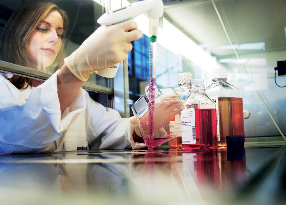

1 HoloMonitor M4 Holographic time-lapse imaging cytometry For competent and powerful discoveries Monitor and quantify living cells in their natural environment with unrivaled temporal resolution Phase Holographic Imaging


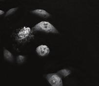
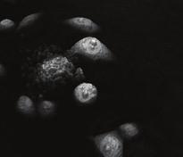
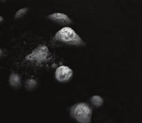
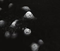
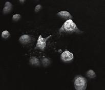
2 Autoimmune diseases Cancer research Stem cell biology Inflammation Gene therapy Toxicological studies Regenerative medicine Applications Unprecedented time-lapse imaging minute by minute and day after day COMPREHENSIVE CELL CULTURE QCA Instantaneously assess your tissue culture integrity Growth rate data for untreated L929 mouse fibroblasts. XY spacial movement plots of murine macrophages M1 phenotype and M2 phenotype. Changes in optical volume over time based on tracking of one cell of interest. Quantitative tissue culture QC metrics within seconds. Cellular growth rate and morphological changes simultaneously. Cellular population statistics comprise many relevant cell-by-cell measurements (size, optical cell volume, thickness, irregularity, etc). CELL MOTILITY Monitor cell movement and morphological changes simultaneously Robust segmentation and tracking of cells of interest. Cell motility speed and cell migration velocity measurements at your fingertips. Non-invasive nature allows subsequent cellular staining for further analysis upon conclusion of holographic imaging. CELL CYCLE KINETICS Analyze cells through rounds of replication with full confidence No perturbation to the natural cell state and function with label-free analysis. Robust measurement of mitosis and cytokinesis based on reliable automated segmentation. Cytometric data with effortless relationship between images and quantitative data. Results comparable to classic cytometric DNA cell cycle studies using fluorescent DNA stains. CELL DEATH Advanced studies with real-time observation of critical events Etoposide-treated DU145 cell line: changes in optical thickness and volume during the death process in one individual cell. Continuous 2D and 3D visual observation of cellular death in second intervals. Large portfolio of quantitative morphological parameters: optical cell volume, thickness, area, irregularity, eccentricity and many more. Single cell tracking and population data analysis. Ideal suitability for studying drug-induced cell death. RARE OR TRANSIENT CELLULAR EVENTS Visualize previously unseen and analyze formerly undetected Unsurpassed temporal resolution with speed of image acquisition of up to 1 image per second. Morphological and quantitative tracking of cells of interest over multiple days. Cell-by-cell population data analysis. L929 mouse fibroblasts: a rare giant budding multinuclear cell.


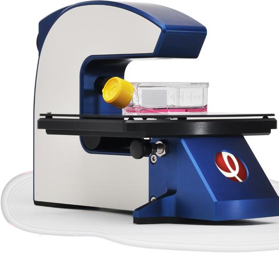
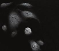
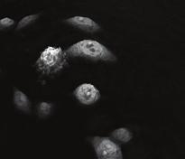
3 HoloMonitor M4 Holographic time-lapse imaging cytometry Quantitative Label-free Long-term Diode laser Reference beam Sample beam Objective Image sensor Hologram HOLOGRAPHIC MICROSCOPY HoloMonitor M4 utilizes the principle of holographic microscopy. A low-power laser beam is split into two, one illuminating the sample and the other providing a reference beam. Laser light passing through the sample is affected by intra-cellular structures causing a phase shift of the illuminating light. Once combined, the two beams create an interference pattern which is recorded by a digital image sensor. The recorded interference pattern the hologram is then processed computationally to produce a holographic image. ROBUST SEGMENTATION The foundation of quantitative analysis is the ability to identify discrete events for quantification: In the line profile of the traditional phase contrast image the background value cannot be accurately determined and a characteristic bright halo around the edge of the cells is present. This type of image does not lend itself for reliable segmentation. HOLOMONITOR SEGMENTATION µm Intensity PHASE CONTRAST DIGITAL AUTOFOCUS The fine focusing is done entirely in software, after recording. The digitally recorded interference pattern is computationally processed to create holographic images over a range of focal distances. From this temporary stack of images, HoloMonitor M4 automatically selects the best in focus image to produce the final holographic image. Alternatively, users may manually select the focal distance to focus on a plane of interest. In contrast, holographic images can be quantified as they reflect the optical thickness of the cell and optical density variations in the specimen. Additionally, holographic images have a background level of zero and the intensity of the events measured as positive values. HoloMonitor methodology enables reliable segmentation seen in the image as yellow cellular boundaries defined by a proprietary software algorithm.


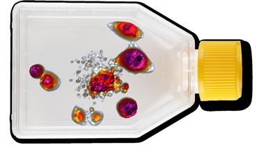
4 An essential and versatile tool for label-free kinetic cell analysis HIGH TEMPORAL RESOLUTION Label-free cell cultures are continuously monitored and analyzed in their natural environment. With the ability to acquire one image per second, both short-term monitoring of transient events in second intervals, and multi-day studies with images captured at user-defined intervals, are possible. HOLOMETRICS Quantitative phase shift measurements are translated by sophisticated software algorithms into morphological parameters optical cell volume, thickness, texture and many more. AREA THICKNESS VOLUME TEXTURE IRREGULARITY 3:48 0:00 LABEL-FREE SAMPLES As samples are analyzed unstained, no sample preparation is required, and most importantly, cellular function is not altered by toxic stains. 3:52 06:08 06:18 06:22 06:26 06:52 5:16 LONG-TERM EVALUATION The incubator-tolerant design makes HoloMonitor M4 especially well-suited for long-term kinetic cellular analysis. The low intensity, single wavelength laser generates no heat and reduces the risk of photodamage to an absolute minimum. 5:32 MOTORIZED STAGE HoloMonitor M4 may optionally be equipped with a motorized stage. The high precision stage allows HoloMonitor M4 to record time-lapse movies at multiple locations, in parallel. Sample locations may be within the same culture or in different in cultures. The stage control software is fully integrated in the HoloMonitor software. After sample locations have been graphically programed, time-lapse movies will be automatically recorded at each location. FLEXIBILITY IN EXPERIMENTAL DESIGN Many different sample vessels can be analyzed from chamber slides to Petri dishes and culture flasks. RELIABLE AND COST-EFFECTIVE OPERATION A new and innovative mechanical design together with intuitive software interface makes HoloMonitor M4 operation simple and reliable.


5 Simple workflow Intuitive user interface Proprietary software HoloStudio RECORD TIME-LAPSE Position the vessel on the mechanical stage and record a time-lapse. The total recording time and time interval between image captures are operator-determined. VIEW IMAGES View images and time-lapse movies in gray scale and color, 2D and 3D. Color variations represent differences in optical thickness. Adjusting threshold allows digital filtering of cells of interest. SEGMENT CELLS Choose one of many available segmentation strategies and fine-tune by adjusting background threshold and cell size. TRACK CELLS Select individual cells to simultaneously track cell movement and changes in cell morphology over time. Individual cell movements are displayed in spatial plots with related quantitative parameters. ANALYZE AND DISPLAY DATA Perform comprehensive automated analysis with options to display quantitative and morphometric features in two dimensional histograms and scattergrams. Data verification is done based on images (example mitotic cells). EXPORT The time-lapse movies can be viewed and effortlessly exported. The acquired images can be easily saved in common image file formats. Additionally, data can be exported as XML, to Excel or easily processed in multiple freeware programs.


6 HoloMonitor gives a totally new dimension to our work TECHNICAL SPECIFICATIONS Sample stage Fixed, manual XY-stage or motorized XYZ-stage Light source External laser unit, 635 nm Sample illumination 635 nm, 0.2 mw/cm 2 Objective 20 Lateral resolution 1 μm Field of view 0.25 mm 2 Working distance mm HoloMonitor offers unique imaging capabilities that greatly enhance our understanding of cell behavior, previously unachievable by other technologies. Ed Luther, Supervisor of Core Imaging and Cytometry Facility, Northeastern University, Boston, USA HoloMonitor gives a totally new dimension to our work. Prof. Stina Oredsson, Department of Biology, Lund University, Sweden Autofocusing range Maximum image rate Image size Travel range Manual XY-stage Motorized XYZ-stage Dimensions (L W H) Weight 1.5 mm 1 image/s pixel mm mm mm 2.5 kg HoloMonitor allows non-invasive monitoring of cancer cells and their response to treatment, without the need for prior cell extraction, staining or exposing cells to harmful light sources. Dr. Maria Falck Miniotis, Researcher, Malmö University, Sweden HoloMonitor M4 allows us to rapidly measure how cell motility is affected by various treatments, without removing the cells from our incubator. Dr. Sung-Kyun Ko, Researcher, Korea Research Institute of Bioscience & Biotechnology, South Korea Requirements Cells Cell culture vessels Computer Incubator Operating temperature Monolayer of adherent T25, 6-well, Petri, IBIDI Windows 7/8 64-bit, 8GB RAM Access port for cabling C 180 mm Operating humidity Max 95% 250 mm 160 mm Regulatory compliance Low voltage directive 2006/95/EC, Electromagnetic compatibility 2004/108/EC EN :2001 EN :2007 For research use only. Not for use in diagnostic procedures. Phase Holographic Imaging PHI AB HoloMonitor and HoloMetrics are registered trademarks of PHI Scheelevägen 22, Lund, Sweden info@phiab.se Phase Holographic Imaging PHI AB All rights reserved
HoloMonitor M4. For powerful discoveries in your incubator
 HoloMonitor M4 For powerful discoveries in your incubator HoloMonitor offers unique imaging capabilities that greatly enhance our understanding of cell behavior, previously unachievable by other technologies
User Manual for HoloStudio M4 2.5 with HoloMonitor M4. Phase Holographic Imaging
 User Manual for HoloStudio M4 2.5 with HoloMonitor M4 Phase Holographic Imaging 1 2 HoloStudio M4 2.5 Software instruction manual 2013 Phase Holographic Imaging AB 3 Contact us: Phase Holographic Imaging
Dynamic Phase-Shifting Microscopy Tracks Living Cells
 from photonics.com: 04/01/2012 http://www.photonics.com/article.aspx?aid=50654 Dynamic Phase-Shifting Microscopy Tracks Living Cells Dr. Katherine Creath, Goldie Goldstein and Mike Zecchino, 4D Technology
1. Editorial. N 9 June Content
 N 9 June 2010 Content 1. Editorial 2. Timelapse: news and updates 3. n vivo rodent imaging setup available in Epalinges 4. 2010 Workshops 5. Spotlight on mage Stitching 1. Editorial We welcome new and
Life Science Instrumentation. New Generation. Light Sheet Fluorescence Microscope. Alph
 Life Science Instrumentation Light Sheet Fluorescence Microscope New Generation Alph Modular Light Sheet Microscope Alpha 3 is a new generation of light sheet fluorescence microscope addressing the needs
IncuCyte ZOOM Scratch Wound Processing Overview
 IncuCyte ZOOM Scratch Wound Processing Overview The IncuCyte ZOOM Scratch Wound assay utilizes the WoundMaker-IncuCyte ZOOM-ImageLock Plate system to analyze both 2D-migration and 3D-invasion in label-free,
IncuCyte ZOOM Scratch Wound Processing Overview
 IncuCyte ZOOM Scratch Wound Processing Overview The IncuCyte ZOOM Scratch Wound assay utilizes the WoundMaker-IncuCyte ZOOM-ImageLock Plate system to analyze both 2D-migration and 3D-invasion in label-free,
Introduction. INSTRUCTION MANUAL CAT XL, 6500-XLCORE, 6500-FL Evos-XL, Evos-XL/Core, Evos-FL
 1 INSTRUCTION MANUAL CAT. 6500-XL, 6500-XLCORE, 6500-FL Evos-XL, Evos-XL/Core, Evos-FL Introduction Experience faster results and easier cell imaging with an EVOS imaging system! An EVOS system is the
Practical work no. 3: Confocal Live Cell Microscopy
 Practical work no. 3: Confocal Live Cell Microscopy Course Instructor: Mikko Liljeström (MIU) 1 Background Confocal microscopy: The main idea behind confocality is that it suppresses the signal outside
Working Simultaneously. The Next Level of TIRF Microscopy. cell^tirf Illuminator Motorized Total Internal Reflection Fluorescence
 cell^tirf Illuminator Motorized Total Internal Reflection Fluorescence Four individually aligned illumination beams for simultaneous multi-color TIRF imaging Working Simultaneously The Next Level of TIRF
Imaging Photometer and Colorimeter
 W E B R I N G Q U A L I T Y T O L I G H T. /XPL&DP Imaging Photometer and Colorimeter Two models available (photometer and colorimetry camera) 1280 x 1000 pixels resolution Measuring range 0.02 to 200,000
LSM 780 Confocal Microscope Standard Operation Protocol
 LSM 780 Confocal Microscope Standard Operation Protocol Basic Operation Turning on the system 1. Sign on log sheet according to Actual start time 2. Check Compressed Air supply for the air table 3. Switch
 07 Setting Place a specimen, and select a fluorescence dye. The FV10i automatically selects the most suitable imaging conditions based on the fluorescence dye selection. Set Image mapping menu Just click
High-sensitivity. optical molecular imaging and high-resolution digital X-ray. In-Vivo Imaging Systems
 High-sensitivity optical molecular imaging and high-resolution digital X-ray In-Vivo Imaging Systems In vivo imaging solutions available in several packages Carestream Molecular Imaging offers a selection
Confocal Laser Scanning Microscopy
 Name of the Core Facility: Confocal Laser Scanning Microscopy CORE Forschungszentrum Immunologie Mainz Welcome to the CSLM Core Facility: The CLSM Core Facility enables working groups to incorporate high
Products - Microarray Scanners - Laser Scanners - InnoScan 900 Series and MAPIX Software
 Products - Microarray Scanners - Laser Scanners - InnoScan 900 Series and MAPIX Software Arrayit offers the world s only next generation microarray scanning technology, with proprietary rotary motion control,
IN Cell Analyzer 2000
 GE Healthcare IN Cell Analyzer 2000 Cell analysis just got easier Impressively enabling for cell analysis Developed with meticulous attention to the needs of the entire high-content imaging workflow,
Fast Laser Raman Microscope RAMAN
 Fast Laser Raman Microscope RAMAN - 11 www.nanophoton.jp Fast Raman Imaging A New Generation of Raman Microscope RAMAN-11 developed by Nanophoton was created by combining confocal laser microscope technology
Technical Benefits of the
 innovation in microvascular assessment Technical Benefits of the Moor Instruments moorflpi-2 moorflpi-2 More Info: Measurement Principle laser speckle contrast analysis Measurement 85nm Laser Wavelength
Parallel Digital Holography Three-Dimensional Image Measurement Technique for Moving Cells
 F e a t u r e A r t i c l e Feature Article Parallel Digital Holography Three-Dimensional Image Measurement Technique for Moving Cells Yasuhiro Awatsuji The author invented and developed a technique capable
ImageXpress Micro XLS Widefield High Content Screening System. Imaging with a vision.
 ImageXpress Micro XLS Widefield High Content Screening System Imaging with a vision www.moleculardevices.com The ImageXpress Micro Widefield High Content Screening System is the ultimate combination of
Locating Molecules Using GSD Technology Project Folders: Organization of Experiment Files...1
 .....................................1 1 Project Folders: Organization of Experiment Files.................................1 2 Steps........................................................................2
Fast Laser Raman Microscope RAMAN
 Fast Laser Raman Microscope RAMAN - 11 www.nanophoton.jp Fast Raman Imaging A New Generation of Raman Microscope RAMAN-11 developed by Nanophoton was created by combining confocal laser microscope technology
IncuCyte ZOOM Fluorescent Processing Overview
 IncuCyte ZOOM Fluorescent Processing Overview The IncuCyte ZOOM offers users the ability to acquire HD phase as well as dual wavelength fluorescent images of living cells producing multiplexed data that
Using Autofocus in NIS-Elements
 Using Autofocus in NIS-Elements Overview This technical note provides an overview of the available autofocus routines in NIS-Elements, and describes the necessary steps for using the autofocus functions.
Cellular Bioengineering Boot Camp. Image Analysis
 Cellular Bioengineering Boot Camp Image Analysis Overview of the Lab Exercises Microscopy and Cellular Imaging The purpose of this laboratory exercise is to develop an understanding of the measurements
Fast, high-contrast imaging of animal development with scanned light sheet based structured-illumination microscopy
 nature methods Fast, high-contrast imaging of animal development with scanned light sheet based structured-illumination microscopy Philipp J Keller, Annette D Schmidt, Anthony Santella, Khaled Khairy,
Colony Imaging with powerful Analysis Software
 TM Imaging with powerful Analysis Software TM Accurate Compact Fast We re not going to interpret your results, but we ll do everything to get you there From image acquisition to data visualisation, straight
Camera Overview. Digital Microscope Cameras for Material Science: Clear Images, Precise Analysis. Digital Cameras for Microscopy
 Digital Cameras for Microscopy Camera Overview For Materials Science Microscopes Digital Microscope Cameras for Material Science: Clear Images, Precise Analysis Passionate about Imaging: Olympus Digital
μscope Microscopy Software
 μscope Microscopy Software Pixelink μscope Essentials (ES) Software is an easy-to-use robust image capture tool optimized for productivity. Pixelink μscope Standard (SE) Software had added features, making
Camera Overview. Digital Microscope Cameras for Material Science: Clear Images, Precise Analysis. Digital Cameras for Microscopy
 Digital Cameras for Microscopy Camera Overview For Materials Science Microscopes Digital Microscope Cameras for Material Science: Clear Images, Precise Analysis Passionate about Imaging: Olympus Digital
The Next Level of TIRF Microscopy. cell^tirf Illuminator Motorized Total Internal Reflection Fluorescence
 cell^tirf Illuminator Motorized Total Internal Reflection Fluorescence Four individually aligned illumination beams for simultaneous multi-color TIRF imaging The Next Level of TIRF Microscopy Mario Faretta,
Quality Performance, Innovative Design
 Dimensions Confocal Laser Scanning Biological Microscope Table size (mm): 1400(W) 800(D) * Table is not available from Olympus. Avoid placing the controller directly on the floor. Dimensions / Weight /
Fastest high definition Raman imaging. Fastest Laser Raman Microscope RAMAN
 Fastest high definition Raman imaging Fastest Laser Raman Microscope RAMAN - 11 www.nanophoton.jp Observation A New Generation in Raman Observation RAMAN-11 developed by Nanophoton was newly created by
OPTIV CLASSIC 321 GL TECHNICAL DATA
 OPTIV CLASSIC 321 GL TECHNICAL DATA TECHNICAL DATA Product description The Optiv Classic 321 GL offers an innovative design for non-contact measurement. The benchtop video-based measuring machine is equipped
AxioCam MRc 5 A World of Digital Possibilities
 Microscopy from Carl Zeiss AxioCam MRc 5 A World of Digital Possibilities More flexibility and more performance in microscope camera technology Impressive Performance A trend setter in digital microscopy,
NPTEL VIDEO COURSE PROTEOMICS PROF. SANJEEVA SRIVASTAVA
 HANDOUT LECTURE-31 MICROARRAY WORK-FLOW: IMAGE SCANNING AND DATA PROCESSING Slide 1: This module contains the summary of the discussion with Mr. Pankaj Khanna, an application specialist from Spinco Biotech,
the image analysis people
 Clemex CMT Computerized Microhardness Testing Clemex CMT (automatic) the image analysis people Modular Solutions Clemex CMT (automatic) Clemex CMT (semi-automatic) Clemex CMT Lite When you need a microhardness
Prime Scientific CMOS Camera Processing Tools for Super-Resolution Microscopy
 Technical Note Prime Scientific CMOS Camera Processing Tools for Super-Resolution Microscopy Prime Scientific CMOS cameras provide the highest levels of sensitivity which make them ideal for low-light
In-Vivo IMAGING SYSTEMS. A complete line of high resolution optical & X-ray systems for pre-clinical imaging
 In-Vivo IMAGING SYSTEMS A complete line of high resolution optical & X-ray systems for pre-clinical imaging In-Vivo Imaging Systems Carestream is a strong, successful, multi-billion dollar, international
capabilities today. Flexibility for tomorrow.
 capabilities today. Flexibility for tomorrow. NEW CellStream benchtop flow cytometry system with Amnis detection technology inside The life science business of Merck KGaA, Darmstadt, Germany operates as
Camera Overview. Digital Microscope Cameras for Material Science: Clear Images, Precise Analysis. Digital Cameras for Microscopy
 Digital Cameras for Microscopy Camera Overview For Materials Science Microscopes Digital Microscope Cameras for Material Science: Clear Images, Precise Analysis Passionate about Imaging: Olympus Digital
Opterra II Multipoint Scanning Confocal Microscope. Innovation with Integrity
 Opterra II Multipoint Scanning Confocal Microscope Enabling 4D Live-Cell Fluorescence Imaging through Speed, Sensitivity, Viability and Simplicity Innovation with Integrity Fluorescence Microscopy The
FLUORESCENCE MICROSCOPY. Matyas Molnar and Dirk Pacholsky
 FLUORESCENCE MICROSCOPY Matyas Molnar and Dirk Pacholsky 1 The human eye perceives app. 400-700 nm; best at around 500 nm (green) Has a general resolution down to150-300 μm (human hair: 40-250 μm) We need
#P Quality Measures for Imaging-based Cellular Assays
 Abstract #P1224 - Quality Measures for Imaging-based Cellular Assays Ilya Ravkin, Vitra Bioscience, Inc. Z-factor and related measures are useful in estimating assay variability in HTS caused by assay
Observing Microorganisms through a Microscope LIGHT MICROSCOPY: This type of microscope uses visible light to observe specimens. Compound Light Micros
 PHARMACEUTICAL MICROBIOLOGY JIGAR SHAH INSTITUTE OF PHARMACY NIRMA UNIVERSITY Observing Microorganisms through a Microscope LIGHT MICROSCOPY: This type of microscope uses visible light to observe specimens.
Studying of Reflected Light Optical Laser Microscope Images Using Image Processing Algorithm
 IRAQI JOURNAL OF APPLIED PHYSICS Fatema H. Rajab Al-Nahrain University, College of Engineering, Department of Laser and Optoelectronic Engineering Studying of Reflected Light Optical Laser Microscope Images
WHITE PAPER FAST PROTEIN INTERACTION BINDING CURVES WITH INO S F-HS CONFOCAL MICROSCOPE
 WHITE PAPER FAST PROTEIN INTERACTION BINDING CURVES WITH INO S F-HS CONFOCAL MICROSCOPE Christian Tardif, Jean-Pierre Bouchard Pascal Gallant, Sebastien Roy, Ozzy Mermut September 2017 Introduction Protein-protein
Megapixel FLIM with bh TCSPC Modules
 Megapixel FLIM with bh TCSPC Modules The New SPCM 64-bit Software Abstract: Becker & Hickl have recently introduced version 9.60 of their SPCM TCSPC data acquisition software. SPCM version 9.60 not only
contents TABLE OF The SECOM platform Applications - sections Applications - whole cells Features Integrated workflow Automated overlay
 S E C O M TABLE OF contents The SECOM platform 4 Applications - sections 5 Applications - whole cells 8 Features 9 Integrated workflow 12 Automated overlay ODEMIS - integrated software Specifications 13
Image Analysis for Fluorescence
 Image Analysis for Fluorescence Terminology Table Image Analysis Macro Colocalization Intensity Dye AFI The extraction of meaningful information from digital images by means of digital image processing
In our previous lecture, we understood the vital parameters to be taken into consideration before data acquisition and scanning.
 Interactomics: Protein Arrays & Label Free Biosensors Professor Sanjeeva Srivastava MOOC NPTEL Course Indian Institute of Technology Bombay Module 7 Lecture No 34 Software for Image scanning and data processing
Ionscope SICM. About Ionscope. Scanning Ion Conductance Microscopy. Ionscope A brand of Openiolabs Limited
 SICM About is a brand of openiolabs Ltd, headquartered in Cambridge UK, is the world-leader in (SICM), a rapidly emerging Scanning Probe Microscopy (SPM) technique which allows nanoscale topographical
Light Microscopy. Upon completion of this lecture, the student should be able to:
 Light Light microscopy is based on the interaction of light and tissue components and can be used to study tissue features. Upon completion of this lecture, the student should be able to: 1- Explain the
High Resolution BSI Scientific CMOS
 CMOS, EMCCD AND CCD CAMERAS FOR LIFE SCIENCES High Resolution BSI Scientific CMOS Prime BSI delivers the perfect balance between high resolution imaging and sensitivity with an optimized pixel design and
Chapter 2 The Study of Microbial Structure: Microscopy and Specimen Preparation
 Chapter 2 The Study of Microbial Structure: Microscopy and Specimen Preparation 1 Lenses and the Bending of Light light is refracted (bent) when passing from one medium to another refractive index a measure
Development of a High-speed Super-resolution Confocal Scanner
 Development of a High-speed Super-resolution Confocal Scanner Takuya Azuma *1 Takayuki Kei *1 Super-resolution microscopy techniques that overcome the spatial resolution limit of conventional light microscopy
OLYMPUS Digital Cameras for Materials Science Applications: Get the Best out of Your Microscope
 Digital Cameras for Microscopy Camera Overview For Materials Science Microscopes OLYMPUS Digital Cameras for Materials Science Applications: Get the Best out of Your Microscope Passionate About Imaging
Cytell Cell Imaging System
 GE Healthcare Life Sciences Data file 29-0866-95 AA Cell analysis and imaging Cytell Cell Imaging System The Cytell Cell Imaging System (Fig 1) combines the functionalities of a digital microscope, an
Camera Overview. Olympus Digital Cameras for Materials Science Applications: For Clear and Precise Image Analysis. Digital Cameras for Microscopy
 Digital Cameras for Microscopy Camera Overview For Materials Science Microscopes Olympus Digital Cameras for Materials Science Applications: For Clear and Precise Image Analysis Passionate about Imaging
Multi-channel imaging cytometry with a single detector
 Multi-channel imaging cytometry with a single detector Sarah Locknar 1, John Barton 1, Mark Entwistle 2, Gary Carver 1 and Robert Johnson 1 1 Omega Optical, Brattleboro, VT 05301 2 Philadelphia Lightwave,
Multifluorescence The Crosstalk Problem and Its Solution
 Multifluorescence The Crosstalk Problem and Its Solution If a specimen is labeled with more than one fluorochrome, each image channel should only show the emission signal of one of them. If, in a specimen
Invisible sophistication. Visible simplicity. CS Welcome to the simplicity of compact panoramic imaging
 Invisible sophistication. Visible simplicity. CS 8100 Welcome to the simplicity of compact panoramic imaging Introducing the CS 8100 The Carestream Dental Factor Humanized technology We keep our technology
Y N C R O S C O P Y A DIVISION OF THE SYNOPTICS GROUP
 S Y N C R O S C O P Y A DIVISION OF THE SYNOPTICS GROUP THE PROBLEM: As a microscopist you often have to work with samples that are difficult to focus. When viewing a 3-D sample using an optical microscope
Nanosurf easyscan 2 FlexAFM
 Nanosurf easyscan 2 FlexAFM Your Versatile AFM System for Materials and Life Science www.nanosurf.com The new Nanosurf easyscan 2 FlexAFM scan head makes measurements in liquid as simple as measuring in
Proudly serving laboratories worldwide since 1979 SPECIFICATIONS
 www.ietltd.com Proudly serving laboratories worldwide since 1979 SPECIFICATIONS Scan RDI Specifications System Components Main analytical console Laser Module CRT Printer Data Manager Motorized stage (option)
DIGITAL-MICROSCOPY CAMERA SOLUTIONS USB 3.0
 DIGITAL-MICROSCOPY CAMERA SOLUTIONS USB 3.0 PixeLINK for Microscopy Applications PixeLINK will work with you to choose and integrate the optimal USB 3.0 camera for your microscopy project. Ideal for use
Zeiss 880 Training Notes Zen 2.3
 Zeiss 880 Training Notes Zen 2.3 1 Turn on the HXP 120V Lamp 2 Turn on Main Power Switch Turn on the Systems PC Switch Turn on the Components Switch. 3 4 5 Turn on the PC and log into your account. Start
Sensor Fusion Enables Comprehensive Analysis of Laser Processing in Additive Manufacturing
 MKS Instruments 1 of 6 Sensor Fusion Enables Comprehensive Analysis of Laser Processing in Additive Manufacturing By Kevin Kirkham, Senior Manager, Product Development, Ophir Sensor: "A device that detects
Exercise questions for Machine vision
 Exercise questions for Machine vision This is a collection of exercise questions. These questions are all examination alike which means that similar questions may appear at the written exam. I ve divided
Nikon Eclipse Ti2-E Widefield/Spinning Disk Confocal Microscope Standard Operation Protocol
 Nikon Eclipse Ti-E Widefield/Spinning Disk Confocal Microscope Standard Operation Protocol Please sign on the log sheet before switching on system. Turn on system Turn on A only if confocal mode or laser
Training Guide for Carl Zeiss LSM 5 LIVE Confocal Microscope
 Training Guide for Carl Zeiss LSM 5 LIVE Confocal Microscope AIM 4.2 Optical Imaging & Vital Microscopy Core Baylor College of Medicine (2017) Power ON Routine 1 2 Verify that main power switches on the
LVEM 25. Low Voltage Electron Microscope Fast Compact Powerful.... your way to electron microscopy
 LVEM 25 Low Voltage Electron Microscope Fast Compact Powerful... your way to electron microscopy INTRODUCING THE LVEM 25 High Contrast & High Resolution Unmatched contrast of biologic and light material
The First True Color Confocal Scanner
 The First True Color Confocal Scanner White color and infrared confocal images: the advantages of white color and confocality together for better fundus images. The infrared to see what our eye is not
Manual. Cell Border Tracker. Jochen Seebach Institut für Anatomie und Vaskuläre Biologie, WWU Münster
 Manual Cell Border Tracker Jochen Seebach Institut für Anatomie und Vaskuläre Biologie, WWU Münster 1 Cell Border Tracker 1. System Requirements The software requires Windows XP operating system or higher
Operation Guide for the Leica SP2 Confocal Microscope Bio-Imaging Facility Hunter College October 2009
 Operation Guide for the Leica SP2 Confocal Microscope Bio-Imaging Facility Hunter College October 2009 Introduction of Fluoresence Confocal Microscopy The first confocal microscope was invented by Princeton
Zeiss 780 Training Notes
 Zeiss 780 Training Notes Turn on Main Switch, System PC and Components Switches 780 Start up sequence Do you need the argon laser (458, 488, 514 nm lines)? Yes Turn on the laser s main power switch and
Stereotopix Research. Precision Pathology. Highthroughput. pathology. powered by newcast. Advantages of Stereotopix : RUO
 Precision Pathology Highthroughput pathology Stereotopix Research powered by newcast RUO Researchers use quantitative microscopy in many ways with the goal of producing high-quality, quantitative results
T H E J O U R N A L O F C E L L B I O L O G Y
 T H E J O U R N A L O F C E L L B I O L O G Y Supplemental material Sikirzhytski et al., http://www.jcb.org/cgi/content/full/jcb.201401090/dc1 Figure S1. Behavior and organization of K-fibers in PtK 2
Diskovery Spinning Disk Guide
 Diskovery Spinning Disk Guide qbi.microscopy@uq.edu.au Getting started The microscope and its peripherals (Fig. 1a) should always be turned on, but if they are not, turn them on in the following way: 1.
LaserBeam ProfilingSolutions. IRLaserBeam Profiler
 LaserBeam ProfilingSolutions IRLaserBeam Profiler TABLE OF CONTENTS PRODUCT DESCRIPTION LASERDEC CL200 TECHNICAL DATA DIMENSIONS LASERDEC CL500 TECHNICAL DATA DIMENSIONS LASERDEC CR200 TECHNICAL DATA DIMENSIONS
Infra Red Interferometers
 Infra Red Interferometers for performance testing of infra-red materials and optical systems Specialist expertise in testing, analysis, design, development and manufacturing for Optical fabrication, Optical
Introduction Approach Work Performed and Results
 Algorithm for Morphological Cancer Detection Carmalyn Lubawy Melissa Skala ECE 533 Fall 2004 Project Introduction Over half of all human cancers occur in stratified squamous epithelia. Approximately one
Product Information Version 1.1. ZEISS Xradia 410 Versa Submicron X-ray Imaging: Bridge the Gap in Lab-based Microscopy
 Product Information Version 1.1 ZEISS Xradia 410 Versa Submicron X-ray Imaging: Bridge the Gap in Lab-based Microscopy A Workhorse Solution for Your 3D Submicron Imaging Xradia 410 Versa bridges the gap
In this talk I will be talking about improving the accuracy of S phase estimation from cytometric data containing DNA content. A new method of interpo
 In this talk I will be talking about improving the accuracy of S phase estimation from cytometric data containing DNA content. A new method of interpolation, parabolic splines (PS), for Probability State
A Rapid Image Acquisition Method for Focus Stacking in Microscopy
 A Rapid Image Acquisition Method for Focus Stacking in Microscopy Douglas Clark* and Brian Brown Paedia LLC, 327 Lake Street, San Francisco, CA 94118 * dclark@paedia.com Introduction Modern image acquisition
Nikon Instruments Europe
 Nikon Instruments Europe Recommendations for N-SIM sample preparation and image reconstruction Dear customer, We hope you find the following guidelines useful in order to get the best performance out of
DU-897 (back illuminated)
") IMAGING Andor s ixon EM + DU-897 back illuminated EMCCD has single photon detection capability without an image intensifier, combined with greater than 90% QE of a back-illuminated sensor. Containing a
IMAGING Andor s ixon EM + DU-897 back illuminated EMCCD has single photon detection capability without an image intensifier, combined with greater than 90% QE of a back-illuminated sensor. Containing a
Automated Imaging Technology to Simplify Your Workflow!
 Automated Imaging Technology to Simplify Your Workflow! BioSpectrum Imaging System Imaging Made Easy for Chemiluminescence Bioluminescence Colorimetric Fluorescence MegaCam 810 Camera OptiChemi 600 Camera
Bringing Answers to the Surface
 3D Bringing Answers to the Surface 1 Expanding the Boundaries of Laser Microscopy Measurements and images you can count on. Every time. LEXT OLS4100 Widely used in quality control, research, and development
Opterra. Multipoint Scanning Confocal Microscope. Innovation with Integrity. Cell-Friendly, High-Speed, High-Resolution Imaging
 Opterra Multipoint Scanning Confocal Microscope Cell-Friendly, High-Speed, High-Resolution Imaging Innovation with Integrity Fluorescence Microscopy Opterra Multipoint Scanning Confocal Microscope Superior
The First True Color Confocal Scanner on the Market
 The First True Color Confocal Scanner on the Market White color and infrared confocal images: the advantages of white color and confocality together for better fundus images. The infrared to see what our
attocfm I for Surface Quality Inspection NANOSCOPY APPLICATION NOTE M01 RELATED PRODUCTS G
 APPLICATION NOTE M01 attocfm I for Surface Quality Inspection Confocal microscopes work by scanning a tiny light spot on a sample and by measuring the scattered light in the illuminated volume. First,
Microvasculature on a chip: study of the Endothelial Surface Layer and the flow structure of Red Blood Cells
 Supplementary Information Microvasculature on a chip: study of the Endothelial Surface Layer and the flow structure of Red Blood Cells Daria Tsvirkun 1,2,5, Alexei Grichine 3,4, Alain Duperray 3,4, Chaouqi
LSM 710 Confocal Microscope Standard Operation Protocol
 LSM 710 Confocal Microscope Standard Operation Protocol Basic Operation Turning on the system 1. Switch on Main power switch 2. Switch on System / PC power button 3. Switch on Components power button 4.
3. are adherent cells (ie. cells in suspension are too far away from the coverslip)
") Before you begin, make sure your sample... 1. is seeded on #1.5 coverglass (thickness = 0.17) 2. is an aqueous solution (ie. fixed samples mounted on a slide will not work - not enough difference in refractive
Before you begin, make sure your sample... 1. is seeded on #1.5 coverglass (thickness = 0.17) 2. is an aqueous solution (ie. fixed samples mounted on a slide will not work - not enough difference in refractive
Imaging Retreat - UMASS Customized real-time confocal and 2-photon imaging
 Imaging Retreat - UMASS 2012 Customized real-time confocal and 2-photon imaging Mike Sanderson Department of Microbiology and Physiological Systems University of Massachusetts Medical School Thanks for
Bi/BE 227 Winter Assignment #3. Adding the third dimension: 3D Confocal Imaging
 Bi/BE 227 Winter 2016 Assignment #3 Adding the third dimension: 3D Confocal Imaging Schedule: Jan 20: Assignment Jan 20-Feb 8: Work on assignment Feb 10: Student PowerPoint presentations. Goals for this
The First True-Color Wide-Field Confocal Scanner
 The First True-Color Wide-Field Confocal Scanner 2 Company Profile CenterVue designs and manufactures highly automated medical devices for the diagnosis and management of ocular pathologies, including
Microscopy from Carl Zeiss
 Microscopy from Carl Zeiss Contents Page Contents... 1 Introduction... 1 Starting the System... 2 Introduction to ZEN Efficient Navigation... 5 Setting up the microscope... 10 Configuring the beam path