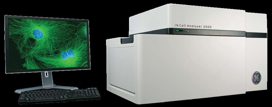
IN Cell Analyzer 2000
|
|
|
- Duane Bradley
- 6 years ago
- Views:
Transcription


1 GE Healthcare IN Cell Analyzer 2000 Cell analysis just got easier
2
3 Impressively enabling for cell analysis Developed with meticulous attention to the needs of the entire high-content imaging workflow, the IN Cell Analyzer 2000 makes even the most challenging high-content assays an everyday reality. From investigative microscopy to automated high-content screening From organelles to cells to tissues to whole organisms From fixed end-point assays to extended live cell studies High-performance hardware, state-of-the-art software, and expert instrument and applications support are harmonized to transform your cellular workflow. System components have been subjected to rigorous accelerated lifetime testing using GE s Six Sigma performance guidelines to ensure reliability. Behind the scenes, our team of dedicated experts is ready and willing to support you. Together with the security of Bio InSite remote support software, this customer focused approach underscores our commitment to help you get the most out of your cellular analysis. IN Cell Analyzer 2000 high-content analysis system provides the performance you need when you need it. High-content analysis provides multiplexed, quantitative data based on automated cell imaging, allowing you to answer complex questions rapidly, in a true biological context

 See ordering information for more details.")
 A wide range of additional objectives (2 to 100 ) including high numerical aperture (NA) options (up to 0.")

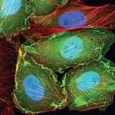
4 Building on experience and excellence Designed with you in mind IN Cell Analyzer 2000 can be configured to your specification through a range of optional modules and accessories, allowing you to build the instrument you need now, or upgrade as your needs evolve. The standard instrument includes: Software autofocus Hardware autofocus Image restoration options Selected polychroics, filters, and objectives Automated objective, correction collar and polychroic changing High performance CCD camera ( pixels) See ordering information for more details. Optional modules and accessories include: Environmental control Liquid handling Transmitted light imaging Temperature control Slide imaging Additional 3-D image restoration options Large chip CCD camera ( pixels) A wide range of additional objectives (2 to 100 ) including high numerical aperture (NA) options (up to 0.95 NA) High-speed without compromising imaging quality IN Cell Analyzer 2000 delivers accurate, high-speed imaging through a combination of proprietary optics and fast hardware and software autofocus for screening applications. A bright light source reduces exposure times to further maximize speed without compromising image quality and cell health. Confocal-like images can be obtained with the rapid image restoration options. Mouse intestine tissue section (Invitrogen) acquired with the 40 /0.6 NA objective and the large chip CCD camera. Alexa Fluor 350 wheat germ agglutinin (blue) stains the mucus of goblet cells. Alexa Fluor 568 phalloidin (red) stains actin concentrated in the brush border. Nuclei are stained with SYTOX Green nucleic acid stain. Bovine pumonary artery cells stained for actin (red), tubulin (green) and nuclei (blue), and imaged with the 40 /0.6 NA objective. Micronuclei induced by etoposide treatment of a CHO-derived cell line. Cells are stained with Hoechst nuclear dye. Inset shows a close-up of three cells that have formed micronuclei. (20 /0.45NA objective).


, or treated with cytochalasin D (c).")
5 Transforming images into data rapidly and easily IN Cell Investigator combines the elements you need for analysis and interpretation into one powerful software package. Developed in collaboration with high-content users, IN Cell Investigator s multifunctional tools enable you to get started quickly, whatever your experience level with image analysis, and whatever your biological field of interest. IN Cell Analysis Modules: Ideal for the new user, a range of fully validated, multifunctional and ready-to-use analysis modules (including the Multitarget Analysis Module for enhanced user-definition). IN Cell Developer Toolbox: Ideal for unique or challenging assays, a flexible tool using interactive guides to create customized protocols for specific biologies without the need to understand programming or scripting. Spotfire DecisionSite : An interactive visual analysis tool to simplify data interpretation. (a) (b) (c) (d) Images transformed into data with IN Cell Investigator. Quantitation of actin microfilaments in a CHO-derived cell line. Images acquired from cells that were either untreated (a), or treated with cytochalasin D (c). Colored bitmaps (b and d) show the results of feature identification following analysis of (a) and (c), respectively, with IN Cell Investigator software. Segmentation outlines are color-coded to identify actin fibers between 1 and 10 μm in length (yellow), and actin fibers > 10 μm in length (red). The results confirm that cytochalasin D treatment induces a significant decrease in actin fiber length. Managing the data mountain IN Cell Miner HCM (high-content management) provides advanced content management for highcontent applications, allowing you to manage huge amounts of complex image, analysis, and experimental data with ease. Building on EMC Documentum, the leading electronic content management software platform, IN Cell Miner HCM provides the performance, reliability, and scalability required to prevent data bottlenecks and enable an efficient workflow. A range of automation options For automation of high-content assays, IN Cell Analyzer 2000 has been specifically designed to be compatible with a wide range of automation options. Robots purchased from us as an option with the instrument are supported by a full integration program. Alternatively, the instrument may be interfaced with your own solutions. For more information on IN Cell Investigator, IN Cell Miner HCM and automation options, visit


 before acquiring")
 (b) (c) (d) High performance large chip CCD camera An optional new large chip CCD camera (2048 2048 pixel array) enables")


6 Imaging feature highlights IN Cell Analyzer 2000 builds on our extensive experience in cellular and subcellular imaging and analysis. As such, the instrument incorporates innovative features to help you gain insights and speed you never thought possible. Preview scan This function allows you to quickly preview a selected area of your sample at any magnification before starting an acquisition run. By choosing the region of interest, you can avoid imaging unwanted areas and significantly increase speed. The region of interest can be any size, from the entire plate or slide, the entire well, or a portion of a well: simply zoom in and out to identify the region of interest and optimize the imaging position by changing the position of the field-of-view. Preview scan of Hoechst stained nuclei in a neurite outgrowth assay at 10 magnification. Preview scan quickly locates the region of interest and reveals the current field-of-view over area devoid of cells (a and b). It is easy to re-position the field of view to avoid artifacts or regions of non-uniform cell density (c and d) before acquiring images. (a) (b) (c) (d) High performance large chip CCD camera An optional new large chip CCD camera ( pixel array) enables superior optical zooming and captures approximately four-times the field-of-view compared with the standard chip camera. Coupled with a long-life widefield illumination source that is two-times brighter than a conventional xenon lamp, the large chip camera acquires high-quality images that are evenly illuminated across the entire fieldof-view. With the large chip camera, you can capture many more cells in a single image, giving more statistically robust results in a single pass, as well as artifact-free detection and quantitation of large structures or rare events. Standard chip camera: CoolSNAP ES2, 1.4 Mp camera captures 23 cells at 40 magnification Large chip camera: CoolSNAP K4, a new 4.2 Mp camera, captures 95 cells at 40 and four times as much information improving productivity and throughput


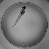
7 Whole-well imaging The combination of a 2 objective and large chip CCD camera enables rapid capture of an entire well with a single image (96-well plate). In cases where cells are unevenly distributed or differentially affected by test treatments, collection of data from every cell in the well provides more robust statistical results. Whole-well imaging also enables the user to image large tissue sections and whole organisms quickly without the need for image stitching. Extended structures, organs, and other gross morphological features can be analyzed in their entirety with no risk of stitching artifacts. Capturing data from the entire population of a well ensures that rare events will not be missed particularly valuable for applications such as lineage tracing, colony counting, and toxicity testing. Stem cell colonies*. Zebra fish embryo*. * Image on right captured with 2 objective and large chip CCD camera. This is only a portion of the entire image shown on the left. Mouse kidney section mounted on glass slide*. Slide imaging IN Cell Analyzer 2000 offers a simple and fast solution to acquisition of images from slides. An intuitive graphical user interface transforms the slide imaging workflow. The Preview scan mode helps you quickly locate the region of interest prior to acquisition at a higher magnification. Smart software autofocus significantly shortens run times and overlapping images can be stitched easily using IN Cell Investigator software. Slide imaging for high-content analysis is truly enabled. The slide imaging workflow. Mouse intestine imaged at 4 following a rapid Preview scan
 and axial (Z) dimensions.")
, co-stained with Hoechst 33342 nuclear dye (blue) and Texas Red conjugated phalloidin (red) following treatment")
 works to recover the original information, making maximum use of")

8 Imaging modes IN Cell Analyzer 2000 includes a choice of six imaging modes to ensure you get the most quantitatively accurate images possible from the system for your sample type. These image restoration options are capable of removing noise, increasing contrast, and improving resolution in both the lateral (X-Y) and axial (Z) dimensions. Image acquired without deconvolution Image acquired with deconvolution Image restoration improves resolution and contrast. Images of the AKT1-EGFP cell line stably expressing AKT1 fused upstream of EGFP (green), co-stained with Hoechst nuclear dye (blue) and Texas Red conjugated phalloidin (red) following treatment of the cells with IGF-1. Images acquired with the 40 /0.60 NA objective and the standard chip CCD camera. Unlike confocal approaches, which discard light emanating from above and below the focal plane, image restoration (deconvolution) works to recover the original information, making maximum use of signal from dim samples. Image restoration is based on a realistic model of the lens system, and therefore provides more accurate results than image enhancement techniques like nearest neighbor de-blurring and sharpening provided by commercial image editing packages. In addition, Z-axis projection provides a virtually extended depth-of-field to increase lateral resolution without missing features that fall above or below the focal plane. This is particularly useful when working with high magnification, high numerical aperture lenses, which provide very thin optical sections. Transmitted light imaging Transmitted light imaging allows label-free monitoring of cell health, movement, morphology, and growth, and can be particularly beneficial for extended time-course studies where fluorescent markers could have a toxic effect. Whether you need images for analysis, or simply want to include them in a Mouse intestine tissue section. Fluorescent (left- hand side) and transmitted light (right-hand side) images of the same field-of-view were acquired with a 40 /0.9 NA objective. publication, the IN Cell Analyzer 2000 simplifies the process, offering a range of modes to ensure you have the flexibility to get the highest quality images possible from your samples. Choose from: Bright-field Differential interference contrast (DIC) Phase-contrast The high quality of transmitted light images, combined with advanced image analysis tools for segmentation, make label-free cell quantitation a reality. For added information and convenience, transmitted light images can also be analyzed in combination with images acquired in fluorescent channels in the standard IN Cell Investigator software.
9 One instrument to meet all your needs The designed-in features and functions ensure IN Cell Analyzer 2000 provides all the performance you need for your screening and research needs. Screening applications Key function System feature Application examples Speed Sensitivity and resolution Reliability Large chip CCD camera Bright optics with powerful light source Fast hardware autofocus Fast online sufficient cell count function Compatible with 384- and 1536-well plates Rapid slide imaging Bright optics with powerful light source High-performance CCD camera Wide range of objectives to suit sensitivity requirements of varied applications (2 to 100 with high NA options) Rapid image restoration options higher contrast and improved resolution even with dim samples Z-axis projection and image restoration combine to provide virtually extended depth of field for high magnifications Robust design and construction using Six Sigma principles for excellent build quality Stage accuracy and repeatability Rapid hardware and software autofocus ensure desired plane of interest is always in focus Online sufficient cell counting to rapidly secure statistically reliable data collection Large chip CCD camera maximizes data collection for statistically robust results Compound screening Predictive toxicology RNAi screening Lead optimization Slide-based arrays (eg. tissue microarrays, sirnas) Phenotypic profiling in screens requiring resolution of subcellular features Rapid detection of low abundance probes and weak fluorescent sensors Micronuclei screening Demanding screening applications with long run-times Shared facilities meeting the demands of multiple users or screening programs
10 Research applications Key function System feature Application examples Resolution and sensitivity High NA/high magnification objectives (eg. 100 /0.9 NA) provide thin optical section and maximum resolution in lateral and axial dimensions Bright optics with powerful light source to detect low abundance probes and weak fluorescent sensors Rapid image restoration options higher contrast and improved resolution even with dim samples Z-axis projection and image restoration combine to provide virtually extended depth of field for high magnifications Protein localization Functional studies Antibody characterization Neurite outgrowth and neuronal function Target identification Phenotypic characterization of test compounds Live-cell assays Rare event analysis Variable temperature control (up to 42 C) Environmental control module Three transmitted light modes Accurate X-Y staging better cell tracking Hardware optimized for optical performance reduced exposure times Cell tracking software Fast hardware autofocus Whole-well imaging using a large chip CCD camera and 2 objective Preview scan rapid location of region of interest Image overlap for stitching Compatible with all well densities from uni well to 1536-well plates Insect cells Heat-shock studies Time-course analysis Temperature-sensitive mutations Cell migration Cell lineage/cycle studies Stem cells Kinetic studies Predictive toxicology Stem cell assays Spheroids Colony assays Small organisms Tissue studies Lineage analysis 3-D analysis Accurate Z-staging Quantitatively accurate 3-D stacks with confocal-like image quality Image restoration options for maximum resolution in lateral and axial dimensions Spheroids Tissue sections Small organisms (eg. zebrafish, C. elegans, Xenopous oocytes) Stacks suitable for 3-D volume rendering and reconstruction of organelles, tissues etc Slide imaging Intuitive graphical user interface transforms workflow Preview scan mode rapid location of region of interest Smart software autofocus algorithm for accuracy and exceptional speed Automated objective changing Image stitching Investigative microscopy Small organisms Cell arrays Tissue arrays
11 Ordering information Instruments Order Code IN Cell Analyzer 2000, standard chip CCD camera ( square pixels, 6.4 µm 6.4 µm pixel size) Includes: hardware autofocus, software autofocus, NA objective, NA objective with automated spherical aberration collar (ASAC) correction, standard polychroics, filter set, 2D deconvolution software, computer, monitor, keyboard, and mouse IN Cell Analyzer 2000, large chip CCD camera ( square pixels, 7.4 µm 7.4 µm pixel size) Includes: hardware autofocus, software autofocus, NA objective, NA objective with automated spherical aberration collar (ASAC) correction, standard polychroics, filter set, 2D deconvolution software, computer, monitor, keyboard, and mouse Modules and accessories Order Code IN Cell Analyzer NA Objective Lens IN Cell Analyzer NA Objective Lens IN Cell Analyzer NA Objective Lens IN Cell Analyzer 2000 ASAC NA Lens IN Cell Analyzer 2000 ASAC NA Lens IN Cell Analyzer 2000 ASAC NA Lens IN Cell Analyzer 2000 ASAC NA Lens IN Cell Analyzer 2000 ASAC NA Lens IN Cell Analyzer D Deconvolution Software IN Cell Analyzer 2000 Transmitted Light Module IN Cell Analyzer 2000 Slide Handling Module (2) IN Cell Analyzer 2000 Live Cell Package A Includes: Temperature Control and Environmental Control Modules IN Cell Analyzer 2000 Live Cell Package B Includes: Temperature Control and Liquid Handling Modules IN Cell Analyzer 2000 Live Cell Package C Includes: Temperature Control, Liquid Handling, and Environmental Control Modules IN Cell Investigator, single seat license IN Cell Miner HCM, academic use includes 3 client and 1 server licenses IN Cell Miner HCM, commercial use includes 5 client and 1 server licenses A polychroic that enables imaging of common fluorescent proteins (including CFP and YFP) individually or as a FRET pair is also available. Please inquire for details
12 Impressively enabling for cell analysis let us prove it to you IN Cell Analyzer 2000 delivers the high-performance hardware, state of-the-art software, and expert support you need to transform your cellular workflow. But don t just take our word for it contact us now to learn more about just how easy your cell analysis could be. For local office contact information, visit: GE Healthcare Bio-Sciences Corp 800 Centennial Avenue P.O. Box 1327 Piscataway, NJ USA GE, imagination at work, and GE monogram are trademarks of General Electric Company. Bio Insite is a trademark of GE Healthcare companies. All third party trademarks are the property of their respective owners General Electric Company All rights reserved. First published April 2009 GE Healthcare Bio-Sciences AB, Björkgatan 30, Uppsala, Sweden GE Healthcare UK Ltd, Amersham Place, Little Chalfont, Buckinghamshire, HP7 9NA, UK GE Healthcare Europe GmbH, Munzinger Strasse 5, D Freiburg, Germany GE Healthcare Bio-Sciences KK, Sanken Bldg , Hyakunincho, Shinjuku-ku, Tokyo , Japan AA 04/2009
Cytell Cell Imaging System
 GE Healthcare Life Sciences Data file 29-0866-95 AA Cell analysis and imaging Cytell Cell Imaging System The Cytell Cell Imaging System (Fig 1) combines the functionalities of a digital microscope, an
IN Cell Analyzer 6500HS
 GE Healthcare IN Cell Analyzer 6500HS IN Cell Analyzer 6500HS is a fully-automated confocal cell imaging system from GE Healthcare. Building on the capabilities of earlier IN Cell Analyzer systems, it
ImageXpress Micro XLS Widefield High Content Screening System. Imaging with a vision.
 ImageXpress Micro XLS Widefield High Content Screening System Imaging with a vision www.moleculardevices.com The ImageXpress Micro Widefield High Content Screening System is the ultimate combination of
New Amersham Hyperfilm
 GE Healthcare The clear choice for reliable results With Comparative Data New Amersham Hyperfilm Are you looking for the right film with the clearest results? As a scientist with a passion for research,
Light Microscopy. Upon completion of this lecture, the student should be able to:
 Light Light microscopy is based on the interaction of light and tissue components and can be used to study tissue features. Upon completion of this lecture, the student should be able to: 1- Explain the
HoloMonitor M4. For powerful discoveries in your incubator
 HoloMonitor M4 For powerful discoveries in your incubator HoloMonitor offers unique imaging capabilities that greatly enhance our understanding of cell behavior, previously unachievable by other technologies
Desalting using ÄKTA start
 GE Healthcare Life Sciences Desalting using ÄKTA start Training cue card This protocol will help you understand the practical principles of desalting chromatography by taking you step-by-step through the
Opterra II Multipoint Scanning Confocal Microscope. Innovation with Integrity
 Opterra II Multipoint Scanning Confocal Microscope Enabling 4D Live-Cell Fluorescence Imaging through Speed, Sensitivity, Viability and Simplicity Innovation with Integrity Fluorescence Microscopy The
HoloMonitor. Phase. For competent and powerful discoveries. Holographic time-lapse imaging cytometry
 HoloMonitor M4 Holographic time-lapse imaging cytometry For competent and powerful discoveries Monitor and quantify living cells in their natural environment with unrivaled temporal resolution Phase Holographic
ANSWER KEY Lab 2 (IGB): Bright Field and Fluorescence Optical Microscopy and Sectioning
: Bright Field and Fluorescence Optical Microscopy and Sectioning") Phys598BP Spring 2016 University of Illinois at Urbana-Champaign ANSWER KEY Lab 2 (IGB): Bright Field and Fluorescence Optical Microscopy and Sectioning Location: IGB Core Microscopy Facility Microscope:
Phys598BP Spring 2016 University of Illinois at Urbana-Champaign ANSWER KEY Lab 2 (IGB): Bright Field and Fluorescence Optical Microscopy and Sectioning Location: IGB Core Microscopy Facility Microscope:
Introduction. INSTRUCTION MANUAL CAT XL, 6500-XLCORE, 6500-FL Evos-XL, Evos-XL/Core, Evos-FL
 1 INSTRUCTION MANUAL CAT. 6500-XL, 6500-XLCORE, 6500-FL Evos-XL, Evos-XL/Core, Evos-FL Introduction Experience faster results and easier cell imaging with an EVOS imaging system! An EVOS system is the
Multifluorescence The Crosstalk Problem and Its Solution
 Multifluorescence The Crosstalk Problem and Its Solution If a specimen is labeled with more than one fluorochrome, each image channel should only show the emission signal of one of them. If, in a specimen
Automated Cellular Imaging and Analysis System
 Automated Cellular Imaging and Analysis System CELLULAR IMAGING AND ANALYSIS FOR SCREENING AUTOMATED ACQUISITION AUTOMATED ANALYSIS HIGH RESOLUTION The ImageXpress TM 5000A automated cellular imaging and
contents TABLE OF The SECOM platform Applications - sections Applications - whole cells Features Integrated workflow Automated overlay
 S E C O M TABLE OF contents The SECOM platform 4 Applications - sections 5 Applications - whole cells 8 Features 9 Integrated workflow 12 Automated overlay ODEMIS - integrated software Specifications 13
Confocal Microscope. Confocal Microscope C2
 Confocal Microscope Confocal Microscope C2 Confocal Microscope An essential microscopy laboratory instrument The C2 confocal microscope system comprises a new generation of Nikon confocal instruments designed
Digital Camera Technologies for Scientific Bio-Imaging. Part 2: Sampling and Signal
 Digital Camera Technologies for Scientific Bio-Imaging. Part 2: Sampling and Signal Yashvinder Sabharwal, 1 James Joubert 2 and Deepak Sharma 2 1. Solexis Advisors LLC, Austin, TX, USA 2. Photometrics
High-sensitivity. optical molecular imaging and high-resolution digital X-ray. In-Vivo Imaging Systems
 High-sensitivity optical molecular imaging and high-resolution digital X-ray In-Vivo Imaging Systems In vivo imaging solutions available in several packages Carestream Molecular Imaging offers a selection
Nature Methods: doi: /nmeth Supplementary Figure 1. Schematic of 2P-ISIM AO optical setup.
 Supplementary Figure 1 Schematic of 2P-ISIM AO optical setup. Excitation from a femtosecond laser is passed through intensity control and shuttering optics (1/2 λ wave plate, polarizing beam splitting
In-Vivo IMAGING SYSTEMS. A complete line of high resolution optical & X-ray systems for pre-clinical imaging
 In-Vivo IMAGING SYSTEMS A complete line of high resolution optical & X-ray systems for pre-clinical imaging In-Vivo Imaging Systems Carestream is a strong, successful, multi-billion dollar, international
Very short introduction to light microscopy and digital imaging
 Very short introduction to light microscopy and digital imaging Hernan G. Garcia August 1, 2005 1 Light Microscopy Basics In this section we will briefly describe the basic principles of operation and
Last updated: May 2014 Y.DeGraaf
 FLINDERS MICROSCOPY BIOMEDICAL SERVICES AVAILABLE MICROSCOPES AND SPECIFICATIONS & INFORMATION REGARDING TRAINING FOR NEW USERS Last updated: May 2014 Y.DeGraaf If you have new staff or students (Honours/Masters
Supplementary Information. Stochastic Optical Reconstruction Microscopy Imaging of Microtubule Arrays in Intact Arabidopsis thaliana Seedling Roots
 Supplementary Information Stochastic Optical Reconstruction Microscopy Imaging of Microtubule Arrays in Intact Arabidopsis thaliana Seedling Roots Bin Dong 1,, Xiaochen Yang 2,, Shaobin Zhu 1, Diane C.
Why and How? Daniel Gitler Dept. of Physiology Ben-Gurion University of the Negev. Microscopy course, Michmoret Dec 2005
 Why and How? Daniel Gitler Dept. of Physiology Ben-Gurion University of the Negev Why use confocal microscopy? Principles of the laser scanning confocal microscope. Image resolution. Manipulating the
Fast, high-contrast imaging of animal development with scanned light sheet based structured-illumination microscopy
 nature methods Fast, high-contrast imaging of animal development with scanned light sheet based structured-illumination microscopy Philipp J Keller, Annette D Schmidt, Anthony Santella, Khaled Khairy,
Leica_Dye_Finder :53 Uhr Seite 6 Dye Finder LAS AF
 Dye Finder LAS AF Dye Finder Multicolor live cell fluorescence microscopy is limited by the availability of spectrally separable fluorescent dyes. Fluorescent dyes (or spectral GFP variants) with incongruent
Camera Overview. Digital Microscope Cameras for Material Science: Clear Images, Precise Analysis. Digital Cameras for Microscopy
 Digital Cameras for Microscopy Camera Overview For Materials Science Microscopes Digital Microscope Cameras for Material Science: Clear Images, Precise Analysis Passionate about Imaging: Olympus Digital
DeltaVision OMX SR. DeltaVision OMX SR microscope. gelifesciences.com
 Data file 29-1467-19 AA Super-resolution microscopy DeltaVision OMX SR The DeltaVision OMX SR system is a compact multimode imaging platform that delivers super-resolution images of live cells. Incorporating
Camera Overview. Digital Microscope Cameras for Material Science: Clear Images, Precise Analysis. Digital Cameras for Microscopy
 Digital Cameras for Microscopy Camera Overview For Materials Science Microscopes Digital Microscope Cameras for Material Science: Clear Images, Precise Analysis Passionate about Imaging: Olympus Digital
Opterra. Multipoint Scanning Confocal Microscope. Innovation with Integrity. Cell-Friendly, High-Speed, High-Resolution Imaging
 Opterra Multipoint Scanning Confocal Microscope Cell-Friendly, High-Speed, High-Resolution Imaging Innovation with Integrity Fluorescence Microscopy Opterra Multipoint Scanning Confocal Microscope Superior
Systematic Workflow via Intuitive GUI. Easy operation accomplishes your goals faster than ever.
 Systematic Workflow via Intuitive GUI Easy operation accomplishes your goals faster than ever. 16 With the LEXT OLS4100, observation or measurement begins immediately once the sample is placed on the stage.
Camera Overview. Digital Microscope Cameras for Material Science: Clear Images, Precise Analysis. Digital Cameras for Microscopy
 Digital Cameras for Microscopy Camera Overview For Materials Science Microscopes Digital Microscope Cameras for Material Science: Clear Images, Precise Analysis Passionate about Imaging: Olympus Digital
Dynamic Phase-Shifting Microscopy Tracks Living Cells
 from photonics.com: 04/01/2012 http://www.photonics.com/article.aspx?aid=50654 Dynamic Phase-Shifting Microscopy Tracks Living Cells Dr. Katherine Creath, Goldie Goldstein and Mike Zecchino, 4D Technology
3D light microscopy techniques
 3D light microscopy techniques The image of a point is a 3D feature In-focus image Out-of-focus image The image of a point is not a point Point Spread Function (PSF) 1D imaging 2D imaging 3D imaging Resolution
Nature Neuroscience: doi: /nn Supplementary Figure 1. Optimized Bessel foci for in vivo volume imaging.
 Supplementary Figure 1 Optimized Bessel foci for in vivo volume imaging. (a) Images taken by scanning Bessel foci of various NAs, lateral and axial FWHMs: (Left panels) in vivo volume images of YFP + neurites
Light microscopy BMB 173, Lecture 14, Feb. 21, 2018
 Light microscopy The Structural Biology Continuum Next two lectures: Light microscopy Many slides taken from Scott Fraser, Murphy s Fundamentals of light microscopy, Alberts Molecular Biology of the Cell,
Advanced Optical Microscopy lecture. 03. December 2012 Kai Wicker
 Advanced Optical Microscopy lecture 03. December 2012 Kai Wicker Today: Optical transfer functions (OTF) and point spread functions (PSF) in incoherent imaging. 1. Quick revision: the incoherent wide-field
Nikon Instruments Europe
 Nikon Instruments Europe Recommendations for N-SIM sample preparation and image reconstruction Dear customer, We hope you find the following guidelines useful in order to get the best performance out of
v tome x m microfocus CT
 GE Inspection Technologies v tome x m microfocus CT Uniting premium 3D metrology and inspection with quality and speed. gemeasurement.com/ct x plore precision CT line Inspect with precision, power, and
Low Voltage Electron Microscope
 LVEM 25 Low Voltage Electron Microscope fast compact powerful Delong America FAST, COMPACT AND POWERFUL The LVEM 25 offers a high-contrast, high-throughput, and compact solution with nanometer resolutions.
LVEM 25. Low Voltage Electron Mictoscope. fast compact powerful
 LVEM 25 Low Voltage Electron Mictoscope fast compact powerful FAST, COMPACT AND POWERFUL The LVEM 25 offers a high-contrast, high-throughput, and compact solution with nanometer resolutions. All the benefits
IncuCyte ZOOM Fluorescent Processing Overview
 IncuCyte ZOOM Fluorescent Processing Overview The IncuCyte ZOOM offers users the ability to acquire HD phase as well as dual wavelength fluorescent images of living cells producing multiplexed data that
CLEMEX intelligent microscopy
 CLEMEX intelligent microscopy Vision PE 5.0 Advanced Image Analysis Experience in Image Analysis Research and Quality Control Solutions With Vision PE, Clemex provides a powerful image analysis solution
microscopy A great online resource Molecular Expressions, a Microscope Primer Partha Roy
 Fundamentals of optical microscopy A great online resource Molecular Expressions, a Microscope Primer http://micro.magnet.fsu.edu/primer/index.html Partha Roy 1 Why microscopy Topics Functions of a microscope
INTRODUCTION TO OPTICAL MICROSCOPY
 Experimental Biophysics TEK265, FYST23, TNF060, FAF010F Lab Exercise Supervisor: Karl Adolfsson Written by Peter Jönsson and Jason Beech Updated by Henrik Persson, Karl Adolfsson and Zhen Li karl.adolfsson@ftf.lth.se
Bringing Answers to the Surface
 3D Bringing Answers to the Surface 1 Expanding the Boundaries of Laser Microscopy Measurements and images you can count on. Every time. LEXT OLS4100 Widely used in quality control, research, and development
ScanArray Overview. Principle of Operation. Instrument Components
 ScanArray Overview The GSI Lumonics ScanArrayÒ Microarray Analysis System is a scanning laser confocal fluorescence microscope that is used to determine the fluorescence intensity of a two-dimensional
Life Science Instrumentation. New Generation. Light Sheet Fluorescence Microscope. Alph
 Life Science Instrumentation Light Sheet Fluorescence Microscope New Generation Alph Modular Light Sheet Microscope Alpha 3 is a new generation of light sheet fluorescence microscope addressing the needs
WaveMaster IOL. Fast and accurate intraocular lens tester
 WaveMaster IOL Fast and accurate intraocular lens tester INTRAOCULAR LENS TESTER WaveMaster IOL Fast and accurate intraocular lens tester WaveMaster IOL is a new instrument providing real time analysis
The Flexibility of Choice
 Digital Microscope Cameras Camera Overview Color, B&W and Universal Release Information 08.08 The Flexibility of Choice 2 THE FLEXIBILITY OF CHOICE Your project needs the right camera It is always preferable
FluorChem M MultiFluor System
 FluorChem M MultiFluor System Advancing Effortless Multiplex Western Blot Imaging Multiplex Western Analysis FluorChem M Imaging System FluorChem M sets a new standard for quantitative multiplex Western
Seishi IKAMI* Takashi KOBAYASHI** Yasutake TANAKA* and Akira YAMAGUCHI* Abstract. 2. System configuration. 1. Introduction
 Development of a Next-generation CCD Imager for Life Sciences Research Seishi IKAMI* Takashi KOBAYASHI** Yasutake TANAKA* and Akira YAMAGUCHI* Abstract We have developed a next-generation CCD-based imager
VivaTome. Discover the Dynamics of Life. The Entry-level System that Captures Dynamic Processes with Outstanding Image Quality.
 Microscopy from Carl Zeiss VivaTome Discover the Dynamics of Life The Entry-level System that Captures Dynamic Processes with Outstanding Image Quality. Innovative Technology Captures Dynamic Processes
Illumination Correction tutorial
 Illumination Correction tutorial I. Introduction The Correct Illumination Calculate and Correct Illumination Apply modules are intended to compensate for the non uniformities in illumination often present
A 4 Megapixel camera with 6.5μm pixels, Prime BSI captures highly. event goes undetected.
 PRODUCT DATASHEET Prime BSI SCIENTIFIC CMOS CAMERA Can a camera single-handedly differentiate your product against competitors? With the Prime BSI, the answer is a resounding yes. Instrument builders no
BD LSRFortessa X-20. Special Order Product. Designed for limited space and boundless potential
 BD LSRFortessa X-2 Special Order Product Designed for limited space and boundless potential Next generation high performance cell analyzer The BD LSRFortessa X-2 cell analyzer delivers high performance,
Media Cybernetics White Paper Spherical Aberration
 Media Cybernetics White Paper Spherical Aberration Brian Matsumoto, University of California, Santa Barbara Introduction Digital photomicrographers assume that lens aberrations are corrected by the microscope
Confocal Microscope. Confocal Microscope C2
 Confocal Microscope Confocal Microscope C2 Confocal Microscope An essential microscopy laboratory insturument The C2 confocal microscope system comprises a new generation of Nikon confocal instruments
Y N C R O S C O P Y A DIVISION OF THE SYNOPTICS GROUP
 S Y N C R O S C O P Y A DIVISION OF THE SYNOPTICS GROUP THE PROBLEM: As a microscopist you often have to work with samples that are difficult to focus. When viewing a 3-D sample using an optical microscope
MECOS-C2 microscopy systems
 MECOS-C2 microscopy systems Microscopy systems of the MECOS-C2 family production LLC "Medical computer Systems (MECOS)" belong to a class of scanning microscopes-analyzers and are intended for: Increase
MAKE SURE YOUR SLIDES ARE CLEAN (TOP & BOTTOM) BEFORE LOADING DO NOT LOAD SLIDES DURING SOFTWARE INITIALIZATION
 BEFORE LOADING DO NOT LOAD SLIDES DURING SOFTWARE INITIALIZATION") Olympus VS120-L100 Slide Scanner Standard Operating Procedure Startup 1) Red power bar switch (behind monitor) 2) Computer 3) Login: UserVS120 account (no password) 4) Double click: WAIT FOR INITIALIZATION
Olympus VS120-L100 Slide Scanner Standard Operating Procedure Startup 1) Red power bar switch (behind monitor) 2) Computer 3) Login: UserVS120 account (no password) 4) Double click: WAIT FOR INITIALIZATION
GALILEO TMA CK 4500 HTS Tissue Microarray Platform
 GALILEO TMA CK 4500 HTS Tissue Microarray Platform Tissue Microarray (TMA) A Block Of Samples From Hundreds Of Blocks (S. M. Hewitt, M.D., Ph.D., Tissue Array Research Program, LP, CCR, NCI, NIH) TMA technology
Confocal Laser Scanning Microscopy
 Name of the Core Facility: Confocal Laser Scanning Microscopy CORE Forschungszentrum Immunologie Mainz Welcome to the CSLM Core Facility: The CLSM Core Facility enables working groups to incorporate high
Products - Microarray Scanners - Laser Scanners - InnoScan 900 Series and MAPIX Software
 Products - Microarray Scanners - Laser Scanners - InnoScan 900 Series and MAPIX Software Arrayit offers the world s only next generation microarray scanning technology, with proprietary rotary motion control,
Low Voltage Electron Microscope
 LVEM5 Low Voltage Electron Microscope Nanoscale from your benchtop LVEM5 Delong America DELONG INSTRUMENTS COMPACT BUT POWERFUL The LVEM5 is designed to excel across a broad range of applications in material
Spectral Imaging with the Opterra Multipoint Scanning Confocal
 Spectral Imaging with the Opterra Multipoint Scanning Confocal Outline Opterra design overview Scan Modes Light Path Spectral Imaging with Opterra Drosophila larva heart. Opterra Design Overview Supravideo
Point Spread Function. Confocal Laser Scanning Microscopy. Confocal Aperture. Optical aberrations. Alternative Scanning Microscopy
 Bi177 Lecture 5 Adding the Third Dimension Wide-field Imaging Point Spread Function Deconvolution Confocal Laser Scanning Microscopy Confocal Aperture Optical aberrations Alternative Scanning Microscopy
ADVANCED METHODS FOR CONFOCAL MICROSCOPY II. Jean-Yves Chatton Sept. 2006
 ADVANCED METHODS FOR CONFOCAL MICROSCOPY II Jean-Yves Chatton Sept. 2006 Workshop outline Confocal microscopy of living cells and tissues X-Z scanning Time series Bleach: FRAP, photoactivation Emission
Nikon. King s College London. Imaging Centre. N-SIM guide NIKON IMAGING KING S COLLEGE LONDON
 N-SIM guide NIKON IMAGING CENTRE @ KING S COLLEGE LONDON Starting-up / Shut-down The NSIM hardware is calibrated after system warm-up occurs. It is recommended that you turn-on the system for at least
NPTEL VIDEO COURSE PROTEOMICS PROF. SANJEEVA SRIVASTAVA
 LECTURE-31 MICROARRAY WORK-FLOW: IMAGE SACNNING AND DATA PROCESSING TRANSCRIPT Welcome to the proteomics course. In today s lecture we will talk about microarray work-flow the image scanning and processing.
WaveMaster IOL. Fast and Accurate Intraocular Lens Tester
 WaveMaster IOL Fast and Accurate Intraocular Lens Tester INTRAOCULAR LENS TESTER WaveMaster IOL Fast and accurate intraocular lens tester WaveMaster IOL is an instrument providing real time analysis of
Super High Vertical Resolution Non-Contact 3D Surface Profiler BW-S500/BW-D500 Series
 Super High Vertical Resolution Non-Contact 3D Surface Profiler BW-S500/BW-D500 Series Nikon's proprietary scanning-type optical interference measurement technology achieves 1pm* height resolution. * Height
Improving the Collection Efficiency of Raman Scattering
 PERFORMANCE Unparalleled signal-to-noise ratio with diffraction-limited spectral and imaging resolution Deep-cooled CCD with excelon sensor technology Aberration-free optical design for uniform high resolution
Observing Microorganisms through a Microscope LIGHT MICROSCOPY: This type of microscope uses visible light to observe specimens. Compound Light Micros
 PHARMACEUTICAL MICROBIOLOGY JIGAR SHAH INSTITUTE OF PHARMACY NIRMA UNIVERSITY Observing Microorganisms through a Microscope LIGHT MICROSCOPY: This type of microscope uses visible light to observe specimens.
Point Spread Function Estimation Tool, Alpha Version. A Plugin for ImageJ
 Tutorial Point Spread Function Estimation Tool, Alpha Version A Plugin for ImageJ Benedikt Baumgartner Jo Helmuth jo.helmuth@inf.ethz.ch MOSAIC Lab, ETH Zurich www.mosaic.ethz.ch This tutorial explains
Aberrations and adaptive optics for biomedical microscopes
 Aberrations and adaptive optics for biomedical microscopes Martin Booth Department of Engineering Science And Centre for Neural Circuits and Behaviour University of Oxford Outline Rays, wave fronts and
High resolution extended depth of field microscopy using wavefront coding
 High resolution extended depth of field microscopy using wavefront coding Matthew R. Arnison *, Peter Török #, Colin J. R. Sheppard *, W. T. Cathey +, Edward R. Dowski, Jr. +, Carol J. Cogswell *+ * Physical
Super Resolution Microscope N-SIM E. Super Resolution Microscope
 Super Resolution Microscope N-SIM E Super Resolution Microscope Explore Nano world with Nikon N-SIM E is a streamlined, affordable superresolution system that provides double the resolution of conventional
IncuCyte ZOOM Scratch Wound Processing Overview
 IncuCyte ZOOM Scratch Wound Processing Overview The IncuCyte ZOOM Scratch Wound assay utilizes the WoundMaker-IncuCyte ZOOM-ImageLock Plate system to analyze both 2D-migration and 3D-invasion in label-free,
Microscopy Techniques that make it easy to see things this small.
 Microscopy Techniques that make it easy to see things this small. What is a Microscope? An instrument for viewing objects that are too small to be seen easily by the naked eye. Dutch spectacle-makers Hans
High Resolution BSI Scientific CMOS
 CMOS, EMCCD AND CCD CAMERAS FOR LIFE SCIENCES High Resolution BSI Scientific CMOS Prime BSI delivers the perfect balance between high resolution imaging and sensitivity with an optimized pixel design and
Resolution. Diffraction from apertures limits resolution. Rayleigh criterion θ Rayleigh = 1.22 λ/d 1 peak at 2 nd minimum. θ f D
 Microscopy Outline 1. Resolution and Simple Optical Microscope 2. Contrast enhancement: Dark field, Fluorescence (Chelsea & Peter), Phase Contrast, DIC 3. Newer Methods: Scanning Tunneling microscopy (STM),
Camera Overview. Olympus Digital Cameras for Materials Science Applications: For Clear and Precise Image Analysis. Digital Cameras for Microscopy
 Digital Cameras for Microscopy Camera Overview For Materials Science Microscopes Olympus Digital Cameras for Materials Science Applications: For Clear and Precise Image Analysis Passionate about Imaging
Chapter 18 Optical Elements
 Chapter 18 Optical Elements GOALS When you have mastered the content of this chapter, you will be able to achieve the following goals: Definitions Define each of the following terms and use it in an operational
Working Simultaneously. The Next Level of TIRF Microscopy. cell^tirf Illuminator Motorized Total Internal Reflection Fluorescence
 cell^tirf Illuminator Motorized Total Internal Reflection Fluorescence Four individually aligned illumination beams for simultaneous multi-color TIRF imaging Working Simultaneously The Next Level of TIRF
Center for Microscopy and Image Analysis Axio Scan.Z1 Operating Manual
 No index entries found. Center for Microscopy and Image Analysis Axio Scan.Z1 Operating Manual Table of contents 1. Starting procedure... 3 1.1. Turn on hardware... 3 1.2. Starting ZEN blue... 4 2. Load
GE Healthcare. Senographe 2000D Full-field digital mammography system
 GE Healthcare Senographe 2000D Full-field digital mammography system Digital has arrived. The Senographe 2000D Full-Field Digital Mammography (FFDM) system gives you a unique competitive advantage. That
SpectraMax i3x Multi-Mode Detection Platform. Explore a wealth of applications in one future-ready system
 SpectraMax i3x Multi-Mode Detection Platform Explore a wealth of applications in one future-ready system Benefits User-upgradeable application modules including cellular imaging Sensitivity across spectrum
Colony Imaging with powerful Analysis Software
 TM Imaging with powerful Analysis Software TM Accurate Compact Fast We re not going to interpret your results, but we ll do everything to get you there From image acquisition to data visualisation, straight
Pixel shift in fluorescence microscopy
 Pixel shift in fluorescence microscopy 1. Introduction Multicolor imaging in fluorescence microscopy is typically performed by sequentially acquiring images of different colors. An overlay of these images
Chapter 2 The Study of Microbial Structure: Microscopy and Specimen Preparation
 Chapter 2 The Study of Microbial Structure: Microscopy and Specimen Preparation 1 Lenses and the Bending of Light light is refracted (bent) when passing from one medium to another refractive index a measure
In our previous lecture, we understood the vital parameters to be taken into consideration before data acquisition and scanning.
 Interactomics: Protein Arrays & Label Free Biosensors Professor Sanjeeva Srivastava MOOC NPTEL Course Indian Institute of Technology Bombay Module 7 Lecture No 34 Software for Image scanning and data processing
Physics 3340 Spring Fourier Optics
 Physics 3340 Spring 011 Purpose Fourier Optics In this experiment we will show how the Fraunhofer diffraction pattern or spatial Fourier transform of an object can be observed within an optical system.
Imaging Introduction. September 24, 2010
 Imaging Introduction September 24, 2010 What is a microscope? Merriam-Webster: an optical instrument consisting of a lens or combination of lenses for making enlarged images of minute objects; especially:
Image Capture TOTALLAB
 1 Introduction In order for image analysis to be performed on a gel or Western blot, it must first be converted into digital data. Good image capture is critical to guarantee optimal performance of automated
A 3D Profile Parallel Detecting System Based on Differential Confocal Microscopy. Y.H. Wang, X.F. Yu and Y.T. Fei
 Key Engineering Materials Online: 005-10-15 ISSN: 166-9795, Vols. 95-96, pp 501-506 doi:10.408/www.scientific.net/kem.95-96.501 005 Trans Tech Publications, Switzerland A 3D Profile Parallel Detecting
Bio 407. Applied microscopy. Introduction into light microscopy. José María Mateos. Center for Microscopy and Image Analysis
 Center for Microscopy and Image Analysis Bio 407 Applied Introduction into light José María Mateos Fundamentals of light Compound microscope Microscope composed of an objective and an additional lens (eyepiece,
Automated Imaging Technology to Simplify Your Workflow!
 Automated Imaging Technology to Simplify Your Workflow! BioSpectrum Imaging System Imaging Made Easy for Chemiluminescence Bioluminescence Colorimetric Fluorescence MegaCam 810 Camera OptiChemi 600 Camera
Nature Methods: doi: /nmeth Supplementary Figure 1
 . Supplementary Figure 1 Schematics and characterization of our AO two-photon fluorescence microscope. (a) Essential components of our AO two-photon fluorescence microscope: Ti:Sapphire laser; optional
Redefining Gel and Blot Imaging
 Redefining Gel and Blot Imaging PXi AND PXi TOUCH Gel and blot imaging made easy Syngene imaging systems are recognised world-wide as high quality, high performance instruments for the capture and analysis
Basics of Light Microscopy and Metallography
 ENGR45: Introduction to Materials Spring 2012 Laboratory 8 Basics of Light Microscopy and Metallography In this exercise you will: gain familiarity with the proper use of a research-grade light microscope