Quality control of microarrays
|
|
|
- Milton Hoover
- 6 years ago
- Views:
Transcription
1 Quality control of microarrays Solveig Mjelstad Angelskår Intoduction to Microarray technology September 2009
2 Overview of the presentation 1. Image analysis 2. Quality Control (QC) general concepts 3. Examples: QC of Illumina arrays QC of Agilent arrays 4. Outlier detection QC on between-arrays level


3 Results from scanning: Image analysis cy3 Pseudo-color overlay cy5 Spot color yellow Signal strength Control = perturbed Gene expression unchanged red Control < perturbed induced green Control > perturbed repressed
4 Processing of images Addressing or gridding Assigning coordinates to each of the spots Segmentation Classification of pixels either as foreground or as background Intensity extraction (for each spot) Foreground fluorescence intensity pairs (R, G) Background intensities Quality measures

5 Addressing and gridding The basic structure is known from the producer Parameters: The overall position of the array in the image Separation between rows and cols of grids Separation between rows and cols of spots within each grid

6 Segmentation Segmentation methods : Fixed circle segmentation Adaptive circle segmentation Adaptive shape segmentation
7 Intensity extraction I The total amount of hybridization for a spot is proportional to the total fluorescence at the spot Spot intensity = sum of pixel intensities within the spot mask To create ratios we can either use Sum of pixel intensities within the spot mask Mean of pixel intensities within the spot mask Median of pixel intensities within the spot mask
8 Intensity extraction II For most commercial platforms the software extracts signals and digitalize signal intensities automatically For other platforms/scanners it is wise to do a manual visual inspection of spots, and output values to ensure good quality data
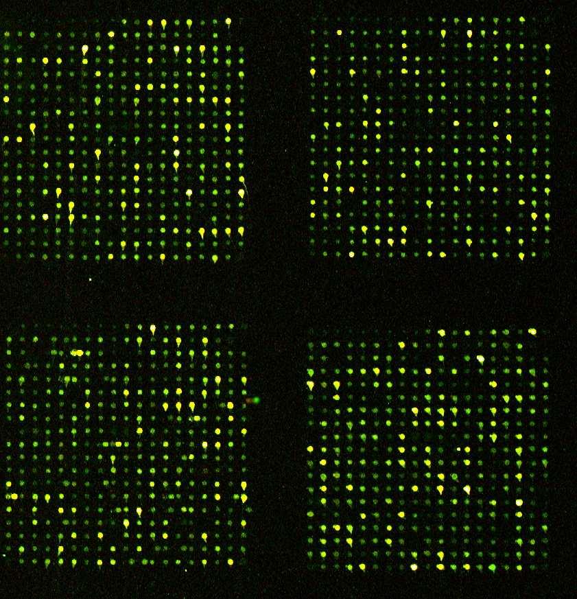
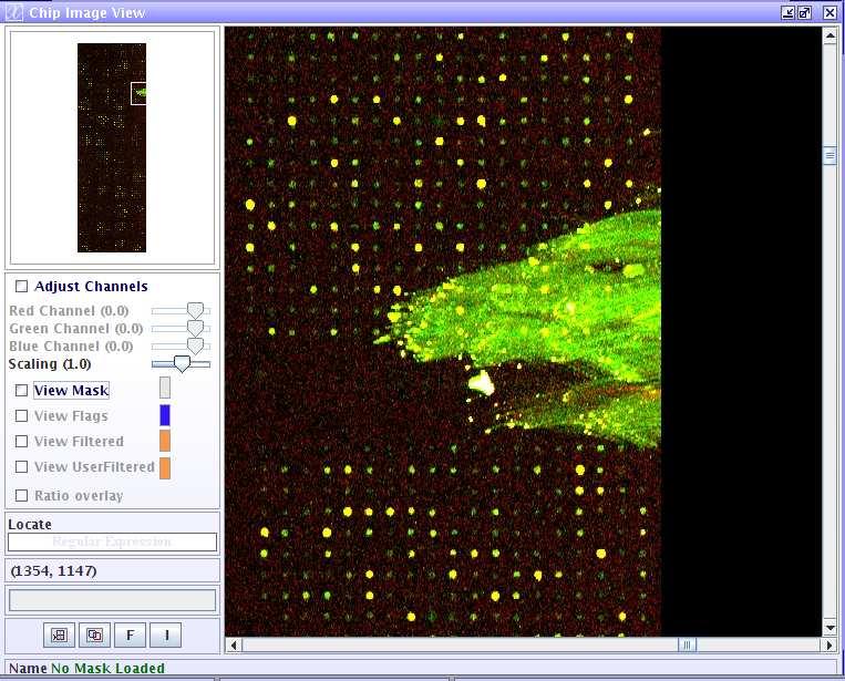
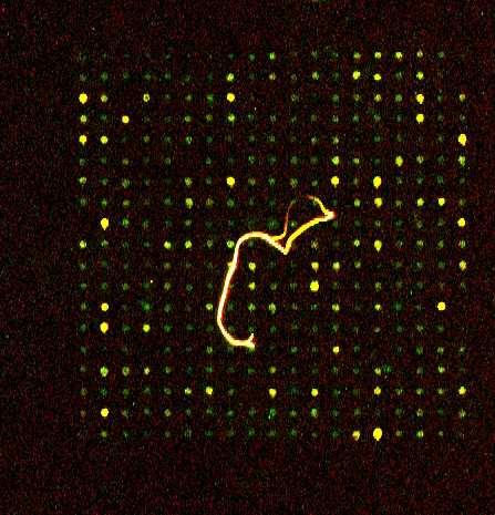
9 Visual inspection of images Comet tails Dust
10 Background intensity Motivation : spot s measured intensity includes a contribution of non-specific hybridization and other chemicals on the glass Estimation of background intensity : Local background, global background, negative controls ScanAlyze ImaGene Spot, GenePix Some findings suggests that the binding of fluorescent dyes to negative control spots is lower than the binding to the glass slide
11 Why quality control? Many steps that influence the data: Sampling Extraction Labelling Hybridization Scanning Extraction of data Trustworthy data Are we measuring biological differences? Or other technical differences? Are the arrays uniform enough to compare against each other?
12 Levels of quality control Array level Assess each spot and surroundings. Quality of expression levels of a particular spot Control spots Spike ins Flags Uniformity between probes of the same type Experiment level Comparing all arrays in the experiment to identify outliers and batch effects
13 Quality measures of signals How good are foreground and background measurements? Spot size Circularity measure Uniformity Population outlier Spot intensity relative to background Based on these measurements, one can flag a spot Different image softwares have different measures that is uses to flag potential unreliable spots
14 Flags Flagging is used to mark spots, probes, etc that should be paid attention to, have bad quality Automated process for commercial platforms Yes/no thresholds Custom platform: more manual process Different types of flags Spot size Circularity Uniformity Signal strength Flags used to filter data and remove bad spots Don t flag too many spots!! Be discerning!
15 Different controls on the array Process-controls Labelling controls, several steps Hybridization controls (stringency controls) Positive and negative control spots Grid control (positive spots) Background controls (negative/empty spots)

16 Spots used for placing grid
17 Spike ins RNA Spike-ins are positive controls for monitoring the microarray workflow from sample amplification and labeling to microarray processing RNA optimized to anneal to their complementary probes on the microarray, platform specific kits Amplified and labeled together with RNA, known concentrations You have to add it yourself! Confidence in the experimental data is increased when it is compared to control transcripts of known concentrations and ratios Help in normalizing data

18 Spike-ins used in QC
19 Summary I Helps to have knowledge of how particular image analysis software works In particular which methods are used for signal extraction Which spot quality checks are done (flags), as this can be used to improve the overall quality of the array during preprocessing Visual inspection of scan image(s) and plots of spacial distribution of signals may help identify problem arrays Decision of whether to subtract background or not is important for the identification of differentially expressed genes
20 Summary II Quality control of microarrays Array level Spot quality Control spots Flags Experment level Next presentation!
Microarray Data Pre-processing. Ana H. Barragan Lid
 Microarray Data Pre-processing Ana H. Barragan Lid Hybridized Microarray Imaged in a microarray scanner Scanner produces fluorescence intensity measurements Intensities correspond to levels of hybridization
Preparation of Sample Hybridization Scanning and Image Analysis
 Preparation of Sample Hybridization Scanning and Image Analysis Sample preparation 1. Design experiment 2. Perform experiment Question? Replicates? Test? mutant wild type 3. Precipitate RNA 4. Label RNA
GenePix Application Note
 GenePix Application Note Biological Relevance of GenePix Results Shawn Handran, Ph.D. and Jack Y. Zhai, Ph.D. Axon Instruments, Inc. 3280 Whipple Road, Union City, CA 94587 Last Updated: Aug 22, 2003.
Low-level Analysis. cdna Microarrays. Lecture 2 Low Level Gene Expression Data Analysis. Stat 697K, CS 691K, Microbio 690K
 Lecture 2 Low Level Gene Expression Data nalysis Stat 697K, CS 691K, icrobio 690K Statistical Challenges odel variation of data not related to gene expression Compare expression for the same gene across
Automatic gene expression estimation from microarray images. Daniel O. Dantas Adviser: : Junior Barrera
 Automatic gene expression estimation from microarray images Daniel O. Dantas Adviser: : Junior Barrera IME-USP Summary Introduction Problem definition Solution strategy Image segmentation Signal estimation
Developed by BioDiscovery, Inc. DualChip evaluation software User Manual Version 1.1
 Developed by BioDiscovery, Inc. DualChip evaluation software User Manual Version 1.1 1 Table of contents 1. INTRODUCTION...3 2. SCOPE OF DELIVERY...4 3. INSTALLATION PROCEDURES...5 3.1. SYSTEM REQUIREMENTS...
In our previous lecture, we understood the vital parameters to be taken into consideration before data acquisition and scanning.
 Interactomics: Protein Arrays & Label Free Biosensors Professor Sanjeeva Srivastava MOOC NPTEL Course Indian Institute of Technology Bombay Module 7 Lecture No 34 Software for Image scanning and data processing
NPTEL VIDEO COURSE PROTEOMICS PROF. SANJEEVA SRIVASTAVA
 HANDOUT LECTURE-31 MICROARRAY WORK-FLOW: IMAGE SCANNING AND DATA PROCESSING Slide 1: This module contains the summary of the discussion with Mr. Pankaj Khanna, an application specialist from Spinco Biotech,
Steps involved in microarray analysis after the experiments
 Steps involved in microarray analysis after the experiments Scanning slides to create images Conversion of images to numerical data Processing of raw numerical data Further analysis Clustering Integration
GenePix Application Note
 GenePix Application Note Determining the Signal-to-Noise Ratio and Optimal Photomultiplier gain setting in the GenePix 4000B Siobhan Pickett, M.S., Sean Carriedo, Ph.D. and Chang Wang, Ph.D. Axon Instruments,
NPTEL VIDEO COURSE PROTEOMICS PROF. SANJEEVA SRIVASTAVA
 LECTURE-31 MICROARRAY WORK-FLOW: IMAGE SACNNING AND DATA PROCESSING TRANSCRIPT Welcome to the proteomics course. In today s lecture we will talk about microarray work-flow the image scanning and processing.
Computational Genomics. High-throughput experimental biology
 Computational Genomics 10-810/02 810/02-710, Spring 2009 Gene Expression Analysis Data pre-processing processing Eric Xing Lecture 15, March 4, 2009 Reading: class assignment Eric Xing @ CMU, 2005-2009
Automated cdna microarray image segmentation
 Automated cdna microarray image segmentation Author Liew, Alan Wee-Chung, Yan, Hong Published 2007 Conference Title Proceedings of the International Symposium on Computational Models for Life Sciences
ScanArray Overview. Principle of Operation. Instrument Components
 ScanArray Overview The GSI Lumonics ScanArrayÒ Microarray Analysis System is a scanning laser confocal fluorescence microscope that is used to determine the fluorescence intensity of a two-dimensional
MICROARRAY IMAGE ANALYSIS PROGRAM
 Revision submitted for publication to Loyola Schools Review, 13 November 2002 MICROARRAY IMAGE ANALYSIS PROGRAM Paul Ignatius D. Echevarria Jerome C. Punzalan John Paul C. Vergara Department of Information
Product Information. Introduction
 Microarray Scanner Calibration Slide To quantitatively analyze scanners performance and output, adjust and fine-tune scanners, and perform comparative analysis for multiple scanner units. To verify scanners
Instructions for Mapping * µarray Images using GenePix 5.0
 Instructions for Mapping * µarray Images using GenePix 5.0 Preliminary Information Make sure that the GenePix 5.0 software has been installed on your computer and you have the USB hardware dongle that
Products - Microarray Scanners - Laser Scanners - InnoScan 900 Series and MAPIX Software
 Products - Microarray Scanners - Laser Scanners - InnoScan 900 Series and MAPIX Software Arrayit offers the world s only next generation microarray scanning technology, with proprietary rotary motion control,
Scan slides (Axon Genepix 4200AL)
") Page 1 Scan slides (Axon Genepix 4200AL) We need to scan the slides on both channels (Cy3 and Cy5) to obtain a 16-bit grayscale TIFF file for each. Typically these files are about 20-26Mb per channel,
Page 1 Scan slides (Axon Genepix 4200AL) We need to scan the slides on both channels (Cy3 and Cy5) to obtain a 16-bit grayscale TIFF file for each. Typically these files are about 20-26Mb per channel,
Scanning and Image Processing -by Steve Clough
 Scanning and Image Processing -by Steve Clough cdna microarrays use two dyes with well separated emission spectra such as Cy3 and Cy5 to allow direct comparisons on single slide GSI Lumonics Ratio of Expression
IncuCyte ZOOM Fluorescent Processing Overview
 IncuCyte ZOOM Fluorescent Processing Overview The IncuCyte ZOOM offers users the ability to acquire HD phase as well as dual wavelength fluorescent images of living cells producing multiplexed data that
Feature Level Data. Outline. Affymetrix GeneChip Design. Affymetrix GeneChip arrays Two color platforms
 Feature Level Data Outline Affymetrix GeneChip arrays Two color platforms Affymetrix GeneChip Design 5 3 Reference sequence TGTGATGGTGCATGATGGGTCAGAAGGCCTCCGATGCGCCGATTGAGAAT GTACTACCCAGTCTTCCGGAGGCTA
EmbryoCellect. RHS Scanning and Analysis Instructions. for. Genepix Pro Software
 EmbryoCellect RHS Scanning and Analysis Instructions for Genepix Pro Software EmbryoCellect Genepix Pro Scanning and Analysis Technical Data Sheet Version 1.0 October 2015 1 Copyright Reproductive Health
Analysing data from Illumina BeadArrays
 The bead Analysing data from Illumina BeadArrays Each silica bead is 3 microns in diameter Matt Ritchie Department of Oncology University of Cambridge, UK 4th September 008 700,000 copies of same probe
Traffic Sign Recognition Senior Project Final Report
 Traffic Sign Recognition Senior Project Final Report Jacob Carlson and Sean St. Onge Advisor: Dr. Thomas L. Stewart Bradley University May 12th, 2008 Abstract - Image processing has a wide range of real-world
An Improved Bernsen Algorithm Approaches For License Plate Recognition
 IOSR Journal of Electronics and Communication Engineering (IOSR-JECE) ISSN: 78-834, ISBN: 78-8735. Volume 3, Issue 4 (Sep-Oct. 01), PP 01-05 An Improved Bernsen Algorithm Approaches For License Plate Recognition
Image Database and Preprocessing
 Chapter 3 Image Database and Preprocessing 3.1 Introduction The digital colour retinal images required for the development of automatic system for maculopathy detection are provided by the Department of
ImageJ: Introduction to Image Analysis 3 May 2012 Jacqui Ross
 Biomedical Imaging Research Unit School of Medical Sciences Faculty of Medical and Health Sciences The University of Auckland Private Bag 92019 Auckland 1142, NZ Ph: 373 7599 ext. 87438 http://www.fmhs.auckland.ac.nz/sms/biru/.
IncuCyte ZOOM Scratch Wound Processing Overview
 IncuCyte ZOOM Scratch Wound Processing Overview The IncuCyte ZOOM Scratch Wound assay utilizes the WoundMaker-IncuCyte ZOOM-ImageLock Plate system to analyze both 2D-migration and 3D-invasion in label-free,
Image and video processing
 Image and video processing Processing Colour Images Dr. Yi-Zhe Song The agenda Introduction to colour image processing Pseudo colour image processing Full-colour image processing basics Transforming colours
Spotxel 1.7 Microarray Image and Data Analysis Software User s Guide
 Spotxel 1.7 Microarray Image and Data Analysis Software User s Guide 27 April 2017 - Rev 7 Spotxel is only intended for research and not intended or approved for diagnosis of disease in humans or animals.
Image Analysis for Fluorescence
 Image Analysis for Fluorescence Terminology Table Image Analysis Macro Colocalization Intensity Dye AFI The extraction of meaningful information from digital images by means of digital image processing
Illumination Correction tutorial
 Illumination Correction tutorial I. Introduction The Correct Illumination Calculate and Correct Illumination Apply modules are intended to compensate for the non uniformities in illumination often present
CSE 564: Scientific Visualization
 CSE 564: Scientific Visualization Lecture 5: Image Processing Klaus Mueller Stony Brook University Computer Science Department Klaus Mueller, Stony Brook 2003 Image Processing Definitions Purpose: - enhance
Donuts, Scratches and Blanks: Robust Model-Based Segmentation of Microarray Images
 Donuts, Scratches and Blanks: Robust Model-Based Segmentation of Microarray Images Qunhua Li 1,a, Chris Fraley 1,a, Roger E. Bumgarner 2,b, Ka Yee Yeung 2,c, Adrian E. Raftery 1,a Technical Report no.
DISCRIMINANT FUNCTION CHANGE IN ERDAS IMAGINE
 DISCRIMINANT FUNCTION CHANGE IN ERDAS IMAGINE White Paper April 20, 2015 Discriminant Function Change in ERDAS IMAGINE For ERDAS IMAGINE, Hexagon Geospatial has developed a new algorithm for change detection
CS 445 HW#2 Solutions
 1. Text problem 3.1 CS 445 HW#2 Solutions (a) General form: problem figure,. For the condition shown in the Solving for K yields Then, (b) General form: the problem figure, as in (a) so For the condition
Abstract. comment reviews reports deposited research refereed research interactions information
 http://genomebiology.com/21/2/11/research/47.1 Research Sources of nonlinearity in cdna microarray expression measurements Latha Ramdas*, Kevin R Coombes, Keith Baggerly, Lynne Abruzzo, W Edward Highsmith,
Instructions for Howto Scan µarrays
 Instructions for Howto Scan µarrays Introduction After probing the µarray slides with samples, one is now ready to scan them. To scan a µarrays slide is too convert the biological information trapped on
Microarray Image Analysis: Background Estimation using Region and Filtering Techniques
 Microarray Image Analysis: Background Estimation using Region and Filtering Techniques Anders Bengtsson December 9, 2003 Abstract This report examines properties of two main methods used for local background
Material analysis by infrared mapping: A case study using a multilayer
 Material analysis by infrared mapping: A case study using a multilayer paint sample Application Note Author Dr. Jonah Kirkwood, Dr. John Wilson and Dr. Mustafa Kansiz Agilent Technologies, Inc. Introduction
Biometrics Final Project Report
 Andres Uribe au2158 Introduction Biometrics Final Project Report Coin Counter The main objective for the project was to build a program that could count the coins money value in a picture. The work was
Instruction Manual. Mark Deimund, Zuyi (Jacky) Huang, Juergen Hahn
 Huang, Juergen Hahn") Instruction Manual Mark Deimund, Zuyi (Jacky) Huang, Juergen Hahn This manual is for the program that implements the image analysis method presented in our paper: Z. Huang, F. Senocak, A. Jayaraman, and
Instruction Manual Mark Deimund, Zuyi (Jacky) Huang, Juergen Hahn This manual is for the program that implements the image analysis method presented in our paper: Z. Huang, F. Senocak, A. Jayaraman, and
Acute Lymphocytic Leukemia Detection and Classification (ALLDC) System
 System") Acute Lymphocytic Leukemia Detection and Classification (ALLDC) System Jamila Harbi, PhD Computer Science Dept. College of Science Al- Mustansiriyah University Baghdad, Iraq Rana Ali Computer Science Dept.
Acute Lymphocytic Leukemia Detection and Classification (ALLDC) System Jamila Harbi, PhD Computer Science Dept. College of Science Al- Mustansiriyah University Baghdad, Iraq Rana Ali Computer Science Dept.
Introduction to Image Analysis with
 Introduction to Image Analysis with PLEASE ENSURE FIJI IS INSTALLED CORRECTLY! WHAT DO WE HOPE TO ACHIEVE? Specifically, the workshop will cover the following topics: 1. Opening images with Bioformats
Introduction Approach Work Performed and Results
 Algorithm for Morphological Cancer Detection Carmalyn Lubawy Melissa Skala ECE 533 Fall 2004 Project Introduction Over half of all human cancers occur in stratified squamous epithelia. Approximately one
NHSC/PACS Web Tutorials Running the PACS Spectrometer pipeline for CHOP/NOD Mode. PACS-301 Level 0 to 1 processing
 NHSC/PACS s Running the PACS Spectrometer pipeline for CHOP/NOD Mode page 1 PACS-301 Level 0 to 1 processing Prepared by Dario Fadda September 2012 Introduction This tutorial will guide you through the
Computer Vision. Howie Choset Introduction to Robotics
 Computer Vision Howie Choset http://www.cs.cmu.edu.edu/~choset Introduction to Robotics http://generalrobotics.org What is vision? What is computer vision? Edge Detection Edge Detection Interest points
HoloMonitor. Phase. For competent and powerful discoveries. Holographic time-lapse imaging cytometry
 HoloMonitor M4 Holographic time-lapse imaging cytometry For competent and powerful discoveries Monitor and quantify living cells in their natural environment with unrivaled temporal resolution Phase Holographic
The Bead. beadarray: : An R Package for Illumina BeadArrays. Bead Preparation and Array Production. Beads in Wells. Mark Dunning -
 beadarray: : An R Package for Illumina BeadArrays Mark Dunning - md392@cam.ac.uk PhD Student - Computational Biology Group, Department of Oncology - University of Cambridge Address The Bead Probe 23 b
Segmentation of Microscopic Bone Images
 International Journal of Electronics Engineering, 2(1), 2010, pp. 11-15 Segmentation of Microscopic Bone Images Anand Jatti Research Scholar, Vishveshvaraiah Technological University, Belgaum, Karnataka
FIJI/Image J for Quantification Hands on session
 FIJI/Image J for Quantification Hands on session Dr Paul McMillan Biological Optical Microscopy Platform Hands on demonstrations FIJI set up Line Profile Thresholding Area of stain Cell confluence Nuclei
Version 6. User Manual OBJECT
 Version 6 User Manual OBJECT 2006 BRUKER OPTIK GmbH, Rudolf-Plank-Str. 27, D-76275 Ettlingen, www.brukeroptics.com All rights reserved. No part of this publication may be reproduced or transmitted in any
CoE4TN4 Image Processing. Chapter 3: Intensity Transformation and Spatial Filtering
 CoE4TN4 Image Processing Chapter 3: Intensity Transformation and Spatial Filtering Image Enhancement Enhancement techniques: to process an image so that the result is more suitable than the original image
Teton Photography Group
 Overview general post-processing (editing) workflow for serious photographers Focus on processes more than software Examples using Adobe Lightroom and Photoshop Teton Photography Group January 2016 Emphasis
THEORY AND APPROACHES TO AUTOMATED IMAGE ANALYSIS IN DIGITAL PATHOLOGY
 THEORY AND APPROACHES TO AUTOMATED IMAGE ANALYSIS IN DIGITAL PATHOLOGY Kyle Takayama, MS Charles River Laboratories EVERY STEP OF THE WAY EVERY STEP OF THE WAY MORPHOMETRY Measurements or counts performed
Introduction to DSP ECE-S352 Fall Quarter 2000 Matlab Project 1
 Objective: Introduction to DSP ECE-S352 Fall Quarter 2000 Matlab Project 1 This Matlab Project is an extension of the basic correlation theory presented in the course. It shows a practical application
Colony Imaging with powerful Analysis Software
 TM Imaging with powerful Analysis Software TM Accurate Compact Fast We re not going to interpret your results, but we ll do everything to get you there From image acquisition to data visualisation, straight
ROBOT VISION. Dr.M.Madhavi, MED, MVSREC
 ROBOT VISION Dr.M.Madhavi, MED, MVSREC Robotic vision may be defined as the process of acquiring and extracting information from images of 3-D world. Robotic vision is primarily targeted at manipulation
Application of Machine Vision Technology in the Diagnosis of Maize Disease
 Application of Machine Vision Technology in the Diagnosis of Maize Disease Liying Cao, Xiaohui San, Yueling Zhao, and Guifen Chen * College of Information and Technology Science, Jilin Agricultural University,
Chapter 3 Part 2 Color image processing
 Chapter 3 Part 2 Color image processing Motivation Color fundamentals Color models Pseudocolor image processing Full-color image processing: Component-wise Vector-based Recent and current work Spring 2002
Fingerprints - Formation - Fingerprints are a reproduction of friction skin ridges that are on the palm side of fingers and thumbs
 Fingerprints - Formation - Fingerprints are a reproduction of friction skin ridges that are on the palm side of fingers and thumbs - these skin surfaces have been designed by nature to provide our bodies
prf_estimate.pl David Makovoz, 10/15/04 Table of Contents
 prf_estimate.pl 1 prf_estimate.pl David Makovoz, 10/15/04 Table of Contents prf_estimate.pl... 1 1 Overview... Input....1 Input Data.... Namelist Configuration file... 3.3 Quality Control Mask Images...
Overview. Corrosion detection improvement of oil and gas pipelines with industrial radiography method by using image processing.
 detection improvement of oil and gas pipelines with industrial radiography method by using image processing Alireza Karimian (Engineering faculty of Isfahan university, Isfahan,, Iran ) Sepideh Yazdani
Technical Aspects in Digital Pathology
 Technical Aspects in Digital Pathology Yukako Yagi, PhD yyagi@mgh.harvard.edu Director of the MGH Pathology Imaging & Communication Technology Center Assistant Professor of Pathology, Harvard Medical School
Experiment 1: Fraunhofer Diffraction of Light by a Single Slit
 Experiment 1: Fraunhofer Diffraction of Light by a Single Slit Purpose 1. To understand the theory of Fraunhofer diffraction of light at a single slit and at a circular aperture; 2. To learn how to measure
Statistics 101: Section L Laboratory 10
 Statistics 101: Section L Laboratory 10 This lab looks at the sampling distribution of the sample proportion pˆ and probabilities associated with sampling from a population with a categorical variable.
Proposed Method for Off-line Signature Recognition and Verification using Neural Network
 e-issn: 2349-9745 p-issn: 2393-8161 Scientific Journal Impact Factor (SJIF): 1.711 International Journal of Modern Trends in Engineering and Research www.ijmter.com Proposed Method for Off-line Signature
Raster Based Region Growing
 6th New Zealand Image Processing Workshop (August 99) Raster Based Region Growing Donald G. Bailey Image Analysis Unit Massey University Palmerston North ABSTRACT In some image segmentation applications,
VLSI Implementation of Impulse Noise Suppression in Images
 VLSI Implementation of Impulse Noise Suppression in Images T. Satyanarayana 1, A. Ravi Chandra 2 1 PG Student, VRS & YRN College of Engg. & Tech.(affiliated to JNTUK), Chirala 2 Assistant Professor, Department
GALILEO TMA CK 4500 HTS Tissue Microarray Platform
 GALILEO TMA CK 4500 HTS Tissue Microarray Platform Tissue Microarray (TMA) A Block Of Samples From Hundreds Of Blocks (S. M. Hewitt, M.D., Ph.D., Tissue Array Research Program, LP, CCR, NCI, NIH) TMA technology
Microarray BASICA: Background Adjustment, Segmentation, Image Compression and Analysis of Microarray Images
 EURASIP Journal on Applied Signal Processing 24:1, 92 17 c 24 Hindawi Publishing Corporation Microarray BASICA: Background Adjustment, Segmentation, Image Compression and Analysis of Microarray Images
Automatic Licenses Plate Recognition System
 Automatic Licenses Plate Recognition System Garima R. Yadav Dept. of Electronics & Comm. Engineering Marathwada Institute of Technology, Aurangabad (Maharashtra), India yadavgarima08@gmail.com Prof. H.K.
Arcturus XT Laser Capture Microdissection System AutoScanXT Software Module. User Manual
 Arcturus XT Laser Capture Microdissection System AutoScanXT Software Module User Manual For Research Use Only. Not intended for any animal or human therapeutic or diagnostic use. Information in this document
Tutorial document written by Vincent Pelletier and Maria Kilfoil 2007.
 Tutorial document written by Vincent Pelletier and Maria Kilfoil 2007. Overview This code finds and tracks round features (usually microscopic beads as viewed in microscopy) and outputs the results in
Assessments Using Spike-In Experiments
 Assessments Using Spike-In Experiments Rafael A Irizarry, Department of Biostatistics JHU rafa@jhu.edu http://www.biostat.jhsph.edu/~ririzarr http://www.bioconductor.org A probe set = 11-20 PM,MM pairs
Segmentation of Liver CT Images
 Segmentation of Liver CT Images M.A.Alagdar 1, M.E.Morsy 2, M.M.Elzalabany 3 1,2,3 Electronics And Communications Department-.Faculty Of Engineering Mansoura University, Egypt. Abstract In this paper we
Supporting Information: Electron Microscopic Visualization of Protein Assemblies on Flattened DNA Origami
 Supporting Information: Electron Microscopic Visualization of Protein Assemblies on Flattened DNA Origami Leena Mallik, Soma Dhakal, Joseph Nichols, Jacob Mahoney, Anne M. Dosey, Shuoxing Jiang ǂ, Roger
SINCE2011 Singapore International NDT Conference & Exhibition, 3-4 November 2011
 SINCE2011 Singapore International NDT Conference & Exhibition, 3-4 November 2011 Automated Defect Recognition Software for Radiographic and Magnetic Particle Inspection B. Stephen Wong 1, Xin Wang 2*,
HoloMonitor M4. For powerful discoveries in your incubator
 HoloMonitor M4 For powerful discoveries in your incubator HoloMonitor offers unique imaging capabilities that greatly enhance our understanding of cell behavior, previously unachievable by other technologies
NR601. VAHTS TM mrna-seq V2 Library Prep Kit for Illumina
 NR601 VAHTS TM mrna-seq V2 Library Prep Kit for Illumina v Vazyme Biotech Co., Ltd Website: www.vazyme.com Order: global@vazyme.com Support: support@vazyme.com Service: service@vazyme.com SYSTEMS www.vazyme.com
Reference Free Image Quality Evaluation
 Reference Free Image Quality Evaluation for Photos and Digital Film Restoration Majed CHAMBAH Université de Reims Champagne-Ardenne, France 1 Overview Introduction Defects affecting films and Digital film
Princeton ELE 201, Spring 2014 Laboratory No. 2 Shazam
 Princeton ELE 201, Spring 2014 Laboratory No. 2 Shazam 1 Background In this lab we will begin to code a Shazam-like program to identify a short clip of music using a database of songs. The basic procedure
Crossword: A Fully Automated Algorithm for the Segmentation and Quality Control of Protein Microarray Images
 Crossword: A Fully Automated Algorithm for the Segmentation and Quality Control of Protein Microarray Images The MIT Faculty has made this article openly available. Please share how this access benefits
BacklightFly Manual.
 BacklightFly Manual http://www.febees.com/ Contents Start... 3 Installation... 3 Registration... 7 BacklightFly 1-2-3... 9 Overview... 10 Layers... 14 Layer Container... 14 Layer... 16 Density and Design
AUTOMATED MALARIA PARASITE DETECTION BASED ON IMAGE PROCESSING PROJECT REFERENCE NO.: 38S1511
 AUTOMATED MALARIA PARASITE DETECTION BASED ON IMAGE PROCESSING PROJECT REFERENCE NO.: 38S1511 COLLEGE : BANGALORE INSTITUTE OF TECHNOLOGY, BENGALURU BRANCH : COMPUTER SCIENCE AND ENGINEERING GUIDE : DR.
Study and Analysis of various preprocessing approaches to enhance Offline Handwritten Gujarati Numerals for feature extraction
 International Journal of Scientific and Research Publications, Volume 4, Issue 7, July 2014 1 Study and Analysis of various preprocessing approaches to enhance Offline Handwritten Gujarati Numerals for
7-2 Mean, Median, Mode, and Range. IWBAT find the mean, median, mode, and range of a data set.
 IWBAT find the mean, median, mode, and range of a data set. mean median mode range outlier Vocabulary WRITE: The mean is the sum of the data values divided by the number of data items. The median is the
Image Processing - License Plate Localization and Letters Extraction *
 OpenStax-CNX module: m33156 1 Image Processing - License Plate Localization and Letters Extraction * Cynthia Sung Chinwei Hu Kyle Li Lei Cao This work is produced by OpenStax-CNX and licensed under the
Influence of Dictionary Size on the Lossless Compression of Microarray Images
 Influence of Dictionary Size on the Lossless Compression of Microarray Images Robert Bierman 1, Rahul Singh 1 Department of Computer Science, San Francisco State University, San Francisco, CA bierman@sfsu.edu,
Uncertainty in CT Metrology: Visualizations for Exploration and Analysis of Geometric Tolerances
 Uncertainty in CT Metrology: Visualizations for Exploration and Analysis of Geometric Tolerances Artem Amirkhanov 1, Bernhard Fröhler 1, Michael Reiter 1, Johann Kastner 1, M. Eduard Grӧller 2, Christoph
Exercise questions for Machine vision
 Exercise questions for Machine vision This is a collection of exercise questions. These questions are all examination alike which means that similar questions may appear at the written exam. I ve divided
Retina. last updated: 23 rd Jan, c Michael Langer
 Retina We didn t quite finish up the discussion of photoreceptors last lecture, so let s do that now. Let s consider why we see better in the direction in which we are looking than we do in the periphery.
Chem466 Lecture Notes. Spring, 2004
 Chem466 Lecture Notes Spring, 004 Overview of the course: Many of you will use instruments for chemical analyses in lab. settings. Some of you will go into careers (medicine, pharmacology, forensic science,
DataCapture Transcript Module Getting Started Guide
 DataCapture Transcript Module Getting Started Guide Version: 6.6 Written by: Product Documentation, R&D Date: February 2011 ImageNow and CaptureNow are registered trademarks of Perceptive Software, Inc.
Photographing Long Scenes with Multiviewpoint
 Photographing Long Scenes with Multiviewpoint Panoramas A. Agarwala, M. Agrawala, M. Cohen, D. Salesin, R. Szeliski Presenter: Stacy Hsueh Discussant: VasilyVolkov Motivation Want an image that shows an
TotalLab Quant v12.3. Product Specification: 1D Analysis Module
 Product Specification: TotalLab Quant v12.3 1D Analysis Module General Fully automatic, single button press complete image analysis within area of interest if required Instant access to refinement of any
Improved Accuracy of Spot Search on HPV DNA Microarray Chip
 , pp.182-186 http://dx.doi.org/10.14257/astl.2017.143.38 Improved Accuracy of Spot Search on HPV DNA Microarray Chip Jae-Hong Min 1, Chan-Young Park 2,3, Yu-Seop Kim,2,3, Hye-Jeong Song 3, Jong-Dae Kim
Multiplexing as Essential Tool for Modern Biology
 Multiplexing as Essential Tool for Modern Biology Bio-Plex Seminar, Debrecen, 2012. Gyula Csanádi, PhD. The "Age of "-omics" Studying interrelationships at different level of complexity Genes - Unveiling
Image Processing for feature extraction
 Image Processing for feature extraction 1 Outline Rationale for image pre-processing Gray-scale transformations Geometric transformations Local preprocessing Reading: Sonka et al 5.1, 5.2, 5.3 2 Image