The ultimate in macro fluorescence im The new MVX10 MacroView from Olym
|
|
|
- Jack Elliott
- 5 years ago
- Views:
Transcription

1

, was a significant breakthrough since proteins can now be labelled without influencing their")
2 The ultimate in macro fluorescence im The new MVX10 MacroView from Olym m Researchers are interested in the impact of gene expression and protein function not only at the cellular level but also within whole tissues, organs and even organisms. Hence organisms like C. elegans, Drosophila, Zebrafish, Xenopus, Mouse or the plant Arabidopsis are used as biological models for in vivo studies in a vast field of research applications. The introduction of the naturally fluorescent protein markers, such as Green Fluorescent Protein (GFP), was a significant breakthrough since proteins can now be labelled without influencing their function. The perfect microscope for fluorescence observation in intact organisms must combine maximum detection sensitivity at the lowe magnifications with a high magnification zoom for the re of fine details within organs, tissues and even cells. The Olympus MVX10 MacroView brings both of these factors together with many other unique features to bridge the g between macro and micro observation, providing unprecedented brightness, resolution and precision. High fluorescence efficiency plus stereo observation Seamless observation from 4x to 125x Zoom factor up to 31 times Long WD for observation at optimum magnification Maximum specimen protection due to short exposure time Complete system solutions for optimized recordings 1


3 aging. pus. 2


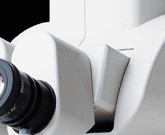
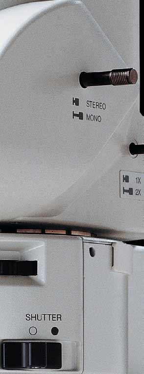
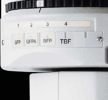
4 Bright fluorescence imaging with seamless m High fluorescence efficiency plus stereo observation Up until now, stereo microscopes have been the instruments of choice for fluorescence observation at low magnifications. For the stereoscopic effect, two optical paths are used one for the left and one for the right eye. Stereo microscopy though, is not very well suited to imaging the weak light generated by fluorescence, since the light collected by the objective is split in two. The Olympus MVX10 MacroView on the other hand, employs a single-zoom optical path with a large diameter, which is optimized to collect light with unprecedented efficiency and resolution at all magnifications. From fluorescent observation of whole organisms such as zebrafish at low magnification to the detailed observation of gene expression at the cellular level at high magnification the MVX10 helps you to see it all. What s more, the MVX10 features a unique pupil division mechanism in the light-path to mimic the effect of stereo microscopy. So you can get the best of both worlds high light efficiency and stereo observation in one system just by moving a slider. This puts the MVX10 in a class of its own. Zebrafish spinal cord expressing green fluorescent protein Dedicated to fluorescence All components of the light path contribute to the phenomenal fluorescence performance of the MVX10. Using the latest technologies and new materials, the MVX10 objectives produce almost zero autofluorescence. gether with very high numerical apertures this results in an extremely good signal-to-noise (S/N) ratio, ensuring excellent contrast for observation of even the faintest fluorescence signals. Moreover, the S/N ratio is further enhanced by two novel proprietary features: A new coating technique gives the Olympus HQ filters an exceptional edge steepness and very low autofluorescence. All the filter cubes are equipped to absorb stray light. Light collection efficiency is also maximized with an aspherical fluorescence collector, which bundles the light for minimum intensity loss. Reflected light fluorescence unit + fluorescence mirror unit 3


5 acro to micro zooming. Smooth and Parfocal objectives for seamless observation from macro to micro A unique objective line The MVX10 provides the same working distance and large field of view as stereo microscopes, but with much higher resolution due to the increased numerical aperture (NA). Specially designed for the MVX10, the 0.63x, 1x and 2x planapochromatic objectives produce the highest image quality. All three objectives are pupil-corrected for best image flatness and show high transmission to NIR and superior chromatic aberration correction. This produces great flexibility for efficient, fast and precise fluorescence observation, screening and imaging from low to high magnification over time. Dynamic The 0.63x objective has a maximum field of view of 55 mm, making it easy to track fast-moving specimens over time. With its exceptionally high NA of 0.15, fluorescence from large objects, such as whole embryos, can be viewed with perfect brightness at all magnifications. Objective lineup Gentle The peerless NA and S/N ratio values of all the optical components mean that specimens can be exposed to fluorescent light for shorter periods. This is also true at near-infrared wavelengths where the MVX10 has superior transmission properties and thus fluorochromes throughout the entire spectrum can be used with minimal sample damage. From macro to micro Using the 2-position revolving nosepiece with the 0.63x and 2x objectives expands the usable zoom range up to 31. The objectives are parfocal corrected, making refocusing after objective switching very quick and easy. Only a small amount of fine focusing is necessary to return to the optimal focus position, making macro to micro changes seamless. The 2x objective is also equipped with an additional correction collar to adjust the image quality independently of the specimen medium. 2 mm 100µm Purkinje cell of sliced mouse brain with Lucifer Yellow injected, at 0.63x (left) and 12.5x (right) magnification Long working distance (WD) ensures more efficiency in screening and observation In comparison with stereomicroscopes, the MVX 10 provides the same working distance and a much higher NA (65mm WD and maximum 0.25 NA when using a 1x objective). This makes fluorescence screening and verification of gene expression especially efficient, improves speed and precision, reduces judgment errors, and eliminates the need to switch back and forth between a stereomicroscope and inverted microscope. 4


 The Principal Olive (Medial Accessory Olive) was stimulated to visualize the neuronal circuit structure. The images were acquired using the MVX10 (MVPLAPO 2XC and 6.")

. The numbers above the images represent zoom magnification, and the numbers below the images represent the time after stimulation.")
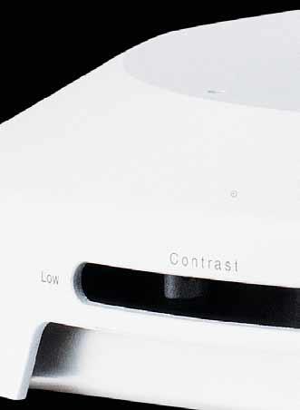
6 Use MVX10 for Optical Membrane Voltage Recording - From Sample Preparation to Recording With optimal fluorescence light throughput, the MVX10 is highly effective for optical membrane voltage recordings requiring the detention of minute changes in fluorescence. It can be used for optical recordings at high speeds and high signal-to-noise ratios as well as utilized in the preparation of brain slices, tissue blocks, isolated hearts, in vivo animals, and other biological specimens. The interchangeable fluorescence filter cube unit in the MVX10 enables recordings using various kinds of fluorescent probe F/F msec after stimulation 25msec after stimulation Optical Recording of Neuronal Circuits in Mice Cerebella An isolated P7 mouse cerebellum was stained with membrane voltage-sensitive dye (Di-2 ANEPEQ, Invitrogen Corp.) The Principal Olive (Medial Accessory Olive) was stimulated to visualize the neuronal circuit structure. The images were acquired using the MVX10 (MVPLAPO 2XC and 6.3X Zoom) and a high-speed imaging system (MiCAM02-HR, Brainvision Inc.) at 200 frames per second, 192 X 128 pixels of spatial resolution, and 10 times averaging. Individual pixel size at this magnification is approximately 7-15 microns/pixel. The pseudo colors in the above image sample display both the intensity and propagation of electrical activity resulting from electrode stimulation of inferior 30 msec olivary nuclei (indicated by arrow). The numbers above the images represent zoom magnification, and the numbers below the images represent the time after stimulation. The waves (upper right) reflect the changes in fluorescence corresponding to the red-, black-, and blue-circled points on the image. The detailed structure of neuronal circuits can be recorded at high spatial and temporal resolutions using the MVX10 and membrane voltagesensitive dye. Dr. Akiko Arata Laboratory for Memory and Learning, Neuronal Circuit Mechanisms Research Group RIKEN, Brain Science Institute F/F msec 1.00msec 2.00msec 3.00msec 4.00msec 30 msec Elapsed time 5.00msec Optical Recording of Neural Activity with Membrane Voltage- Sensitive Dyes These images show the propagation of neural activity in a mouse hippocampus slice (400-micron thickness) resulting from electrical stimulation in the Schaffer collateral region. Membrane voltage-sensitive dye (Di-4 ANEPPS, Invitrogen Corp.) was used to image the minute changes in fluorescence. The images were acquired using the MXV10 (MVPLAPO2XC and 6.3X Zoom) and a high-speed imaging system (MiCAM ULTIMA-L, Brainvision Inc.) at 10,000 frames per second, 100 X 100 pixels of spatial resolution, and 6 times averaging. Individual pixel size at this magnification is approximately 8 microns/pixel. The pseudo colors in the above image sample display both the intensity and propagation of electrical activity resulting from electrode stimulation. The numbers below the images represent frame numbers and time after stimulation. The waves reflect the changes in fluorescence corresponding to the red-, black-, and blue-squared points on the image. Optimal signal-to-noise ratios can be recorded at extremely high speeds with MVX10. Dr. Yuko Sekino and Dr. Akihiro Fukushima Division of Neuronal Network, Department of Basic Medical Sciences The Institute of Medical Science, University of kyo Illuminators for various observation methods High-level transmitted light illumination base SZX2-ILLB This illumination base provides optimal contrast adjustment for detailed observation of transparent specimens. With a single action, the user can select a high or low contrast setting. Oblique illumination is also provided. Large stand SZX2-STL This stable stand with large base provides a broad working space for observing large specimens. 5
7 MVX10 System Diagram C MOUNT DIGITAL CAMERA, C MOUNT VIDEO CAMERA WHN10X-H Eyepiece MVX-TTRS Tilting trinocular tube MVX-CA 2X MVX-TLU Magnification changer 2x Tube lens MVX-TV 0.63XC MVX-TV 1XC C mount camera port with 0.63x lens C mount camera port with 1x lens U-MGFP/XL U-MGFPA/XL U-MCFPHQ/XL U-MGFPHQ/XL U-MYFPHQ/XL U-MRFPHQ/XL U-MF/XL Mirror unit U-DP1XC U-DP MVX-RFA Dual port 1x Dual port Coaxial fluorescence illuminator U-LH100HG U-RFL-T 100W mercury lamp housing Power source for 100 mercury lamp U-LH100HGAPO 100W mercury apo lamp housing MVX-ZB10 MVX10 zoom body U-RX-T 32ND6 32ND12 32ND25 32ND50 U-LH75XE ND Filters 75W xenon lamp housing U-RX-T Power source for 75W xenon lamp MVX-2RE Revolving nosepiece A φ25 FILTER MVPLAPO 0.63X MVPLAPO 1X MVPLAPO 2XC 0.63x objective 1x objective 2x objective SZX2-AN Rotatable anal φ30.5 FILTER LG-FAD HLL301 Filter adapter Spot lens LG-DI Dual flexible light guide LG-DFI LG-PS2 Dual combination light guide Light source SZX-LGR66 Ring light guide adapter Analyzer Polarizer A LG-R66 Ring light guide LG-R66PL HS/FS Analyzer and Polarizer set for LG-R66 SZX2-FOFH SZX-FOA2 Fine focusing unit for heavy loading Motorized focus unit <Accessories for stands> SZX-MDCU SZX-MDHSW Control unit Hand switch U-ACAD4515 AC adapter SP-FL SZ-SC SZH-SG Stage plate for fluorescence Cup stage Gliding stage SZH-P400 BH2-SH U-SRG U-SRP SZX-R Square mechanical stage Circular rotatable stage SZX-MDFSW 400mm pillar Drop prevention collar Foot Switch SZH-P mm pillar SZ2-FO Focusing unit SZX-STAD1 JAPAN SZX-STAD1 SZH-STAD1 BX stage adapter type 1 BH stage adapter type 1 B MIU-IBCSZX/ MIU-IBCSZXF MATS-U55SZX2A CO2 incubator B C SZX2-ILLB SZX2-ILLD High-level transmitted light illumination base Thermo plate B C BF/DF transmitted light illumination base SZX-PO SZX2-STL Large stand SZX-STAD2 Simple polarizer Damper for SZX2 base Frame for incubator SZX-CL Manipulator adapter BX stage adapter type 2 SZX2-DMP MS-SZX2A NO-SZX2A Ø45 FILTER B U-LS30-5 6V30W lamp socket Large stage plate SZX-TLGAD C Transmitted light guide adapter LG-SF minimize environmental impact, Olympus employs ecological glass that is free of lead and other harmful substances in the eyepieces, head, zoom body, and objective lenses. Light guide 6 A U-SIC4R/L Large square mechanical stage
8 Printed in Japan M1584E-1107B
Research Macro Zoom System Microscope
 Research Macro Zoom System Microscope Researchers are interested in the impact of gene expression and protein function not only at the cellular level but also within whole tissues, organs and even organisms.
Research Macro Zoom System Microscope MVX10. MacroView. The First True Macro Fluorescence Imaging System
 Research Macro Zoom System Microscope MVX10 MacroView The First True Macro Fluorescence Imaging System High-Precision Macro Fluorescence Imaging The MVX10 MacroView from Olympus Researchers are interested
Life Science Instrumentation. New Generation. Light Sheet Fluorescence Microscope. Alph
 Life Science Instrumentation Light Sheet Fluorescence Microscope New Generation Alph Modular Light Sheet Microscope Alpha 3 is a new generation of light sheet fluorescence microscope addressing the needs
Axio Zoom.V16 The Fluorescence Zoom Microscope for Large Fields
 Product Information Interactive PDF internet-link video/animation Release 1.0 It s About Brilliance. Because Only the Best Is Good Enough In Brief The Advantages The Applications In 1994, the molecular
Nature Methods: doi: /nmeth Supplementary Figure 1. Schematic of 2P-ISIM AO optical setup.
 Supplementary Figure 1 Schematic of 2P-ISIM AO optical setup. Excitation from a femtosecond laser is passed through intensity control and shuttering optics (1/2 λ wave plate, polarizing beam splitting
Practical work no. 3: Confocal Live Cell Microscopy
 Practical work no. 3: Confocal Live Cell Microscopy Course Instructor: Mikko Liljeström (MIU) 1 Background Confocal microscopy: The main idea behind confocality is that it suppresses the signal outside
EXC500p-- PATHOLOGY MICROSCOPE. EXC500hd -- HD DIGITAL PATHOLOGY MICROSCOPE. EXC500r -- RESEARCH MICROSCOPE EXC500-LABORATORY SCOPE
 EXC500p-- PATHOLOGY MICROSCOPE EXC500hd -- HD DIGITAL PATHOLOGY MICROSCOPE EXC500r -- RESEARCH MICROSCOPE EXC500-LABORATORY SCOPE The EXC500 Pathology and Laboratory Microscope is the most optically advanced
Introduction. Laboratory Equipment & Supplies. Model 1333PHi Shown (Phase Contrast) (2) Eyepieces (Eyecups installed) Diopter Adjustment Mechanism
 (2) Eyepieces (Eyecups installed) Diopter Adjustment Mechanism") Introduction With the invention of the microscope in the early 17th century, it was made possible to view objects which were too small for the human eye to see. As the microscope evolved, the structure
Introduction With the invention of the microscope in the early 17th century, it was made possible to view objects which were too small for the human eye to see. As the microscope evolved, the structure
Imaging Introduction. September 24, 2010
 Imaging Introduction September 24, 2010 What is a microscope? Merriam-Webster: an optical instrument consisting of a lens or combination of lenses for making enlarged images of minute objects; especially:
OPELCO OPtical ELements COrporation LB Objective Series for Biological Use
 LB Objective Series for Biological Use 105 Executive Drive Suite 100 Dulles, VA 20166-9558 Tel: (703) 471-0080 S PLAN APOCHROMAT OBJECTIVES These objectives compensate for three wavelength of chromatic
Marine Invertebrate Zoology Microscope Introduction
 Marine Invertebrate Zoology Microscope Introduction Introduction A laboratory tool that has become almost synonymous with biology is the microscope. As an extension of your eyes, the microscope is one
Light Microscopy. Upon completion of this lecture, the student should be able to:
 Light Light microscopy is based on the interaction of light and tissue components and can be used to study tissue features. Upon completion of this lecture, the student should be able to: 1- Explain the
Discover Another Dimension
 Discover Another Dimension Digital Microscope DSX110 For All Conventional Microscope Users, This Is an Olympus Proposal for the Next Generation of Microscopes. Free-angle Wide-zoom Digital Microscope Olympus
User Manual. Trinocular Metallurgical Microscope. MicroscopeNet.com
 User Manual Trinocular Metallurgical Microscope Model M83MPTR MicroscopeNet.com Table of Contents i. Caution.. 1 ii. Care and Maintenance... 2 1. Components Illustration..... 3 2. Installation...4 3. Operation
High-sensitivity. optical molecular imaging and high-resolution digital X-ray. In-Vivo Imaging Systems
 High-sensitivity optical molecular imaging and high-resolution digital X-ray In-Vivo Imaging Systems In vivo imaging solutions available in several packages Carestream Molecular Imaging offers a selection
Chapter 2 The Study of Microbial Structure: Microscopy and Specimen Preparation
 Chapter 2 The Study of Microbial Structure: Microscopy and Specimen Preparation 1 Lenses and the Bending of Light light is refracted (bent) when passing from one medium to another refractive index a measure
Sunny Instruments. RX50 Biological Microscope
 Sunny Instruments RX50 Biological Microscope After years of research and development in optical technology field, RX50 biological microscope is designed to present a safe, comfortable and efficiency observation
OMM300. Inverted Metallurgical Microscope
 OMM300 Inverted Metallurgical Microscope Instruction Manual Please read the instructions carefully before operating CONTENTS Safety 2 Parts List 2 Features 3 Assembly 5 Operation 7 Maintenance 9 Specifications
Observing Microorganisms through a Microscope LIGHT MICROSCOPY: This type of microscope uses visible light to observe specimens. Compound Light Micros
 PHARMACEUTICAL MICROBIOLOGY JIGAR SHAH INSTITUTE OF PHARMACY NIRMA UNIVERSITY Observing Microorganisms through a Microscope LIGHT MICROSCOPY: This type of microscope uses visible light to observe specimens.
Systematic Workflow via Intuitive GUI. Easy operation accomplishes your goals faster than ever.
 Systematic Workflow via Intuitive GUI Easy operation accomplishes your goals faster than ever. 16 With the LEXT OLS4100, observation or measurement begins immediately once the sample is placed on the stage.
FLUORESCENCE MICROSCOPY. Matyas Molnar and Dirk Pacholsky
 FLUORESCENCE MICROSCOPY Matyas Molnar and Dirk Pacholsky 1 The human eye perceives app. 400-700 nm; best at around 500 nm (green) Has a general resolution down to150-300 μm (human hair: 40-250 μm) We need
Polarizing Microscope BX51-P
 Polarizing Microscope BX51-P Unsurpassed optics render polarized light images sharper than ever before. Olympus is proud to introduce the BX51-P, the new polarizing microscope with superb performance in
Operation Guide for the Leica SP2 Confocal Microscope Bio-Imaging Facility Hunter College October 2009
 Operation Guide for the Leica SP2 Confocal Microscope Bio-Imaging Facility Hunter College October 2009 Introduction of Fluoresence Confocal Microscopy The first confocal microscope was invented by Princeton
Therefore, all descriptions and illustrations in this instruction manual, including all specifications are subject to change without notice.
 We are constantly endeavouring to improve our instruments and to adapt them to the requirements of modern research techniques and testing methods. This involves modification to the mechanical structure
b. Turn the power switch and key to on position for blue laser.
 OLYMPUS FLUOVIEW 300 CONFOCAL MICOSCOPE OPERATION PROCEDURE 1. Turn ON microscope in this order: 1) Turn on mercury lamp (Note: once the mercury lamp is turned off, DO NOT turn it back on for at least
TABLE OF CONTENTS. Safety notes i. Care and Maintenance. ii. 1. Components Illustration Installation of Components.. 4
 TABLE OF CONTENTS Safety notes i Care and Maintenance. ii 1. Components Illustration... 1 2. Installation of Components.. 4 2.1 Installation Diagram... 4 2.2 Installation Procedures 5 3. Operation...11
Microscope anatomy, image formation and resolution
 Microscope anatomy, image formation and resolution Ian Dobbie Buy this book for your lab: D.B. Murphy, "Fundamentals of light microscopy and electronic imaging", ISBN 0-471-25391-X Visit these websites:
INSTRUCTIONS FOR COURSE WORK 4 (AxioVert) Instructor: Anne Vaahtokari (MIU) 1. Purpose of the work
 Instructor: Anne Vaahtokari (MIU) 1. Purpose of the work") INSTRUCTIONS FOR COURSE WORK 4 (AxioVert) Instructor: Anne Vaahtokari (MIU) 1. Purpose of the work In this work, you will get familiar with an inverted epifluorescence microscope. Also, you will learn
INSTRUCTIONS FOR COURSE WORK 4 (AxioVert) Instructor: Anne Vaahtokari (MIU) 1. Purpose of the work In this work, you will get familiar with an inverted epifluorescence microscope. Also, you will learn
Lecture 23 MNS 102: Techniques for Materials and Nano Sciences
 Lecture 23 MNS 102: Techniques for Materials and Nano Sciences Reference: #1 C. R. Brundle, C. A. Evans, S. Wilson, "Encyclopedia of Materials Characterization", Butterworth-Heinemann, Toronto (1992),
Microscopy: Fundamental Principles and Practical Approaches
 Microscopy: Fundamental Principles and Practical Approaches Simon Atkinson Online Resource: http://micro.magnet.fsu.edu/primer/index.html Book: Murphy, D.B. Fundamentals of Light Microscopy and Electronic
User Manual. Digital Compound Binocular LED Microscope. MicroscopeNet.com
 User Manual Digital Compound Binocular LED Microscope Model MD82ES10 MicroscopeNet.com Table of Contents i. Caution... 1 ii. Care and Maintenance... 2 1. Components Illustration... 3 2. Installation...
MICROSCOPE LAB. Resolving Power How well specimen detail is preserved during the magnifying process.
 AP BIOLOGY Cells ACTIVITY #2 MICROSCOPE LAB OBJECTIVES 1. Demonstrate proper care and use of a compound microscope. 2. Identify the parts of the microscope and describe the function of each part. 3. Compare
Olympus Fluoview 1000S Spectral Confocal Microscope Introduction to the NRI-MCDB Microscopy Facility Spectral Confocal Microscope
 Olympus Fluoview 1000S Spectral Confocal Microscope Introduction to the NRI-MCDB Microscopy Facility Spectral Confocal Microscope Improved Optics More Lasers 405 diode 440 diode 488 Argon 515 Argon 559
STEINDORFF METALLURGICAL MICROSCOPE. NYMCS-620 Instruction Manual
 METALLURGICAL MICROSCOPE NYMCS-620 Instruction Manual It is recommended strongly that you study this manual thoroughly before using the microscope. Retain this manual in an easily accessible place near
MICROSCOPY FOR THE DEVELOPMENTAL BIOLOGY STUDENT...
 MICROSCOPY FOR THE DEVELOPMENTAL BIOLOGY STUDENT... You will be using two configurations of microscope during the course of the semester to observe specimens and record your results: compound microscopes
microscopy A great online resource Molecular Expressions, a Microscope Primer Partha Roy
 Fundamentals of optical microscopy A great online resource Molecular Expressions, a Microscope Primer http://micro.magnet.fsu.edu/primer/index.html Partha Roy 1 Why microscopy Topics Functions of a microscope
Rapid Adaptive Optical Recovery of Optimal Resolution over Large Volumes
 SUPPLEMENTARY MATERIAL Rapid Adaptive Optical Recovery of Optimal Resolution over Large Volumes Kai Wang, Dan Milkie, Ankur Saxena, Peter Engerer, Thomas Misgeld, Marianne E. Bronner, Jeff Mumm, and Eric
ImageXpress Micro XLS Widefield High Content Screening System. Imaging with a vision.
 ImageXpress Micro XLS Widefield High Content Screening System Imaging with a vision www.moleculardevices.com The ImageXpress Micro Widefield High Content Screening System is the ultimate combination of
Instruction Manual T Binocular Acromat Research Scope T Trinocular Acromat Research Scope
 Research Scope Instruction Manual T-29031 Binocular Acromat Research Scope T-29041 Trinocular Acromat Research Scope T-29032 Binocular Semi-Plan Research Scope T-29042 Trinocular Semi-Plan Research Scope
Introduction to Light Microscopy. (Image: T. Wittman, Scripps)
") Introduction to Light Microscopy (Image: T. Wittman, Scripps) The Light Microscope Four centuries of history Vibrant current development One of the most widely used research tools A. Khodjakov et al. Major
Introduction to Light Microscopy (Image: T. Wittman, Scripps) The Light Microscope Four centuries of history Vibrant current development One of the most widely used research tools A. Khodjakov et al. Major
CAPTURING IMAGES ON THE HIGH-MAGNIFICATION MICROSCOPE
 University of Virginia ITC Academic Computing Health Sciences CAPTURING IMAGES ON THE HIGH-MAGNIFICATION MICROSCOPE Introduction The Olympus BH-2 microscope in ACHS s microscope lab has objectives from
Light field photography and microscopy
 Light field photography and microscopy Marc Levoy Computer Science Department Stanford University The light field (in geometrical optics) Radiance as a function of position and direction in a static scene
Microbiology Laboratory 2
 Microbiology Laboratory 2 Microscopy Background Microorganisms are too small to be seen with the naked eye. Thus a microscope is used to magnify objects so they can be observed. A lens consists of one
A Ray of Light. Fluorescence Light Sources. Fluorescence Illumination Mercury, Xenon and LED
 A Ray of Light Fluorescence Light Sources Fluorescence Illumination Mercury, Xenon and LED 2 MOOD IMAGE 1 CONTENTS THE LIGHT OF LIFE Pushing research forward The advent of fluorescence microscopy has enabled
Biological Microscope. Biological Microscope ECLIPSE E100
 Biological Microscope Biological Microscope ECLIPSE E100 Bright LED illumination, superb optical quality, durability and ergonomic touches, all exceed your expectations High-intensity LED Eco-illumination
VISUAL PHYSICS ONLINE DEPTH STUDY: ELECTRON MICROSCOPES
 VISUAL PHYSICS ONLINE DEPTH STUDY: ELECTRON MICROSCOPES Shortly after the experimental confirmation of the wave properties of the electron, it was suggested that the electron could be used to examine objects
Nikon Ti-E Microscope Manual. Rightmire Hall Ohio State University. Director: Tony Brown Rightmire
 Nikon Ti-E Microscope Manual Rightmire Hall Ohio State University Director: Tony Brown Rightmire 060 292-1205 brown.2302@osu.edu Facility Manager: Paula Monsma Rightmire 062 293-0939 292-1367 monsma.1@osu.edu
a) How big will that physical image of the cells be your camera sensor?
 How big will that physical image of the cells be your camera sensor?") 1. Consider a regular wide-field microscope set up with a 60x, NA = 1.4 objective and a monochromatic digital camera with 8 um pixels, properly positioned in the primary image plane. This microscope is
1. Consider a regular wide-field microscope set up with a 60x, NA = 1.4 objective and a monochromatic digital camera with 8 um pixels, properly positioned in the primary image plane. This microscope is
Microscopy Techniques that make it easy to see things this small.
 Microscopy Techniques that make it easy to see things this small. What is a Microscope? An instrument for viewing objects that are too small to be seen easily by the naked eye. Dutch spectacle-makers Hans
Unit Two Part II MICROSCOPY
 Unit Two Part II MICROSCOPY AVERETT 1 0 /9/2013 1 MICROSCOPES Microscopes are devices that produce magnified images of structures that are too small to see with the unaided eye Humans cannot see objects
Practical Light Microscopy
 Biomedical & X-ray Physics Kjell Carlsson Important: Study the preparatory exercises carefully before the lab session starts! Practical Light Microscopy Laboratory instructions for course SK2500/01, Physics
Basics of Light Microscopy and Metallography
 ENGR45: Introduction to Materials Spring 2012 Laboratory 8 Basics of Light Microscopy and Metallography In this exercise you will: gain familiarity with the proper use of a research-grade light microscope
Camera Overview. Digital Microscope Cameras for Material Science: Clear Images, Precise Analysis. Digital Cameras for Microscopy
 Digital Cameras for Microscopy Camera Overview For Materials Science Microscopes Digital Microscope Cameras for Material Science: Clear Images, Precise Analysis Passionate about Imaging: Olympus Digital
SWIFT SERIES M2252DGL MICROSCOPE
 SWIFT SERIES M2252DGL MICROSCOPE The M2252DGL Series is ideal for elementary to high school classrooms. Built to withstand student use, this series has locked-on eyepieces, objectives, illuminator housing
2/4/15. Brightfield Microscopy! It s all about Magnification..! or is it?!
 Brightfield Microscopy It s all about Magnification.. or is it? 1 What actually does go into chosing a microscope Choice depends on what you need the microscope to do. Do you want to magnify stained specimens?
Things to check before start-up.
 Byeong Cha Page 1 11/24/2009 Manual for Leica SP2 Confocal Microscope Enter you name, the date, the time, and the account number in the user log book. Things to check before start-up. Make sure that your
ML7520 ML7530 DIOPTER ADJUSTMENT RING BINOCULAR BODY, INCLINED 30. (a) Field Iris Control Lever. (c) Filter Slots EYEPIECES, KHW10X
 Field Iris Control Lever. (c) Filter Slots EYEPIECES, KHW10X") JAPAN DIOPTER ADJUSTMENT RING BINOCULAR BODY, INCLINED 30 (a) Field Iris Control Lever (c) Filter Slots EYEPIECES, KHW10X ANALYZER CONTROL LEVER (b) Aperture Iris Control Lever LIGHT SOURCE HOUSING VERTICAL
JAPAN DIOPTER ADJUSTMENT RING BINOCULAR BODY, INCLINED 30 (a) Field Iris Control Lever (c) Filter Slots EYEPIECES, KHW10X ANALYZER CONTROL LEVER (b) Aperture Iris Control Lever LIGHT SOURCE HOUSING VERTICAL
Spectral Imaging with the Opterra Multipoint Scanning Confocal
 Spectral Imaging with the Opterra Multipoint Scanning Confocal Outline Opterra design overview Scan Modes Light Path Spectral Imaging with Opterra Drosophila larva heart. Opterra Design Overview Supravideo
The light microscope
 What is a microscope? The microscope is an essential tool in modern biology. It allows us to view structural details of organs, tissue, and cells not visible to the naked eye. The microscope should always
STRUCTURE OF THE MICROSCOPE
 STRUCTURE OF THE MICROSCOPE Use the word list to label the microscope below: Light Source Coarse adjustment knob Diaphragm Stage Clips Objectives Fine Adjustment Knob Base Stage Stage Clips Arm Revolving
The microscope is useful in making observations and collecting data in scientific experiments. Microscopy involves three basic concepts:
 AP BIOLOGY Chapter 6 NAME DATE Block MICROSCOPE LAB PART I: COMPOUND MICROSCOPE OBJECTIVES: After completing this exercise you should be able to: Demonstrate proper care and use of a compound microscope.
TECHNICAL SPECIFICATION SCHEDULE
 LAMPIRAN Q5 MEMBEKAL, MENGHANTAR, MEMASANG DAN KOMISYEN METALLURGICAL MICROSCOPE WITH COMPUTER ATTACHMENT KE P.P.KEJ. B AHAN & SUMBER MINERAL, UNIVERSITI SAINS MALAYSIA, KAMPUS KEJURUTERAAN Quotation No:
BA310POL ADVANCED POLARIZATION MICROSCOPE
 BA310POL ADVANCED POLARIZATION MICROSCOPE Based on the success of its popular BA Microscope Series for Bio-Medical applications, Motic is pleased to introduce the new BA310POL, an extremely powerful yet
MICROSCOPY and CELL STRUCTURE
 MICROSCOPY and CELL STRUCTURE Readings: Review pp. 69-71, and Fig. 4.1 on p. 65 in your text (POHS, 5 th ed.). Introduction: Biologists rely on many different types of microscopic techniques to find out
Systems Biology. Optical Train, Köhler Illumination
 McGill University Life Sciences Complex Imaging Facility Systems Biology Microscopy Workshop Tuesday December 7 th, 2010 Simple Lenses, Transmitted Light Optical Train, Köhler Illumination What Does a
Zoom Stereo Microscope NYMCS-360 Instruction Manual
 Zoom Stereo Microscope NYMCS-60 Instruction Manual This manual is written for stereo microscope NYMCS-60. To ensure the safety, obtain optimum performance and to familiarize yourself fully with the use
Name Date Block LAB: Exploring Plant & Animal Cells
 Name Date Block LAB: Exploring Plant & Animal Cells Background Information: One of the first scientists to look at cells under a microscope was an English scientist by the name of Robert Hooke. He viewed
Camera Overview. Digital Microscope Cameras for Material Science: Clear Images, Precise Analysis. Digital Cameras for Microscopy
 Digital Cameras for Microscopy Camera Overview For Materials Science Microscopes Digital Microscope Cameras for Material Science: Clear Images, Precise Analysis Passionate about Imaging: Olympus Digital
ANSWER KEY Lab 2 (IGB): Bright Field and Fluorescence Optical Microscopy and Sectioning
: Bright Field and Fluorescence Optical Microscopy and Sectioning") Phys598BP Spring 2016 University of Illinois at Urbana-Champaign ANSWER KEY Lab 2 (IGB): Bright Field and Fluorescence Optical Microscopy and Sectioning Location: IGB Core Microscopy Facility Microscope:
Phys598BP Spring 2016 University of Illinois at Urbana-Champaign ANSWER KEY Lab 2 (IGB): Bright Field and Fluorescence Optical Microscopy and Sectioning Location: IGB Core Microscopy Facility Microscope:
Introduction. INSTRUCTION MANUAL CAT XL, 6500-XLCORE, 6500-FL Evos-XL, Evos-XL/Core, Evos-FL
 1 INSTRUCTION MANUAL CAT. 6500-XL, 6500-XLCORE, 6500-FL Evos-XL, Evos-XL/Core, Evos-FL Introduction Experience faster results and easier cell imaging with an EVOS imaging system! An EVOS system is the
Proudly serving laboratories worldwide since 1979 SPECIFICATIONS
 www.ietltd.com Proudly serving laboratories worldwide since 1979 SPECIFICATIONS Scan RDI Specifications System Components Main analytical console Laser Module CRT Printer Data Manager Motorized stage (option)
OM FL400. Reflected Light Fluorescence Microscope. Instruction Manual. Please read instructions carefully before using microscope.
 OM FL400 Reflected Light Fluorescence Microscope Instruction Manual Please read instructions carefully before using microscope. Contents Safety ---------------------------------------------- 2 Parts List
Using the Nikon TE2000 Inverted Microscope
 Wellcome Trust Centre for Human Genetics Molecular Cytogenetics and Microscopy Core Using the Nikon TE2000 Inverted Microscope Fluorescence image acquisition using Scanalytic s IPLab software and the B&W
Martin J. Booth, Delphine Débarre and Alexander Jesacher. Adaptive Optics for
 Martin J. Booth, Delphine Débarre and Alexander Jesacher Adaptive Optics for Over the last decade, researchers have applied adaptive optics a technology that was originally conceived for telescopes to
Life Science Chapter 2 Study Guide
 Key concepts and definitions Waves and the Electromagnetic Spectrum Wave Energy Medium Mechanical waves Amplitude Wavelength Frequency Speed Properties of Waves (pages 40-41) Trough Crest Hertz Electromagnetic
Working Simultaneously. The Next Level of TIRF Microscopy. cell^tirf Illuminator Motorized Total Internal Reflection Fluorescence
 cell^tirf Illuminator Motorized Total Internal Reflection Fluorescence Four individually aligned illumination beams for simultaneous multi-color TIRF imaging Working Simultaneously The Next Level of TIRF
Nature Methods: doi: /nmeth Supplementary Figure 1
 . Supplementary Figure 1 Schematics and characterization of our AO two-photon fluorescence microscope. (a) Essential components of our AO two-photon fluorescence microscope: Ti:Sapphire laser; optional
Microscopy. Danil Hammoudi.MD
 Microscopy Danil Hammoudi.MD Care and Handling of the Microscope: A microscope is a delicate piece of equipment and should be treated with care. Use two hands when carrying the microscope. Place one hand
MML-High Resolution 5M Series
 Fixed Magnification Series -High Resolution 5M Series High-resolution models that possess the best contrast and NA of all Series. Image acquisition with even higher image quality is realized by combining
Motorized Axio Observer Start-up instructions
 Start-up instructions 1. If using fluorescence turn on Fluorescent light source. TL light Source (Hal 100) 2. Turn on microscope using switch on lower left side of the microscope. 3. If imaging, turn on
Variable microinspection system. system125
 Variable microinspection system system125 Variable micro-inspection system Characteristics Large fields, high NA The variable microinspection system mag.x system125 stands out from conventional LD inspection
Ocular Lenses. Head. Arm. Objective Lenses. Slide Holder Stage. On / Off Switch. Condenser with Iris Diaphragm. Light Intensity Control
 BIOLOGY 211: HUMAN ANATOMY & PHYSIOLOGY ********************************************************************************************************* USE OF THE LIGHT MICROSCOPE **********************************************************************************************************
Inverted Research Microscope. Inverted Research Microscope ECLIPSE Ts2R
 Inverted Research Microscope Inverted Research Microscope ECLIPSE Ts2R E a s y t o w o r k w i t h Simple operations Control buttons on the Ts2R microscope are intuitively located for a streamlined workflow.
In-Vivo IMAGING SYSTEMS. A complete line of high resolution optical & X-ray systems for pre-clinical imaging
 In-Vivo IMAGING SYSTEMS A complete line of high resolution optical & X-ray systems for pre-clinical imaging In-Vivo Imaging Systems Carestream is a strong, successful, multi-billion dollar, international
Light field sensing. Marc Levoy. Computer Science Department Stanford University
 Light field sensing Marc Levoy Computer Science Department Stanford University The scalar light field (in geometrical optics) Radiance as a function of position and direction in a static scene with fixed
Specifications Optical system Main body Focusing. Dimensional Diagram. Fixed Stage Microscope for Electrophysiological Research
 Specifications Optical system Main body Focusing Nosepiece CFI60 and CFI5 infinity optical system I-shaped, external power supply Via nosepiece up/down movement Manual coaxial coarse/fine focus knobs (on
Nikon E800 Operating Instructions.
 Nikon E800 Operating Instructions. You can request electronic copies of this manual by contacting lshats@jhsph.edu Copies are also available on the JHU MMI Department web site. Please send your comments
Fast Laser Raman Microscope RAMAN
 Fast Laser Raman Microscope RAMAN - 11 www.nanophoton.jp Fast Raman Imaging A New Generation of Raman Microscope RAMAN-11 developed by Nanophoton was created by combining confocal laser microscope technology
1 Co Localization and Working flow with the lsm700
 1 Co Localization and Working flow with the lsm700 Samples -1 slide = mousse intestine, Dapi / Ki 67 with Cy3/ BrDU with alexa 488. -1 slide = mousse intestine, Dapi / Ki 67 with Cy3/ no BrDU (but with
BASICS IN BIOIMAGING AND OPTICS PLATFORM EPFL SV PTBIOP LIGHT MICROSCOPY
 BASICS IN LIGHT MICROSCOPY OVERVIEW 1. Motivation 2. Basic in optics 3. How microscope works 4. Illumination and resolution 5. Microscope optics 6. Contrasting methods -2- MOTIVATION Why do we need microscopy?
Applications of Optics
 Nicholas J. Giordano www.cengage.com/physics/giordano Chapter 26 Applications of Optics Marilyn Akins, PhD Broome Community College Applications of Optics Many devices are based on the principles of optics
PHYSICS. Chapter 35 Lecture FOR SCIENTISTS AND ENGINEERS A STRATEGIC APPROACH 4/E RANDALL D. KNIGHT
 PHYSICS FOR SCIENTISTS AND ENGINEERS A STRATEGIC APPROACH 4/E Chapter 35 Lecture RANDALL D. KNIGHT Chapter 35 Optical Instruments IN THIS CHAPTER, you will learn about some common optical instruments and
The Nature of Light. Light and Energy
 The Nature of Light Light and Energy - dependent on energy from the sun, directly and indirectly - solar energy intimately associated with existence of life -light absorption: dissipate as heat emitted
Very short introduction to light microscopy and digital imaging
 Very short introduction to light microscopy and digital imaging Hernan G. Garcia August 1, 2005 1 Light Microscopy Basics In this section we will briefly describe the basic principles of operation and
Components of the Microscope
 Swift M3 Microscope The Swift M3 is a versatile microscope designed for both microscopic (high magnification, small field of view) and macroscopic (low magnification, large field of view) applications.
LSM 510 META in Chang Gung University
 Content LSM 510 META in Chang ung University LSM 510 META 路 理 The features and applications of LSM 510 META 01-09 Introduction of the hardware 10-12 Fluorescence observation in conventional microscope
Ryf AG Bettlachstrasse Grenchen tel fax Industrial Microscopes
 Ryf AG Bettlachstrasse 2 2540 Grenchen tel 032 654 21 00 fax 032 654 21 09 www.ryfag.ch Industrial Microscopes The ECLIPSE microscope body has been modularized to meet industrial microscope applications
Fluorescent Imaging. Description and Theory of Operation. System Components
 Concept Tech Note 4 Fluorescent Imaging Description and Theory of Operation System Components The IVIS Spectrum, IVIS 200 Series Imaging System, and IVIS Lumina offer built-in fluorescence imaging capability
Camera Overview. Digital Microscope Cameras for Material Science: Clear Images, Precise Analysis. Digital Cameras for Microscopy
 Digital Cameras for Microscopy Camera Overview For Materials Science Microscopes Digital Microscope Cameras for Material Science: Clear Images, Precise Analysis Passionate about Imaging: Olympus Digital
The fully equipped all-round compound microscope for schools, training and laboratories
 Compound microscope OBE-1 The fully equipped all-round compound microscope for schools, training and laboratories The OBE series is a range of high-quality, fully-equipped compound microscopes, which can