Imaging the brain at ultra-high resolution using 3D FatNavs
|
|
|
- Ferdinand Michael Harrell
- 6 years ago
- Views:
Transcription

1 Imaging the brain at ultra-high resolution using 3D FatNavs Daniel Gallichan Centre d Imagerie BioMédicale EPFL, Lausanne, Switzerland

2 Overview Introduction How motion affects MRI scans Ways we can track motion Methods to correct for motion Our approach: 3D FatNavs Results

3 Motion in MRI If the object moves during the scan the image will be affected! The effect is different to motion blur in photography Head-motion is a simpler problem as can be considered rigid-body Photograph MRI Zaitsev et al, NeuroImage 2006
4 The kind of motion is important for MRI No motion Photograph MRI Large, smooth motion
5 The kind of motion is important for MRI No motion Photograph MRI Small, rough motion

6 So how do real subjects move? MRI Zaitsev et al, NeuroImage 2006 Not the most realistic motion Most motion-correction studies ask volunteers to make large deliberate motion
7 So how do real subjects move? Preliminary data from 31 subjects

8 The kind of image is also important for MRI No motion MRI No motion MRI Small, rough motion

9 The kind of image is also important for MRI
10 The kind of image is also important for MRI Similar motion during two scans Without Fat Suppression Howarth et al, NeuroImage 2006 With Fat Suppression
11 Clearly, motion is an issue for MRI So how can it be corrected for?


12 Motion-correction in MRI For 3D structural neuroimaging Measurement of motion: MR Navigators vs External markers Welch et al, MRM 2001 Tisdall et al, MRM 2012 Zaitsev et al, MRM 2006 Maclaren et al, PLOS ONE 2012 Application of correction: Prospective vs Retrospective Scanner coordinates updated during scan Correct 3D k-space offline after the acquisition
13 Example of high-resolution motion-correction 0.6 mm isotropic MP-RAGE at 7T, from Maclaren et al, PloS One (2012) MoCo off MoCo on
14 Example of high-resolution motion-correction 0.6 mm isotropic MP-RAGE at 7T, from Maclaren et al, PloS One (2012) - Requires line-of-sight between camera and marker - Existing MR navigator-based methods yet to be demonstrated at this resolution take too long to acquire - Our aim: a high-resolution navigator as quickly as possible, with minimal influence on host sequence MoCo on
 Advantages of a FatNav - Minimal impact to spin")
 1.")

15 A fat-based MR navigator for motion-correction Water Image (32 s GRE at 2x2x2mm) Advantages of a FatNav - Minimal impact to spin history of host sequence 1,2 - Inherently sparse image suitable for very high acceleration 3,4 Fat Image (32 s GRE at 2x2x2mm) 1. Ari and Kraft, Proc ISMRM van der Kouwe et al, Proc ISMRM Gallichan et al, Proc ISMRM Skare et al, MRM 2015
16 Natural sparsity of fat images allows GRAPPA at very high acceleration factors Gallichan et al, proc ISMRM 2014 Water image R = 2x2 = 4 R = 6x6 = 36 R = 10x10 = 100 R = 14x14 = 196 R = 18x18 = 324 Fat image


17 Implementation All data from 7T head-only system (Siemens) with 32ch RF receive array (Nova Medical Inc.) MP2RAGE T 1 -weighted TSE T 2 -weighted GRE T 2 *-weighted

18 Tracking of motion with FatNavs Small deliberate motion
19 Retrospective correction of k-space Translations can be accounted for by simple multiplication of each k-space plane by a phase ramp Rotations require rotation of the k-space planes
20 Retrospective correction of k-space Need to interpolate back onto Cartesian grid before FFT Implemented using NUFFT (from Jeffrey Fessler s Matlab toolbox)


21 Example: MP2RAGE 330 µm x 330 µm x 1 mm, acquired in 37 mins Gallichan et al, MRM 2016 RMS RMS motion: motion: mm, mm, degrees degrees
22 Example: MP2RAGE 330 µm x 330 µm x 1 mm, acquired in 37 mins Zoom from same dataset as previous slide Improvement subtle, but could be critical for quantitative measurements (e.g. cortical thickness) or identifying very small features No MoCo Gallichan et al, MRM 2016

23 Example: MP2RAGE 330 µm x 330 µm x 1 mm, acquired in 37 mins Zoom from same dataset as previous slide Improvement subtle, but could be critical for quantitative measurements (e.g. cortical thickness) or identifying very small features With MoCo Gallichan et al, MRM 2016
24 Example: MP2RAGE 330 µm x 330 µm x 1 mm, acquired in 37 mins Vessel contrasts also available from the same MP2RAGE dataset No Moco Minimum Intensity Projection over 15 mm slab of INV 2 contrast Maximum Intensity Projection over 80 mm slab of INV 2 contrast


25 Example: MP2RAGE 330 µm x 330 µm x 1 mm, acquired in 37 mins Vessel contrasts also available from the same MP2RAGE dataset With Moco Minimum Intensity Projection over 15 mm slab of INV 2 contrast Maximum Intensity Projection over 80 mm slab of INV 2 contrast

26 So how do real subjects move? Preliminary data from 31 subjects

27 Usefulness in standard cases Subject who moved the least No MoCo
28 Usefulness in standard cases Subject who moved the least With MoCo
29 Usefulness in standard cases Subject who moved the most No MoCo

30 Usefulness in standard cases Subject who moved the most With MoCo

31 Usefulness in standard cases Small deliberate motion No MoCo
32 Usefulness in standard cases Small deliberate motion With MoCo
33 Usefulness in standard cases Small deliberate motion Correction not complete May be improved by: Fixing motion-corrupted GRAPPA calibration data Iterative NUFFT to account for k- space gaps and overlaps Autofocusing (Atkinson 1997, Loktyushin 2013)

34 Latest work: motion-correction enabled ultra-high resolution imaging

35
36

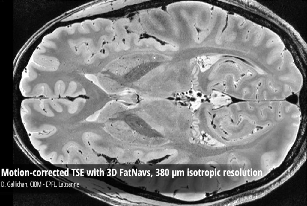
37 Motion-corrected GRE with 3D FatNavs, 380 μm isotropic resolution


38 The hippocampus From Neurocomic M. Farinella and H. Ros
")

39 1 mm resolution (1000 nl) 500 µm resolution (125 nl) 380 µm resolution (55 nl) Manually segmented hippocampus


")
40 1 mm resolution (1000 nl) 500 µm resolution (125 nl) 350 µm resolution (43 nl)

![) changes in motion-parameters and B0-field changes [1] Kober et al, MRM](/docs-images/74/70824121/images/41-2.jpg "2011 T2*-weighted GRE 250 µm x 250 µm x 1.")
41 Hot off the press Gretsch et al, ISMRM 2016 Integration of dual-echo FatNavs with FIDnavs 1 Sensitive to respiratory and cardiac (!) changes in motion-parameters and B0-field changes [1] Kober et al, MRM 2011 T2*-weighted GRE 250 µm x 250 µm x 1.00 mm Minimum-intensity projection over 9 mm slab
42 Outlook Make more widely available! Distinguish brain regions by cortical contrast on a single-subject basis
43 Outlook Make more widely available! Distinguish brain regions by cortical contrast on a single-subject basis Identify pathologies where ultra-high resolution may be beneficial Cryptogenic epilepsy Hippocampal abnormalities in early Alzheimer s Substantia nigra changes in Parkinson s


44 Outlook Make more widely available! Distinguish brain regions by cortical contrast on a single-subject basis Identify pathologies where ultra-high resolution may be beneficial Analyse many datasets to assess impact of motion-correction Centre d Imagerie BioMédicale
Methods. Experimental Stimuli: We selected 24 animals, 24 tools, and 24
 Methods Experimental Stimuli: We selected 24 animals, 24 tools, and 24 nonmanipulable object concepts following the criteria described in a previous study. For each item, a black and white grayscale photo
Works-in-Progress package Version 1.0. For the SIEMENS Magnetom. Installation and User s Guide NUMARIS/4VA21B. January 22, 2003
 Works-in-Progress package Version 1.0 For the Installation and User s Guide NUMARIS/4VA21B January 22, 2003 Section of Medical Physics, University Hospital Freiburg, Germany Contact: Klaus Scheffler PhD,
Page 1 of 9. Protocol: adult_other_adni3_study_human_ge_3t_25w_ _ _1. 3 Plane Localizer. 3 Plane Localizer PATIENT POSITION
 3 Localizer FOV 26.0 Slice Thickness 5.0 Slice Spacing 0.0 Freq 256 Phase 128 3-PLANE 3 Localizer Unswap Phase Correction Gradient Echo Imaging Options Seq, Fast Recon All Images Contrast Yes/ 3 Localizer
MRI Summer Course Lab 2: Gradient Echo T1 & T2* Curves
 MRI Summer Course Lab 2: Gradient Echo T1 & T2* Curves Experiment 1 Goal: Examine the effect caused by changing flip angle on image contrast in a simple gradient echo sequence and derive T1-curves. Image
Simultaneous Multi-Slice (Slice Accelerated) Diffusion EPI
 Diffusion EPI") Simultaneous Multi-Slice (Slice Accelerated) Diffusion EPI Val M. Runge, MD Institute for Diagnostic and Interventional Radiology Clinics for Neuroradiology and Nuclear Medicine University Hospital Zurich
Simultaneous Multi-Slice (Slice Accelerated) Diffusion EPI Val M. Runge, MD Institute for Diagnostic and Interventional Radiology Clinics for Neuroradiology and Nuclear Medicine University Hospital Zurich
Encoding of inductively measured k-space trajectories in MR raw data
 Downloaded from orbit.dtu.dk on: Apr 10, 2018 Encoding of inductively measured k-space trajectories in MR raw data Pedersen, Jan Ole; Hanson, Christian G.; Xue, Rong; Hanson, Lars G. Publication date:
TimTX TrueShape. The parallel transmit architecture of the future. Answers for life.
 www.siemens.com/trueshape TimTX TrueShape The parallel transmit architecture of the future. The product/feature (mentioned herein) is not commercially available. Due to regulatory reasons its future availability
Diffusion and Functional MRI of the Spinal Cord Methods and Clinical Applications
 Diffusion and Functional MRI of the Spinal Cord Methods and Clinical Applications Susceptibility artifacts in DTI of the spinal cord J. Cohen-Adad Q-space imaging and axon diameter measurements Functional
2015 Spin echoes and projection imaging
 1. Spin Echoes 1.1 Find f0, transmit amplitudes, and shim settings In order to acquire spin echoes, we first need to find the appropriate scanner settings using the FID GUI. This was all done last week,
Improve Image Quality of Transversal Relaxation Time PROPELLER and FLAIR on Magnetic Resonance Imaging
 Journal of Physics: Conference Series PAPER OPEN ACCESS Improve Image Quality of Transversal Relaxation Time PROPELLER and FLAIR on Magnetic Resonance Imaging To cite this article: N Rauf et al 2018 J.
NIH Public Access Author Manuscript Int J Cardiovasc Imaging. Author manuscript; available in PMC 2008 May 26.
 NIH Public Access Author Manuscript Published in final edited form as: Int J Cardiovasc Imaging. 2001 August ; 17(4): 287 296. A comparison of prospective and retrospective respiratory navigator gating
3T Unlimited. ipat on MAGNETOM Allegra The Importance of ipat at 3T. medical
 3T Unlimited ipat on MAGNETOM Allegra The Importance of ipat at 3T s medical ipat on MAGNETOM Allegra The Importance of ipat at 3T The rise of 3T MR imaging Ultra High Field MR (3T) has flourished during
ACRIN 6686 / RTOG 0825
 ACRIN 6686 (RTOG 0825) Advanced MRI Imaging Manual ACRIN 6686 / RTOG 0825 A phase III double blind placebo controlled trial of conventional chemoradiation and adjuvant temozolomide plus bevacizumab vs
ISSN X CODEN (USA): PCHHAX. The role of dual spin echo in increasing resolution in diffusion weighted imaging of brain
: PCHHAX. The role of dual spin echo in increasing resolution in diffusion weighted imaging of brain") Available online at www.derpharmachemica.com ISSN 0975-413X CODEN (USA): PCHHAX Der Pharma Chemica, 2016, 8(17):15-20 (http://derpharmachemica.com/archive.html) The role of in increasing resolution in
Available online at www.derpharmachemica.com ISSN 0975-413X CODEN (USA): PCHHAX Der Pharma Chemica, 2016, 8(17):15-20 (http://derpharmachemica.com/archive.html) The role of in increasing resolution in
Medical Imaging. X-rays, CT/CAT scans, Ultrasound, Magnetic Resonance Imaging
 Medical Imaging X-rays, CT/CAT scans, Ultrasound, Magnetic Resonance Imaging From: Physics for the IB Diploma Coursebook 6th Edition by Tsokos, Hoeben and Headlee And Higher Level Physics 2 nd Edition
Attenuation Correction in Hybrid MR-BrainPET Imaging
 Mitglied der Helmholtz-Gemeinschaft Attenuation Correction in Hybrid MR-BrainPET Imaging Elena Rota Kops Institute of Neuroscience and Biophysics Medicine Brain Imaging Physics Interactions of 511 kev
KEYENCE VKX LASER-SCANNING CONFOCAL MICROSCOPE Standard Operating Procedures (updated Oct 2017)
") KEYENCE VKX LASER-SCANNING CONFOCAL MICROSCOPE Standard Operating Procedures (updated Oct 2017) 1 Introduction You must be trained to operate the Laser-scanning confocal microscope (LSCM) independently.
KEYENCE VKX LASER-SCANNING CONFOCAL MICROSCOPE Standard Operating Procedures (updated Oct 2017) 1 Introduction You must be trained to operate the Laser-scanning confocal microscope (LSCM) independently.
High-Resolution Corrosion Monitoring for Reliable Assessment of Infrastructure
 19 th World Conference on Non-Destructive Testing 2016 High-Resolution Corrosion Monitoring for Reliable Assessment of Infrastructure André Lamarre 1 1 Olympus Scientific Solutions Americas, Quebec City,
Applications Guide. Spectral Editing with SVS. (Works-in-Progress) MAGNETOM TaTs and Verio Systems (3T)
 MAGNETOM TaTs and Verio Systems (3T)") Applications Guide Spectral Editing with SVS (Works-in-Progress) MAGNETOM TaTs and Verio Systems (3T) syngo MR Numaris 4 VB17A June 2009 Version 1.1 WIP #529 Important Note This document provides a description
Applications Guide Spectral Editing with SVS (Works-in-Progress) MAGNETOM TaTs and Verio Systems (3T) syngo MR Numaris 4 VB17A June 2009 Version 1.1 WIP #529 Important Note This document provides a description
Hardware. MRI System. MRI system Multicoil Microstrip. Part1
 Hardware MRI system Multicoil Microstrip MRI System Part1 1 The MRI system is made up of a variety of subsystems. the Operator Workspace Gradient Driver subsystem The Physiological Acquisition Controller
Practical Image and Video Processing Using MATLAB
 Practical Image and Video Processing Using MATLAB Chapter 1 Introduction and overview What will we learn? What is image processing? What are the main applications of image processing? What is an image?
technology meets pathology Institute of Pathology, Charité Universitätsmedizin Berlin, Berlin, Germany 3 Overview
 ASSESSMENT OF TECHNICAL PARAMETERS A. Alekseychuk 1, N. Zerbe 2, Y. Yagi 3 1 Computer Vision and Remote Sensing, TU Berlin, Berlin, Germany 2 Institute of Pathology, Charité Universitätsmedizin Berlin,
MRI Phase Mismapping Image Artifact Correction
 American Journal of Biomedical Engineering 2016, 6(4): 115-123 DOI: 10.5923/j.ajbe.20160604.02 MRI Phase Mismapping Image Artifact Correction Ashraf A. Abdallah 1,*, Mawia A. Hassan 2 1 Medical Engineering
Toward an Augmented Reality System for Violin Learning Support
 Toward an Augmented Reality System for Violin Learning Support Hiroyuki Shiino, François de Sorbier, and Hideo Saito Graduate School of Science and Technology, Keio University, Yokohama, Japan {shiino,fdesorbi,saito}@hvrl.ics.keio.ac.jp
Noninvasive Blood Flow Mapping with Arterial Spin Labeling (ASL) Paul Kyu Han and Sung-Hong Park
 Paul Kyu Han and Sung-Hong Park") Noninvasive Blood Flow Mapping with Arterial Spin Labeling (ASL) Paul Kyu Han and Sung-Hong Park Department of Bio and Brain Engineering, Korea Advanced Institute of Science and Technology (KAIST), Daejeon,
Noninvasive Blood Flow Mapping with Arterial Spin Labeling (ASL) Paul Kyu Han and Sung-Hong Park Department of Bio and Brain Engineering, Korea Advanced Institute of Science and Technology (KAIST), Daejeon,
FRAUNHOFER INSTITUTE FOR INTEGRATED CIRCUITS IIS. MANUAL PANORAMIC MICROSCOPY WITH istix
 FRAUNHOFER INSTITUTE FOR INTEGRATED CIRCUITS IIS MANUAL PANORAMIC MICROSCOPY WITH istix CLINICAL DIAGNOSTICS AND MATERIAL SCIENCES IMPROVED BY DIGITAL MICROSCOPY B A C K G R O U N D Due to a high grade
Lesson 06: Pulse-echo Imaging and Display Modes. These lessons contain 26 slides plus 15 multiple-choice questions.
 Lesson 06: Pulse-echo Imaging and Display Modes These lessons contain 26 slides plus 15 multiple-choice questions. These lesson were derived from pages 26 through 32 in the textbook: ULTRASOUND IMAGING
Pulse Sequences: Rapid Gradient Echo
 Pulse Sequences: Rapid Gradient Echo M229 Advanced Topics in MRI Holden H. Wu, Ph.D. 2018.04.17 Department of Radiological Sciences David Geffen School of Medicine at UCLA Class Business Office hours -
High Energy Digital Radiography & 3D-CT for Industrial Systems
 DIR 2007 - International Symposium on Digital industrial Radiology and Computed Tomography, June 25-27, 2007, Lyon, France High Energy Digital Radiography & 3D-CT for Industrial Systems Non-Destructive
Annual Ceiling Amount: $
 High Field Magnetic Resonance Imaging Center Research MRI Suite, Basement Floor of Building 203 VAMC 4150 Clement Street, San Francisco, CA 94121 MR STUDY APPLICATION for Research Users Application date:
Software and Hardware in CCTA. Elly Castellano PhD
 Software and Hardware in CCTA Elly Castellano PhD Outline technical requirements for coronary CTA the modern cardiac CT scanner ECG-gating technology image reconstruction algorithms 2 Technical requirements
Postprocessing of nonuniform MRI
 Postprocessing of nonuniform MRI Wolfgang Stefan, Anne Gelb and Rosemary Renaut Arizona State University Oct 11, 2007 Stefan, Gelb, Renaut (ASU) Postprocessing October 2007 1 / 24 Outline 1 Introduction
Field Simulation Software to Improve Magnetic Resonance Imaging
 Field Simulation Software to Improve Magnetic Resonance Imaging a joint project with the NRI in South Korea CST Usergroup Meeting 2010 Darmstadt Institute for Biometry and Medicine Informatics J. Mallow,
160-slice CT SCANNER / New Standard for the Future
 TECHNOLOGY HISTORY For over 130 years, Toshiba has been a world leader in developing technology to improve the quality of life. Our 50,000 global patents demonstrate a long, rich history of leading innovation.
Pulse Sequence Design Made Easier
 Pulse Sequence Design Made Easier Gregory L. Wheeler, BSRT(R)(MR) MRI Consultant gurumri@gmail.com 1 2 Pulse Sequences generally have the following characteristics: An RF line characterizing RF Pulse applications
Sub-millimeter Wave Planar Near-field Antenna Testing
 Sub-millimeter Wave Planar Near-field Antenna Testing Daniёl Janse van Rensburg 1, Greg Hindman 2 # Nearfield Systems Inc, 1973 Magellan Drive, Torrance, CA, 952-114, USA 1 drensburg@nearfield.com 2 ghindman@nearfield.com
Supplementary Figure 1
 Supplementary Figure 1 Left aspl Right aspl Detailed description of the fmri activation during allocentric action observation in the aspl. Averaged activation (N=13) during observation of the allocentric
Enhancing Gray-to-White Matter Contrast in 3T T1 Spin-Echo Brain Scans by Optimizing Flip Angle
 AJNR Am J Neuroradiol 26:2000 2004, September 2005 Enhancing Gray-to-White Matter Contrast in 3T T1 Spin-Echo Brain Scans by Optimizing Flip Angle Bernd L. Schmitz, Georg Grön, Florian Brausewetter, Martin
SIEMENS MAGNETOM Skyra syngo MR D13
 Page 1 of 12 SIEMENS MAGNETOM Skyra syngo MR D13 \\USER\CIND\StudyProtocols\PTSA\*ep2d_M0Map_p2_TE15 TA:7.9 s PAT:2 Voxel size:2.5 2.5 3.0 mm Rel. SNR:1.00 :epfid Properties Routine Contrast Prio Recon
Lab 8 6.S02 Spring 2013 MRI Projection Imaging
 1. Spin Echos 1.1 Find f0, TX amplitudes, and shim settings In order to acquire spin echos, we first need to find the appropriate scanner settings using the FID GUI. This was all done last week, but these
Digital Photographic Imaging Using MOEMS
 Digital Photographic Imaging Using MOEMS Vasileios T. Nasis a, R. Andrew Hicks b and Timothy P. Kurzweg a a Department of Electrical and Computer Engineering, Drexel University, Philadelphia, USA b Department
k y 2k y,max k x 2k x,max
 EE225E/BIOE265 Spring 2012 Principles of MRI Miki Lustig Assignment 5 Due Feb 26, 2012 1. Finish reading Nishimura Ch. 5. 2. For the 16 turn spiral trajectory, plotted below, what is the a) Spatial resolution,
SECTION I - CHAPTER 2 DIGITAL IMAGING PROCESSING CONCEPTS
 RADT 3463 - COMPUTERIZED IMAGING Section I: Chapter 2 RADT 3463 Computerized Imaging 1 SECTION I - CHAPTER 2 DIGITAL IMAGING PROCESSING CONCEPTS RADT 3463 COMPUTERIZED IMAGING Section I: Chapter 2 RADT
Jordan Journal of Physics
 Volume 9, Number 2, 2016. pp. 103-108 ARTICLE Jordan Journal of Physics Robust High Resolution Fat-Water Separation in the Abdomen during Free-Breathing by Self-Gated 2D Radial TrueFISP Imaging Riad S.
Digital Image Processing
 What is an image? Digital Image Processing Picture, Photograph Visual data Usually two- or three-dimensional What is a digital image? An image which is discretized, i.e., defined on a discrete grid (ex.
Image Enhancement in spatial domain. Digital Image Processing GW Chapter 3 from Section (pag 110) Part 2: Filtering in spatial domain
 Part 2: Filtering in spatial domain") Image Enhancement in spatial domain Digital Image Processing GW Chapter 3 from Section 3.4.1 (pag 110) Part 2: Filtering in spatial domain Mask mode radiography Image subtraction in medical imaging 2 Range
Image Enhancement in spatial domain Digital Image Processing GW Chapter 3 from Section 3.4.1 (pag 110) Part 2: Filtering in spatial domain Mask mode radiography Image subtraction in medical imaging 2 Range
HISTORY. CT Physics with an Emphasis on Application in Thoracic and Cardiac Imaging SUNDAY. Shawn D. Teague, MD
 CT Physics with an Emphasis on Application in Thoracic and Cardiac Imaging Shawn D. Teague, MD DISCLOSURES 3DR- advisory committee CT PHYSICS WITH AN EMPHASIS ON APPLICATION IN THORACIC AND CARDIAC IMAGING
TITLE: Prostate Cancer Detection Using High-Spatial Resolution MRI at 7.0 Tesla: Correlation with Histopathologic Findings at Radical Prostatectomy
 Award Number: W81XWH-11-1-0253 TITLE: Prostate Cancer Detection Using High-Spatial Resolution MRI at 7.0 Tesla: Correlation with Histopathologic Findings at Radical Prostatectomy PRINCIPAL INVESTIGATOR:
Operation of a Single Pass, Bunch-by-bunch x-ray Beam Size Monitor for the CESR Test Accelerator Research Program. October 3, 2012
 Operation of a Single Pass, Bunch-by-bunch x-ray Beam Size Monitor for the CESR Test Accelerator Research Program October 3, 2012 Goals Goals For This Presentation: 1.Provide an overview of the efforts
Pulse Sequence Design and Image Procedures
 Pulse Sequence Design and Image Procedures 1 Gregory L. Wheeler, BSRT(R)(MR) MRI Consultant 2 A pulse sequence is a timing diagram designed with a series of RF pulses, gradients switching, and signal readout
A machine vision system for scanner-based laser welding of polymers
 A machine vision system for scanner-based laser welding of polymers Zelmar Echegoyen Fernando Liébana Laser Polymer Welding Recent results and future prospects for industrial applications in a European
Digital Radiographic Inspection replacing traditional RT and 3D RT Development
 Digital Radiographic Inspection replacing traditional RT and 3D RT Development Iploca Novel Construction Meeting 27&28 March 2014 Geneva By Jan van der Ent Technical Authority International Contents Introduction
BOLD fmri: signal source, data acquisition, and interpretation
 BOLD fmri: signal source, data acquisition, and interpretation Cheryl Olman 4 th year student, Department of Neuroscience and Center for Magnetic Resonance Research Discussion series Week 1: Biological
QUANTITATIVE COMPUTERIZED LAMINOGRAPHY. Suzanne Fox Buchele and Hunter Ellinger
 QUANTITATIVE COMPUTERIZED LAMINOGRAPHY Suzanne Fox Buchele and Hunter Ellinger Scientific Measurement Systems, Inc. 2201 Donley Drive Austin, Texas 78758 INTRODUCTION Industrial computerized-tomography
Breast Tomosynthesis. Bob Liu, Ph.D. Department of Radiology Massachusetts General Hospital And Harvard Medical School
 Breast Tomosynthesis Bob Liu, Ph.D. Department of Radiology Massachusetts General Hospital And Harvard Medical School Outline Physics aspects of breast tomosynthesis Quality control of breast tomosynthesis
1. Figure A' is similar to Figure A. Which transformations compose the similarity transformation that maps Figure A onto Figure A'?
 Exit Ticket Sample Solutions 1. Figure A' is similar to Figure A. Which transformations compose the similarity transformation that maps Figure A onto Figure A'? Figure A Figure A' We first take a dilation
Synchrotron X-ray tomographic microscopy Theory vs. practice
 Synchrotron X-ray tomographic microscopy Theory vs. practice Federica Marone Swiss Light Source, Paul Scherrer Institut, Villigen, Switzerland Theory Radon transform Rf x = Beer-Lambert law I E = I 0 (E)e
Standards for Imaging Endpoints in Clinical Trials: Standardization and Optimization of Image Acquisitions: Magnetic Resonance
 FDA Workshop April 13, 2010 Standards for Imaging Endpoints in Clinical Trials: Standardization and Optimization of Image Acquisitions: Magnetic Resonance Edward F. Jackson, PhD Professor and Chief, Section
Taylor Hanayik. John E. Richards. Department of Psychology, University of South Carolina. March, 2018
 Preprocessing and processing pipeline for fmri for faces and houses study Taylor Hanayik John E. Richards Department of Psychology, University of South Carolina March, 2018 Corresponding author: Taylor
Noise Characteristics of the FORE+OSEM(DB) Reconstruction Method for the MiCES PET Scanner
 Reconstruction Method for the MiCES PET Scanner") Noise Characteristics of the FORE+OSEM(DB) Reconstruction Method for the MiCES PET Scanner Kisung Lee, Member, IEEE, Paul E. Kinahan, Senior Member, Robert S. Miyaoka, Member, IEEE, Jeffrey A. Fessler,
Noise Characteristics of the FORE+OSEM(DB) Reconstruction Method for the MiCES PET Scanner Kisung Lee, Member, IEEE, Paul E. Kinahan, Senior Member, Robert S. Miyaoka, Member, IEEE, Jeffrey A. Fessler,
Cardiac MR. Dr John Ridgway. Leeds Teaching Hospitals NHS Trust, UK
 Cardiac MR Dr John Ridgway Leeds Teaching Hospitals NHS Trust, UK Cardiac MR Physics for clinicians: Part I Journal of Cardiovascular Magnetic Resonance 2010, 12:71 http://jcmr-online.com/content/12/1/71
Rapid Non linear Image Scanning Microscopy, Supplementary Notes
 Rapid Non linear Image Scanning Microscopy, Supplementary Notes Calculation of theoretical PSFs We calculated the electrical field distribution using the wave optical theory developed by Wolf 1, and Richards
Center for Imaging and Sensing (CIS)
") Center for Imaging and Sensing (CIS) Raunak Borwankar, Ian Costanzo, Gene Bogdanov, Sasidhar Tadanki, Reinhold Ludwig ECE Department 100 Institute Road Worcester, MA 01609 Phone: 508-831-5231 October 10,
Center for Imaging and Sensing (CIS) Raunak Borwankar, Ian Costanzo, Gene Bogdanov, Sasidhar Tadanki, Reinhold Ludwig ECE Department 100 Institute Road Worcester, MA 01609 Phone: 508-831-5231 October 10,
Tribometrics. Version 2.11
 Tribometrics Version 2.11 Table of Contents Tribometrics... 1 Version 2.11... 1 1. About This Document... 4 1.1. Conventions... 4 2. Introduction... 5 2.1. Software Features... 5 2.2. Tribometrics Overview...
Structured-Light Based Acquisition (Part 1)
") Structured-Light Based Acquisition (Part 1) CS635 Spring 2017 Daniel G. Aliaga Department of Computer Science Purdue University Passive vs. Active Acquisition Passive + Just take pictures + Does not intrude
Structured-Light Based Acquisition (Part 1) CS635 Spring 2017 Daniel G. Aliaga Department of Computer Science Purdue University Passive vs. Active Acquisition Passive + Just take pictures + Does not intrude
A Mathematical model for the determination of distance of an object in a 2D image
 A Mathematical model for the determination of distance of an object in a 2D image Deepu R 1, Murali S 2,Vikram Raju 3 Maharaja Institute of Technology Mysore, Karnataka, India rdeepusingh@mitmysore.in
DIGITAL-MICROSCOPY CAMERA SOLUTIONS USB 3.0
 DIGITAL-MICROSCOPY CAMERA SOLUTIONS USB 3.0 PixeLINK for Microscopy Applications PixeLINK will work with you to choose and integrate the optimal USB 3.0 camera for your microscopy project. Ideal for use
Reconstruction of Non-Cartesian MRI Data
 G16.448 Practical Magnetic Resonance Imaging II Sacler Institute of Biomedical Sciences New Yor Universit School of Medicine Reconstruction of Non-Cartesian MRI Data Ricardo Otazo PhD ricardo.otazo@numc.org
SIGNA Explorer Lift revives our MR
 Seiji Shiotani, MD, PhD Seirei Fuji Hospital in Fuji City, Shizuoka, Japan Masayoshi Sugimura Seirei Fuji Hospital in Fuji City, Shizuoka, Japan SIGN Explorer Lift revives our MR The clinical usefulness
Supplementary Information
 Supplementary Information Simultaneous whole- animal 3D- imaging of neuronal activity using light field microscopy Robert Prevedel 1-3,10, Young- Gyu Yoon 4,5,10, Maximilian Hoffmann,1-3, Nikita Pak 5,6,
Regan Mandryk. Depth and Space Perception
 Depth and Space Perception Regan Mandryk Disclaimer Many of these slides include animated gifs or movies that may not be viewed on your computer system. They should run on the latest downloads of Quick
Digital Imaging CT & MR
 Digital Imaging CT & MR January 22, 2008 Digital Radiography, CT and MRI generate images in a digital format What is a Digital Image? A digital image is made up of picture elements, pixels row by column
SUPER RESOLUTION INTRODUCTION
 SUPER RESOLUTION Jnanavardhini - Online MultiDisciplinary Research Journal Ms. Amalorpavam.G Assistant Professor, Department of Computer Sciences, Sambhram Academy of Management. Studies, Bangalore Abstract:-
Development and Application of 500MSPS Digitizer for High Resolution Ultrasonic Measurements
 Indian Society for Non-Destructive Testing Hyderabad Chapter Proc. National Seminar on Non-Destructive Evaluation Dec. 7-9, 2006, Hyderabad Development and Application of 500MSPS Digitizer for High Resolution
PD233: Design of Biomedical Devices and Systems
 PD233: Design of Biomedical Devices and Systems (Lecture-8 Medical Imaging Systems) (Imaging Systems Basics, X-ray and CT) Dr. Manish Arora CPDM, IISc Course Website: http://cpdm.iisc.ac.in/utsaah/courses/
SmartExam helps to banish most repeat knee studies at Desert Medical Imaging
 I s s u e 3 2 - J u l y 2 0 0 7 F i e l d Strength Publication for the Philips MRI Community SmartExam helps to banish most repeat knee studies at Desert Medical Imaging Total scan automation boosts efficiency,
Toward Non-stationary Blind Image Deblurring: Models and Techniques
 Toward Non-stationary Blind Image Deblurring: Models and Techniques Ji, Hui Department of Mathematics National University of Singapore NUS, 30-May-2017 Outline of the talk Non-stationary Image blurring
DEVELOPMENT AND APPLICATION OF AN EXTENDED GEOMETRIC MODEL FOR HIGH RESOLUTION PANORAMIC CAMERAS
 DEVELOPMENT AND APPLICATION OF AN EXTENDED GEOMETRIC MODEL FOR HIGH RESOLUTION PANORAMIC CAMERAS D. Schneider, H.-G. Maas Dresden University of Technology Institute of Photogrammetry and Remote Sensing
Edge-Raggedness Evaluation Using Slanted-Edge Analysis
 Edge-Raggedness Evaluation Using Slanted-Edge Analysis Peter D. Burns Eastman Kodak Company, Rochester, NY USA 14650-1925 ABSTRACT The standard ISO 12233 method for the measurement of spatial frequency
Project: Sudoku solver
 Project: Sudoku solver Write a program that finds the sudoku square in the image, detects the 81 fields, and identifies the number in the fields that have a number. The output should be a 9x9 matrix with
For customers in USA This device complies with Part 15 of the FCC rules. Operation is subject to the following two conditions:
 User manual For customers in North and South America For customers in USA This device complies with Part 15 of the FCC rules. Operation is subject to the following two conditions: (1) This device may not
TGR EDU: EXPLORE HIGH SCHOOL DIGITAL TRANSMISSION
 TGR EDU: EXPLORE HIGH SCHL DIGITAL TRANSMISSION LESSON OVERVIEW: Students will use a smart device to manipulate shutter speed, capture light motion trails and transmit their digital image. Students will
(N)MR Imaging. Lab Course Script. FMP PhD Autumn School. Location: C81, MRI Lab B0.03 (basement) Instructor: Leif Schröder. Date: November 3rd, 2010
MR Imaging. Lab Course Script. FMP PhD Autumn School. Location: C81, MRI Lab B0.03 (basement) Instructor: Leif Schröder. Date: November 3rd, 2010") (N)MR Imaging Lab Course Script FMP PhD Autumn School Location: C81, MRI Lab B0.03 (basement) Instructor: Leif Schröder Date: November 3rd, 2010 1 Purpose: Understanding the basic principles of MR imaging
(N)MR Imaging Lab Course Script FMP PhD Autumn School Location: C81, MRI Lab B0.03 (basement) Instructor: Leif Schröder Date: November 3rd, 2010 1 Purpose: Understanding the basic principles of MR imaging
Introduction. Chapter 16 Diagnostic Radiology. Primary radiological image. Primary radiological image
 Introduction Chapter 16 Diagnostic Radiology Radiation Dosimetry I Text: H.E Johns and J.R. Cunningham, The physics of radiology, 4 th ed. http://www.utoledo.edu/med/depts/radther In diagnostic radiology
Optimization of Energy Modulation Filter for Dual Energy CBCT Using Geant4 Monte-Carlo Simulation
 Original Article PROGRESS in MEDICAL PHYSICS 27(3), Sept. 2016 http://dx.doi.org/10.14316/pmp.2016.27.3.125 pissn 2508-4445, eissn 2508-4453 Optimization of Energy Modulation Filter for Dual Energy CBCT
12/21/2016. Siemens Medical Systems Research Agreement Philips Healthcare Research Agreement AAN and ASN Committees
 Joseph V. Fritz, PhD Nandor Pintor, MD Dent Neurologic Institute ASN 2017 Friday, January 20, 2017 Siemens Medical Systems Research Agreement Philips Healthcare Research Agreement AAN and ASN Committees
2014 M.S. Cohen all rights reserved
 2014 M.S. Cohen all rights reserved mscohen@g.ucla.edu IMAGE QUALITY / ARTIFACTS SYRINGOMYELIA Source http://gait.aidi.udel.edu/res695/homepage/pd_ortho/educate/clincase/syrsco.htm Surgery is usually recommended
BACKGROUND. ** 78% of all MRI scanners have Image Quality problems. *** *** 25% of all Multi-Channel RF coils have at least one bad channel.
 Range of Results from over 534 ACR-mandated Annual MRI Performance Evaluations on over 204 Magnets from 8 Vendors Spanning a 10-year Period Moriel NessAiver, Ph.D. - Simply Physics - Baltimore, MD moriel@simplyphysics.com
IMAGE MANAGEMENT PLAN FOR ACRIN PA 4003 Evaluation of the Ability of a Novel [ 18 F] amyloid ligand ([ 18 F-AV-45]) to distinguish patients with a
![IMAGE MANAGEMENT PLAN FOR ACRIN PA 4003 Evaluation of the Ability of a Novel [ 18 F] amyloid ligand ([ 18 F-AV-45]) to distinguish patients with a](/thumbs/76/73395541.jpg "IMAGE MANAGEMENT PLAN FOR ACRIN PA 4003 Evaluation of the Ability of a Novel [ 18 F] amyloid ligand ([ 18 F-AV-45]) to distinguish patients with a") IMAGE MANAGEMENT PLAN FOR ACRIN PA 4003 Evaluation of the Ability of a Novel [ 18 F] amyloid ligand ([ 18 F-AV-45]) to distinguish patients with a clinical diagnosis of Alzheimer s disease from cognitively
IMAGE MANAGEMENT PLAN FOR ACRIN PA 4003 Evaluation of the Ability of a Novel [ 18 F] amyloid ligand ([ 18 F-AV-45]) to distinguish patients with a clinical diagnosis of Alzheimer s disease from cognitively
Gradient Spoiling. Average balanced SSFP magnetization Reduce sensitivity to off-resonance. FFE, FISP, GRASS, GRE, FAST, Field Echo
 Gradient Spoiling Average balanced SSFP magnetization Reduce sensitivity to off-resonance FFE, FISP, GRASS, GRE, FAST, Field Echo 1 Gradient-Spoiled Sequence (GRE, FFE, FISP, GRASS) RF TR G z G y G x Signal
Year 6 Visual Arts Unit 2017 Colour and Tone Term: Week:
 Term: 1 2 3 4 Week: 1 2 3 4 5 6 7 8 9 10 11 OUTCOMES Making: investigates subject matter in an attempt to represent likenesses of things in the world - makes artworks for different audiences, assembling
ZEISS LSM 710 CONFOCAL MICROSCOPE USER MANUAL
 ZEISS LSM 710 CONFOCAL MICROSCOPE USER MANUAL START THE SYSTEM... 2 START ZEN SOFTWARE... 3 SET THE TEMPERATURE AND THE CO2 CONTROLLERS... OBSERVATION AT OCULARS... 5 STATIF PRESENTATION... 6 ACQUIRE ONE
Quintic Hardware Tutorial Camera Set-Up
 Quintic Hardware Tutorial Camera Set-Up 1 All Quintic Live High-Speed cameras are specifically designed to meet a wide range of needs including coaching, performance analysis and research. Quintic LIVE
Supplementary Material
 Supplementary Material Orthogonal representation of sound dimensions in the primate midbrain Simon Baumann, Timothy D. Griffiths, Li Sun, Christopher I. Petkov, Alex Thiele & Adrian Rees Methods: Animals
Advanced MSK MRI Protocols at 3.0T. Garry E. Gold, M.D. Associate Professor Department of Radiology Stanford University
 Advanced MSK MRI Protocols at 3.0T Garry E. Gold, M.D. Associate Professor Department of Radiology Stanford University Outline Why High Field for MSK? SNR and Relaxation Times Technical Issues Example
Background (~EE369B)
") Background (~EE369B) Magnetic Resonance Imaging D. Nishimura Overview of NMR Hardware Image formation and k-space Excitation k-space Signals and contrast Signal-to-Noise Ratio (SNR) Pulse Sequences 13
Background (~EE369B) Magnetic Resonance Imaging D. Nishimura Overview of NMR Hardware Image formation and k-space Excitation k-space Signals and contrast Signal-to-Noise Ratio (SNR) Pulse Sequences 13
Confocal Application Notes Vol. 5 July 2010
 Tile Scan Prepared by Myriam Gastard, PhD Application and Technical Support Group, Leica Microsystems, Inc. In this issue of our Confocal Application Notes, proper set up of the Tile function enables you
Modeling and Synthesis of Aperture Effects in Cameras
 Modeling and Synthesis of Aperture Effects in Cameras Douglas Lanman, Ramesh Raskar, and Gabriel Taubin Computational Aesthetics 2008 20 June, 2008 1 Outline Introduction and Related Work Modeling Vignetting
GE Healthcare. Discovery MR T. Simply powerful. Powerfully simple.
 GE Healthcare Discovery MR750 3.0T Simply powerful. Powerfully simple. Break free. The breast images you need in only two sequences. A complete liver study in a 15-minute time slot. Routine fmri with shorter
Image Quality/Artifacts Frequency (MHz)
") The Larmor Relation 84 Image Quality/Artifacts (MHz) 42 ω = γ X B = 2πf 84 0.0 1.0 2.0 Magnetic Field (Tesla) 1 A 1D Image Magnetic Field Gradients Magnet Field Strength Field Strength / Gradient Coil
The Larmor Relation 84 Image Quality/Artifacts (MHz) 42 ω = γ X B = 2πf 84 0.0 1.0 2.0 Magnetic Field (Tesla) 1 A 1D Image Magnetic Field Gradients Magnet Field Strength Field Strength / Gradient Coil