GE Healthcare. Discovery MR T. Simply powerful. Powerfully simple.
|
|
|
- Allison Johnston
- 6 years ago
- Views:
Transcription

1 GE Healthcare Discovery MR T Simply powerful. Powerfully simple.
2 Break free. The breast images you need in only two sequences. A complete liver study in a 15-minute time slot. Routine fmri with shorter paradigms and greater activation. Changing What ifs to Why nots. And that s just for starters. Because the Discovery MR T lets you break the bonds of traditional 3.0T MR scanning. Powerfully. Precisely. Productively. It s MR of unprecedented power. Pure and simple.


3 The Discovery MR750 enables a robust, comprehensive liver study in a 15-minute time slot. Conduct routine, accurate, repeatable 5-minute fmri studies with 60% more slices in the same scan time as before. High-resolution and multi-contrast acquisition, as well as real-time reconstruction, allow you to acquire the breast images you need in only two sequences.


4 Breaking today s MR boundaries. To give you every edge.


5

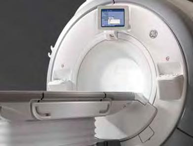
6 Routine exams made faster. Advanced exams routine. And every scan a study in speed. Introducing the Discovery MR750 Express Exam. Ready. Set. iroc (In-Room Operator Console). Faster exam setup at your fingertips. Mounted conveniently on the front of the magnet, the high-resolution color console of the Discovery MR750 consolidates patient set-up information and operator controls in one place that s easy to see no matter where you re standing. View patient, system and scan information, control and select parameters, change scanner configurations, and initiate scans in real time right in the room. Save footsteps by eliminating multiple trips to and from the control room. When AutoStart is selected on the iroc, the system will start scanning automatically when the scan room door is closed. Dual-sided scanner control panels. Within reach. Control the scanner from either side of the patient table and easily access cardiac or peripheral gating leads and IV lines. Backlit buttons indicate the next logical step in the exam process, and a built-in trackball mouse makes scrolling and clicking easy. IntelliTouch patient positioning. Start scanning in just two simple steps. The Discovery MR750 setup is so automated, you can position patients in just two simple steps, in as little as 30 seconds. Along with the use of the detachable table, IntelliTouch patient positioning shortens in-room set-up time by up to 70% over fixed-table designs. To operate, simply press the IntelliTouch strip on either side of the patient table at the landmark location, then press the Advance to Scan button to move the patient safely into the bore. Express Patient Table. Transfer patients once to speed throughput. Easily docked and undocked by a single technologist, the GE-exclusive Express detachable patient table boosts your You can fully prepare patients for their exams outside the scan room on the Discovery MR750 Express Patient Table. scanner efficiency and productivity by minimizing time between scans. Single transfers of patients directly onto the MR table create a more comfortable patient experience and increase exam productivity. In the event of an emergency, just one technologist can undock the table and safely transport the patient out of the room in under 30 seconds.


, the")


7 Two 32-channel surface coil connections integrated at the end of the table simplify patient preparation outside the scanning room. Easy-to-use IntelliTouch strips on each side of the patient table enable fast and accurate landmarking and patient positioning. The Advance to Scan button automatically moves the patient into scanning position. Go. With a simple, one-handed motion, the integrated arm boards can be optimally positioned to support the patient for injections and transport. With the high-resolution iroc (In-Room Operator Console), the technologist can complete patient setup right in the scan room for a streamlined exam. Located to the left and right of the scanner bore, dual-sided controls enable the scanner to be operated from either side of the patient table.


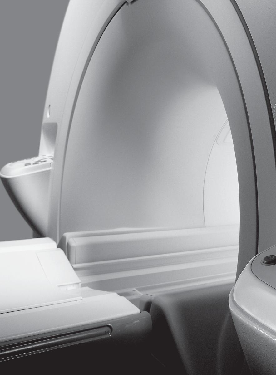
8 Swifter. Smoother. Simpler. The Discovery MR750 performs just as well for your patients as it does for you. Making a faster, easier, more comfortable MR experience. Focus on patients. Reduce patients anxiety by preparing for the exam outside the scan room on the detachable Express Patient Table. In the event of an emergency, the table can be undocked and removed from the scan room in just 30 seconds. With automated acquisition, processing, and networking, entire exams with the Discovery MR750 can be completed with just a few steps. Get patients in and out of the scan room quickly with highly streamlined workflow made possible by easier patient positioning, quicker exam setup, automated protocols, and faster scanning.


9 Spend more time at the patient s side by completing the exam setup while in the room. Answer questions, address concerns, offer reassurance, and provide an extra level of comfort all without leaving the room. Keep patients refreshed and comfortable by adjusting the airflow and lighting in the bore with multiple control settings.
10 No boundaries.

11 Just frontiers.
12 VIBRANT-Flex Combining multiple series into one, VIBRANT-Flex reduces exam times and ensures can t-miss homogeneous fat suppression over both breasts in the axial or sagittal plane. By utilizing self-calibrating ARC parallel imaging and eliminating the need for manual bilateral shimming, your productivity benefits on the Discovery MR750 are substantial. Water only Fat only In-phase Out-of-phase Cube TM Reformat submillimeter isotropic 3D volume image data from a single Cube acquisition into any plane with no gaps or loss of resolution. The innovative, selfcalibrating parallelimaging registration technique ARC secures rapid, virtually artifact-free imaging. Time of Flight Time of Flight (TOF) imaging on the Discovery MR750 generates better contrast between flowing blood and surrounding tissue and shortens scan time. Images with small vessel detail and subtleties of the blood flow are rapidly captured in high definition with no need for contrast injection for more confident diagnoses.
13 IDEAL Even in the most challenging areas of anatomy, this unique water and fat separation technique provides consistent, uniform fat suppression virtually every time from patient to patient, technologist to technologist. Water only Fat only In-phase Out-of-phase Industry-leading gradient performance and exceptional magnet homogeneity enable high-resolution, off-center musculoskeletal imaging.
14 LAVA-Flex Providing uniform, consistent fat suppression across the entire field of view, LAVA-Flex is a volumetric technique that produces four contrasts in just one acquisition, for confident diagnoses and fewer repeat exams. Its 3D nature allows for multi-plane reformats of exceptional quality. Case 1 Water only Fat only In-phase Case 2 Out-of-phase Coronal reformat Sagittal reformat 3D MERGE Generate excellent gray-white matter contrast in the spinal cord without sacrificing SNR. This high-definition, 3D application now enables you to do reformats in different planes.
15 Multi-Plane PROPELLER 2.0 The Discovery MR750 extends the remarkable motion-correction capabilities of PROPELLER 2.0 to all imaging planes. It consistently delivers contrast-rich, motion, and susceptibility artifact-free T2, T2 FLAIR, and DWI images. BrainWave With a two-fold improvement in overall signal stability, 60% more slices, and more SNR, this suite of functional MRI (fmri) applications for neurofunctional brain mapping and DTI consistently provides exceptional performance, making the challenging neuro exam routine. IDEAL Whole Spine With its ability to obtain both in-phase and out-of-phase images in one acquisition, IDEAL can be used to scan the entire spine to better identify and locate pathology. Diffusion Tensor Imaging (DTI)/FiberTrak With the Discovery MR750, DTI performance moves to a whole new level with more than twice the slices, alternatively more diffusion directions or shorter scan times. DTI/FiberTrak lets you visualize white matter in the brain as eigenvectors, ADC, fractional anisotropy maps, or 3D fiber tracts projections.


16 Start from a position of power. Powerful magnet. Powerful gradients. Powerful processing. Stunning new technologies take 3.0T MR imaging to a new level.
 gives you convenient control of")
17 Your window on power. The iroc (In-Room Operator Console) gives you convenient control of the system s exceptional technological capabilities.

18 3.OT The strongest whole-body gradients. The gradients the most powerful in the industry deliver unmatched 50 mt/m amplitude and 200 T/m/s slew-rate on each axis simultaneously. The result: faster, higher accuracy, and more reproducible scans. A new advanced thermal management system enables up to 60% more anatomical coverage and resolution per unit time compared to conventional designs. The 3.0T magnet. Built to last and built to deliver. The Discovery MR750 is built around the third generation, short-bore, superconducting 3.0T magnet proven to deliver high homogeneity for excellent results even in large or off-center FOV imaging, fat saturation techniques, and high-performance applications such as cardiac, fmri, diffusion tensor, and spectroscopy. Higher performance with real-time system optimization. PERFORM 2.0 automatically fine-tunes system SAR modeling, personalizing it for every patient. It provides up to a 17% gain in scanning efficiency and a proprietary gradient waveform algorithm, managing limitations due to peripheral nerve stimulation (PNS). Push protocol parameters and acquisition speeds to the limit without risking patient safety or slowing down the system. Unique gradient architecture for faster acquisitions. Heat extraction for fewer slowdowns. The advanced thermal management system increases reliability five-fold over previous generations of 3.0T systems.

19 Exclusive Optical RF system for greater SNR. Designed with OpTix, a GE-exclusive optical RF technology, the Discovery MR750 is engineered to increase signal clarity and maximize signal intensity for cleaner, crisper images. This next-generation digital receiver produces 4x dynamic range and its in-room integrated optical data architecture boosts the system signal-to-noise ratio by up to 27% over conventional non-optical receivers. Accelerated parallel imaging. A data-driven parallel-imaging technique, ARC TM utilizes a full 3D data kernel for more accurate reconstruction and improved image quality. It inspires a broader use of parallel imaging by eliminating misregistration errors and related artifacts and is less sensitive to tight fields-of-view. With the Discovery MR750, acquisition times are reduced confidently. Faster reconstructions. Reconstruction processing gains open the door for data-intensive 32-channel studies once thought impractical on 3.0T MR systems. A powerful volume reconstruction engine enables real-time image generation even from large, 3D parallel-imaging datasets. Anatomy-optimized RF coils and arrays. High-density arrays focus coil elements around the anatomy of interest and provide extended coverage where needed, for optimal image quality in every procedure. Open, flexible RF architecture provides you with coil options. Easily scalable for future expansion. Built on an exclusive data pipeline, the scalable architecture can be expanded to support 128 channels of simultaneous data acquisition.

20 Faster throughput can mean better ROI and more referrals. Invest in a Discovery MR750 and we ll make sure you keep your edge for a long time to come. Tilt the field in your favor. Count on GE to provide your Discovery MR750 with one of the largest, most highly trained service forces in the industry. Rated #1 in Overall Service Performance in the 2007 IMV ServiceTrak * MR report, our field engineers are rigorously trained on the unique service capabilities of the Discovery MR750 that help ensure proactive monitoring, reliability, and uptime. In addition, a wide range of learning tools teaches your imaging professionals how to use the advanced capabilities on the Discovery MR750 with skill and finesse. Exclusive to GE Healthcare, unique MR Masters Series Physicians Training provides an avenue to learn about the latest MR techniques directly from world-renowned radiologists. And our exclusive Continuum is the promise that you ll outrun obsolescence with the ability to upgrade your Discovery MR750 to future technology without replacing your magnet. *IMV is a leading independent third-party research firm and owner of auntminnie.com.
21
22 2008 General Electric Company All rights reserved. General Electric Company reserves the right to make changes in specifications and features shown herein, or discontinue the product described at any time without notice or obligation. GE, GE Monogram, ARC, Continuum, Cube and Discovery are trademarks of General Electric Company. A General Electric Company, doing business as GE Healthcare. Healthcare Re-imagined GE is dedicated to helping you transform healthcare delivery by driving critical breakthroughs in biology and technology. Our expertise in medical imaging and information technologies, medical diagnostics, patient monitoring systems, drug discovery, and biopharmaceutical manufacturing technologies is enabling healthcare professionals around the world to discover new ways to predict, diagnose and treat disease earlier. We call this model of care Early Health. The goal: to help clinicians detect disease earlier, access more information and intervene earlier with more targeted treatments, so they can help their patients live their lives to the fullest. Re-think, Re-discover, Re-invent, Re-imagine. GE Healthcare 3000 North Grandview Waukesha, WI U.S.A. imagination at work MR EN-US
CARING DESIGN. PRACTICAL TECHNOLOGY.
 GE Healthcare CARING DESIGN. PRACTICAL TECHNOLOGY. Brivo * MR355 1.5T Inspire ITS ELEGANT DESIGN AND WARM LIGHT ARE INTRIGUING. Brivo MR355 Inspire product manager Every piece of equipment you own represents
GE Healthcare. Essential for life. Senographe Essential Full-Field Digital Mammography system
 GE Healthcare Essential for life Senographe Essential Full-Field Digital Mammography system Excellence in FFDM is a process. An ongoing quest, fueled by our continuing breakthroughs in breast cancer detection
Vivid S5. Cardiovascular ultrasound system. GE Healthcare. Davis Medical
 GE Healthcare Vivid S5 Cardiovascular ultrasound system Versatility. It s a new design concept one that leverages our miniaturization expertise gained from the Vivid i and our performance expertise of
Delivering Better Patient Care with SIGNA Architect
 Delivering etter Patient Care with SIGNA Architect As a regional leader in outpatient-based diagnostic imaging, the radiologists and staff at Inova Fairfax MRI Center are focused on one thing: the patient.
GE Healthcare. Precision 500D. Digital radiography and fluoroscopy system
 GE Healthcare Precision 500D Digital radiography and fluoroscopy system What do you look for in a classical digital R&F system? Exceptional image quality at low dose, certainly. Increased clinical productivity
GE Healthcare LOGIQ P3. Staying ahead of the curve
 GE Healthcare LOGIQ P3 Staying ahead of the curve Ultrasound Expertise. Enhanced. GE is a trusted partner of many healthcare providers around the world. GE s TruScan technology, incorporating SmartScan,
GE Healthcare. Senographe 2000D Full-field digital mammography system
 GE Healthcare Senographe 2000D Full-field digital mammography system Digital has arrived. The Senographe 2000D Full-Field Digital Mammography (FFDM) system gives you a unique competitive advantage. That
SIGNA Architect. Make the unimaginable the expected. Issue Spotlight. Issue Spotlight
 SIGNA Architect Make the unimaginable the expected The potential for MR is now even more astonishing. Introducing SIGNA Architect 3.0T, the most advanced and intuitive engineering in MR technology from
Surpass the unimaginable and make it the expected.
 GE Healthcare Surpass the unimaginable and make it the expected. SIGNA Artist Imagine what MR can be. Artist is not yet CE marked. May not be marketed or placed into service until made to comply with CE
160-slice CT SCANNER / New Standard for the Future
 TECHNOLOGY HISTORY For over 130 years, Toshiba has been a world leader in developing technology to improve the quality of life. Our 50,000 global patents demonstrate a long, rich history of leading innovation.
Liver imaging beyond expectations with Ingenia
 Publication for the Philips MRI Community Issue 47 2012/3 Liver imaging beyond expectations with Ingenia Contributed by John Penatzer, RT, MR clinical product specialist, Cleveland, OH, USA Publication
GE Healthcare. Vivid S5. Cardiovascular ultrasound system
 GE Healthcare Vivid S5 Cardiovascular ultrasound system Versatility. It s a new design concept one that leverages our miniaturization expertise gained from the Vivid i and our performance expertise of
Brilliance in everything Philips CT products and services
 Brilliance in everything Philips CT products and services Ready for anything No one does more than Philips to help you gain the productivity you need with a comprehensive approach to CT that marries significant
SIGNA Explorer Lift revives our MR
 Seiji Shiotani, MD, PhD Seirei Fuji Hospital in Fuji City, Shizuoka, Japan Masayoshi Sugimura Seirei Fuji Hospital in Fuji City, Shizuoka, Japan SIGN Explorer Lift revives our MR The clinical usefulness
GE Healthcare. Performa. High-performance breast imaging
 GE Healthcare Performa High-performance breast imaging Moving mammography forward. And patients faster. GE Healthcare s unparalleled leadership across mammography begins with a deep understanding of breast
Multi-Access Biplane Lab
 Multi-Access Biplane Lab Advanced technolo gies deliver optimized biplane imaging Designed in concert with leading physicians, the Infinix VF-i/BP provides advanced, versatile patient access to meet the
Advanced MSK MRI Protocols at 3.0T. Garry E. Gold, M.D. Associate Professor Department of Radiology Stanford University
 Advanced MSK MRI Protocols at 3.0T Garry E. Gold, M.D. Associate Professor Department of Radiology Stanford University Outline Why High Field for MSK? SNR and Relaxation Times Technical Issues Example
The shortest distance to diagnosis Philips BrightView SPECT
 The shortest distance to diagnosis Philips Closer is better Simplicity is seeing something better right from the start. And is a completely new vision of what patient care can be, in a system as compact
3T Unlimited. ipat on MAGNETOM Allegra The Importance of ipat at 3T. medical
 3T Unlimited ipat on MAGNETOM Allegra The Importance of ipat at 3T s medical ipat on MAGNETOM Allegra The Importance of ipat at 3T The rise of 3T MR imaging Ultra High Field MR (3T) has flourished during
Background (~EE369B)
") Background (~EE369B) Magnetic Resonance Imaging D. Nishimura Overview of NMR Hardware Image formation and k-space Excitation k-space Signals and contrast Signal-to-Noise Ratio (SNR) Pulse Sequences 13
Background (~EE369B) Magnetic Resonance Imaging D. Nishimura Overview of NMR Hardware Image formation and k-space Excitation k-space Signals and contrast Signal-to-Noise Ratio (SNR) Pulse Sequences 13
TimTX TrueShape. The parallel transmit architecture of the future. Answers for life.
 www.siemens.com/trueshape TimTX TrueShape The parallel transmit architecture of the future. The product/feature (mentioned herein) is not commercially available. Due to regulatory reasons its future availability
1.5T HIGH FIELD SMALL ANIMAL MRI
 1.5T HIGH FIELD SMALL ANIMAL MRI Designed Specifically for Veterinarians TECHNICAL GUIDE ADVANCING THE ART AND SCIENCE OF VETERINARY MRI The PetVet is the only high-field MRI system designed specifically
Discover the new Prestige and experience 3D/4D imaging beyond your imagination.
 3D/4D Beyond Imagination The Prestige ultrasound imaging system represents the pinnacle of more than a decade of technological advancement in 3D/4D ultrasound imaging at MEDISON. Inheriting a tradition
2D, 3D CT Intervention, and CT Fluoroscopy
 2D, 3D CT Intervention, and CT Fluoroscopy SOMATOM Definition, Definition AS, Definition Flash Answers for life. Siemens CT Vision Siemens CT Vision The justification for the existence of the entire medical
Beyond the Scan Issue Spotlight
 GEHEALTHCARE.COM/MR 84 AUTUMN 2011 According to Merriam-Webster, the word humanize means, to represent as human, attribute human qualities to. It can be used in so many ways to humanize your brand, company,
[Maestro Class] MAGNETOM Harmony The New Degree of Perfection
![[Maestro Class] MAGNETOM Harmony The New Degree of Perfection](/thumbs/90/102664938.jpg "[Maestro Class] MAGNETOM Harmony The New Degree of Perfection") [Maestro Class] MAGNETOM Harmony The New Degree of Perfection MAGNETOM Family The Perfection of Care The aim of Siemens MR is the perfection of care. To create products and services that improve the quality
[Maestro Class] MAGNETOM Harmony The New Degree of Perfection MAGNETOM Family The Perfection of Care The aim of Siemens MR is the perfection of care. To create products and services that improve the quality
Image Quality. HTC Grid High Transmission Cellular Grid provides higher contrast images
 B R E A S T I M A G I N G S O L U T I O N S Setting the benchmark for mammography M-IV Series Innovations in breast imaging The Lorad M-IV Series exemplifies Hologic's commitment to developing advanced
Pulse Sequence Design and Image Procedures
 Pulse Sequence Design and Image Procedures 1 Gregory L. Wheeler, BSRT(R)(MR) MRI Consultant 2 A pulse sequence is a timing diagram designed with a series of RF pulses, gradients switching, and signal readout
Cardiac MR. Dr John Ridgway. Leeds Teaching Hospitals NHS Trust, UK
 Cardiac MR Dr John Ridgway Leeds Teaching Hospitals NHS Trust, UK Cardiac MR Physics for clinicians: Part I Journal of Cardiovascular Magnetic Resonance 2010, 12:71 http://jcmr-online.com/content/12/1/71
Maximum Performance, Minimum Space
 TECHNOLOGY HISTORY For over 130 years, Toshiba has been a world leader in developing technology to improve the quality of life. Our 50,000 global patents demonstrate a long, rich history of leading innovation.
MRI RF-Coils. Innovation with Integrity. Highest sensitivity for your preclinical MRI and MRS applications. Preclinical Imaging
 MRI RF-Coils Highest sensitivity for your preclinical MRI and MRS applications Innovation with Integrity Preclinical Imaging Molecular and Preclinical Imaging Preclinical magnetic resonance imaging of
General Imaging Ultrasound in a Whole New Light. Introducing the
 General Imaging Ultrasound in a Whole New Light. Introducing the It s a Tour de Force Redefining innovation through value and performance It s the dawning of a new day in the world of compact ultrasound
The Enlightened Choice for High-field MRI
 The Enlightened Choice for High-field MRI ECHELON heralds the dawn of a new standard for 1.5T superconductive MRI. The ECHELON features a small footprint with economics that do not compromise diagnostic
Hardware. MRI System. MRI system Multicoil Microstrip. Part1
 Hardware MRI system Multicoil Microstrip MRI System Part1 1 The MRI system is made up of a variety of subsystems. the Operator Workspace Gradient Driver subsystem The Physiological Acquisition Controller
Image Quality. HTC Grid High Transmission Cellular Grid provides higher contrast images
 B R E A S T I M A G I N G S O L U T I O N S Setting the benchmark for mammography M-IV Series Innovations in breast imaging The Lorad M-IV Series exemplifies Hologic s commitment to developing advanced
MR Basics: Module 8 Image Quality
 Module 8 Transcript For educational and institutional use. This transcript is licensed for noncommercial, educational inhouse or online educational course use only in educational and corporate institutions.
SmartExam helps to banish most repeat knee studies at Desert Medical Imaging
 I s s u e 3 2 - J u l y 2 0 0 7 F i e l d Strength Publication for the Philips MRI Community SmartExam helps to banish most repeat knee studies at Desert Medical Imaging Total scan automation boosts efficiency,
Design Your Performance
 MEDISON has been a leading name in diagnostic ultrasound since its foundation in 1985. As one of the only companies dedicated solely to ultrasound imaging, we have remained at the forefront of research
Control and confidence all around. Philips EP cockpit people focused solutions for heart rhythm care
 Control and confidence all around Philips EP cockpit people focused solutions for heart rhythm care EP cockpit - brings new innovations EP cockpit simplifies your EP lab 1. Improving your EP lab working
Magnetic Resonance Research Facility (MRRF) Resources
 Resources") Magnetic Resonance Research Facility (MRRF) Resources The Magnetic Resonance Research Facility (MRRF) has scanners located in both the hospital and research buildings on the campus of the University of
Magnetic Resonance Research Facility (MRRF) Resources The Magnetic Resonance Research Facility (MRRF) has scanners located in both the hospital and research buildings on the campus of the University of
SIGNA Pioneer: Ultra High Efficiency Gradient System Advancing the gradient technology curve
 GE Healthcare SIGNA Pioneer: Ultra High Efficiency Gradient System Advancing the gradient technology curve NEW TECHNOLOGY 40W Watts spec is irrelevant. 4W LED bulb delivers same brightness as 40W incandescent
Siemens AG, Healthcare Sector. syngo MR D Operator Manual - Breast 0.0.
 Siemens AG, Healthcare Sector Cs2 syngo Breast Operator 2010-2012 MR-05019 630 02 English 06/2012 n.a. Informatik, Manual D13 Cape syngo MR D13 Operator Manual - Breast syngo MR D13 www.siemens.com/healthcare
from signals to sources asa-lab turnkey solution for ERP research
 from signals to sources asa-lab turnkey solution for ERP research asa-lab : turnkey solution for ERP research Psychological research on the basis of event-related potentials is a key source of information
Maximizing clinical outcomes
 Maximizing clinical outcomes Digital Tomosynthesis Dual Energy Subtraction Automated Long Length Imaging Improved image quality at a low dose Xray Xray Patented ISS capture technology promotes high sensitivity
Invisible sophistication. Visible simplicity. CS Welcome to the simplicity of compact panoramic imaging
 Invisible sophistication. Visible simplicity. CS 8100 Welcome to the simplicity of compact panoramic imaging Introducing the CS 8100 The Carestream Dental Factor Humanized technology We keep our technology
High Field MRI: Technology, Applications, Safety, and Limitations
 High Field MRI: Technology, Applications, Safety, and Limitations R. Jason Stafford, Ph.D. The University of Texas M. D. Anderson Cancer Center, Houston, TX Introduction The amount of available signal
Publication for the Philips MRI Community
 FieldStrength Publication for the Philips MRI Community Issue 38 Summer 2009 Hospitals deploy Panorama HFO as an all purpose MRI scanner Panorama High Field Open scanner serves all MRI needs of both Community
Pitfalls and Remedies of MDCT Scanners as Quantitative Instruments
 intensity m(e) m (/cm) 000 00 0 0. 0 50 0 50 Pitfalls and Remedies of MDCT Scanners as Jiang Hsieh, PhD GE Healthcare Technology University of Wisconsin-Madison Root-Causes of CT Number Inaccuracies Nature
Compact yet Sophisticated
 Compact yet Sophisticated Hitachi has brought Open MRI one step further in its evolution, to better assist medical professionals who work at the forefront of healthcare. AIRIS Light MSK offers radiologists
GE Healthcare. Senographe Crystal The choice is crystal clear
 Senographe Crystal The choice is crystal clear Senographe Crystal The choice is crystal clear. The Senographe* Crystal mammography system makes it easy to transition to full-field digital mammography.
Weber State University Radiologic Technology 4603
 Weber State University Radiologic Technology 4603 MRI Physics and Instrumentation Instructor: Rex T. Christensen MHA R.T. (R) (MR) (CT) (ARRT) CIIP Contact Info: E-mail: rexchristensen@weber.edu Phone:
ACRIN 6686 / RTOG 0825
 ACRIN 6686 (RTOG 0825) Advanced MRI Imaging Manual ACRIN 6686 / RTOG 0825 A phase III double blind placebo controlled trial of conventional chemoradiation and adjuvant temozolomide plus bevacizumab vs
Iterative Reconstruction in Image Space. Answers for life.
 Iterative Reconstruction in Image Space Answers for life. Iterative Reconstruction in Image Space * (IRIS) * Please note: IRIS is used as an abbreviation for Iterative Reconstruction in Image Space throughout
Ultimate DR flexibility
 Ultimate DR flexibility to fit your room, workflow, and budget KODAK DIRECTVIEW DR 7500 System KODAK DIRECTVIEW DR 7500 System YOU HAVE NEVER SEEN A DIGITAL RADIOGRAPHY SYSTEM LIKE THIS! Your radiography
MRI SYSTEM COMPONENTS Module One
 MRI SYSTEM COMPONENTS Module One 1 MAIN COMPONENTS Magnet Gradient Coils RF Coils Host Computer / Electronic Support System Operator Console and Display Systems 2 3 4 5 Magnet Components 6 The magnet The
Ysio Max. The most direct way to the image. Answers for life.
 Ysio Max The most direct way to the image www.siemens.com/ysio-max Answers for life. 2 It s more. It s MAX. MAX Multiple Advances in X-ray It s more than just single features or functions. MAX offers multiple
MUSICA Nerve Center. Artificial Intelligence. Intelligent tools for your Digital Radiography workflow. Fluoroscopy. Workflow Optimization
 Image Quality Bariatric Abdomen Pediatric Imaging Diagnostic Confidence Fluoroscopy Neonatal Imaging Scatter Suppression Dental Full Leg Full Spine Exposure Control Index Artificial Intelligence General
Simultaneous Multi-Slice (Slice Accelerated) Diffusion EPI
 Diffusion EPI") Simultaneous Multi-Slice (Slice Accelerated) Diffusion EPI Val M. Runge, MD Institute for Diagnostic and Interventional Radiology Clinics for Neuroradiology and Nuclear Medicine University Hospital Zurich
Simultaneous Multi-Slice (Slice Accelerated) Diffusion EPI Val M. Runge, MD Institute for Diagnostic and Interventional Radiology Clinics for Neuroradiology and Nuclear Medicine University Hospital Zurich
Changing the Shape of Nuclear Medicine
 TRUTH IN IMAGING Changing the Shape of Nuclear Medicine Multi-Purpose SPECT Scanner Nothing Gets Closer Introducing 360 Body Contour Scanning With 360 degree detector coverage, and unique proximity sensors
Page 1 of 9. Protocol: adult_other_adni3_study_human_ge_3t_25w_ _ _1. 3 Plane Localizer. 3 Plane Localizer PATIENT POSITION
 3 Localizer FOV 26.0 Slice Thickness 5.0 Slice Spacing 0.0 Freq 256 Phase 128 3-PLANE 3 Localizer Unswap Phase Correction Gradient Echo Imaging Options Seq, Fast Recon All Images Contrast Yes/ 3 Localizer
MRI imaging in neuroscience Dr. Thom Oostendorp Lab class: 2 hrs
 MRI imaging in neuroscience Dr. Thom Oostendorp Lab class: 2 hrs 1 Introduction In tomographic imaging techniques, such as MRI, a certain tissue property within a slice is imaged. For each voxel (volume
MRI Systems and Coil Technology
 MRI for Technologists MRI Systems and Coil Technology PROGRAM INFORMATION MRI for Technologists is a training program designed to meet the needs of radiologic technologists entering or working in the field
Point-of-Care Ultrasound in a Whole New Light. Introducing the
 Point-of-Care Ultrasound in a Whole New Light. Introducing the It s a Tour de Force for Point-of-Care Ultrasound Redefin ing innovation through value and performance It s the dawning of a new day in the
Advancing the Art of Endoscopy
 Advancing the Art of Endoscopy Advancing the Art of Endoscopy with an array of opto-digital innovations. OLYMPUS technology continues to advance the art of endoscopy. As the world leader in endoscopy,
Designing an MR compatible Time of Flight PET Detector Floris Jansen, PhD, Chief Engineer GE Healthcare
 GE Healthcare Designing an MR compatible Time of Flight PET Detector Floris Jansen, PhD, Chief Engineer GE Healthcare There is excitement across the industry regarding the clinical potential of a hybrid
GE Healthcare. Dash 2500 The standard of excellence for sub-acuity monitoring
 GE Healthcare Dash 2500 The standard of excellence for sub-acuity monitoring The clinical With monitored patients, even small measurement variations can indicate significant changes in your patient s condition.
dstream architecture The digital revolution in MRI The print quality of this copy is not an accurate representation of the original.
 dstream architecture The digital revolution in MRI The MR world is constantly evolving towards higher levels of performance in terms of better image quality and consistency, faster imaging and processing,
Precision Performance Power
 ODYSSEY HF SERIES ULTRA High Frequency X-Ray Technology Precision Performance Power Innovations in Digital Imaging. TM STEP 1 Select anatomical region STEP 2 Select anatomical view STEP 3 Ready for exposure
ODYSSEY HF SERIES. ULTRA High Frequency X-Ray Technology. Precision... Performance... Power. Innovations in Digital Imaging.
 ODYSSEY HF SERIES ULTRA High Frequency X-Ray Technology Precision... Performance... Power Innovations in Digital Imaging. TM STEP 1 Select anatomical region STEP 2 Select anatomical view STEP 3 Ready for
Detector technology in simultaneous spectral imaging
 Computed tomography Detector technology in simultaneous spectral imaging Philips IQon Spectral CT Z. Romman, I. Uman, Y. Yagil, D. Finzi, N. Wainer, D. Milstein; Philips Healthcare While CT has become
MR Advance Techniques. Flow Phenomena. Class II
 MR Advance Techniques Flow Phenomena Class II Flow Phenomena In this class we will explore different phenomenona produced from nuclei that move during the acquisition of data. Flowing nuclei exhibit different
The Flash IIP Console is the heart of every FCR system. It s designed to maximize productivity in the busiest environments.
 Choose the FCR system that best fits your practice. The FCR XL-2. Perfect for higher-volume environments. It can process up to 94 images per hour yet it fits right into small exam rooms or offices where
DELWORKS DR MEDICAL. take the next step
 DELWORKS DR MEDICAL take the next step DELWORKS MEDICAL DR If you are thinking of taking the next step to digital radiography, consider a DelWorks Medical DR Retrofit Package, the easy and affordable way
The future of nuclear imaging is clear
 Cardius X-ACT The future of nuclear imaging is clear Increased regulations, growing competition, and concerns about radiation exposure are just a sampling of the current challenges facing the nuclear medicine
MR in RTP. MR Data for Treatment Planning: Spatial Accuracy Issues, Protocol Optimization, and Applications (Preview of TG117 Report) Acknowledgements
 Acknowledgements") MR Data for Treatment Planning: Issues, Protocol Optimization, and s (Preview of TG117 Report) Debra H. Brinkmann Mayo Clinic, Rochester MN Acknowledgements TG-117 Use of MRI Data in Treatment Planning
MR Data for Treatment Planning: Issues, Protocol Optimization, and s (Preview of TG117 Report) Debra H. Brinkmann Mayo Clinic, Rochester MN Acknowledgements TG-117 Use of MRI Data in Treatment Planning
GE Healthcare. Senographe Crystal The choice is crystal clear
 Senographe Crystal The choice is crystal clear Senographe Crystal The choice is crystal clear. The Senographe* Crystal mammography system makes it easy to transition to full-field digital mammography.
4 Working With Scan Modes
 4 Working With Scan Modes Scan Modes Overview All of the information in this chapter pertains to live imaging. Many of the controls and functions change when you freeze the scan. For information on using
Architecture of Quality Imaging Mary K. Henne, MS, CNMT, RDMS, RVT Ultrasound Education Specialist GE Healthcare
 Architecture of Quality Imaging Mary K. Henne, MS, CNMT, RDMS, RVT Ultrasound Education Specialist GE Healthcare 2 DOC1292532 Architecture of Quality Imaging Agile Acoustic Architecture E-Series and XDclear
(N)MR Imaging. Lab Course Script. FMP PhD Autumn School. Location: C81, MRI Lab B0.03 (basement) Instructor: Leif Schröder. Date: November 3rd, 2010
MR Imaging. Lab Course Script. FMP PhD Autumn School. Location: C81, MRI Lab B0.03 (basement) Instructor: Leif Schröder. Date: November 3rd, 2010") (N)MR Imaging Lab Course Script FMP PhD Autumn School Location: C81, MRI Lab B0.03 (basement) Instructor: Leif Schröder Date: November 3rd, 2010 1 Purpose: Understanding the basic principles of MR imaging
(N)MR Imaging Lab Course Script FMP PhD Autumn School Location: C81, MRI Lab B0.03 (basement) Instructor: Leif Schröder Date: November 3rd, 2010 1 Purpose: Understanding the basic principles of MR imaging
(C) 2018 BrainAnalyze S.A.S 25 rue du Maréchal Foch, Versailles, France Mob:
 2018 BrainAnalyze S.A.S 25 rue du Maréchal Foch, Versailles, France Mob:") BrainAnalyst Innovative Solution for Neuro-Image Processings Multi-Environment, Multi-Parametric, Fast, Accurate, Automated, Manufacturer Independent BrainAnalyst is the Complete and Innovative Neuro-Image
BrainAnalyst Innovative Solution for Neuro-Image Processings Multi-Environment, Multi-Parametric, Fast, Accurate, Automated, Manufacturer Independent BrainAnalyst is the Complete and Innovative Neuro-Image
WE MAKE RELATIONSHIP FOR LIFE.
 2018 WE MAKE RELATIONSHIP FOR LIFE. www.sanrad.in Contents Corporate Profile 02 Company Profile Rich in heritage and synonymous with healthcare, the field of medicine is deeply ingrained in Sanrad s roots
MRI Metal Artifact Reduction
 MRI Metal Artifact Reduction PD Dr. med. Reto Sutter University Hospital Balgrist Zurich University of Zurich OUTLINE Is this Patient suitable for MR Imaging? Metal artifact reduction Is this Patient suitable
A positioning QA procedure for 2D/2D (kv/mv) and 3D/3D (CT/CBCT) image matching for radiotherapy patient setup
 and 3D/3D (CT/CBCT) image matching for radiotherapy patient setup") JOURNAL OF APPLIED CLINICAL MEDICAL PHYSICS, VOLUME 10, NUMBER 4, FALL 2009 A positioning QA procedure for 2D/2D (kv/mv) and 3D/3D (CT/CBCT) image matching for radiotherapy patient setup Huaiqun Guan,
JOURNAL OF APPLIED CLINICAL MEDICAL PHYSICS, VOLUME 10, NUMBER 4, FALL 2009 A positioning QA procedure for 2D/2D (kv/mv) and 3D/3D (CT/CBCT) image matching for radiotherapy patient setup Huaiqun Guan,
ISSN X CODEN (USA): PCHHAX. The role of dual spin echo in increasing resolution in diffusion weighted imaging of brain
: PCHHAX. The role of dual spin echo in increasing resolution in diffusion weighted imaging of brain") Available online at www.derpharmachemica.com ISSN 0975-413X CODEN (USA): PCHHAX Der Pharma Chemica, 2016, 8(17):15-20 (http://derpharmachemica.com/archive.html) The role of in increasing resolution in
Available online at www.derpharmachemica.com ISSN 0975-413X CODEN (USA): PCHHAX Der Pharma Chemica, 2016, 8(17):15-20 (http://derpharmachemica.com/archive.html) The role of in increasing resolution in
PathStation TM Fits Your Lab
 PathStation TM Fits Your Lab PathStation hood Compact In-hood Imaging system. Supports in-process documentation PathStation Key Features 20 MP Image Capture Great HD Video 8.33 X Optical zoom 4X Digital
FCR XL-2 and FCR XC-2. High-quality digital x-ray that's perfect for your private practice.
 F U J I F I L M D I G I T A L X - R A Y FCR XL-2 and FCR XC-2. High-quality digital x-ray that's perfect for your private practice. So small, yet so powerful. With a compact footprint of less than 2.5
v tome x m microfocus CT
 GE Inspection Technologies v tome x m microfocus CT Uniting premium 3D metrology and inspection with quality and speed. gemeasurement.com/ct x plore precision CT line Inspect with precision, power, and
Magnetic Resonance Imaging Principles, Methods, and Techniques
 Magnetic Resonance Imaging Principles, Methods, and Techniques Perry Sprawls Jr., Emory University Publisher: Medical Physics Publishing Corporation Publication Place: Madison, Wisconsin Publication Date:
Philips Astonish. Key advantages. including improved image quality and
 Philips Key advantages including improved image quality and with AC offers improved image quality, interpretative certainty, diagnostic Alternatively, simplify patient care by exposing patients to reduced
Industry Breakthrough
 Industry Breakthrough Dynamic SPECT Acquisition Quantifying Myocardial Blood Flow Nuclear Cardiology in the 21st Century In the 21st century, most nuclear cameras are still relying on a technology invented
Magnetic Resonance Imaging
 Magnetic Resonance Imaging Principles, Methods, and Techniques Perry Sprawls, Ph.D., FACR, FAAPM, FIOMP Distinguished Emeritus Professor Department of Radiology Emory University Atlanta, Georgia Medical
MR Basics: Module 6 Pulse Sequences
 Module 6 Transcript For educational and institutional use. This transcript is licensed for noncommercial, educational inhouse or online educational course use only in educational and corporate institutions.
Optimized CT metal artifact reduction using the Metal Deletion Technique (MDT)
") Optimized CT metal artifact reduction using the Metal Deletion Technique (MDT) F Edward Boas, Roland Bammer, and Dominik Fleischmann Extended abstract for RSNA 2012 Purpose CT metal streak artifacts are
Optimized CT metal artifact reduction using the Metal Deletion Technique (MDT) F Edward Boas, Roland Bammer, and Dominik Fleischmann Extended abstract for RSNA 2012 Purpose CT metal streak artifacts are
X-RAY MEDICAL EQUIPMENT
 X-RAY MEDICAL EQUIPMENT CHEST RADIOGRAPHY GENERAL RADIOGRAPHY & FLUOROSCOPY RADIOTHERAPY MOBILE HEALTHCARE MAMMOGRAPHY MAMMOSCAN FULL FIELD DIGITAL MAMMOGRAPHY SYSTEM Biopsy Attachment џ MAMMOSCAN an ADANI
Redefining Ergonomics
 Samsung Electronics Co., Ltd. inspires the world and shapes the future with transformative ideas and technologies, redefining the worlds of TVs, smartphones, wearable devices, tablets, cameras, digital
Marco TRS Total Refraction System
 Marco TRS-5100 Total Refraction System TRS-5100: Total Refraction System The TRS has a forehead position detector (blue LED) for reliable vertex measurements. Wide Visual Field (40 ) apertures provide
Receive Arrays and Circuitry
 Receive Arrays and Circuitry Cecilia Possanzini, Ph.D. Philips Healthcare, The Netherlands Email: cecilia.possanzini@philips.com Introduction This session provides an overview of the design principles
2 nd generation TOMOSYNTHESIS
 2 nd generation TOMOSYNTHESIS 2 nd generation DBT true innovation in breast imaging synthesis graphy Combo mode Stereotactic Biopsy Works in progress: Advanced Technology, simplicity and ergonomics Raffaello
The SENSE Ghost: Field-of-View Restrictions for SENSE Imaging
 JOURNAL OF MAGNETIC RESONANCE IMAGING 20:1046 1051 (2004) Technical Note The SENSE Ghost: Field-of-View Restrictions for SENSE Imaging James W. Goldfarb, PhD* Purpose: To describe a known (but undocumented)
The Script of ZST + Presentation. MIS Upstream Marketing Team [ 日期 ]
![The Script of ZST + Presentation. MIS Upstream Marketing Team [ 日期 ]](/thumbs/94/119182132.jpg "The Script of ZST + Presentation. MIS Upstream Marketing Team [ 日期 ]") 1 The Script of ZST + Presentation MIS Upstream Marketing Team [ 日期 ] 1 The Script of ZST + Presentation Since Mindray was founded to develop ultrasound business, core technology has always been the engine
1 The Script of ZST + Presentation MIS Upstream Marketing Team [ 日期 ] 1 The Script of ZST + Presentation Since Mindray was founded to develop ultrasound business, core technology has always been the engine