Synchrotron X-ray tomographic microscopy Theory vs. practice
|
|
|
- Barbra Wilson
- 5 years ago
- Views:
Transcription

1 Synchrotron X-ray tomographic microscopy Theory vs. practice Federica Marone Swiss Light Source, Paul Scherrer Institut, Villigen, Switzerland
2 Theory Radon transform Rf x = Beer-Lambert law I E = I 0 (E)e I E ln I 0 E = Fourier Slice Theorem Filtered Back Projection L f(x) dx L μ E dl μ E dl P θ = f x, y dl y x f(x, y)

3 Practice Standard microtomography setup to digital camera Dust/dirt Response Thickness Zingers Pixels Dynamic range PSF X-ray beam Scintillator Light microscopy objective Mirror Vibrations Monochromaticity Sample size Strong absorption Stability
4 Practice Standard microtomography setup to digital camera Dust/dirt Response Thickness Zingers Pixels Dynamic range PSF X-ray beam Scintillator Light microscopy objective Mirror Vibrations Monochromaticity Sample size Strong absorption Stability
5 Overview Flat field correction Beam stability Scintillator response Ring artifacts Local tomography artifacts Background noise Cupping artifact Sample stability Iterative algorithms Importance of phase contrast
6 Flat field correction Beer-Lambert law I E = I 0 (E)e Measurement μ E dl Flat field correction ln I E I 0 E = L μ E dl

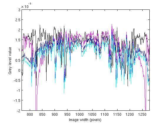
7 Flat field correction Mechanical vibrations Several different frequencies (100 Hz, Hz, 66 Hz, 16 Hz) Intensity instabilities 3 ms exposure time 251 projections Sampled every 3 Movie 25 fps Scan is actually faster

8 Flat field correction Scintillator decay time and afterglow Injection 40x, LSO:Tb 5.9 um, 1% 20x, LAG:Ce 20 um, 1%
9 Flat field correction Scintillator decay time and afterglow 2x, LAG:Ce 20 um, 3.5%

10 Flat field correction Resulting artifacts Ring/band artifacts Non-quantitative Resolution degradation Solutions Average flat-field or intermediate flat-fields Flat-field tracking Adaptive time-dependent normalization Titarenko et al., 2010 Dual camera acquisition Mokso et al., 2013


11 Ring artifacts Inconsistencies in single or multiple channels over an extended range of views

12 Ring artifacts Inhomogeneities in the individual pixel response of detector elements Damaged dirty scintillator screens Beam variations
13 Ring artifacts Correction algorithms Simple to use, minimum amount of tuning parameters Sinogram domain Lines easier to detect Integration in existing pipelines difficult Reconstruction domain (Cartesian/Polar coordinates) Circles/rings difficult to detect Integration straightforward
14 Ring artifacts Correction algorithms Low/high pass filtering Oimoen et al., 2000, Yousuf and Asaduzzaman, 2010 Median/mean/moving average filtering Rivers at al., Boin and Haibel, 2006, Sadi et al., 2010 Transform domain processing Raven, 2010, Münch et al., 2009 Artifact template vector Sijbers and Postnov, 2004, Brun et al., 2009 A priori information Titarenko et al., 2010
15 Ring artifacts Correction algorithms None fully suitable for different (weak, varying intensity, band) rings without distortion and blurring Difficult choice of the optimal parameters Parameters often slice dependent Combination of approaches


16 Local tomography artifacts Large object at low resolution insufficient resolution for features of interest Tesselation time consuming, computationally heavy Larger detectors expensive, inefficient Ill-posed reconstruction problem x-rays CCD
17 Local tomography artifacts
18 Local tomography artifacts


19 Local tomography artifacts Marone et al., SPIE, 2010 Kyrieleis et al., J. Microscopy, 2010

20 Local tomography artifacts

21 Local tomography artifacts

22 Local tomography artifacts 0.5 mm Marone et al., SPIE, 2010


23 Local Original Local tomography artifacts ZP=0 ZP=0.5 ZP=1.5
24 Local tomography artifacts Simple lateral sinogram extension Insensitive to truncation degree Insensitive to position of region of interest Good results for structural analysis Non quantitative Difference in absolute value from slice to slice Calibration points if sample comparison needed More involved algorithms if absolute values needed
25 Local tomography artifacts Many ideas present in the medical imaging community Specific for medical applications Patent protected Various technique for projection completion Smooth continuation Iterative methods (e.g. sparsity, statistical) Computationally heavy Back projection of the first (Hilbert) or second (Lambda) derivative of projections A priori information needed For specific geometries Zoom-in tomography Multiple scans needed


26 Local tomography artifacts 4 mm sample 0.7 mm FOV prj


27 Local tomography artifacts 1501 prj 2251 prj 4501 prj


28 Sample stability 20 mm 20 mm
29 Sample stability Dynamic processes Animal physiology (brain, lungs, insect) Foam rheology Mechanical testing Sintering Annealing Electrochemistry Dose limitation In-vivo experiments Liquid foams surface tension properties Metals temperature change Micro-particles thermal dilation Fuel cell degradation Iterative reconstruction algorithms Few projections Short exposures (noisy projections) Phase contrast

30 Importance of phase contrast Towards in-vivo lung physiology Absorption CNR=1.8 Phase CNR=15.0 Total scan time: 5.4 s G. Lovric et al.,2013

31 Importance of phase contrast Monochromatic Polychromatic Foam physics CNR=4.9 CNR=0.7 CNR=4 Total scan time: 330 ms Phase Edge- enhancement Mokso et al., 2013

32 Importance of phase contrast Distance 1 Distance 2 Phase Edge- enhancement Mixed Guigay et al., 2007 TIE Paganin et al., 2002 Mokso et al., 2013

33 Importance of phase contrast Mixed Distance 1 Phase Edge- enhancement Mokso et al., 2013
34 Summary More advanced algorithms useful for different artifacts: Ring artifacts Local tomography artifacts Flat-field correction Ultrafast tomography Noisy projections -> Advanced phase retrieval algorithms Few projections -> Iterative algorithms

35 Acknoledgments Rajmund TOMCAT group Marco Thank you for your attention!


36 Streaking artifacts Inconsistencies in isolated measurements


37 Streaking artifacts High flux, high energy experiments Scattered x-rays hitting the camera chip directly Large energy deposition relative to the visible light photons Perfectly straight lines at random orientations in the image

38 Streaking artifacts High flux, high energy experiments Scattered x-rays hitting the camera chip directly Large energy deposition relative to visible light photons Perfectly straight lines at random orientations in the image
 Low pass filter for each line in the sinogram Division")

39 Streaking artifacts Correction algorithm (after M. Rivers) Low pass filter for each line in the sinogram Division of the original sinogram by the smooth one Substitute pixels with a grey level value smaller than threshold with average of neighboring pixels
Towards accurate measurements with synchrotron tomography Problems and pitfalls. Robert C. Atwood. Nghia T. Vo, Michael Drakopoulos, Thomas Connolley
 Towards accurate measurements with synchrotron tomography Problems and pitfalls Robert C. Atwood Nghia T. Vo, Michael Drakopoulos, Thomas Connolley Artefacts in Synchrotron X-ray Tomography Rings Rings
MC SIMULATION OF SCATTER INTENSITIES IN A CONE-BEAM CT SYSTEM EMPLOYING A 450 kv X-RAY TUBE
 MC SIMULATION OF SCATTER INTENSITIES IN A CONE-BEAM CT SYSTEM EMPLOYING A 450 kv X-RAY TUBE A. Miceli ab, R. Thierry a, A. Flisch a, U. Sennhauser a, F. Casali b a Empa - Swiss Federal Laboratories for
X-RAY COMPUTED TOMOGRAPHY
 X-RAY COMPUTED TOMOGRAPHY Bc. Jan Kratochvíla Czech Technical University in Prague Faculty of Nuclear Sciences and Physical Engineering Abstract Computed tomography is a powerful tool for imaging the inner
Radionuclide Imaging MII Single Photon Emission Computed Tomography (SPECT)
") Radionuclide Imaging MII 3073 Single Photon Emission Computed Tomography (SPECT) Single Photon Emission Computed Tomography (SPECT) The successful application of computer algorithms to x-ray imaging in
Radionuclide Imaging MII 3073 Single Photon Emission Computed Tomography (SPECT) Single Photon Emission Computed Tomography (SPECT) The successful application of computer algorithms to x-ray imaging in
COMPUTATIONAL IMAGING. Berthold K.P. Horn
 COMPUTATIONAL IMAGING Berthold K.P. Horn What is Computational Imaging? Computation inherent in image formation What is Computational Imaging? Computation inherent in image formation (1) Computing is getting
HISTORY. CT Physics with an Emphasis on Application in Thoracic and Cardiac Imaging SUNDAY. Shawn D. Teague, MD
 CT Physics with an Emphasis on Application in Thoracic and Cardiac Imaging Shawn D. Teague, MD DISCLOSURES 3DR- advisory committee CT PHYSICS WITH AN EMPHASIS ON APPLICATION IN THORACIC AND CARDIAC IMAGING
Digital Images & Image Quality
 Introduction to Medical Engineering (Medical Imaging) Suetens 1 Digital Images & Image Quality Ho Kyung Kim Pusan National University Radiation imaging DR & CT: x-ray Nuclear medicine: gamma-ray Ultrasound
IBEX TECHNOLOGY APPLIED TO DIGITAL RADIOGRAPHY
 WHITE PAPER: IBEX TECHNOLOGY APPLIED TO DIGITAL RADIOGRAPHY IBEX Innovations Ltd. Registered in England and Wales: 07208355 Address: Discovery 2, NETPark, William Armstrong Way, Sedgefield, UK Patents:
HIGH SPEED, HIGH RESOLUTION AND LOW COST DIGITAL RADIOGRAPHY
 HIGH SPEED, HIGH RESOLUTION AND LOW COST DIGITAL RADIOGRAPHY AND COMPUTED TOMOGRAPHY SYSTEM Kasiviswanathan Rangarajan1,2 and T. Jensen 1 Department of Computer Engineering 2 Center for Nondestructive
Niklas Norrby 12/17/2010
 LINKÖPINGS UNIVERSITET Nanotomography Synchrotron radiation course project Niklas Norrby 12/17/2010 Introduction Tomography is a method to image three-dimensional objects by illumination from different
Introduction. Chapter 16 Diagnostic Radiology. Primary radiological image. Primary radiological image
 Introduction Chapter 16 Diagnostic Radiology Radiation Dosimetry I Text: H.E Johns and J.R. Cunningham, The physics of radiology, 4 th ed. http://www.utoledo.edu/med/depts/radther In diagnostic radiology
SPECT Reconstruction & Filtering
 SPECT Reconstruction & Filtering Goals Understand the basics of SPECT Reconstruction Filtered Backprojection Iterative Reconstruction Make informed choices on filter selection and settings Pre vs. Post
Pitfalls and Remedies of MDCT Scanners as Quantitative Instruments
 intensity m(e) m (/cm) 000 00 0 0. 0 50 0 50 Pitfalls and Remedies of MDCT Scanners as Jiang Hsieh, PhD GE Healthcare Technology University of Wisconsin-Madison Root-Causes of CT Number Inaccuracies Nature
Growing Tall Poppies: An Authentic Science Experience
 Growing Tall Poppies: An Authentic Science Experience Introduction A group of Year 10 students from Santa Maria College Working with a program called Growing Tall Poppies with CXS and La Trobe University
Image Deblurring. This chapter describes how to deblur an image using the toolbox deblurring functions.
 12 Image Deblurring This chapter describes how to deblur an image using the toolbox deblurring functions. Understanding Deblurring (p. 12-2) Using the Deblurring Functions (p. 12-5) Avoiding Ringing in
CHAPTER 8 GENERIC PERFORMANCE MEASURES
 GENERIC PERFORMANCE MEASURES M.E. DAUBE-WITHERSPOON Department of Radiology, University of Pennsylvania, Philadelphia, Pennsylvania, United States of America 8.1. INTRINSIC AND EXTRINSIC MEASURES 8.1.1.
Multi-Lateral Shearing Interferometry: Principle and Application on X-ray Laboratory Sources
 Multi-Lateral Shearing Interferometry: Principle and Application on X-ray Laboratory Sources International Symposium on Digital Industrial Radiology and Computed Tomography June 22-25, 2015 Adrien STOLIDI
Related topics Beam hardening, cupping effect, Beam hardening correction, metal artefacts, photon starvation
 Beam hardening and metal artefacts TEP Related topics Beam hardening, cupping effect, Beam hardening correction, metal artefacts, photon starvation Principle X-ray sources produce a polychromatic spectrum
Large-Area CdTe Photon-Counting Pixel Detectors
 Large-Area CdTe Photon-Counting Pixel Detectors Tilman Donath, Application Scientist 22.6.2015, DIR2015, Ghent DECTRIS Ltd. 5400 Baden Switzerland www.dectris.com Agenda 1. Introduction Hybrid Photon Counting
LYNXEYE XE. Innovation with Integrity. High-Resolution Energy-Dispersive Detector for 0D, 1D, and 2D Diffraction XRD
 High-Resolution Energy-Dispersive Detector for 0D, 1D, and 2D Diffraction The is the first energy dispersive 0D, 1D, and 2D detector operating at room temperature for ultra fast X-ray diffraction measurements.
PIXSCAN CT scanner for Small Animal Imaging Based on hybrid pixel detectors
 PIXSCAN CT scanner for Small Animal Imaging Based on hybrid pixel detectors Centre de Physique des Particules de M arseille (CPPM -IN2P3), France S. B a s o lo, A. B o n is s e n t, P. B re u g n o n,
Postprocessing of nonuniform MRI
 Postprocessing of nonuniform MRI Wolfgang Stefan, Anne Gelb and Rosemary Renaut Arizona State University Oct 11, 2007 Stefan, Gelb, Renaut (ASU) Postprocessing October 2007 1 / 24 Outline 1 Introduction
Reconstruction Filtering in Industrial gamma-ray CT Application
 Reconstruction Filtering in Industrial gamma-ray CT Application Lakshminarayana Yenumula *, Rajesh V Acharya, Umesh Kumar, and Ashutosh Dash Industrial Tomography and Instrumentation Section, Isotope Production
Distortion Correction in LODOX StatScan X-Ray Images
 Distortion Correction in LODOX StatScan X-Ray Images Matthew Paul Beets November 27, 2007 X-Ray images produced by the LODOX StatScan machine contain a non-linear distortion in the direction of the beam
COMPUTED TOMOGRAPHY 1
 COMPUTED TOMOGRAPHY 1 Why CT? Conventional X ray picture of a chest 2 Introduction Why CT? In a normal X-ray picture, most soft tissue doesn't show up clearly. To focus in on organs, or to examine the
Applications of the Cracow X-Ray Microprobe in Tomography
 Vol. 115 (2009) ACTA PHYSICA POLONICA A No. 2 Proceedings of the XLII Zakopane School of Physics, Zakopane 2008 Applications of the Cracow X-Ray Microprobe in Tomography J. Bielecki a, S. Bożek a,b, J.
3D light microscopy techniques
 3D light microscopy techniques The image of a point is a 3D feature In-focus image Out-of-focus image The image of a point is not a point Point Spread Function (PSF) 1D imaging 2D imaging 3D imaging Resolution
An Activity in Computed Tomography
 Pre-lab Discussion An Activity in Computed Tomography X-rays X-rays are high energy electromagnetic radiation with wavelengths smaller than those in the visible spectrum (0.01-10nm and 4000-800nm respectively).
REAL-TIME X-RAY IMAGE PROCESSING; TECHNIQUES FOR SENSITIVITY
 REAL-TIME X-RAY IMAGE PROCESSING; TECHNIQUES FOR SENSITIVITY IMPROVEMENT USING LOW-COST EQUIPMENT R.M. Wallingford and J.N. Gray Center for Aviation Systems Reliability Iowa State University Ames,IA 50011
Particle Image Velocimetry
 Markus Raffel Christian E. Willert Steve T. Wereley Jiirgen Kompenhans Particle Image Velocimetry A Practical Guide Second Edition With 288 Figures and 42 Tables < J Springer Contents Preface V 1 Introduction
Gas scintillation Glass GEM detector for high-resolution X-ray imaging and CT
 Gas scintillation Glass GEM detector for high-resolution X-ray imaging and CT Takeshi Fujiwara 1, Yuki Mitsuya 2, Hiroyuki Takahashi 2, and Hiroyuki Toyokawa 2 1 National Institute of Advanced Industrial
DALLA LUCE VISIBILE AI RAGGI X: NUOVI RIVELATORI DI IMMAGINI PER RAGGI X A DISCRIMINAZIONE IN ENERGIA ED APPLICAZIONI
 DALLA LUCE VISIBILE AI RAGGI X: NUOVI RIVELATORI DI IMMAGINI PER RAGGI X A DISCRIMINAZIONE IN ENERGIA ED APPLICAZIONI D. Pacella ENEA - Frascati LIMS, Frascati 14-15 ottobre 2015 Come per la fotografia:
Superfast phase-shifting method for 3-D shape measurement
 Superfast phase-shifting method for 3-D shape measurement Song Zhang 1,, Daniel Van Der Weide 2, and James Oliver 1 1 Department of Mechanical Engineering, Iowa State University, Ames, IA 50011, USA 2
Defining the Optimal Beam Hardening Correction Parameters for CT Dimensional Metrology Applications
 International Conference on Competitive Manufacturing Defining the Optimal Beam Hardening Correction Parameters for CT Dimensional Metrology Applications Y. Tan 1,2, K. Kiekens 1,2, F. Welkenhuyzen 2,
Radiology Physics Lectures: Digital Radiography. Digital Radiography. D. J. Hall, Ph.D. x20893
 Digital Radiography D. J. Hall, Ph.D. x20893 djhall@ucsd.edu Background Common Digital Modalities Digital Chest Radiograph - 4096 x 4096 x 12 bit CT - 512 x 512 x 12 bit SPECT - 128 x 128 x 8 bit MRI -
Pixel Array Detectors: Counting and Integrating
 Pixel Array Detectors: Counting and Integrating Roger Durst, Bruker AXS October 13, 2016 1 The quest for a perfect detector There is, of course, no perfect detector All available detector technologies
PIXSCAN CT-scanner for Small Animal Imaging Based on hybrid pixel detectors
 PIXSCAN CT-scanner for Small Animal Imaging Based on hybrid pixel detectors Centre de Physique des Particules de Marseille (CPPM-IN2P3), France S. Basolo, A. Bonissent, P. Breugnon, J.C. Clemens, P. Delpierre,
Freeze-fixation of bubbles for micro-ct imaging of liquid aerated food emulsions
 Freeze-fixation of bubbles for micro-ct imaging of liquid aerated food emulsions G. van Dalen 1, M. Koster 1, J. Hazekamp 2 1 Unilever Research & Development, Imaging & Spectroscopy, Olivier van Noortlaan
Sensor Fusion Enables Comprehensive Analysis of Laser Processing in Additive Manufacturing
 MKS Instruments 1 of 6 Sensor Fusion Enables Comprehensive Analysis of Laser Processing in Additive Manufacturing By Kevin Kirkham, Senior Manager, Product Development, Ophir Sensor: "A device that detects
Practical work no. 3: Confocal Live Cell Microscopy
 Practical work no. 3: Confocal Live Cell Microscopy Course Instructor: Mikko Liljeström (MIU) 1 Background Confocal microscopy: The main idea behind confocality is that it suppresses the signal outside
Observational Astronomy
 Observational Astronomy Instruments The telescope- instruments combination forms a tightly coupled system: Telescope = collecting photons and forming an image Instruments = registering and analyzing the
PET Performance Evaluation of MADPET4: A Small Animal PET Insert for a 7-T MRI Scanner
 PET Performance Evaluation of MADPET4: A Small Animal PET Insert for a 7-T MRI Scanner September, 2017 Results submitted to Physics in Medicine & Biology Negar Omidvari 1, Jorge Cabello 1, Geoffrey Topping
PERFORMANCE CHARACTERIZATION OF AMORPHOUS SILICON DIGITAL DETECTOR ARRAYS FOR GAMMA RADIOGRAPHY
 12 th A-PCNDT 2006 Asia-Pacific Conference on NDT, 5 th 10 th Nov 2006, Auckland, New Zealand PERFORMANCE CHARACTERIZATION OF AMORPHOUS SILICON DIGITAL DETECTOR ARRAYS FOR GAMMA RADIOGRAPHY Rajashekar
COMPUTED RADIOGRAPHY CHAPTER 4 EFFECTIVE USE OF CR
 This presentation is a professional collaboration of development time prepared by: Rex Christensen Terri Jurkiewicz and Diane Kawamura New Technology https://www.youtube.com/watch?v=ptkzznazb 7U COMPUTED
Maximum Performance, Minimum Space
 TECHNOLOGY HISTORY For over 130 years, Toshiba has been a world leader in developing technology to improve the quality of life. Our 50,000 global patents demonstrate a long, rich history of leading innovation.
SUPER RESOLUTION INTRODUCTION
 SUPER RESOLUTION Jnanavardhini - Online MultiDisciplinary Research Journal Ms. Amalorpavam.G Assistant Professor, Department of Computer Sciences, Sambhram Academy of Management. Studies, Bangalore Abstract:-
An Activity in Computed Tomography
 Pre-lab Discussion An Activity in Computed Tomography X-rays X-rays are high energy electromagnetic radiation with wavelengths smaller than those in the visible spectrum (0.01-10nm and 4000-800nm respectively).
Motion Deblurring of Infrared Images
 Motion Deblurring of Infrared Images B.Oswald-Tranta Inst. for Automation, University of Leoben, Peter-Tunnerstr.7, A-8700 Leoben, Austria beate.oswald@unileoben.ac.at Abstract: Infrared ages of an uncooled
Properties of Neutron Pixel Detector based on Medipix-2 Device
 Properties of Neutron Pixel Detector based on Medipix-2 Device Jan Jakubek, Tomas Holy, Eberhard Lehmann, Stanislav Pospisil, Josef Uher, Jiri Vacik, Daniel Vavrik Abstract - Neutron transmission radiography
Image Quality/Artifacts Frequency (MHz)
") The Larmor Relation 84 Image Quality/Artifacts (MHz) 42 ω = γ X B = 2πf 84 0.0 1.0 2.0 Magnetic Field (Tesla) 1 A 1D Image Magnetic Field Gradients Magnet Field Strength Field Strength / Gradient Coil
The Larmor Relation 84 Image Quality/Artifacts (MHz) 42 ω = γ X B = 2πf 84 0.0 1.0 2.0 Magnetic Field (Tesla) 1 A 1D Image Magnetic Field Gradients Magnet Field Strength Field Strength / Gradient Coil
SEAMS DUE TO MULTIPLE OUTPUT CCDS
 Seam Correction for Sensors with Multiple Outputs Introduction Image sensor manufacturers are continually working to meet their customers demands for ever-higher frame rates in their cameras. To meet this
Charge-Coupled Device (CCD) Detectors pixel silicon chip electronics cryogenics
 Detectors pixel silicon chip electronics cryogenics") Charge-Coupled Device (CCD) Detectors As revolutionary in astronomy as the invention of the telescope and photography semiconductor detectors a collection of miniature photodiodes, each called a picture
Charge-Coupled Device (CCD) Detectors As revolutionary in astronomy as the invention of the telescope and photography semiconductor detectors a collection of miniature photodiodes, each called a picture
Examples of image processing
 Examples of image processing Example 1: We would like to automatically detect and count rings in the image 3 Detection by correlation Correlation = degree of similarity Correlation between f(x, y) and
3D light microscopy techniques
 3D light microscopy techniques The image of a point is a 3D feature In-focus image Out-of-focus image The image of a point is not a point Point Spread Function (PSF) 1D imaging 1 1 2! NA = 0.5! NA 2D imaging
IBEX MATERIALS DETECTION TECHNOLOGY
 WHITE PAPER: IBEX MATERIALS DETECTION TECHNOLOGY IBEX Innovations Ltd. Registered in England and Wales: 07208355 Address: Discovery 2, NETPark, William Armstrong Way, Sedgefield, TS21 3FH, UK Patents held
Nikon Instruments Europe
 Nikon Instruments Europe Recommendations for N-SIM sample preparation and image reconstruction Dear customer, We hope you find the following guidelines useful in order to get the best performance out of
Nikon. King s College London. Imaging Centre. N-SIM guide NIKON IMAGING KING S COLLEGE LONDON
 N-SIM guide NIKON IMAGING CENTRE @ KING S COLLEGE LONDON Starting-up / Shut-down The NSIM hardware is calibrated after system warm-up occurs. It is recommended that you turn-on the system for at least
Physical unsharp mask with structured detection
 This paper was published in Optics Letters and is made available as an electronic reprint with the permission of OSA. The paper can be found at the following URL on the OSA website: https://www.osapublishing.org/ol/abstract.cfm?uri=ol-40-15-3611.
CSPADs: how to operate them, which performance to expect and what kind of features are available
 CSPADs: how to operate them, which performance to expect and what kind of features are available Gabriella Carini, Gabriel Blaj, Philip Hart, Sven Herrmann Cornell-SLAC Pixel Array Detector What is it?
SYLLABUS. 1. Identification of Subject:
 SYLLABUS Date/ Revision : 30 January 2017/1 Faculty : Life Sciences Approval : Dean, Faculty of Life Sciences SUBJECT : Biophysics 1. Identification of Subject: Name of Subject : Biophysics Code of Subject
Noise Characteristics of the FORE+OSEM(DB) Reconstruction Method for the MiCES PET Scanner
 Reconstruction Method for the MiCES PET Scanner") Noise Characteristics of the FORE+OSEM(DB) Reconstruction Method for the MiCES PET Scanner Kisung Lee, Member, IEEE, Paul E. Kinahan, Senior Member, Robert S. Miyaoka, Member, IEEE, Jeffrey A. Fessler,
Noise Characteristics of the FORE+OSEM(DB) Reconstruction Method for the MiCES PET Scanner Kisung Lee, Member, IEEE, Paul E. Kinahan, Senior Member, Robert S. Miyaoka, Member, IEEE, Jeffrey A. Fessler,
Medical Imaging. X-rays, CT/CAT scans, Ultrasound, Magnetic Resonance Imaging
 Medical Imaging X-rays, CT/CAT scans, Ultrasound, Magnetic Resonance Imaging From: Physics for the IB Diploma Coursebook 6th Edition by Tsokos, Hoeben and Headlee And Higher Level Physics 2 nd Edition
Introduction to BioImage Analysis
 Introduction to BioImage Analysis Qi Gao CellNetworks Math-Clinic core facility 22-23.02.2018 MATH- CLINIC Math-Clinic core facility Data analysis services on bioimage analysis & bioinformatics: 1-to-1
ZIMPOL-3: a powerful solar polarimeter
 ZIMPOL-3: a powerful solar polarimeter San Diego, SPIE, 1. July 2010 Renzo Ramelli, IRSOL, Locarno, Switzerland and the ZIMPOL team The ZIMPOL-3 team Silvano Balemi, SUPSI Michele Bianda, IRSOL Ivan Defilippis,
Very short introduction to light microscopy and digital imaging
 Very short introduction to light microscopy and digital imaging Hernan G. Garcia August 1, 2005 1 Light Microscopy Basics In this section we will briefly describe the basic principles of operation and
... In vivo imaging in Nuclear Medicine. 1957: Anger camera (X;Y) X Y
 X Y") József Varga, PhD EMISSION IMAGING BASICS OF QUANTIFICATION Imaging devices Aims of image processing Reconstruction University of Debrecen Department of Nuclear Medicine. In vivo imaging in Nuclear Medicine
József Varga, PhD EMISSION IMAGING BASICS OF QUANTIFICATION Imaging devices Aims of image processing Reconstruction University of Debrecen Department of Nuclear Medicine. In vivo imaging in Nuclear Medicine
ME 6406 MACHINE VISION. Georgia Institute of Technology
 ME 6406 MACHINE VISION Georgia Institute of Technology Class Information Instructor Professor Kok-Meng Lee MARC 474 Office hours: Tues/Thurs 1:00-2:00 pm kokmeng.lee@me.gatech.edu (404)-894-7402 Class
Introduction. Lighting
 &855(17 )8785(75(1'6,10$&+,1(9,6,21 5HVHDUFK6FLHQWLVW0DWV&DUOLQ 2SWLFDO0HDVXUHPHQW6\VWHPVDQG'DWD$QDO\VLV 6,17()(OHFWURQLFV &\EHUQHWLFV %R[%OLQGHUQ2VOR125:$< (PDLO0DWV&DUOLQ#HF\VLQWHIQR http://www.sintef.no/ecy/7210/
Large Field of View, High Spatial Resolution, Surface Measurements
 Large Field of View, High Spatial Resolution, Surface Measurements James C. Wyant and Joanna Schmit WYKO Corporation, 2650 E. Elvira Road Tucson, Arizona 85706, USA jcwyant@wyko.com and jschmit@wyko.com
Enhanced Method for Image Restoration using Spatial Domain
 Enhanced Method for Image Restoration using Spatial Domain Gurpal Kaur Department of Electronics and Communication Engineering SVIET, Ramnagar,Banur, Punjab, India Ashish Department of Electronics and
High Energy Digital Radiography & 3D-CT for Industrial Systems
 DIR 2007 - International Symposium on Digital industrial Radiology and Computed Tomography, June 25-27, 2007, Lyon, France High Energy Digital Radiography & 3D-CT for Industrial Systems Non-Destructive
PET/CT Instrumentation Basics
 / Instrumentation Basics 1. Motivations for / imaging 2. What is a / Scanner 3. Typical Protocols 4. Attenuation Correction 5. Problems and Challenges with / 6. Examples Motivations for / Imaging Desire
Film Replacement in Radiographic Weld Inspection The New ISO Standard
 BAM Berlin Film Replacement in Radiographic Weld Inspection The New ISO Standard 17636-2 Uwe Ewert, Uwe Zscherpel, Mirko Jechow Requests and information to: uwez@bam.de 1 Outline - The 3 essential parameters
High granularity scintillating fiber trackers based on Silicon Photomultiplier
 High granularity scintillating fiber trackers based on Silicon Photomultiplier A. Papa Paul Scherrer Institut, Villigen, Switzerland E-mail: angela.papa@psi.ch Istituto Nazionale di Fisica Nucleare Sez.
Digital Imaging CT & MR
 Digital Imaging CT & MR January 22, 2008 Digital Radiography, CT and MRI generate images in a digital format What is a Digital Image? A digital image is made up of picture elements, pixels row by column
10/3/2012. Study Harder
 This presentation is a professional collaboration of development time prepared by: Rex Christensen Terri Jurkiewicz and Diane Kawamura Study Harder CR detection is inefficient, inferior to film screen
Dynamic Phase-Shifting Microscopy Tracks Living Cells
 from photonics.com: 04/01/2012 http://www.photonics.com/article.aspx?aid=50654 Dynamic Phase-Shifting Microscopy Tracks Living Cells Dr. Katherine Creath, Goldie Goldstein and Mike Zecchino, 4D Technology
TEM Cameras. Digital Cameras for Electron Microscopy
 Digital Imaging Solutions TEM Cameras Side- and bottom-mounted TEM cameras Digital Cameras for Electron Microscopy IMAGING SOLUTIONS FOR ELECTRON MICROSCOPY. BASED ON OPTO-DIGITAL KNOW-HOW. DESIGNED BY
Thorough Small Angle X-ray Scattering analysis of the instability of liquid micro-jets in air
 Supplementary Information Thorough Small Angle X-ray Scattering analysis of the instability of liquid micro-jets in air Benedetta Marmiroli a *, Fernando Cacho-Nerin a, Barbara Sartori a, Javier Pérez
Digital Radiography : Flat Panel
 Digital Radiography : Flat Panel Flat panels performances & operation How does it work? - what is a sensor? - ideal sensor Flat panels limits and solutions - offset calibration - gain calibration - non
Initial Certification
 Initial Certification Nuclear Medical Physics (NMP) Study Guide Part 2 Content Guide and Sample Questions The content of all ABR exams is determined by a panel of experts who select the items based on
10/26/2015. Study Harder
 This presentation is a professional collaboration of development time prepared by: Rex Christensen Terri Jurkiewicz and Diane Kawamura Study Harder CR detection is inefficient, inferior to film screen
LYNXEYE XE-T. < 380 ev. Innovation with Integrity. Energy. Resolution. High-Resolution Position Sensitive Detector with Superb Energy Resolution XRD
 Energy < 380 ev Resolution High-Resolution Position Sensitive Detector with Superb Energy Resolution The is the next generation "Compound Silicon Strip" detector with superb energy resolution for ultrafast
3D imaging of aerated emulsions using X-ray microtomography
 3D imaging of aerated emulsions using X-ray microtomography G. van Dalen, M.W. Koster Unilever Research & Development, Advanced Measurement & Data Modelling, Olivier van Noortlaan 120, NL-3133AT Vlaardingen
X-Ray Transport, Diagnostic, & Commissioning Plans. LCLS Diagnostics and Commissioning Workshop
 X-Ray Transport, Diagnostic, & Commissioning Plans LCLS Diagnostics and Commissioning Workshop *This work was performed under the auspices of the U.S. Department of Energy by the University of California,
Control of Noise and Background in Scientific CMOS Technology
 Control of Noise and Background in Scientific CMOS Technology Introduction Scientific CMOS (Complementary metal oxide semiconductor) camera technology has enabled advancement in many areas of microscopy
Dynamic 4D imaging of foamed bitumen by X-ray micro computed tomography
 Image sources: http://www.ethlife.ethz.ch/archive_articles/120629_mensaumbau_tl/1206 29_Mensaumbau_l.jpg?hires http://files.newsnetz.ch/story/2/5/4/25415795/1/topelement.jpg http://www.wirtschaftsingenieurinnovation.ch/wing/images/ausschreibungen/jobs/luftaufnahme_campu
Studies on MCM D interconnections
 Studies on MCM D interconnections Speaker: Peter Gerlach Department of Physics Bergische Universität Wuppertal D-42097 Wuppertal, GERMANY Authors: K.H.Becks, T.Flick, P.Gerlach, C.Grah, P.Mättig Department
NM Module Section 2 6 th Edition Christian, Ch. 3
 NM 4303 Module Section 2 6 th Edition Christian, Ch. 3 Gas Filled Chamber Voltage Gas filled chamber uses Hand held detectors cutie pie Geiger counter Dose calibrators Cutie pie Chamber voltage in Ionization
ON THE WAY TO DIGITAL RADIOGRAPHY
 The 14 th International Conference of the Slovenian Society for Non-Destructive Testing»Application of Contemporary Non-Destructive Testing in Engineering«September 4-6, 2017, Bernardin, Slovenia More
AGIPD calibration status report
 AGIPD calibration status report A. Allahgholi 2, R. Dinapoli 1, P. Goettlicher 2, M. Gronewald 4, H. Graafsma 2,5, D. Greiffenberg 1, B.H. Henrich 1, H. Hirsemann 2, S. Jack 2, R. Klanner 3, A. Klyuev
5th International Symposium on NDT in Aerospace, 13-15th November 2013, Singapore
 5th International Symposium on NDT in Aerospace, 13-15th November 2013, Singapore Accurate Area and Width Measurements from Neutron Beam Computed Tomography (NBCT) Images of Cooling Holes of a Used Tornado
This document is published in:
 Institutional Repository This document is published in: Bo Yu (ed.) (01). 01 IEEE Nuclear Science Symposium and Medical Imaging Conference (NSS/MIC): Anaheim, California, USA. October 9 - November 3, 01.
Lecture 20: Optical Tools for MEMS Imaging
 MECH 466 Microelectromechanical Systems University of Victoria Dept. of Mechanical Engineering Lecture 20: Optical Tools for MEMS Imaging 1 Overview Optical Microscopes Video Microscopes Scanning Electron
Hardware for High Energy Applications 30 October 2009
 Paper No. 003 09 Hardware for High Energy Applications 30 October 2009 This document was created by the Federal Working Group on Industrial Digital Radiography. Reproduction is authorized. Federal Working
Advances in X-Ray Scintillator Technology Roger D. Durst Bruker AXS Inc.
 Advances in X-Ray Scintillator Technology Roger D. Durst Inc. Acknowledgements T. Thorson, Y. Diawara, E. Westbrook, MBC J. Morse, ESRF C. Summers, Georgia Tech/PTCE B. Wagner, Georgia Tech/PTCE V. Valdna,
Image Denoising Using Different Filters (A Comparison of Filters)
") International Journal of Emerging Trends in Science and Technology Image Denoising Using Different Filters (A Comparison of Filters) Authors Mr. Avinash Shrivastava 1, Pratibha Bisen 2, Monali Dubey 3,
International Journal of Emerging Trends in Science and Technology Image Denoising Using Different Filters (A Comparison of Filters) Authors Mr. Avinash Shrivastava 1, Pratibha Bisen 2, Monali Dubey 3,
LENSES. INEL 6088 Computer Vision
 LENSES INEL 6088 Computer Vision Digital camera A digital camera replaces film with a sensor array Each cell in the array is a Charge Coupled Device light-sensitive diode that converts photons to electrons
Principles of CT scan
 Related topics Detector calibration, saturation, CT acquisition, CT reconstruction Principle X-ray computed tomography consists of using X-rays that are converted to a digital signal by a detector and
X-Ray Detection Using SOI Monolithic Sensors at a Compact High-Brightness X-Ray Source Based on Inverse Compton Scattering
 Abstract #: 1054 Conference: NSS (Oral) Accelerator Technologies and Beam Line Instrumentation X-Ray Detection Using SOI Monolithic Sensors at a Compact High-Brightness X-Ray Source Based on Inverse Compton