Microscope - Exercise 1
|
|
|
- August Oliver
- 6 years ago
- Views:
Transcription
1 Microscope - Exercise 1 Objectives -Familiarize parts and functions of the microscope. -Calculate total magnifications. -Determining the Diameter of the field of view for different magnifications. -Estimate size of an object under the microscope. -Identify parts (organelles) in animal and plant cells.

2 Condenser: It will gather the light from the illuminator and focus it on the specimen lying on the stage. The function of the condenser is to focus the light rays from the light source onto the specimen. Iris diaphragm lever: This allows the amount of light passing through the condenser to be regulated to see the object.
3 Microscope You have to learn how to use the microscope if your going to take Anatomy & Physiology, BI203, or any other science related courses, so get a good hang out of it now. Also, learn the parts and functions of the microscope.
4 Measuring Objects with the Microscope -To focus on an object, you must first center the object to the center of the field of view, and remember to do this every time you switch to a higher magnification. -The field of view refers to the total area that is visible through the microscope. The field of view is circular. The field gets smaller as the magnification increases. It is possible to measure the diameter of the field of view, and that measurement can then be used to construct reasonable estimates of the size of any specimen contained within the field of view.
5 Page 11 Lab Book Known magnification X original um = Diameter of Unknown magnification Field of View Measuring Objects with the Microscope With the 4x objective lens in place, look through the microscope and adjust the ruler so that it lies on a diameter of the field of view. Calculating the Diameter of the Field of View at other Magnifications There is an inverse relationship between the total magnification and the diameter of the field of view i.e., as magnifications increases the diameter of the field decreases in proportion, so the diameter of field of view at different magnification can be calculated mathematically, using the formula.
6 Dimensional Analysis You are converting um (10-6 m) mm (10-3 m) 1um 10-6 m mm 10-3 m = um x 10-6 m x 1 mm = 10-3 aka 1,000 um/mm 1 um 10-3 m
7 Determining the Diameter of the field of view at 40x (low power) (Ruler) With the 4x objective lens (40 magnification) in place, look through the microscope and adjust the ruler so that it lies on a diameter of the field of view. Diameter of the field of view at 40x (low power) which is objective lens 4. To convert the diameter of field to micrometer you just multiply it by 1,000, and you could get whatever you measured your ruler. So lets say we measured our ruler to be 4mm, so we multiply it by 1,000, and we get 4,000 um. Now, we could get other diameter of fields with different magnifications using a formula. For example, say we have to find the diameter of field of 100x? The first thing you do is put 40, which is your total magnification that you started with, over your unknown magnification, then you multiply that by your total micrometers, so for this case its 4,000, and you get a diameter of field of 1600 um. Now, say their asking us for 430x magnification, so what we do is put 40 over 430, and you multiply that by 4,000 which you would get 372 um.
8 Estimate size of an object under the microscope The approach you would take to estimate the size of a microscopic object, if you are given the diameter of field of views at different magnifications. Step 1: Focus on the object at certain magnification and note the diameter of the field of view at that magnification. Step 2: Determine (guesstimate) the percent of the diameter of the field of view is occupied by the object (image). Step 3: Use the following formula.. Percent Object Size = X diameter of field of view = 100
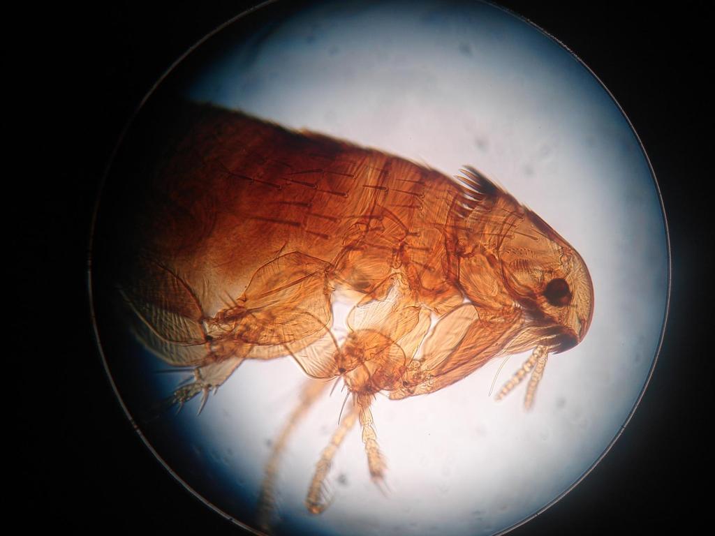
9 Estimate size of an object under the microscope The size of a specimen can be estimated by comparing it to the known diameter of the field of view at any magnification. First, you have to estimate how much of an object is occupying the field of view of the microscope, so your not going to get an accurate answer, but your answer should get an answer that s going to be within a range. Ok, your still using the 4,000 um that you get previously and say you have a flea, and that flea takes up about 40% of your microscope, so what you do is covert the 40% into a decimal by dividing it by 100, and you get.40 then you multiply that by 4,000 um and you should get an answer, which is 1600 um. Now, they can ask you to estimate another size of an object. For example, say the Flea s eye covers about 5%. So you divide that 100, you should get.05 and multiply that by 4,000 um, and you should get 200 um. Note: You could be asked this at different magnifications.
10
11 Your going to be ask to identify parts of both animal and plant cells, such as chloroplast, nucleus of a cell, and those sorts of things, so review them on your own.


12 Human epithelial cell From Rust 1a & 1b You will need to identify the nucleus, cytoplasm, and the cell membrane under a microscope.


13 Elodea Leaf cell From Rust 1c & 1d You will need to identify the cytoplasm, cell wall, and chloroplast under a microscope.
14 What is the function of the chloroplasts?
15 What is the function of the chloroplasts? Photosynthesis to make food for the plant.

16 Calculate the diameter of the field of view occupied by the flea. The diameter field of view is 100x magnification of the microscope is 380 micrometer. You have to estimate the percentage of the flea that occupies the microscope. This is subjective.
17
18 Questions 1. In the diameter of field of view of a light microscope at 40X magnification is 6,000 micrometer (um), what would be the field of view at 400X magnification? 2. An elodea cell was found occupying 40% of field of view s diameter at 400X magnification. At this magnification the field of view of the microscope is 200 micrometer. What is the size of the elodea cell? Remember this is a gesstimate percent of the diameter of the field of view is occupied by the object (image).
19 Questions 1. In the diameter of field of view of a light microscope at 40X magnification is 6,000 micrometer (um), what would be the field of view at 400X magnification? 40 x 6000 = 600 um An elodea cell was found occupying 40% of field of view s diameter at 400X magnification. At this magnification the field of view of the microscope is 200 micrometer. What is the size of the elodea cell? Remember this is a gesstimate percent of the diameter of the field of view is occupied by the object (image). 40 = x 200 = 80 um
Anatomy: Introduction to the Light Microscope
 Anatomy: Introduction to the Light Microscope Background: Microscopes are very important tools in biology. The term microscope can be translated as to view the tiny, because microscopes are used to study
Name Date Block LAB: Exploring Plant & Animal Cells
 Name Date Block LAB: Exploring Plant & Animal Cells Background Information: One of the first scientists to look at cells under a microscope was an English scientist by the name of Robert Hooke. He viewed
The microscope is useful in making observations and collecting data in scientific experiments. Microscopy involves three basic concepts:
 Lab #4 Biology 10 BCC Topic: MICROSCOPE LAB PART I: COMPOUND LIGHT MICROSCOPE OBJECTIVES: After completing this exercise you should be able to: Demonstrate proper care and use of a compound microscope.
CALIBRATION OF MICROSCOPE EYEPIECE GRATICULE
 CALIBRATION OF MICROSCOPE EYEPIECE GRATICULE A typical eyepiece graticule looks like this: It is 10mm in length and each mm is divided into 10 parts So each small division = 0.1mm = 100µm The eyepiece
Visual Anatomy ansd Physiology Lab Manual Pig Version 2nd Edition Sarikas TEST BANK
 Visual Anatomy ansd Physiology Lab Manual Pig Version 2nd Edition Sarikas TEST BANK https://testbankreal.com/download/visual-anatomy-ansd-physiology-labmanual-pig-version-2nd-edition-sarikas-test-bank/
1. A laboratory technique is illustrated in the diagram below. Explain why the coverslip is lowered at an angle.
 1. A laboratory technique is illustrated in the diagram below. Explain why the coverslip is lowered at an angle. 2. Base your answer to the following question on Which laboratory procedure is represented
Microscopes & cells. 2. arm. 3. ocular lens. 4. objective lenses. 5. stage. 6. slide clamp. 7. stage controls
 Microscopes & cells Objectives: At the end of this lab you should be able to: o demonstrate the safe and proper handling of a microscope, including carrying a microscope, slide placement, and storage.
AN INTRODUCTION TO THE MICROSCOPE
 AN INTRODUCTION TO THE MICROSCOPE INTRODUCTION In this exercise you will learn the components and operation of the compound microscope and the dissection microscope. This will be followed by a short exercise
MICROSCOPY and CELL STRUCTURE
 MICROSCOPY and CELL STRUCTURE Readings: Review pp. 69-71, and Fig. 4.1 on p. 65 in your text (POHS, 5 th ed.). Introduction: Biologists rely on many different types of microscopic techniques to find out
History of microscopy
 History of microscopy Introduction Structure of microscope Care of microscope Use of microscope Magnification As we already know cells are microscopic. What does this mean? Scientists were able to see
What you should have learned from the microscope labs.
 What you should have learned from the microscope labs. Microscope Lab 1 Directionality Items appear backwards and inverted On Stage In Microscope NOT!!!! Microscope Lab 1 More Directionality Items move
Perfecting Microscope Skills
 I. Introduction to the Microscope Perfecting Microscope Skills There are different types of microscopes used by biologists depending on the job they wish to accomplish, including dissecting (or "stereoscopic")
BIOLOGY 1101 LAB 2: MICROSCOPES AND CELLS
 BIOLOGY 1101 LAB 2: MICROSCOPES AND CELLS READING: Please read Chapter 4 in your text book to learn about the history of microscopy and basic cell structure. INTRODUCTION: The microscope is an important
Laboratory 2: Microscopy and Observation of Cells authors: Dr. Ruth Dahlquist-Willard & Michael Kunz
 Laboratory 2: Microscopy and Observation of Cells authors: Dr. Ruth Dahlquist-Willard & Michael Kunz Corresponding Readings: Campbell Ch. 4 BIOL-100L Safety Information: We will be using laboratory glassware
MEASURING WITH A MICROSCOPE Size Determination in Compound Light Microscopes
 MEASURING WITH A MICROSCOPE Size Determination in Compound Light Microscopes Name: Per: Date: 1. What do the following pictures represent? Which one is bigger? What s missing? Write your answers next to
What are some of the characteristics of plant and animal cells?
 What are some of the characteristics of plant and animal cells? BACKGROUND Ever since the first microscope was used, biologists have been interested in studying the cellular organization of all living
The microscope is useful in making observations and collecting data in scientific experiments. Microscopy involves three basic concepts:
 AP BIOLOGY Chapter 6 NAME DATE Block MICROSCOPE LAB PART I: COMPOUND MICROSCOPE OBJECTIVES: After completing this exercise you should be able to: Demonstrate proper care and use of a compound microscope.
Ocular Lenses. Head. Arm. Objective Lenses. Slide Holder Stage. On / Off Switch. Condenser with Iris Diaphragm. Light Intensity Control
 BIOLOGY 211: HUMAN ANATOMY & PHYSIOLOGY ********************************************************************************************************* USE OF THE LIGHT MICROSCOPE **********************************************************************************************************
Introduction to the Compound Microscope Cell Structure & Function
 Introduction to the Compound Microscope Cell Structure & Function Revised Fall 2018 Laboratory Safety Lab coat, long pants, closed-toe shoes, safety goggles, and nitrile or latex gloves are required. **You
Objectives Be able to identify dependent and independent variables in experimental designs.
 Experimental design Be able to identify dependent and independent variables in experimental designs. Imagine you ask the question, "Does amount of sleep affect test performance?" Your experiment involves
MICROSCOPE LAB. Resolving Power How well specimen detail is preserved during the magnifying process.
 AP BIOLOGY Cells ACTIVITY #2 MICROSCOPE LAB OBJECTIVES 1. Demonstrate proper care and use of a compound microscope. 2. Identify the parts of the microscope and describe the function of each part. 3. Compare
Match the microscope structures given in the left column with the statements in the right column that identify or describe them.
 49 Prelab for Name Match the microscope structures given in the left column with the statements in the right column that identify or describe them. Key: a. coarse adjustment knob f. turret or nosepiece
Microscope (and The Cell) Lab Exercise #1
 Lab Exercise #1") Lab Safety-General No eating or drinking Only registered students allowed in the class Long hair must be tied back Familiarize yourself with the emergency stations Do not mark on the models Inform me of
Lab Safety-General No eating or drinking Only registered students allowed in the class Long hair must be tied back Familiarize yourself with the emergency stations Do not mark on the models Inform me of
Scale. A Microscope s job in life. The Light Microscope. The Compound Microscope 9/24/12. Compound Microscope Anatomy
 The Study of Microbial Structure: Microscopy and Specimen Preparation Scale A Microscope s job in life 1.Magnify 2. Resolve ability to separate or distinguish between two points 3. Contrast How much or
Microscope Review. 1. A compound light microscope is represented in the diagram below.
 Name Microscope Review Date 1. A compound light microscope is represented in the diagram below. 5. The diagram below represents a hydra as viewed with a compound light microscope. If the hydra moves toward
Laboratory Introduction
 Laboratory Introduction There are two basic categories of microscopes: light microscopes and electron microscopes. Light, or optical, microscopes require light waves to provide the illumination while electron
Microscope & Measuring
 Name: ate: 1. microscope is supplied with 10 and 15 eyepieces, and with 10 and 44 objectives. What is the maximum magnification that can be obtained from this microscope?. 59. 150. 440. 660 3. student
Care and Use of the Compound Light Microscope
 EXERCISE 2 Care and Use of the Compound Light Microscope Time Estimates for Completing This Lab The activities in this laboratory exercise can be completed in 2 to 2.5 hours. Extra time will be required
Basic Microscopy for Plant Biology
 Page 1 of 8 Basic Microscopy for Plant Biology OBJECTIVES After completing this exercise, you should be able to do the following: a. Name the parts of the compound microscope and the functions of each.
MICROSCOPY MICROSCOPE TERMINOLOGY
 1 MICROSCOPY Most of the microorganisms that we talk about in this class are too small to be seen with the naked eye. The instruments we will use to visualize these microbes are microscopes. The laboratory
RP Microscopy revision activities
 RP Microscopy revision activities Aim: Revision of the Microscopy required practical (RP), relating it to the content of the specification including maths skills and working scientifically. Keywords: resolution,
2017 MICROSCOPE REVIEW by Karen L. Lancour RELATIVE SIZE OF MICROBES
 2017 MICROSCOPE REVIEW by Karen L. Lancour RELATIVE SIZE OF MICROBES 1000 millimeters (mm) = 1 meter (m) 1000 micrometers (µm or mcm) = 1 millimeter (mm) 1000 nanometers (nm) = 1 micrometer (mcm) Size
2018 MICROSCOPE REVIEW by Karen L. Lancour RELATIVE SIZE OF MICROBES
 2018 MICROSCOPE REVIEW by Karen L. Lancour RELATIVE SIZE OF MICROBES 1000 millimeters (mm) = 1 meter (m) 1000 micrometers (µm or mcm) = 1 millimeter (mm) 1000 nanometers (nm) = 1 micrometer (mcm) Size
EXERCISE 3 The Microscope
 Instant download and all chapters Solutions Manual Human Anatomy Laboratory Manual with Cat Dissections 7th Edition Marieb Smith https://testbankdata.com/download/solutions-manual-human-anatomy-laboratorymanual-cat-dissections-7th-edition-marieb-smith/
Basic Microscopy. OBJECTIVES After completing this exercise, you should be able to do the following:
 Page 1 of 10 Basic Microscopy OBJECTIVES After completing this exercise, you should be able to do the following: a. Name the parts of the compound microscope and the functions of each. b. Describe how
Microbiology Laboratory 2
 Microbiology Laboratory 2 Microscopy Background Microorganisms are too small to be seen with the naked eye. Thus a microscope is used to magnify objects so they can be observed. A lens consists of one
Objectives: Vocabulary:
 Measuring with a Microscope Author: David Gardner Date Created: Summer 2007 Subject: Biology (and Chemistry) Level: High School Standards: 1: Analysis, Inquiry and Design 4: Physical Setting and Living
Lab One: Techniques for Better Microscope Use
 Name BioPreAP/GT Purpose: Lab One: Techniques for Better Microscope Use Part A: Microscope Parts and Functions *Refer to Fig. 1 to refresh your memory on the parts of the microscope. *The objectives are
The Care and Use of the Microscope. Lab Exercise #4
 Lab Safety No eating or drinking!!! Long hair must be tied back Clean up your workstation before you leave! Return all materials to the storage sites Clean glassware and wipe down countertops Follow directions
Biology Lab #1: Using Microscopes to Observe and Measure Cells
 Biology Lab #1: Using Microscopes to Observe and Measure Cells Make sure you have signed and submitted the CDNIS Safety Contract before you start this experiment! PURPOSE: to review the use of the microscope
Microscope. & Measurements. Do Now
 Do Now Microscope & Measurements How many: 1. Centimeters (cm) in 4 meters (m)? m 2. Decimeters (dm) in 5 meters (m)? dm 3. Centimeters (cm) in 4,000 millimeters (mm) cm 4. Millimeters (mm) in 40 centimeters
Microbiology: Observing Bacteria Laboratory -1. Name Date
 Microbiology: Observing Bacteria Laboratory -1 Name Date Prelab: Part 1 Introduction to the microscope- please read through this handout and label the picture on the next page before starting the lab Care
THE COMPOUND BRIGHTFIELD MICROSCOPE
 THE COMPOUND BRIGHTFIELD MICROSCOPE Microbiology is the study of microscopic organisms that are so small that they are below the limit of vision of the human eye. Bacteria are the smallest of microorganisms
1. A MICROSCOPIC VIEW OF LIFE
 1. A MICROSCOPIC VIEW OF LIFE Objectives The student should be able to: 1. Identify, locate, and give the functions of the major parts of the compound microscope 2. Properly carry, care for, and put away
LAB 3 Use of the Microscope
 LAB 3 Use of the Microscope Introduction In this laboratory you will be learning how to use one of the most important tools in biology the compound light microscope to view a variety of specimens. You
Cells Unit GOALS AND OBJECTIVES. The student will become familiar with the use of the compound microscope.
 Cells Unit GOALS AND OBJECTIVES GOALS The student will become familiar with the use of the compound microscope. The student will become familiar with the basic parts and functions of the cell. OBJECTIVES
USING THE MICROSCOPE TO OBSERVE CELLS
 USING THE MICROSCOPE TO OBSERVE CELLS *****IMPORTANT!!!!! BEFORE VISITING YOUR LEARNING CENTER TO CARRY OUT THIS LAB ACTIVITY PLEASE READ BELOW Before you visit your Learning Center to use the microscope,
STRUCTURE OF THE MICROSCOPE
 STRUCTURE OF THE MICROSCOPE Use the word list to label the microscope below: Light Source Coarse adjustment knob Diaphragm Stage Clips Objectives Fine Adjustment Knob Base Stage Stage Clips Arm Revolving
Microscopy. Danil Hammoudi.MD
 Microscopy Danil Hammoudi.MD Care and Handling of the Microscope: A microscope is a delicate piece of equipment and should be treated with care. Use two hands when carrying the microscope. Place one hand
USING MICROSCOPES. How a Microscope Works
 USING MICROSCOPES One of the ways that technology has boosted science is by helping researchers observe objects that are normally too small or too far away to see. You won t be using telescopes or binoculars
Introduction to Microscopes
 INTRODUCTION TO THE MICROSCOPE Introduction to Microscopes The first microscopes worked by the same basic principle as the ones you will be using in lab. They are light microscopes. Visible light passes
The Microscope. Packet #2. 10/17/2016 9:12:02 PM Ryan Barrow 2012
 1 The Microscope Packet #2 10/17/2016 9:12:02 PM Ryan Barrow 2012 2 Historical Timeline 1609 Galileo Galilei develops a compound microscope with a convex and a concave les. 1665 Robert Hooke publishes
A BRIEF INTRODUCTION TO MICROSCOPY The two key properties of a microscope that allow you to see microbes are resolution and magnification.
 A BRIEF INTRODUCTION TO MICROSCOPY The two key properties of a microscope that allow you to see microbes are resolution and magnification. Magnification refers to the enlargement of the specimen when seen
Figure 3.4 Approximate size of various types of cells. ~10 um. Red Blood Cells = mm 1500 um. Width of penny Pearson Education, Inc.
 Figure 3.4 Approximate size of various types of cells. ~10 um Red Blood Cells 1.5mm 1500 um Width of penny = 1500 Figure 4.3 The limits of resolution (and some representative objects within those ranges)
Key Points Refer to How to Use the Compound Light Microscope :
 MODULE 1 Objective 1.2 Lesson B Introduction to the Microscope Using the Light Microscope and Slide Preparation Course Advanced Biotechnology Unit Biotech Basics Essential Question How do scientists view
Foundations of Math 11: Unit 2 Proportions. The scale factor can be written as a ratio, fraction, decimal, or percentage
 Lesson 2.3 Scale Name: Definitions 1) Scale: 2) Scale Factor: The scale factor can be written as a ratio, fraction, decimal, or percentage Formula: Formula: Example #1: A small electronic part measures
REVIEW FOR TEST ON MONDAY
 1. The diagram below shows an ameba moving out of the high-power field of view of a compound microscope in the direction indicated by the arrow. 4. The diagram below represents two cells next to a metric
1.When an object is sharply focused and the slide is moved towards you, in which direction does the
 image upright or inverted? Name: Date: _ BIOLOGY EXPERIMENT:Class: Using a Compound Light Microscope II: Depth Perception, resolution, field of view MATERIALS: Compound light microscopecolor magazine clipping
Microscope. Dr. Leena Barhate Department of Microbiology M.J.College, Jalgaon
 Microscope Dr. Leena Barhate Department of Microbiology M.J.College, Jalgaon Acknowledgement http://www.cerebromente.org.br/n17/histor y/neurons1_i.htm Google Images http://science.howstuffworks.com/lightmicroscope1.htm
The light microscope
 What is a microscope? The microscope is an essential tool in modern biology. It allows us to view structural details of organs, tissue, and cells not visible to the naked eye. The microscope should always
Lab Book 1. Combined Science. Edexcel GCSE (9 1) Contents
 Contents") dexcel GS (9 1) ombined Science Lab Book 1 ontents quipment list 2 6 B1g ore practical 2: ph and enzymes 12 B1h ore practical 4: Osmosis in potato slices 17 2d ore practical 1: Investigating inks 22 8c
dexcel GS (9 1) ombined Science Lab Book 1 ontents quipment list 2 6 B1g ore practical 2: ph and enzymes 12 B1h ore practical 4: Osmosis in potato slices 17 2d ore practical 1: Investigating inks 22 8c
Marine Invertebrate Zoology Microscope Introduction
 Marine Invertebrate Zoology Microscope Introduction Introduction A laboratory tool that has become almost synonymous with biology is the microscope. As an extension of your eyes, the microscope is one
Ex 1: Introduction to the microscope
 Ex 1: Introduction to the microscope So what exactly is a microorganism? Microorganisms = any living thing that is too small to be seen with the unaided eye fungus protist bacteria virus Parasitic worm
Light Microscopy. Upon completion of this lecture, the student should be able to:
 Light Light microscopy is based on the interaction of light and tissue components and can be used to study tissue features. Upon completion of this lecture, the student should be able to: 1- Explain the
Exercise 2-A MICROSCOPIC TECHNIQUE & EXAMINATION OF MICROORGANISMS
 Exercise 2-A MICROSCOPIC TECHNIQUE & EXAMINATION OF MICROORGANISMS Introduction to Microscopic Technique Microbiology is the science or study of living organisms too small to be seen with the naked eye.
Henry County Schools Fifth Grade Science Scope and Sequence. Standards and Elements
 Classroom Expectations & Procedures 3 weeks Aug 3 Aug 21 Safety S5CS8. Students will understand important features of the process of scientific inquiry. Students will apply the following to inquiry learning
Lab 2 Assignment Part 2: (Due two weeks following the fluorescence lab) (10 points)
 (10 points)") Lab 2 Assignment Part 2: (Due two weeks following the fluorescence lab) (10 points) Each individual should prepare one set of corresponding phase contrast and fluorescent images and an accompanying figure
Lab 2 Assignment Part 2: (Due two weeks following the fluorescence lab) (10 points) Each individual should prepare one set of corresponding phase contrast and fluorescent images and an accompanying figure
tweezers Goggles Scalpel Tongs E G H K J F C L B D A I Aim #1 3 Safety, Instrumentation, Microscope Ruler Beaker Microscope Thermometer Graduated
 Ruler Beaker Microscope Thermometer Bunsen Burner (We use Hot plates) Eye Dropper/ Pipette Test tube Holder tweezers Goggles Scalpel Tongs Graduated cylinder C L B D A I E G H K J F Youtube: Powers of
The Compound Microscope and Calculations
 The Compound Microscope and Calculations The magnifying power of the eyepiece,(a.k.a.: ocular) is (10 x) The magnifying power of the low-power objective is: (40 x) The magnifying power of the medium-power
If your worksheet is completed, get a sticker from a helper. You may check your answers and fix anything you missed.
 T. Tomm Updated 2015 http://sciencespot.net/ Images from http://www.tnmanning.com/id150.htm If your worksheet is completed, get a sticker from a helper. You may check your answers and fix anything you
VISUAL PHYSICS ONLINE DEPTH STUDY: ELECTRON MICROSCOPES
 VISUAL PHYSICS ONLINE DEPTH STUDY: ELECTRON MICROSCOPES Shortly after the experimental confirmation of the wave properties of the electron, it was suggested that the electron could be used to examine objects
Protist Microscope Lab
 Name: Block: Due Date: Protist Microscope Lab Pre-Lab Assignment 1. Fill out the table for question #4 on the second page of your lab packet. (You may use the Biology textbook pages R8 and R9 in the back
Summer Work Packet For Students Entering Algebra 1 Honors
 June 2017 Summer Work Packet For Students Entering Algebra 1 Honors Dear Student, Welcome! I have prepared a summer work packet for you to help you better prepare for your upcoming course, Algebra 1 Honors.
LAB ACTIVITY: USING A MICROSCOPE
 Name: Date: Period: Lab Partner(s): LAB ACTIVITY: USING A MICROSCOPE Objectives: Demonstrate the proper use and care of a compound light microscope and stereomicroscope. Focus the compound light microscope
used for low power magnification of a sample image is 3 dimensional
 MICROSCOPES One of the most important inventions in the advancement of Biology 1. Simple Microscopes ie. magnifying glass, stereoscope (dissecting scope) have a single lens or a pair of lenses combined
Exercise 2-A MICROSCOPIC TECHNIQUE & EXAMINATION OF MICROORGANISMS
 Exercise 2-A MICROSCOPIC TECHNIQUE & EXAMINATION OF MICROORGANISMS Introduction to Microscopic Technique Microbiology is the science or study of living organisms too small to be seen with the naked eye.
THE TELESCOPE. PART 1: The Eye and Visual Acuity
 THE TELESCOPE OBJECTIVE: As seen with the naked eye the heavens are a wonderfully fascinating place. With a little careful watching the brighter stars can be grouped into constellations and an order seen
2/4/15. Brightfield Microscopy! It s all about Magnification..! or is it?!
 Brightfield Microscopy It s all about Magnification.. or is it? 1 What actually does go into chosing a microscope Choice depends on what you need the microscope to do. Do you want to magnify stained specimens?
Biology 29 Cell Structure and Function Spring, 2009 Springer LABORATORY 1: THE LIGHT MICROSCOPE
 Biology 29 Cell Structure and Function Spring, 2009 Springer LABORATORY 1: THE LIGHT MICROSCOPE Prior to lab: 1) Read these instructions (p 1-6) 2) Go through the online tutorial, the microscopy pre-lab
Station 1 Solve the Mystery
 "Micro" (Greek!) refers to tiny, "scope" refers to view or look. Microscopes are tools used to enlarge images of small objects so they can be studied. The compound light microscope is an instrument containing
Biology The Microscope. May 20 1:19 PM. Using a Microscope to Explore the Cell
 Biology 2201 1.2 The Microscope Using a Microscope to Explore the Cell Resolution or Resolving power The ability of the eye, or other instrument, to distinguish between two objects that are close together
Objective In this lab we will cover logistics and introduce techniques for successful examination, preservation, and identification of algae.
 INTRODUCTORY LAB Objective In this lab we will cover logistics and introduce techniques for successful examination, preservation, and identification of algae. Notebook Requirements - 2 charts & 4 drawings
LAB 1 Introduction to Microscopy
 I. Ubiquity of Microorganisms II. Microscopy LAB 1 Introduction to Microscopy I. UBIQUITY OF MICROORGANISMS Microorganisms are ubiquitous; that is, they are present nearly everywhere. In this lab you will
Basics of Light Microscopy and Metallography
 ENGR45: Introduction to Materials Spring 2012 Laboratory 8 Basics of Light Microscopy and Metallography In this exercise you will: gain familiarity with the proper use of a research-grade light microscope
What Type Of Light Microscope Is Used To View Large Objects Such As Flowers
 What Type Of Light Microscope Is Used To View Large Objects Such As Flowers However, preparing objects for SEM and other common imaging methods can explains researcher Jacob Landis, "there are alternatives
Chapter 3 Op,cal Instrumenta,on
 Imaging by an Op,cal System Change in curvature of wavefronts by a thin lens Chapter 3 Op,cal Instrumenta,on 3-1 Stops, Pupils, and Windows 3-4 The Camera 3-5 Simple Magnifiers and Eyepieces 1. Magnifiers
Systems Biology. Optical Train, Köhler Illumination
 McGill University Life Sciences Complex Imaging Facility Systems Biology Microscopy Workshop Tuesday December 7 th, 2010 Simple Lenses, Transmitted Light Optical Train, Köhler Illumination What Does a
Bio 252: Microscopy Study THE COMPOUND MICROSCOPE
 Name: Date: Block: Microscope Number: Bio 252: Microscopy Study THE COMPOUND MICROSCOPE I. Introduction The compound microscope is one of the most important instruments used by biologists today. Through
Lab 2 T. Microbes in Everyday Life; Pure Culture Project; Hand Washing; Light Microscopy
 Microbes in Everyday Life; Pure Culture Project; Hand Washing; Light Microscopy Lab 2 T oday s lab looks at the results of microbial diversity and continues the project of working toward producing a pure
Optics Day 3 Kohler Illumination (Philbert Tsai July 2004) Goal : To build an bright-field microscope with a Kohler illumination pathway
 Goal : To build an bright-field microscope with a Kohler illumination pathway") Optics Day 3 Kohler Illumination (Philbert Tsai July 2004) Goal : To build an bright-field microscope with a Kohler illumination pathway Prepare the Light source and Lenses Set up Light source Use 3 rail
Optics Day 3 Kohler Illumination (Philbert Tsai July 2004) Goal : To build an bright-field microscope with a Kohler illumination pathway Prepare the Light source and Lenses Set up Light source Use 3 rail
Education in Microscopy and Digital Imaging
 Contact Us Carl Zeiss Education in Microscopy and Digital Imaging ZEISS Home Products Solutions Support Online Shop ZEISS International ZEISS Campus Home Interactive Tutorials Basic Microscopy Spectral
Burton's Microbiology for the Health Sciences
 Burton's Microbiology for the Health Sciences Chapter 2. Viewing the Microbial World Chapter 2 Outline Introduction Using the metric system to express the sizes of microbes Microscopes Simple microscopes
How Microscopes Work By Cindy Grigg
 By Cindy Grigg 1 Inventions often lead scientists to make new discoveries. One of the most important discoveries in life science was the microscope. A microscope is used for looking at things too small
A&P 1 Histology Lab Week 1 In-lab Guide Epithelial Tissue ID: Squamous Tissue Lab Exercises with a special section on microscope use
 A&P 1 Histology Lab Week 1 In-lab Guide Epithelial Tissue ID: Squamous Tissue Lab Exercises with a special section on microscope use In this "In-lab Guide", we will be looking at squamous tissue. We will
Seeing in Biology. Resolving Power of Optical Devices. Bio 101 Laboratory 2. Microscope Intro to Cell Cycle Mitosis
 Bio 101 Laboratory 2 Microscope Intro to Cell Cycle Mitosis 1 Seeing in Biology There are many different tools that biologists/anatomists can use to see biological samples at high resolution. Some include:
A&P 1 Histology Lab Week 1 In-lab Guide Epithelial Tissue ID: Squamous Tissue Lab Exercises
 A&P 1 Histology Lab Week 1 In-lab Guide Epithelial Tissue ID: Squamous Tissue Lab Exercises In this "In-lab Guide", we will be looking at squamous tissue. YOU WILL NEED THE IMAGES IN YOUR TEXTBOOK OR LAB
Katarina Logg, Kristofer Bodvard, Mikael Käll. Dept. of Applied Physics. 12 September Optical Microscopy. Supervisor s signature:...
 Katarina Logg, Kristofer Bodvard, Mikael Käll Dept. of Applied Physics 12 September 2007 O1 Optical Microscopy Name:.. Date:... Supervisor s signature:... Introduction Over the past decades, the number
The Stereomicroscope CHAPTER 1
 CHAPTER 1 The Stereomicroscope The stereomicroscope is used in most preliminary forensic examinations. This low magnification microscope provides viewing of samples in a manner that is similar to the view
Wheels Diameter / Conversion of Units
 Note to the teacher On this page, students will learn about the relationships between wheel diameter, circumference, revolutions and distance. They will also convert measurement units and use fractions
GRADE 11-LESSON 2 PHENOMENA RELATED TO OPTICS
 REFLECTION OF LIGHT GRADE 11-LESSON 2 PHENOMENA RELATED TO OPTICS 1.i. What is reflection of light?.. ii. What are the laws of reflection? a...... b.... iii. Consider the diagram at the right. Which one