Technique Guide. 2.4/2.7 mm Locking Tarsal Plates. Talus Plate, Navicular Plate and Cuboid Plate.
|
|
|
- Nelson Dawson
- 6 years ago
- Views:
Transcription

1 Technique Guide 2.4/2.7 mm Locking Tarsal Plates. Talus Plate, Navicular Plate and Cuboid Plate.
2
3 Table of Contents Introduction 2.4/2.7 mm Locking Tarsal Plates 2 AO Principles 4 Indications 5 Clinical Cases 6 Surgical Technique Locking Talus Plate 7 Locking Navicular Plate 13 Locking Cuboid Plate 17 Product Information Plates 21 Screws 22 Instruments 24 Bibliography 27 Image intensifier control Warning This description is not sufficient for immediate application of the instrumentation. Instruction by a surgeon experienced in handling this instrumentation is highly recommended. Synthes 1

4 2.4/2.7 mm Locking Tarsal Plates Locking Talus Plate Angular stability Round locking holes accept 2.4 and 2.7 mm (head LCP 2.4) locking screws and 2.4 mm cortex screws Anatomic profile The plate can be fitted to the talus medially and laterally The ribs between the plate holes facilitate easy bending and contouring Flat plate and screw profile prevents irritation of ligaments and soft tissues Plates can be easily cut to length for the specific fracture pattern or patient anatomy Locking Navicular Plate Angular stability Round locking holes accept 2.4 and 2.7 mm (head LCP 2.4) locking screws and 2.4 mm cortex screws Anatomic profile The plate fits the specific anatomic profile of the navicular bone The ribs between the plate holes facilitate easy bending and contouring Flat plate and screw profile prevents irritation of ligaments and soft tissues Plates can be easily cut to length for the specific fracture pattern or patient anatomy 2 Synthes 2.4/2.7 mm Locking Tarsal Plates Technique Guide
 locking screws and 2.")

5 Locking Cuboid Plate Angular stability Round locking holes accept 2.4 and 2.7 mm (head LCP 2.4) locking screws and 2.4 mm cortex screws Anatomic profile Left and right plates for anatomic fit The ribs between the plate holes facilitate easy bending and contouring Flat plate and screw profile prevents irritation of ligaments and soft tissues Plates can be easily cut to length for the specific fracture pattern or patient anatomy Synthes 3
6 AO Principles In 1958, the AO formulated four basic principles, which have become the guidelines for internal fixation 1,2. These principles as applied to the 2.4 mm/2.7 mm locking tarsal plates, are: Anatomic reduction The locking tarsal plates are plates that can be anatomically shaped to restore proper anatomic reduction of the Cuboid, Tarsal and Navicular bones in the foot. Stable fixation The locking tarsal plates utilize locking technology. Locking the screw to the plate creates a fixed angle construct that is a stronger construct compared to a similar nonlocking plate and screw combination. Ideal for osteopenic bone, the load in a locked construct is shared between the plate and the screw increasing fixation stability. Preservation of blood supply The plates are anatomic and can be contoured for precise application to the bone minimizing soft tissue and bone dissection. The plates are low profile to allow for good soft tissue coverage and improved blood supply to the fracture site. Early active mobilization The locking tarsal plates combined with the proper AO technique provide stable fracture fixation with minimal trauma to vascular supply. This helps to create an improved environment for bone healing, accelerating the patient s return to previous mobility and function. 1 Müller ME, Allgöwer M, Schneider R, Willenegger H (1995) Manual of Internal Fixation. 3rd, expanded and completely revised ed Berlin, Heidelberg, New York: Springer 2 Rüedi TP, Buckley RE, Moran CG (2007) AO Principles of Fracture Management. 2nd expanded ed Stuttgart, New York: Thieme 4 Synthes 2.4/2.7 mm Locking Tarsal Plates Technique Guide
7 Indications The Synthes 2.4 mm/2.7 mm locking tarsal plates are intended for the fixation of fractures, osteotomies, nonunions, replantations, and fusions of the Cuboid, Tarsal and Navicular bones, particularly in osteopenic bone. Synthes 5


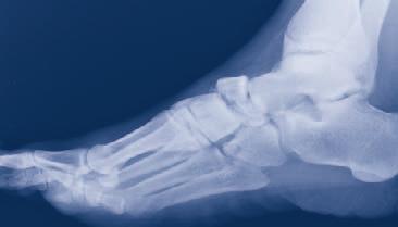
8 Clinical Cases Case 1 Cuboid fracture 18 year old male Fall from roof Preoperative Postoperative Case 2 Talus fracture 44 year old male Motor vehicle accident Preoperative Postoperative Case 3 Navicular fracture 41 year old male Motor vehicle accident Preoperative Postoperative 6 Synthes 2.4/2.7 mm Locking Tarsal Plates Technique Guide
9 Locking Talus Plate Preparation The locking tarsal plates are an addition to the 2.4 mm module of the compact foot set. Required Sets LCP Compact Foot Basic Instrument Set and Screws 2.7 mm, Stainless Steel or LCP Compact Foot Basic Instrument Set and Screws 2.7 mm, Titanium Alloy (TAN) Compact Foot 2.4 (Stainless Steel Implants) or Compact Foot 2.4 (Titanium Implants) Complete the preoperative radiographic assessment and prepare the preoperative plan. Determine the plate and instruments to be used. Synthes 7
10 Locking Talus Plate 1 Approach Two approaches are necessary to treat talus fractures, to allow for good visualization of both the medial and lateral sides of the talus. Medially, a slightly dorsal incision is made along the bisector angle between the anterior and posterior tibialis. This incision is started distally over the tubercle of the tarsal navicular bone and lengthened proximally over the tip of the medial malleolus, if required. Laterally, a longitudinal incision is made; this is called the Ollier approach, from the tip of the lateral malleolus to the dorsolateral part of the talonavicular joint. These incisions are made to the bone, avoiding the dorsal peroneal nerves and vascular structures. 2 Contour plate Instrument Bending Pin for LCP Plates 2.4 and 2.7, with thread Optional instrument Bending/Cutting Pliers Provisional bone fixation can be obtained using k-wires. Independent lag screws can be used for stabilization. 8 Synthes 2.4/2.7 mm Locking Tarsal Plates Technique Guide


11 Medially, the plate fits on the bone with the concavity upward. The posterior portion of the plate is below the medial malleolar facet and the anterior portion runs above the neck, parallel to the talonavicular joint. Laterally, the plate fits on the bone with the concavity downward. The posterior portion of the plate is almost vertical in front of the lateral process and the anterior portion is horizontal, perpendicular to the talonavicular joint. Thread the bending pins or drill guides into the plate on each side of the bend location. Ensure the pins are completely engaged into the plate before bending. Contour the plate. Be careful to avoid overbending and damage to the plate threads. The plates can be cut to length and contoured using the Bending/Cutting pliers for the specific fracture pattern or patient anatomy. Synthes 9

12 Locking Talus Plate 3 Drill and insert 2.4 mm Cortex screw Instruments Drill Bit 1.8 mm, with marking Drill Bit 2.4 mm, length 100/75 mm Handle with quick coupling Stardrive Screwdriver Shaft, T Depth Gauge Universal Drill Guide 2.4 If a combination of cortex screws and locking screws will be used, a cortex screw should be inserted first. Use the 2.4 mm universal drill guide when inserting the cortex screws. Use the 1.8 mm drill bit for the threaded hole and 2.4 mm drill bit for the gliding hole. Drill to the desired depth. Verify drilling depth using image intensification. Remove the drill guide and measure for screw length using the depth gauge. Insert the cortex screw manually with the self-retaining Stardrive screwdriver shaft and handle. 10 Synthes 2.4/2.7 mm Locking Tarsal Plates Technique Guide
13 4 Drill and insert locking screw In the tarsal plates the option of 2.4 mm or 2.7 mm (head LCP 2.4) locking screws can be used. Instruments Handle with quick coupling Stardrive Screwdriver Shaft, T Torque Limiter, 0.8 Nm LCP Drill Sleeve 2.4 for Drill Bits 1.8 mm Drill Bit 1.8 mm, with marking Optional instruments Depth Gauge LCP Drill Sleeve 2.7, for Drill Bits 2.0 mm Drill Bit 2.0 mm with double marking Screw the drill guide into one of the locking holes until it is fully seated. Insert the drill bit through the drill guide to the bone. Synthes 11


14 Locking Talus Plate Caution: Do not start drilling until the drill bit touches the bone. Inserting the drill bit into the drill guide while the drill is running may cause damage to the drill bit or drill guide. Drill to the desired depth. Verify drill depth using image intensification. Determine the screw length directly from the mark on the drill bit and the scale on the threaded drill guide. Alternatively, screw length can be checked by removing the drill guide and using the depth gauge. Insert the locking screw manually with the self-retaining Stardrive screwdriver shaft, Torque Limiter 0.8 Nm and handle. Carefully tighten the locking screw. Excessive force is not necessary to lock the screw to the plate. Repeat for the remaining locking screws. Under image intensification make a final control to ensure that all screws are the correct length and correctly placed. 12 Synthes 2.4/2.7 mm Locking Tarsal Plates Technique Guide


15 Locking Navicular Plate 1 Approach Make a dorsal longitudinal incision from the midneck of the talus towards the base of the second metatarsal. It is important to preserve neurovascular and tendinous structures. It may be necessary to open the talonavicular joint capsule to allow visualization of the joint. To minimize the potential for vascular damage, strip only a small segment of the capsule from the navicular bone. 2 Contour the Plate Instrument Bending Pin for 2.4/ 2.7 mm Locking Plates Optional instrument Bending/Cutting Pliers Provisional bone fixation can be obtained using K-wires. Independent lag screws can be used for stabilization. The plate is designed to fit the navicular bone in a concave up direction. Thread the bending pins or drill guides into the plate on each side of the bend location. Ensure the pins are completely engaged into the plate before bending. Contour the plate. Be careful to avoid overbending and damage to plate threads. The plates can be contoured using the bending/cutting pliers for the specific fracture pattern or patient anatomy. Synthes 13
16 Locking Navicular Plate 3 Drill and insert 2.4 mm cortex screw Instruments Drill Bit 1.8 mm, with marking Drill Bit 2.4 mm, length 100/75 mm Handle with quick coupling Stardrive Screwdriver Shaft, T Depth Gauge Universal Drill Guide 2.4 If a combination of cortex screws and locking screws will be used, a cortex screw should be inserted first. Use the 2.4 mm universal drill guide when inserting the cortex screws. Use the 1.8 mm drill bit for the threaded hole and 2.4 mm drill bit for the gliding hole. Drill to the desired depth. Verify drill depth using image intensification. Remove the drill guide and measure for screw length using the depth gauge. Insert the cortex screw manually with the self-retaining Stardrive screwdriver shaft and handle. 14 Synthes 2.4/2.7 mm Locking Tarsal Plates Technique Guide


17 4 Drill and insert locking screw In the tarsal plates the option of 2.4 mm or 2.7 mm (head LCP 2.4) locking screws can be used. Instruments Handle with quick coupling Stardrive Screwdriver Shaft, T Torque Limiter, 0.8 Nm LCP Drill Sleeve 2.4 for Drill Bits 1.8 mm Drill Bit 1.8 mm, with marking Optional instruments Depth Gauge LCP Drill Sleeve 2.7, for Drill Bits 2.0 mm Drill Bit 2.0 mm with double marking Screw the drill guide into one of the locking holes until it is fully seated. Insert the drill bit through the drill guide to the bone. Synthes 15
18 Locking Navicular Plate Caution: Do not start drilling until the drill bit touches the bone. Inserting the drill bit into the drill guide while the drill is running may cause damage to the drill bit or drill guide. Drill to the desired depth. Verify drill depth using image intensification. Determine the screw length directly from the mark on the drill bit and the scale on the threaded drill guide. Alternatively, screw length can be checked by removing the drill guide and using the depth gauge. Insert the locking screw manually with the self-retaining Stardrive screwdriver shaft, Torque Limiter 0.8 Nm and handle. Carefully tighten the locking screw. Excessive force is not necessary to lock the screw to the plate. Repeat for the remaining locking screws. Under image intensification make a final control to ensure that all screws are the correct length and correctly placed. 16 Synthes 2.4/2.7 mm Locking Tarsal Plates Technique Guide


19 Locking Cuboid Plate 1 Approach Make a linear dorsolateral incision starting at the sinu tarsi and extending to the base of the fourth metatarsal. Caution, this incision may run parallel to or directly over the sural nerve, and crosses the peroneous tertius, care must be taken to avoid injuring these structures. One of the main objectives of cuboid fracture management is restoration of the lateral column length and articular surface. 2 Contour the Plate Instrument Bending Pin for 2.4/ 2.7 mm Locking Plates Optional instrument Bending/Cutting Pliers Provisional bone fixation can be obtained using K-wires. Independent lag screws can be used for stabilization. The cuboid plate is available in left and right plates to match the anatomy of each foot. The longest arm, with 5 screw holes, is designed to be placed proximally. Thread the bending pins or drill guides into the plate on each side of the bend location. Ensure the pins are completely engaged into the plate before bending. Contour the plate. Be careful to avoid overbending and damage to plate threads. The plates can be contoured using the bending/cutting pliers for the specific fracture pattern or patient anatomy. Synthes 17
20 Locking Cuboid Plate 3 Drill and insert 2.4 mm cortex screw Instruments Drill Bit 1.8 mm, with marking Drill Bit 2.4 mm, length 100/75 mm Handle with quick coupling Stardrive Screwdriver Shaft, T Depth Gauge Universal Drill Guide 2.4 If a combination of cortex screws and locking screws will be used, a cortex screw should be inserted first. Use the 2.4 mm universal drill guide when inserting the cortex screws. Use the 1.8 mm drill bit for the threaded hole and 2.4 mm drill bit for the gliding hole. Drill to the desired depth. Verify drill depth using image intensification. Remove the drill guide and measure for screw length using the depth gauge. Insert the cortex screw manually with the self-retaining Stardrive screwdriver shaft and handle. 18 Synthes 2.4/2.7 mm Locking Tarsal Plates Technique Guide
21 4 Drill and insert locking screw In the tarsal plates the option of 2.4 mm or 2.7 mm (head LCP 2.4) locking screws can be used. Instruments Handle with quick coupling Stardrive Screwdriver Shaft, T Torque Limiter, 0.8 Nm LCP Drill Sleeve 2.4 for Drill Bits 1.8 mm Drill Bit 1.8 mm, with marking Optional instruments Depth Gauge LCP Drill Sleeve 2.7, for Drill Bits 2.0 mm Drill Bit 2.0 mm with double marking Screw the drill guide into one of the locking holes until it is fully seated. Insert the drill bit through the drill guide to the bone. Synthes 19


22 Locking Cuboid Plate Caution: Do not start drilling until the drill bit touches the bone. Inserting the drill bit into the drill guide while the drill is running may cause damage to the drill bit or drill guide. Drill to the desired depth. Verify drill depth using image intensification. Determine the screw length directly from the mark on the drill bit and the scale on the threaded drill guide. Alternatively, screw length can be checked by removing the drill guide and using the depth gauge. Insert the locking screw manually with the self-retaining Stardrive screwdriver shaft, Torque Limiter 0.8 Nm and handle. Carefully tighten the locking screw. Excessive force is not necessary to lock the screw to the plate. Repeat for the remaining locking screws. Under image intensification make a final control to ensure that all screws are the correct length and correctly placed. 20 Synthes 2.4/2.7 mm Locking Tarsal Plates Technique Guide


23 Plates 0X Navicular Plate 2.4, locking 0X Cuboid Plate 2.4, locking, left 0X Cuboid Plate 2.4, locking, right 0X Talus Plate 2.4, locking X=2 stainless steel X=4 titanium Implants (or products" as appropriate) are available nonsterile or sterile packed. Add suffix "S" to article number to order sterile product. Synthes 21
24 Screws LCP Locking Screw Stardrive 2.4 mm, self-tapping Thread diameter 2.4 mm Drill bit for threaded hole 1.8 mm Drill bit for gliding hole 2.4 mm Core diameter 1.9 mm Head diameter 3.5 mm Stardrive T8 2.4 mm locking screws available from 6 mm to 30 mm lengths (2 mm increments) LCP Locking Screw Stardrive 2.7 mm (head LCP 2.4), self-tapping Thread diameter 2.7 mm Drill bit for threaded hole 2.0 mm Drill bit for gliding hole 2.7 mm Core diameter 2.1 mm Head diameter 3.5 mm Stardrive T8 2.7 mm locking screws available from 10 mm to 60 mm lengths (2 mm increments up to 50 mm, 5 mm increments up to 60 mm) 22 Synthes 2.4/2.7 mm Locking Tarsal Plates Technique Guide
25 Cortex Screw Stardrive 2.4 mm, self-tapping Thread diameter 2.4 mm Drill bit for threaded hole 1.8 mm Drill bit for gliding hole 2.4 mm Core diameter 1.7 mm Head diameter 4.0 mm Stardrive T8 2.4 mm cortex screws available from 6 mm to 40 mm lengths (1 mm increments up to 14 mm, 2 mm increments from 16 mm up to 40 mm) Note: For information on fixation principles using conventional and locked plating techniques, please refer to the LCP Locking Compression Plate Technique Guide ( ). Note: The T8 Stardrive recess in the screw head offers improved torque transfer, high strength, and self-retention of screws, when compared to cruciform and hexagonal drives. Please note the Stardrive recess in the surgical report. This will remind the surgeon to have a Stardrive screwdriver available when removing these screws. Synthes 23
26 Instruments Bending Pin for LCP Plates 2.4 and 2.7, with thread Bending/Cutting Pliers Drill Bit 1.8 mm, with marking Drill Bit 2.4 mm, length 100/75 mm Handle with quick coupling Stardrive Screwdriver Shaft, T8 24 Synthes 2.4/2.7 mm Locking Tarsal Plates Technique Guide
27 Depth Gauge Universal Drill Guide Torque Limiter, 0.8 Nm LCP Drill Sleeve 2.4 for Drill Bits 1.8 mm LCP Drill Sleeve 2.7, for Drill Bits 2.0 mm Drill Bit 2.0 mm with double marking Synthes 25
28
29 Bibliography Tarsal Plates ST Hansen Jr (2000) Functional Reconstruction of the Foot and Ankle. Lippincott: Williams and Wilkins Approach Images ED McGlamry (1987) Fundamentals of Foot Surgery. Lippincott: Williams and Wilkins: Synthes 27
30
31
32 Ö öAAáä Synthes GmbH Eimattstrasse 3 CH-4436 Oberdorf Presented by: SE_ AA /2008 Synthes, Inc. or its affiliates All rights reserved Synthes, LCP and Stardrive are trademarks of Synthes, Inc. or its affiliates
Variable Angle LCP Tarsal Plates 2.4/2.7. Navicular Plate and Cuboid Plates.
 Variable Angle LCP Tarsal Plates 2.4/2.7. Navicular Plate and Cuboid Plates. Surgical Technique This publication is not intended for distribution in the USA. Instruments and implants approved by the AO
Variable Angle LCP Mesh Plate 2.4/2.7. Part of the Variable Angle LCP Forefoot/Midfoot System 2.4/2.7.
 Variable Angle LCP Mesh Plate 2.4/2.7. Part of the Variable Angle LCP Forefoot/Midfoot System 2.4/2.7. Surgical Technique This publication is not intended for distribution in the USA. Instruments and implants
Variable Angle LCP Forefoot/Midfoot System 2.4/2.7. Procedure specific plates for osteotomies, arthrodeses and fractures of the foot.
 Instruction for Use Variable Angle LCP Forefoot/Midfoot System 2.4/2.7. Procedure specific plates for osteotomies, arthrodeses and fractures of the foot. Table of Contents Introduction VA-LCP Forefoot/Midfoot
LCP Pilon Plate 2.7/3.5. Surgical Technique
 LCP Pilon Plate 2.7/3.5 Surgical Technique Image intensifier control This description alone does not provide sufficient background for direct use of DePuy Synthes products. Instruction by a surgeon experienced
Technique Guide. Variable Angle LCP Opening Wedge Plates 2.4/2.7. Part of the Variable Angle LCP Forefoot / Midfoot System 2.4 / 2.7.
 Technique Guide Variable Angle LCP Opening Wedge Plates 2.4/2.7. Part of the Variable Angle LCP Forefoot / Midfoot System 2.4 / 2.7. Table of Contents Introduction Variable Angle LCP Opening Wedge Plates
1.5 MM LCP SYSTEM. For treatment of fractures and arthrodeses of canines and felines SURGICAL TECHNIQUE
 1.5 MM LCP SYSTEM For treatment of fractures and arthrodeses of canines and felines SURGICAL TECHNIQUE TABLE OF CONTENTS INTRODUCTION 1.5 mm LCP System 2 AO Principles 4 SURGICAL TECHNIQUE Reduce Fracture
Technique Guide. Quadrilateral Surface Plates 3.5. Part of the Low Profile Pelvic System 3.5.
 Technique Guide Quadrilateral Surface Plates 3.5. Part of the Low Profile Pelvic System 3.5. Table of Contents Introduction Quadrilateral Surface Plates 3.5 2 AO Principles 4 Indications 5 Surgical Technique
Technique Guide. Variable Angle LCP 1 st MTP Fusion Plates 2.4/2.7. Part of the Variable Angle LCP Forefoot / Midfoot System 2.4 / 2.7.
 Technique Guide Variable Angle LCP 1 st MTP Fusion Plates 2.4/2.7. Part of the Variable Angle LCP Forefoot / Midfoot System 2.4 / 2.7. Table of Contents Introduction Variable Angle LCP 1 st MTP Fusion
2.4 mm and 3.0 mm Headless Compression Screws
 For Fixation of Small Bones and Small Bone Fragments 2.4 mm and 3.0 mm Headless s Surgical Technique Table of Contents Introduction 2.4 mm and 3.0 mm Headless 2 Technique Overview 4 AO Principles 5 Indications
Technique Guide. 4.5 mm Cannulated Screws. Part of the Synthes Cannulated Screw System.
 Technique Guide 4.5 mm Cannulated Screws. Part of the Synthes Cannulated Screw System. TableofContents Introduction 4.5 mm Cannulated Screws 2 AO Principles 3 Indications 4 Surgical Technique Surgical
Part of the DePuy Synthes Cannulated Screw System. 3.5 mm Cannulated Screws
 Part of the DePuy Synthes Cannulated Screw System 3.5 mm Cannulated Screws Surgical Technique Table of Contents Introduction 3.5 mm Cannulated Screws 2 AO Principles 3 Indications 4 Surgical Technique
VARIABLE ANGLE LOCKING HAND SYSTEM
 VARIABLE ANGLE LOCKING HAND SYSTEM For fragment-specific fracture fixation with variable angle locking and locking technology SURGICAL TECHNIQUE TABLE OF CONTENTS INTRODUCTION Variable Angle Locking Hand
Technique Guide. Occipito-Cervical Fusion System. Implants and instruments designed to optimize fixation to the occiput.
 Technique Guide Occipito-Cervical Fusion System. Implants and instruments designed to optimize fixation to the occiput. Table of Contents Introduction Overview 2 AO ASIF Principles 4 Indications and Contraindications
Technique Guide. LCP Pilon Plate 2.7/3.5
 Technique Guide LCP Pilon Plate 2.7/3.5 LCP Pilon Plate 2.7/3.5 Table of contents Indications 3 Implants 4 Instruments 5 Surgical technique 6 Implant removal 12 Image intensifier control Warning This
VARIABLE ANGLE LOCKING HAND SYSTEM
 VARIABLE ANGLE LOCKING HAND SYSTEM For fragment-specific fracture fixation with variable angle locking and locking technology Instruments and implants approved by the AO Foundation. This publication is
LCP Pilon Plate 2.7/3.5
 Surgical Technique LCP Locking Compression Plate Original Instruments and Implants of the Association for the Study of Internal Fixation AO/ASIF Table of contents Indications 3 Implants 4 Instruments 5
LCP Pilon Plate 2.7/3.5
 LCP Pilon Plate 2.7/3.5 Surgical Technique This publication is not intended for distribution in the USA. Instruments and implants approved by the AO Foundation. Table of contents Indications 2 Implants
7.0 mm Cannulated Screws
 Part of the DePuy Synthes Cannulated Screw System 7.0 mm Cannulated Screws Surgical Technique Table of Contents Introduction 7.0 mm Cannulated Screws 2 AO Principles 3 Indications 4 Surgical Technique
Instructions for Use. LCP Locking Compression Plate. Combine without Compromise.
 Instructions for Use LCP Locking Compression Plate. Combine without Compromise. Table of Contents LCP: Combine without Compromise 2 AO ASIF Principles of Osteosynthesis 4 Indications and Contraindications
Technique Guide. 7.0 mm Cannulated Screws. Part of the Synthes Cannulated Screw System.
 Technique Guide 7.0 mm Cannulated Screws. Part of the Synthes Cannulated Screw System. Table of Contents Introduction 7.0 mm Cannulated Screws 2 AO Principles 3 Indications 4 Surgical Technique Surgical
DLS Dynamic Locking Screw. Combined with LCP Locking Compression Plate.
 DLS Dynamic Locking Screw. Combined with LCP Locking Compression Plate. Instructions for Use Discontinued June 2016 DSEM/TRM/0517/0844(1) Table of Contents Introduction DLS Dynamic Locking Screw 2 Indications
HCS 2.4/3.0. The countersinkable compression screw.
 Technique Guide HCS 2.4/3.0. The countersinkable compression screw. Table of Contents Introduction Features and Benefits 2 Functional Principle 3 Indications 4 Surgical Technique Hand Scaphoid 5 Foot
VLP FOOT Variable Angle Locked Plating System
 Surgical Technique VLP FOOT Variable Angle Locked Plating System Surgical Technique Table of Contents Product overview Implant selection...2 Introduction...3 System overview...4 Indications...4 Design
3.5 mm and 4.5 mm Curved Locking Compression Plates (LCP )
") For Minimally Invasive Osteosynthesis 3.5 mm and 4.5 mm Curved Locking Compression Plates (LCP ) Surgical Technique Table of Contents Introduction 3.5 mm and 4.5 mm Curved Locking Compression 2 Plates
For Minimally Invasive Osteosynthesis 3.5 mm and 4.5 mm Curved Locking Compression Plates (LCP ) Surgical Technique Table of Contents Introduction 3.5 mm and 4.5 mm Curved Locking Compression 2 Plates
2.4 mm Cannulated Screw. An integral part of the Synthes Cannulated Screw System (CSS).
.") 2.4 mm Cannulated Screw. An integral part of the Synthes Cannulated Screw System (CSS). Surgical Technique This publication is not intended f distribution in the USA. and implants approved by the AO Foundation.
2.4 mm Cannulated Screw. An integral part of the Synthes Cannulated Screw System (CSS). Surgical Technique This publication is not intended f distribution in the USA. and implants approved by the AO Foundation.
Small Fragment Locking Compression Plate (LCP ) System Stainless Steel and Titanium TECHNIQUE GUIDE
 System Stainless Steel and Titanium TECHNIQUE GUIDE") Small Fragment Locking Compression Plate (LCP ) System Stainless Steel and Titanium TECHNIQUE GUIDE R Original Instruments and Implants of the Association for the Study of Internal Fixation AO ASIF Introduction
Small Fragment Locking Compression Plate (LCP ) System Stainless Steel and Titanium TECHNIQUE GUIDE R Original Instruments and Implants of the Association for the Study of Internal Fixation AO ASIF Introduction
3.5 mm Cannulated Screw Technique Guide
 3.5 mm Cannulated Screw Technique Guide An Integral Part of the SYNTHES Cannulated Screw System Original Instruments and Implants of the Association for the Study of Internal Fixation AO ASIF The 3.5 mm
URS Degen. Top loading pedicle screw system for posterior stabilization.
 URS Degen. Top loading pedicle screw system for posterior stabilization. Technique Guide This publication is not intended for distribution in the USA. Table of Contents Introduction URS Degen 2 AO Principles
HCS 1.5. The countersinkable compression screw.
 HCS 1.5. The countersinkable compression screw. Surgical Technique This publication is not intended for distribution in the USA. Instruments and implants approved by the AO Foundation. Table of Contents
Occipito-Cervical Fusion System. Implants and instruments designed to optimize fixation to the occiput.
 Occipito-Cervical Fusion System. Implants and instruments designed to optimize fixation to the occiput. Technique Guide This publication is not intended for distribution in the USA. Instruments and implants
Technique Guide. Synapse System. An enhanced set of instruments and implants for posterior stabilization of the cervical and upper thoracic spine.
 Technique Guide Synapse System. An enhanced set of instruments and implants for posterior stabilization of the cervical and upper thoracic spine. Table of Contents Introduction Synapse System 2 AO Principles
ACLP Anterior Cervical Locking Plate System TECHNIQUE GUIDE
 ACLP Anterior Cervical Locking Plate System TECHNIQUE GUIDE Instruments and implants approved by the AO Foundation ACLP Anterior Cervical Locking Plate System The ACLP System is designed to reduce the
OPERATIVE TECHNIQUE RIVAL VIEW PLATING SYSTEM. foot & ankle reconstruction procedures
 OPERATIVE TECHNIQUE RIVAL VIEW PLATING SYSTEM foot & ankle reconstruction procedures INTRODUCTION 3 SYSTEM DESCRIPTION 3 TECHNICAL DETAILS 4 SALES AND MARKETING CONFIGURATION 5 OPERATIVE TECHNIQUE 7 OPERATIVE
6.5 mm and 7.3 mm Cannulated Screws Technique Guide
 6.5 mm and 7.3 mm Cannulated Screws Technique Guide An Integral Part of the SYNTHES Cannulated Screw System Original Instruments and Implants of the Association for the Study of Internal Fixation AO ASIF
MatrixMANDIBLE Preformed Reconstruction Plates. Preshaped to the mandibular anatomy.
 MatrixMANDIBLE Preformed Reconstruction Plates. Preshaped to the mandibular anatomy. Technique Guide CMF Matrix Table of Contents Introduction MatrixMANDIBLE Preformed Reconstruction Plates 2 AO Principles
OCCIPITO-CERVICAL FUSION SYSTEM Implants and instruments designed to optimize fixation to the occiput
 OCCIPITO-CERVICAL FUSION SYSTEM Implants and instruments designed to optimize fixation to the occiput Instruments and implants approved by the AO Foundation. This publication is not intended for distribution
Expert HAN. Expert Hindfoot Arthrodesis Nail.
 Expert HAN. Expert Hindfoot Arthrodesis Nail. Technique Guide Expert Nailing System Table of Contents Introduction Expert Hindfoot Arthrodesis Nail 2 AO Principles 4 Indications 5 Surgical Technique Preoperative
Technique Guide. LCP Dynamic Helical Hip System (DHHS). Part of the Synthes Large Fragment LCP System.
. Part of the Synthes Large Fragment LCP System.") Technique Guide LCP Dynamic Helical Hip System (DHHS). Part of the Synthes Large Fragment LCP System. Table of Contents Introduction LCP Dynamic Helical Hip System (DHHS) 2 AO Principles 4 Indications
Technique Guide LCP Dynamic Helical Hip System (DHHS). Part of the Synthes Large Fragment LCP System. Table of Contents Introduction LCP Dynamic Helical Hip System (DHHS) 2 AO Principles 4 Indications
Cerclage Passer. For minimally invasive application of cerclage cables.
 Cerclage Passer. For minimally invasive application of cerclage cables. Handling Technique Cable application This publication is not intended for distribution in the USA. Instruments and implants approved
VECTRA. SURGICAL TECHNIQUE. Anterior cervical plate system. This publication is not intended for distribution in the USA.
 VECTRA. Anterior cervical plate system. This publication is not intended for distribution in the USA. SURGICAL TECHNIQUE Contents Indications and contraindications Implants Vario Case Instruments Surgical
OPERATIVE TECHNIQUE RIVAL REDUCE FRACTURE PLATING SYSTEM. foot & ankle trauma procedures
 OPERATIVE TECHNIQUE RIVAL REDUCE FRACTURE PLATING SYSTEM foot & ankle trauma procedures INTRODUCTION 3 SYSTEM DESCRIPTION 3 TECHNICAL DETAILS 4 SALES AND MARKETING CONFIGURATION 5 OPERATIVE TECHNIQUE 7
VECTRA SURGICAL TECHNIQUE. Anterior cervical plate system. This publication is not intended for distribution in the USA.
 VECTRA Anterior cervical plate system This publication is not intended for distribution in the USA. SURGICAL TECHNIQUE Image intensifier control This description alone does not provide sufficient background
5th Metatarsal Fracture System Surgical Technique
 5th Metatarsal Fracture System Surgical Technique 5th Metatarsal Fracture System 5th Metatarsal Fracture System The 5th Metatarsal Fracture System (AR-8956S) is a uniquely designed screw and plate system
Synthes Kirschner Wires and Cerclage Wires. Multifunctional devices for temporary fixation, tension band, cerclage wiring and percutaneous pinning.
 Technique Guide Synthes Kirschner Wires and Cerclage Wires. Multifunctional devices for temporary fixation, tension band, cerclage wiring and percutaneous pinning. Image intensifier control Warning This
ACCS Anterior Cervical Compression System TECHNIQUE GUIDE
 ACCS Anterior Cervical Compression System TECHNIQUE GUIDE Original Instruments and Implants of the Association for the Study of Internal Fixation AO ASIF ACCS Anterior Cervical Compression System The Anterior
VariAx. Foot & Ankle. Foot Locking Plate System
 VariAx Foot Locking Plate System Operative Technique Foot & Ankle Trauma and Deformity Correction Polyaxial Locking Technology Comprehensive Calcaneal Fracture Plates VariAx 2 Plates VariAx Foot Locking
Universal Humeral Nail
 990210009 INDEX Indications Preoperative Planning Patient Position Surgical Technique - Step 1 Open Humerus - Step 2 Calibrate The Nail - Step 3 Insert Nail - Step 4 Proximal Locking - Step 5 Assemble
Technique Guide. Cable System. For Orthopaedic Trauma Surgery.
 Technique Guide Cable System. For Orthopaedic Trauma Surgery. Table of Contents Introduction Overview 2 AO Principles 4 Indications and Contraindications 5 Surgical Technique Standard Cerclage Technique
Cannulated Percutaneous Guiding System. For percutaneous placement of 3.5 mm pelvic and cortex screws in the pelvic area.
 Cannulated Percutaneous Guiding System. For percutaneous placement of 3.5 mm pelvic and cortex screws in the pelvic area. Technique Guide This publication is not intended for distribution in the USA. Instruments
Distal Fibula Plate SURGICAL TECHNIQUE
 MAXLOCK EXTREME Distal Fibula Plate SURGICAL TECHNIQUE Contents Key Design Features 3 Surgical Technique 4 Implants and Instruments 8 Proper surgical procedures and techniques are the responsibility of
Fibula Plating System
 ANATOMIC LOCKED PLATING SYSTEM Fibula Plating System Securing optimal fixation through versatile locked and compression plating technology Contents Surgeon Design Team 2 Introduction 3 Anatomic Fibula
Vectra, Vectra-T and Vectra-One. Anterior cervical plating for spinal fusion.
 Vectra, Vectra-T and Vectra-One. Anterior cervical plating for spinal fusion. Technique Guide Instruments and implants approved by the AO Foundation Table of Contents Introduction Vectra, Vectra-T and
OPERATIVE TECHNIQUE RIVAL BITE HEADED CANNULATED AND HEADLESS COMPRESSION SCREWS. foot & ankle applications
 OPERATIVE TECHNIQUE RIVAL BITE HEADED CANNULATED AND HEADLESS COMPRESSION SCREWS foot & ankle applications INTRODUCTION 3 SYSTEM DESCRIPTION 3 TECHNICAL DETAILS 4 SALES AND MARKETING CONFIGURATION 6 OPERATIVE
The Percutaneous Reduction Forceps Technique Guide
 The Percutaneous Reduction Forceps Technique Guide Indications + Product Overview Introduction The Percutaneous Reduction Forceps The Percutaneous Reduction Forceps facilitate standard technique for fixation
Occipito-Cervical Fusion System
 Implants and Instruments designed to enhance Fixation to the Occiput Occipito-Cervical Fusion System Surgical Technique Image intensifier control This description alone does not provide sufficient background
Titanium Modular Hand System. For fractures, replantations and reconstruction of the hand.
 Titanium Modular Hand System. For fractures, replantations and reconstruction of the hand. Plates precontoured for anatomic fit Instruments colorcoded for easy recognition Modules for 1.0 mm to 2.4 mm
Distal Radius System 2.5
 SURGICAL TECHNIQUE STEP BY STEP Distal Radius System 2.5 APTUS Wrist 2 Distal Radius System 2.5 Contents 3 Introduction 3 Product Materials 3 Indications 3 Contraindications 3 Color Coding 3 Possible Combination
Integra. Capture Screw System SURGICAL TECHNIQUE
 Integra Capture Screw System SURGICAL TECHNIQUE Table of Contents Indications... 2 Contraindications... 2 System Description... 2 System Features... 2 Cannulated Low-Profile Screws (AC-Series) Overview...
Distal Medial Tibia Plate Surgical Technique
 Locking Compression Technology by aap 1 Disclaimer This surgical technique is exclusively intended for medical professionals, especially physicians, and therefore may not be regarded as a source of information
3.5 MM low-profile cortex screws
 3.5 MM low-profile cortex screws Low-profle fxation option compatible with all Trauma 3.5 mm plates MechanicallY comparable To DEPUY SYNTHES TRAUMA standard 3.5 MM cortex screws low-profile option To MiniMiZe
Integra. Stainless Headed Compression Screw System SURGICAL TECHNIQUE
 Integra Stainless Headed Compression Screw System SURGICAL TECHNIQUE Table of Contents Design Rationale...2 Indications...2 Contraindications...2 Surgical Technique Step 1: Inserting Guide Wire... 3 Step
WRIST SYSTEM. ARIX Volar Distal Radius Locking Plate System
 WRIST SYSTEM A Contents 3 4 7 14 15 16 19 21 Indications Product Overview Features & Benefits Ordering Information - Screws -2.5mm Self-Tapping Cortical Screws (Non-Locking) -2.5mm Self-Tapping Locking
Lag Screw Device TECHNIQUE GUIDE. Indicated for symphyseal fracture fixation of the mandible. Instruments and implants approved by the AO Foundation
 Lag Screw Device TECHNIQUE GUIDE Indicated for symphyseal fracture fixation of the mandible Instruments and implants approved by the AO Foundation Lag Screw Device Indicated for symphyseal fracture fixation
Table of Contents 2-6. Introduction. Indications Surgical Technique. Ordering Information 15-24
 Table of Contents Introduction Product information ExtremiFix Midsize Large Screw Offering Headless Screw Characteristics Design Features & Benefits Instrumentation Technical Details Calibrated Drill Bits
Instruments for removing Synthes screws. Screw Extraction Set. Handling Technique
 Instruments for removing Synthes screws Screw Extraction Set Handling Technique Image intensifier control This description alone does not provide sufficient background for direct use of DePuy Synthes products.
Technique Guide. Modular Sternal Cable System. Flexibility and strength in sternal closure and repair.
 Technique Guide Modular Sternal Cable System. Flexibility and strength in sternal closure and repair. Table of Contents Introduction Overview 2 Indications and Contraindications 3 Surgical Technique A.
Digital Compression Screw
 Digital Compression Screw Surgical Technique Contents Product The BioPro Digital Compression Screw is a stainless steel lag screw designed for digital fusions. Table of contents Indications & Contraindications
VariAx Foot Locking Plate System
 VariAx Foot Locking Plate System Operative Technique Trauma and Deformity Correction Polyaxial Locking Technology Comprehensive Calcaneal Fracture Plates Table of Contents 1. Introduction 3 2. Overview
Orthopedic Bone Nail System Universal Humeral Nail
 Orthopedic Bone Nail System Universal Humeral Nail Surgical Technique Manual Note: The surgical procedures should be performed under the guidance of qualified skilled orthopedic surgeons, and this surgical
BioDrive Micro Screw System
 At Biomet, engineering excellence is our heritage and our passion. For over 25 years, through various divisions worldwide, we have applied the most advanced engineering and manufacturing technology to
MEDIALMAX System SURGICAL TECHNIQUE
 MAXLOCK EXTREME MEDIALMAX System SURGICAL TECHNIQUE Contents The MEDIALMAX Advantage Design Feature Various flex options POCKETLOCK Technology Anatomic design Advantage Provides the surgeon with multiple
Cable System. For Orthopaedic Trauma Surgery.
 Cable System. For Orthopaedic Trauma Surgery. Surgical Technique This publication is not intended for distribution in the USA. Instruments and implants approved by the AO Foundation. Image intensifier
MaxTorque. surgical technique. Cannulated Screw System. Foot & Ankle. OrthoHelix Technology
 MaxTorque Cannulated Screw System OrthoHelix Technology surgical technique Foot & Ankle 2 M A X T O R Q U E C A N N U L A T E D S C R E W S Y S T E M Table of Contents Advantages 3 Indications 4 Contraindications
Locking Small Fragment
 Locking Small Fragment Page 1 Locking Small Fragment Table of contents Implants 5 Operation technique 7 Inserting drill guides 7 Drilling 7 Length measurement 7 Inserting the measured screw 8 Different
3.5 mm/ 2.7 mm LCP. PanCarPaL arthrodesis S PLate. BroCHUre. For treatment of carpal joints in dogs
 3.5 mm/ 2.7 mm LCP PanCarPaL arpal arthrodesis S PLate For treatment of carpal joints in dogs BroCHUre PLate DeSIGn the DePuy Synthes Vet 3.5 mm/2.7 mm LCP Pancarpal arthrodesis plate is intended for the
MATRIX COMBO PLATING SYSTEM. Streamlined set for craniofacial and mandibular trauma and reconstruction
 MATRIX COMBO PLATING SYSTEM Streamlined set for craniofacial and mandibular trauma and reconstruction MATRIXCOMBO PLATING SET MATRIXCOMBO PLATING SET (01.503.400) The aim of surgical fracture treatment
Technique Guide. Synapse System. An enhanced set of implants and instruments for posterior stabilization of the cervical and upper thoracic spine.
 Technique Guide Synapse System. An enhanced set of implants and instruments for posterior stabilization of the cervical and upper thoracic spine. Image intensifier control Warning This description alone
The Titanium Cannulated Lateral Entry Femoral Recon Nail. Expert nailing system with radiolucent instrumentation.
 The Titanium Cannulated Lateral Entry Femoral Recon Nail. Expert nailing system with radiolucent instrumentation. Technique Guide EXPERT Nailing System Table of Contents Introduction Titanium Cannulated
VariAx DistalFibula. Foot & Ankle. Locking Plate System. Operative Technique
 VariAx DistalFibula Locking Plate System Operative Technique Foot & Ankle Distal Fibula Fracture Repair Polyaxial Locking Technology Low Profile Design VariAx 2 Color Coded Screws and Instruments VariAx
Zimmer Natural Nail System
 Zimmer Natural Nail System Cephalomedullary Nail Surgical Technique Compact Case - Short Nails Only STANDARD Zimmer Natural Nail System Cephalomedullary Nail Surgical Technique - Standard 1 Zimmer Natural
Zimmer Natural Nail System
 Zimmer Natural Nail System Cephalomedullary Nail Surgical Technique Compact Case- Short Nails Only SMALL Zimmer Natural Nail System Cephalomedullary Nail Technique - Small 1 Zimmer Natural Nail System
Zimmer Natural Nail System. Cephalomedullary Nail Surgical Technique SMALL
 Zimmer Natural Nail System Cephalomedullary Nail Surgical Technique SMALL Zimmer Natural Nail System Cephalomedullary Nail Technique - Small 1 Zimmer Natural Nail System Cephalomedullary Nail Surgical
3.5 mm/2.7 mm LCP. Pancarpal Arthrodesis Plate. For treatment of carpal joints in dogs
 3.5 mm/2.7 mm LCP Pancarpal Arthrodesis Plate For treatment of carpal joints in dogs Plate Design The DePuy Synthes Vet 3.5 mm/2.7 mm LCP Pancarpal Arthrodesis plate is intended for the treatment of hyperextension
Synapse System. An enhanced set of instruments and implants for posterior stabilization of the upper spine.
 Synapse System. An enhanced set of instruments and implants for posterior stabilization of the upper spine. Technique Guide 100º Instruments and implants approved by the AO Foundation Table of Contents
ISO Plate SURGICAL TECHNIQUE
 MINI MAXLOCK EXTREME ISO Plate SURGICAL TECHNIQUE Contents Table of Contents Key Design Features 2 Surgical Technique 3 Implants and Instruments 8 Key Design Features The MINI MAXLOCK EXTREME ISO (Intraosseous
Pangea Degenerative Spine System. Top Loading Preassembled Pedicle Screw System for Posterior Stabilization of the Thoracolumbar Spine.
 Technique Guide Pangea Degenerative Spine System. Top Loading Preassembled Pedicle Screw System for Posterior Stabilization of the Thoracolumbar Spine. Contents Introduction AO ASIF Principles 4 Indications
Top Loading Pedicle Screw and Hook System for Posterior Stabilization. URS System. Surgical Technique
 Top Loading Pedicle Screw and Hook System for Posterior Stabilization URS System Surgical Technique Image intensifier control This description alone does not provide sufficient background for direct use
CHARLOTTE CLAW Plate Compression Locking Arthrodesis by Wright SURGICAL TECHNIQUE
 CHARLOTTE CLAW Plate Compression Locking Arthrodesis by Wright SURGICAL TECHNIQUE Contents Chapter 1 1 Chapter 2 2 2 2 3 4 5 6 Appendix A 7 Preoperative Planning Surgical Technique Calcaneal Cuboid Arthrodesis
CHARLOTTE. Multi-Use Compression Screw
 CHARLOTTE Multi-Use Compression Screw CHARLOTTE multi-use compression screw surgical technique SURGICAL ADVISORS ROBERT ANDERSON, MD BRUCE COHEN, MD W. HODGES DAVIS, MD Proper surgical procedures and techniques
TORNIER MAXLOCK EXTREME. Clavicle Plating System SURGICAL TECHNIQUE
 TORNIER MAXLOCK EXTREME Clavicle Plating System SURGICAL TECHNIQUE 2 Table of Contents: Key Design Features...4 Surgical Technique...5 Implants & Instruments...9 3 Key Design Features There are 7 anatomically
Small Plate and Screw System SURGICAL TECHNIQUE
 MINI MAXLOCK EXTREME Small Plate and Screw System SURGICAL TECHNIQUE Contents Key Design Features 2 Surgical Technique 3 Implants and Instruments 9 Proper surgical procedures and techniques are the responsibility
DART-FIRE. Small Screw System SURGICAL TECHNIQUE
 DART-FIRE Small Screw System SURGICAL TECHNIQUE DART-FIRE Small Screw System Surgical Technique Contents Chapter 1 4 Chapter 2 6 Chapter 3 7 Appendix 1 10 Appendix 2 12 Introduction Intended Use DART-FIRE
For Minimally Invasive Application of Cerclage Wires. Cerclage Passer. Surgical Technique
 For Minimally Invasive Application of Cerclage Wires Cerclage Passer Surgical Technique Table of Contents Introduction Cerclage Passer 2 Surgical Technique Preparation 4 Insert Cerclage Passer 5 Connect
Anterior Cervical Plate SURGICAL TECHNIQUE GUIDE. Surgeon Driven Innovation
 Anterior Cervical Plate SURGICAL TECHNIQUE GUIDE Surgeon Driven Innovation 1 The Snowmass Anterior Cervical Plate System is intended for the surgical treatment and correction of traumatic and pathologic
Instruments for Removing DePuy Synthes Screws. Screw Removal Set
 Instruments for Removing DePuy Synthes Screws Screw Removal Set Surgical Technique Table of Contents Introduction Screw Removal Set 2 Surgical Technique Preoperative Planning and Preparation 6 Removal
ORTHOLINK. 2 - Hole Wedge Osteotomy Plate SURGICAL TECHNIQUE
 MAXLOCK EXTREME ORTHOLINK 2 - Hole Wedge Osteotomy Plate SURGICAL TECHNIQUE Contents Key Design Features 3 Surgical Technique 4 Implants and Instruments 7 Proper surgical procedures and techniques are
SURGICAL TECHNIQUE GUIDE
 SURGICAL TECHNIQUE GUIDE DANGER indicates an imminently hazardous situation which, if not avoided, will result in death or serious injury. WARNING indicates a potentially hazardous situation which, if
Technique Guide MTP Arthrodesis System
 Technique Guide MTP Arthrodesis System The CheckMATE MTP Arthrodesis System features low-profile, anatomically pre-contoured Bone Plates with either a combination of locking and non-locking holes or all-locking
SpeedTip CCS 5.0, 7.0
 SURGICAL TECHNIQUE STEP BY STEP SpeedTip CCS 5.0, 7.0 Cannulated Compression Screws APTUS 2 SpeedTip CCS 5.0, 7.0 Cannulated Compression Screws Contents 3 Introduction Product Materials Indications Contraindications
UJAM AA O RTHO PTY LTD 100 HYDE PARK C LO SE HYDE PARK 2196 W W W.UJAM AAO RTHO.C O M
 UJAM AA O RTHO PTY LTD 100 HYDE PARK C LO SE HYDE PARK 2196 W W W.UJAM AAO RTHO.C O M 4.0mm Cannulated Screw Set 112730000 112730100 Guide Wire, φ1.25 112730200 Guide Sleeve, φ1.25/φ3.0 110190300 Drill