SOMATOM Sensation 4 Computed Tomography System for Multislice Spiral Scanning
|
|
|
- Alban Turner
- 6 years ago
- Views:
Transcription

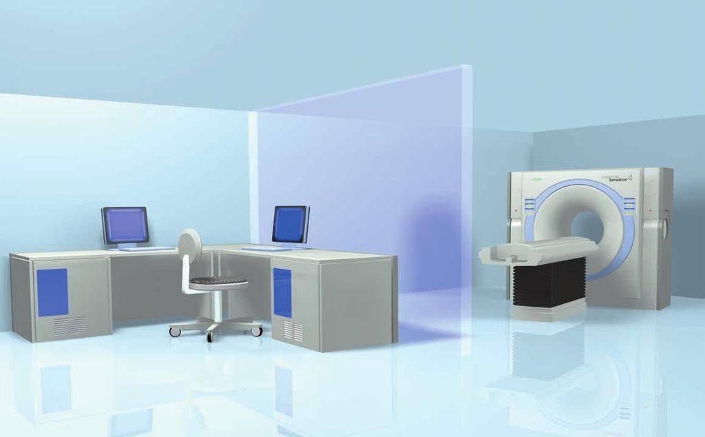
1 SOMATOM Sensation 4 Computed Tomography System for Multislice Spiral Scanning
2 SOMATOM Sensation 4 Computed Tomography System for Multislice Spiral Scanning Siemens has been a global player in CT for more than 25 years, having set lots of milestones. The SOMATOM CT success history is a synonym for technological firsts, clinical leadership, customer satisfaction and investment protection. Our objective is to expand our market leadership role with the introduction of CT products that are based on new cutting edge technology embedded in optimized workflow concepts resulting in outstanding productivity and high patient throughput. The 4-slice-system SOMATOM Sensation 4 is designed to keep pace with the future. The open architecture of the entire system, including the user interface and detector design philosophy, is ready to take advantage of new improvements and technologies as they become available, protecting your investment for years to come. 2 SOMATOM Sensation 4

3 3

4 4


5 Introduction 6 Technology and Data 8 Volume Acquisition System SureView Spiral Image Reconstruction Spiral performance: Examples of typical acquisition times Topogram Sequence Image Quality WorkStream 13 Patient Handling Processing Clinical Applications 20 CARE Solutions 21 System Configuration Choices 22 System Options 23 Installation 24 5

. * optional syngo application platform Provides easy and intuitive operation, common look and feel and participates in the Siemens unique multimodality interconnectivity.")
6 Introduction Technology and Data Introduction WorkStream Outstanding volume acquisition Up to 8 slices/s with 0.5 s* rotation time. Outstanding temporal resolution Down to 125 ms/slice based on 0.5 s* rotation time and dedicated patented reconstruction algorithms. Minimum possible radiation exposure With maximum possible acquisition system efficiency using Ultra Fast Ceramic (UFC ) Detectors, optimized application protocols and real-time tube current modulation (CARE Dose*). * optional syngo application platform Provides easy and intuitive operation, common look and feel and participates in the Siemens unique multimodality interconnectivity. Fully integrated workstream Customizable Multislice workflow helping to fully exploit the new capabilities and speed. Up to 1.5 slices/s reconstruction, instant data access on shared data base for advanced data evaluation and quantification for total examination times < 10 min for routine applications. Advanced evaluation tools Rapid 3D-based semi-automated viewing, analysis, quantification and customizable documentation, fully integrated into a seamless workflow. 6 SOMATOM Sensation 4

, enhanced 3D functionality (Volume Rendering Technique syngo VRT*),")
7 Introduction Clinical Applications HeartView CT* New Frontiers in Cardiac Imaging 0.5 s/360 rotation time, ECG-synchronized acquisition of the heart, the coronary arteries and the vascular system, dedicated Cardiac CT evaluation (syngo Calcium Scoring*, syngo Argus*, syngo Vessel View*). Dedicated evaluation and enhanced viewing tools Rapid 3D-based visualization of pulmonary nodules (syngo LungCARE*), enhanced 3D functionality (Volume Rendering Technique syngo VRT*), enhanced perspective visualization (syngo Fly Through*). CARE Solutions Software package for minimum possible radiation exposure in a wide application range. Thin slices, lowdose scanning protocols or fast volume scanning will extend CT-applications. * optional 7
8 Technology and Data Volume Acquisition System Gantry Aperture 70 cm Scan field 50 cm Tilt ±30 Rotational times HiSpeed* 0.5 s Standard:0.75, 1.0, 1.5 s Temporal resolution down to 125 ms* Continuously rotating tube-detector unit with optimized geometry for high-resolution data acquisition across the entire scan field. Data acquisition system Number of detector rows 8 Elements 5376 Channels per slice 1344 Number of projections up to 2320 (1/360 ) Speed and efficiency based on Ultra Fast Ceramic (UFC) Designed to effectively suppress scattered radiation for precision quantitative CT High frequency generator Max. power Tube assembly Tube Tube current Tube voltages Anode heat storage capacity Focal Spot sizing according to IEC 336/ kw High performance CT X-ray tube Computer controlled monitoring of anode temperature Multifan principle with Flying Focal Spot DURA Akron B ma 80, 120, 140 kv 5.3 MHU 0.5 x 0.7 mm/7 0.8 x 1.2 mm/7 CARE Filter (tube and prefiltration) Tube equivalent to 5.5 mm Al Prefiltration device 1.6 mm PTFE 0.6 mm Ti (body mode) equivalent to 5.5 mm Al * optional 8 SOMATOM Sensation 4


 0.25 2.")
9 Technology and Data SureView Spiral Image Reconstruction The Siemens patented solution is pioneering isotropic Volume CT Imaging designed for no compromises in image quality: Free selection of the pitch Slice width independent of the pitch Image noise and patient dose independent of the pitch Real-time reconstruction up to 1.5 slices/s Reconstructed slice widths: HiRes* 0.5, 0.75 mm Standard: 1.0, 1.25, 1.5, 2, 3, 4, 5, 6, 7, 8, 10 mm Slice increment mm Pitch Factor (Volume Pitch) (1 8) Spiral scan time max. 100 s Scan length max. 157 cm * optional 9


10 Technology and Data Spiral performance: Examples of typical acquisition times Aorta 600 mm coverage, 4 x 2.5 mm collimation at e.g. 120 kv, 130 mas in 21 s Angio Head 60 mm coverage, 4 x 1.0 mm collimation at e.g. 120 kv, 90 mas in 7 s Upper Abdomen 300 mm coverage 4 x 2.5 mm collimation at e.g. 120 kv, 165 mas in 10 s Peripheral Angio 900 mm coverage, 4 x 2.5 mm collimation at e.g. 120 kv, 130 mas in 31 s 10 SOMATOM Sensation 4
11 Technology and Data Topogram Technology and Data Sequence Length Scan times Views mm s a.p., p.a., lateral CARE Topo Real-Time Topogram Manual interruption possible once desired anatomy has been imaged Reconstructed slice widths: HiRes*:0.5, 0.75 mm Standard:1.0, 2.5, 5, 8, 10 mm Number of uninterrupted scans 100 per range Max. number of images 400 per range Max. number of ranges 9 in Autorange Scan cycle time s (scan time s) (±10%) Acquisition with or without table feed Automatic clustering of scans * optional 11
12 Technology and Data Image Quality Low-contrast detectability Low-contrast detectability is the ability to see a small object (mm) with a certain contrast difference (HU) in a particular phantom (Ø) with a certain slice width with a particular patient dose (mgy) Spiral Phantom Object size Contrast diff. Dose at the Surface Technique Sequence Phantom Object size Contrast diff. Dose at the Surface Technique CATPHAN (20 cm) 5 mm 3 HU 17 mgy* 120 kv 10 mm slice width CATPHAN (20 cm) 5 mm 3 HU 21 mgy* 120 kv 10 mm slice width *Air KERMA, measured on the surface of the phantom High-contrast resolution 0% MTF 30 lp/cm ±10% 0.17 mm 2% MTF 24 lp/cm ±10% 0.21 mm Technique 150 ma 120 kv 0.75 s 1 mm Homogeneity Cross-field uniformity in a 20 cm water phantom Dose, CTDI 100 values Phantom KV Ø (mgy/100 mas) 16 cm A (Special head mode) B cm A B A: at center B: 1 cm below surface Technique max. ±4HU typ. ±2HU Phantom positioned near center of rotation 2 x 10 mm collimation 360 rotation PMMA-Phantom Absorbed dose for reference material air Max. deviation ±30% typically less than 15% values according to IEC SOMATOM Sensation 4

13 WorkStream SOMATOM WorkStream is one of the SOMATOM Sensation 4 key features it consists of two consoles, the Navigator and the Wizard*, with fast access to a common data base. Data produced by Volume Scanning can thus be processed smoothly and efficiently. The revolutionary, easy-to-use and intuitive syngo application platform helps streamlining clinical throughput by optimizing up-front patient logistics and ensures easy evaluation of complex volumetric images. Navigator Primarily in charge of the actual scanning procedure, deals with examination functions such as registration, scheduling, protocol selection, reconstruction and standard evaluation applications including multiplanar reconstructions, 3D, CT Angio and other advanced software packages. Wizard* Primarily takes care of multiplanar reconstructions, 3D, CT Angio and other advanced software packages for CT-specific post processing evaluations. * optional 13
 Distance between Gantry front and Table base Optional with installation 200 kg/450")
 - Freely recordable - 30 API text pairs Patient registration Online registration Preregistration of")
14 WorkStream Patient Handling Patient Table Max. table load Table speed Vertical table travel range Vertical travel speed Scanable range (metal-free) Distance between Gantry front and Table base Optional with installation 200 kg/450 lbs mm/s cm (at table top) mm/s 157 cm 40 cm 64 cm Lateral light marker Horizontal and vertical laser light, which controls the isocentric position of the patient. Patient communication Integrated patient intercom Automatic Patient Instruction (API) - Freely recordable - 30 API text pairs Patient registration Online registration Preregistration of patients Patient information from HIS/RIS via DICOM Get worklist Fast trauma protocols Emergency patient registration 14 SOMATOM Sensation 4


15 15
16 WorkStream Processing Real-time display (RTD) Slice thickness HiRes*: 0.5, 0.75 mm Standard: mm Scan field 50 cm Recon field 5 50 cm Recon time up to 1.5 images/s Recon matrix 512 x 512 HU scale 1024 to Extended HU scale to Freely selectable slice thickness for prospective and/or retrospective reconstruction Immediate image display parallel to spiral acquisition (e.g. for trauma), in interpolated 512 x 512 matrix Image display: Standard monitor Monitor size 21 Monitor resolution 1,280 x 1,024 Image display matrix 1,024 x 1,024 Pixel size min mm CINE Display Display of image sequences interactively with mouse-controlled rate or automatically Max. image rate > 10/s Windowing Window width and center freely selectable Single window Double window (e.g. bone/soft tissue) Organ-specific window settings for soft tissue and bone windows Filming Digital film documentation, connection to a suitable digital camera Connection via DICOM Basic print Automatic filming Filming interactively Filming parallel to other activities Independent scanning and documentation Freely selectable positioning of images onto film sheet Configurable image text Printing Documentation on postscript printer supported Image Transfer/Networking Interface for transmitting medical images and information in the DICOM industrial standard. Permits communication between devices from different manufacturers. DICOM Storage (send/receive) DICOM Query/Retrieve DICOM Basic print DICOM Get worklist (HIS/RIS) DICOM Storage Commitment * optional 16 SOMATOM Sensation 4


17 Image Storage Main storage Raw Data Capacity Archiving CD-R MOD DICOM* MOD Pioneer read-only* * optional 36 GB 60,000 images 72 GB 650 MB 1,100 images 5.2 GB drive 2.3/4.1 GB cartridge 4000/7500 images 1.7 GB Evaluation Tools Parallel evaluation of more than 10 Regions of Interest (ROI) - Circle - Irregular - Polygonal Statistical evaluation - Area/Volume - Standard deviation - Mean value - Min/max values - Histogram Profile cuts - Horizontal - Vertical - Oblique Distance measurement Angle measurement Online measurement of a 5 x 5 pixel size ROI Freely selectable positioning of coordinate system Crosshair Image annotation and labeling Dynamic Evaluation Evaluation of contrast enhancement in organs and tissues Calculation of - Time-density curves (up to 5 ROI s) - Peak-enhancement images - Time-to-peak images 17
18 WorkStream Processing 2D post processing Image zoom and pan Image manipulations - Averaging, subtraction - Reversal of gray-scale values - Mirroring Image filter functions - PFO: Posterio Fossa Optimization - LCE: Low Contrast Enhancement - HCE: High Contrast Enhancement - ASA: Advanced Smoothing Algorithm Real-Time MPR Real-time multiplanar reformatting of secondary views Viewing perspectives - sagittal - coronal - paraxial - oblique - double oblique - freehand (curvilinear) CT-Angiography MIP: Maximum Intensity Projection Evaluation of spiral images and display of vessels, vascular anomalies, aneurysms, plaques, and stenoses 18 SOMATOM Sensation 4


 and")

19 3D SSD SSD: Shaded Surface Display Three-dimensional display of surfaces with different density values: - Soft tissues - Bones - Contrast-enhanced vessels Volume Measurements Measurements of various tissues and organs with HU based region growth algorithms and interactive ROI definition. syngo VRT* (Volume Rendering Technique) Advanced 3D functionality as extension to the basic 3D viewer containing Volume Rendering Technique (VRT) and advanced editing functions (icon-based presets). syngo Fly Through* Software for perspective visualization of vessels, airways and intestinal organs. (prerequisite: Wizard) * optional 19
20 Clinical Applications syngo Dental* Reformatting of panoramic slices and paraxial sections through the lower and upper jaw for analysis in connection with implantation surgery. syngo Osteo* Quantitative determination of bone mineral density (BMD) of the vertebrae. Osteo CT measurement is standardized to the ESP Phantom (ESP: European Spine Phantom). syngo Pulmo* Quantitative evaluation of the lung tissue. syngo Fusion* Spatial alignment and visualization of 2 different data sets of one patient, generated on different modalities or with different acquisition times. Provides optimal diagnosis by fusion of morphological data with functional information. syngo Perfusion* Evaluation of dynamic data of the brain following contrast bolus injection. Aids in the assessment of cerebral perfusion irregularities. HeartView CT* ECG-synchronized true isotropic volume acquisition using prospective ECG triggered or retrospective ECG-gating mode. 0.5 s* rotation time and patented reconstruction algorithms to achieve a temporal resolution up to 125 ms. Basis for 3D Cardiac reconstructions, e.g. CT Angiography of the coronary vessels and Calcium Scoring. Quality control tools enable retrospective ECGviewing and interactions as well as computer assisted heart phase definition. The ECG trace used for gating of the CT images is supplied by an external ECG monitor. (Siemens approved ECG monitor and interface) syngo Calcium Scoring* Application for estimating the amount of calcium in CT images obtained with HeartView CT*. syngo Calcium Scoring calculates different scores (e.g. Agatston scores, volumetric scores) within user-defined regions for up to four coronary regions. syngo Vessel View* Dedicated software, compatible for CT and MR data, for semi-automated vessel and lesion quantification (prerequisite: Wizard). 20 SOMATOM Sensation 4
.")
21 CARE Solutions syngo Argus* Dedicated software, compatible for CT and MR data, for virtual 4D-viewing and semi-automated quantification of ventricular function (prerequisite: Wizard). syngo LungCARE* Software on Wizard for rapid 3D-based visualization of pulmonary nodules, with minimum possible radiation exposure (prerequisite: Wizard). The application packages HeartView CS* and CI* are complete workflow solutions for Calcium Scoring. (HeartView CS*: HeartView CT, syngo Calcium Scoring and advanced cardiac imaging) (HeartView CI*: HeartView CT, syngo Calcium Scoring, syngo Vessel View, syngo Argus) * optional CARE Dose* Real-time dose modulated acquisition to adapt the tube current during one scan rotation. Reduce dose for anterior and posterior views (with low attenuation) and adapt dose for lateral projections (with larger attenuation). Pedriatric Protocols With 80 kv selection and a large range of mas settings adapting the exposure to child s weight and age. HandCARE Part of CARE Vision CT*, the Interventional Fluoroscopy package. Online dose reduction algorithm to reduce radiation exposure to the operator and patient during an intervention. ECG Pulsing Dose modulated Cardio spiral for dose reduction in systolic heart phase, part of the HeartView CT* package. CARE Bolus* Scan mode for contrast bolus triggered data acquisition. * optional 21
 High-Resolution (HiRes) package (30 lp/cm, 0.")
22 System Configuration Choices SOMATOM Sensation 4 Rotation time down to 0.75 s/360 SOMATOM WorkStream including Navigator Advanced package* HiSpeed (rotation time down to 0.5 s/360 ) High-Resolution (HiRes) package (30 lp/cm, 0.5 mm slice thickness) SOMATOM WorkStream including Navigator and Wizard CARE Solutions - CARE Dose - CARE Bolus - Pedriatric Protocols 3D Power Package - Auto 3D - 3D-VRT - Enhanced Processor and Graphics Power - Prepared for Advanced 3D Applications * optional 22 SOMATOM Sensation 4
 via modem of ISDN/analog router.")

23 System Options Export devices MOD (Magneto-optical-disc) CD-Recorder Remote Access Remote service (diagnostics) via modem of ISDN/analog router. Image display: Flat screen Monitor size 18 Monitor resolution 1,280 x 1,024 Image display matrix 1,024 x 1,024 Pixel size min mm syngo 3D Workstation Leonardo Advanced Multi-Modality 3D workstation connected via DICOM. 23
24 Installation Dimensions Components Height Width Length Weight mm mm mm kg Gantry Patient table Operator s console Power cabinet Cooling system w/w 200 w/w 900 w/a 400 w/a Computer system 484/ /30 Power consumption Computer on System on standby w/w w/a scanning (120 kv, 260 ma, 80 s) w/w w/a w/w = water/water 4 kva / 1.1 kw 7 kva / 5 kw 10 kva / 7.5 kw 48 kva 50 kva w/a = water/air w/w = water/water w/a = water/air (optional) Examination room environment Temperature range C Relative air humidity % without condensation Cooling Max. heat dissipation Including cooling system 13.5 kw scanning 9.5 kw standby Power supply Nominal voltage 3/N~ V in 20 V steps Nominal line frequency 50; 60 Hz Line impedance mohm (dependent on voltage) Nominal power connection kva (fuse 100 A) Protection against input power fluctuation Interruptions X-ray 5 ms Controllers 20 ms Image Reconstruction System, 20 ms, Navigator and Wizard 300 s optional with UPS Fluctuation Nominal voltage ±10% Nominal Frequency ±5% Electromagnetic compatibility In compliance with IEC Emissions class A, CISPR11 Emissions class according to IEC Surface area for installation System 30 m 2 Emissions class according to IEC SOMATOM Sensation 4

25 25
26 Siemens reserves the right to modify the design and specifications contained herein without prior notice. Please contact your local Siemens Sales representative for the most current information. Some options and functionality will not be available immediately on product release. Where certain options and functionality are not available on delivery, these will be delivered as part of subsequent software or hardware releases. Please confirm availability and timing with your Siemens representative. Note: Original images always lose a certain amount of detail when reproduced. Siemens AG Medical Solutions Henkestr. 127, D Erlangen Germany Telephone: Internet: SiemensMedical.com Siemens AG Medical Solutions Computed Tomography Siemensstr. 1, D Forchheim Germany Telephone: Order No. A91001-M2110-G Printed in Germany CCA XXXXX WS XXXXX.
SOMATOM Esprit A Bundle of Energy
 SOMATOM Esprit A Bundle of Energy DATA SOMATOM Esprit An economical CT scanner designed for...... Excellent spiral image quality... A wide range of clinical applications... Value performance and reliabilty
Data. Take the Lead in CT SOMATOM Sensation 64
 Data Take the Lead in CT SOMATOM Sensation 64 Imaging of this quality, sharpness, speed and gives us the opportunity to study the human anatomy at a level that has only been dreamt about. Werner A. Bautz,
Trusted Performance Without Compromise. SOMATOM Sensation Datasheet for 64-slice configuration / syngo CT 2007S. Answers for life.
 Trusted Performance Without Compromise SOMATOM Sensation Datasheet for 64-slice configuration / syngo CT 2007S Answers for life. 2 SOMATOM Sensation Trusted Performance Without Compromise In the CT world,
The Most Popular CT in the World *
 The Most Popular CT in the World * SOMATOM Emotion Datasheet for 16-slice configuration syngo CT 2009E Answers for life. * Based on the number of systems sold worldwide 2 SOMATOM Emotion Over 6,500 Emotion
2D, 3D CT Intervention, and CT Fluoroscopy
 2D, 3D CT Intervention, and CT Fluoroscopy SOMATOM Definition, Definition AS, Definition Flash Answers for life. Siemens CT Vision Siemens CT Vision The justification for the existence of the entire medical
Maximum Performance, Minimum Space
 TECHNOLOGY HISTORY For over 130 years, Toshiba has been a world leader in developing technology to improve the quality of life. Our 50,000 global patents demonstrate a long, rich history of leading innovation.
Iterative Reconstruction in Image Space. Answers for life.
 Iterative Reconstruction in Image Space Answers for life. Iterative Reconstruction in Image Space * (IRIS) * Please note: IRIS is used as an abbreviation for Iterative Reconstruction in Image Space throughout
siemens.com/luminos-fusion Luminos Fusion The 2-in-1 system that fits your needs and fits your budget
 siemens.com/luminos-fusion Luminos Fusion The 2-in-1 system that fits your needs and fits your budget Luminos Fusion The 2-in-1 system that fits your needs and fits your budget Luminos Fusion provides
160-slice CT SCANNER / New Standard for the Future
 TECHNOLOGY HISTORY For over 130 years, Toshiba has been a world leader in developing technology to improve the quality of life. Our 50,000 global patents demonstrate a long, rich history of leading innovation.
Research Support. Dual-Source CT: What is it and How Do I Test it? Cynthia H. McCollough, Ph.D.
 Dual-Source CT: What is it and How Do I Test it? Cynthia H. McCollough, Ph.D. CT Clinical Innovation Center Department of Radiology Mayo Clinic College of Medicine Rochester, MN Research Support National
Philips XPER FD10C R7.0.4
 Philips XPER FD10C R7.0.4 Reconditioned 2005 System- Upgraded to R7 in Oct 2010 The Allura Xper FD10 (Ceiling) single-plane cardiovascular system is comprised of a ceiling mounted C-arm stand and digital
abc MHRA Philips Mx8000 IDT CT scanner technical evaluation September 2004 Best choice best practice nww.medical-devices.nhs.
 abc September 2004 MHRA 04099 Philips Mx8000 IDT CT scanner technical evaluation Best choice best practice www.mhra.gov.uk nww.medical-devices.nhs.uk About MHRA evaluation reports. What you can expect.
GE Healthcare. Senographe 2000D Full-field digital mammography system
 GE Healthcare Senographe 2000D Full-field digital mammography system Digital has arrived. The Senographe 2000D Full-Field Digital Mammography (FFDM) system gives you a unique competitive advantage. That
NeuViz 16 Computed Tomography. Elevating routine imaging for exceptional results
 NeuViz 16 Computed Tomography Elevating routine imaging for exceptional results Essence NeuViz 16 Raising the bar on clinical utility in routine imaging. Get more. More clinical information for patients.
NEWTOM GO 2D GREAT.VISION
 CEFLA s.c. Via Selice Provinciale 23/a 40026 Imola Italy t. +39 045 8202727 045 583500 info@newtom.it newtom.it 05/2018 NGO2GB181S00 According to the standards in force, in extra-eu areas the availability
Multi-Access Biplane Lab
 Multi-Access Biplane Lab Advanced technolo gies deliver optimized biplane imaging Designed in concert with leading physicians, the Infinix VF-i/BP provides advanced, versatile patient access to meet the
TOPICS: CT Protocol Optimization over the Range of Patient Age & Size and for Different CT Scanner Types: Recommendations & Misconceptions
 CT Protocol Optimization over the Range of Patient Age & Size and for Different CT Scanner Types: Recommendations & Misconceptions TOPICS: Computed Tomography Quick Overview CT Dosimetry Effects of CT
Q3D. Speak to a 3D Specialist. CBCT 3D / Panoramic Imaging GENERAL DIMENSIONS. Suni Imaging Product Lines GET.
 GENERAL Q3D Q3D Ceph Exposure Time FOV Voxel Size Focal Spot Target Angle Tube Voltage Tube Current Line Voltage Warranty Panoramic CT 9 to 17 sec 9 to 17 sec 4 to 12 sec 7.7/14.5 sec 7.7/14.5 sec 4 x
New spectral benefi ts, proven low dose
 New spectral benefi ts, proven low dose Philips MicroDose mammography SI, technical data sheet Philips MicroDose SI with single-shot spectral imaging is a fullfi eld digital mammography solution that delivers
SAFIRE. Sinogram Affirmed Iterative Reconstruction. Answers for life.
 Neuro Thoracic Abdominal Abdominal Cardiovascular Pediatric SAFIRE Sinogram Affirmed Iterative Reconstruction Answers for life. SAFIRE * (Sinogram Affirmed Iterative Reconstruction) * The information
CHAPTER 2 COMMISSIONING OF KILO-VOLTAGE CONE BEAM COMPUTED TOMOGRAPHY FOR IMAGE-GUIDED RADIOTHERAPY
 14 CHAPTER 2 COMMISSIONING OF KILO-VOLTAGE CONE BEAM COMPUTED TOMOGRAPHY FOR IMAGE-GUIDED RADIOTHERAPY 2.1 INTRODUCTION kv-cbct integrated with linear accelerators as a tool for IGRT, was developed to
Data. microcat +SPECT
 Data microcat +SPECT microcat at a Glance Designed to meet the throughput, resolution and image quality requirements of academic and pharmaceutical research, the Siemens microcat sets the standard for
C506-E064. Full digital system. Printed in Japan A-NS
 C506-E064 Full digital system Printed in Japan 6295-08807-30A-NS Full digital system Highest Image Quality in Its Class Comprehensive Full-Digital System FLEXAVISION is a full-digital R/F system equipped
Maximizing clinical outcomes
 Maximizing clinical outcomes Digital Tomosynthesis Dual Energy Subtraction Automated Long Length Imaging Improved image quality at a low dose Xray Xray Patented ISS capture technology promotes high sensitivity
QC Testing for Computed Tomography (CT) Scanner
 Scanner") QC Testing for Computed Tomography (CT) Scanner QA - Quality Assurance All planned and systematic actions needed to provide confidence on a structure, system or component. all-encompassing program, including
QC Testing for Computed Tomography (CT) Scanner QA - Quality Assurance All planned and systematic actions needed to provide confidence on a structure, system or component. all-encompassing program, including
Evaluation Report. Sixteen Slice CT Scanner Comparison Report Version (Free to the NHS) ImPACT report. MDA Evaluation Report MDA 03021
 ImPACT report. MDA Evaluation Report MDA 03021") February 2003 Evaluation Report NUMBER MDA 03021 Sixteen Slice CT Scanner Comparison Report Version 8 ImPACT report MDA Evaluation Report MDA 03021 Crown Copyright 100 (Free to the NHS) WHAT YOU CAN EXPECT
February 2003 Evaluation Report NUMBER MDA 03021 Sixteen Slice CT Scanner Comparison Report Version 8 ImPACT report MDA Evaluation Report MDA 03021 Crown Copyright 100 (Free to the NHS) WHAT YOU CAN EXPECT
ARCO Rk.5 Digital Mobile C-Arm
 Product Data ARCO Rk.5 Digital Mobile C-Arm Product Data 1 Easy to move INTRODUCTION Designed with the latest technology throughout, the ARCO Rk.5 set new standards for the excellence image quality ease
Automated dose control in multi-slice CT. Nicholas Keat Formerly ImPACT, St George's Hospital, London
 Automated dose control in multi-slice CT Nicholas Keat Formerly ImPACT, St George's Hospital, London Introduction to presentation CT contributes ~50+ % of all medical radiation dose Ideally all patients
Multislice HELICAL CT SCANNER
 Multislice HELICAL CT SCANNER Product Data No. MPDCT0237EAE APPLICATION The Asteion TM is a multislice Helical CT scanner that supports whole-body scanning. The system generates 5.3 slices per second using
China Resources Wandong Medical Equipment Co., Ltd. High Frequency 50kW Digital RF System - HF51-5
 China Resources Wandong Medical Equipment Co., Ltd. High Frequency 50kW Digital RF System - HF51-5 #3, No.9, Jiuxianqiaodong Road, Chaoyang District, Beijing 100015, P.R. China E-mail: international@wandong.com.cn
Image Quality and Dose. Image Quality and Dose. Image Quality and Dose Issues in MSCT. Scanner parameters affecting IQ and Dose
 Image Quality and Dose Issues in MSCT Image Quality and Dose Image quality Image noise Spatial resolution Contrast Artefacts Speckle and sharpness S. Edyvean St. George s Hospital London SW17 0QT Radiation
PLD5600A High Frequency Digital Gastrointestinal &DR System(630mA)
") PLD5600A High Frequency Digital Gastrointestinal &DR System(630mA) Application: Full support perspective, gastrointestinal spot film, GI (barium meal, barium enema), orthopedic photography, pediatrics
PLD5600A High Frequency Digital Gastrointestinal &DR System(630mA) Application: Full support perspective, gastrointestinal spot film, GI (barium meal, barium enema), orthopedic photography, pediatrics
Ysio Max. The most direct way to the image. Answers for life.
 Ysio Max The most direct way to the image www.siemens.com/ysio-max Answers for life. 2 It s more. It s MAX. MAX Multiple Advances in X-ray It s more than just single features or functions. MAX offers multiple
Radionuclide Imaging MII Single Photon Emission Computed Tomography (SPECT)
") Radionuclide Imaging MII 3073 Single Photon Emission Computed Tomography (SPECT) Single Photon Emission Computed Tomography (SPECT) The successful application of computer algorithms to x-ray imaging in
Radionuclide Imaging MII 3073 Single Photon Emission Computed Tomography (SPECT) Single Photon Emission Computed Tomography (SPECT) The successful application of computer algorithms to x-ray imaging in
COST EFFECTIVE FLAT PANEL DIGITAL RADIOGRAPHY UPGRADE SOLUTIONS
 COST EFFECTIVE FLAT PANEL DIGITAL RADIOGRAPHY UPGRADE SOLUTIONS DRive is a digital imaging DR hardware & Software solution designed for General Radiography of anatomy. It intended to replace film/screen
Report to 64 slice CT scanner comparison report version 13. September Health and social care working together.
 Report 05068 32 to 64 slice CT scanner comparison report version 13 September 2005 www.pasa.nhs.uk/cep Health and social care working together About evaluation reports The Centre for Evidence-based Purchasing
Truly flexible to meet your clinical needs
 Truly flexible to meet your clinical needs 2 Adapting to meet your needs Flexible Fast and responsive Excellent image quality Designed with ergonomic efficiency Equipped with dose management tools 3 Three
Evaluation Report. Single Slice CT Scanner Comparison Report Version (Free to the NHS) ImPACT report. MDA Evaluation Report MDA 03022
 ImPACT report. MDA Evaluation Report MDA 03022") February 2003 Evaluation Report NUMBER MDA 03022 Single Slice CT Scanner Comparison Report Version 8 ImPACT report MDA Evaluation Report MDA 03022 Crown Copyright 100 (Free to the NHS) WHAT YOU CAN EXPECT
February 2003 Evaluation Report NUMBER MDA 03022 Single Slice CT Scanner Comparison Report Version 8 ImPACT report MDA Evaluation Report MDA 03022 Crown Copyright 100 (Free to the NHS) WHAT YOU CAN EXPECT
Overview of Safety Code 35
 Common Quality Control Procedures for All s Quality Control Procedures Film All s Daily Quality Control Tests Equipment Warm-up (D1) According to manufacturers instructions Can include auto calibration(d1)
Multislice HELICAL CT SCANNER
 Multislice HELICAL CT SCANNER Product Data No. MPDCT0290EA APPLICATION Activion TM 16 is a 16-slice Helical CT system that supports whole-body scanning. The system generates a minimum of 32 slices per
Digital radiography. bucky table and wall stand as a dual detector or wireless system. Amadeo DR Systems
 Amadeo DR Systems with dicom PACS DX-R Software X-ray Systems for the Future Digital radiography with Amadeo R-DR including bucky table and wall stand as a dual detector or wireless system High contrast
Best choice in all DR. Jumong M. SG HealthCare Jumong M Digital radiography specifications
 Best choice in all DR Jumong M SG HealthCare Jumong M Digital radiography specifications Higher workflow efficiency Jumong M Designed for high efficiency with minimum investment Fully featured and designed
Software and Hardware in CCTA. Elly Castellano PhD
 Software and Hardware in CCTA Elly Castellano PhD Outline technical requirements for coronary CTA the modern cardiac CT scanner ECG-gating technology image reconstruction algorithms 2 Technical requirements
Luminos drf Max. Taking 2-in-1 to the MAX in radiography and fluoroscopy. siemens.com/luminos-drf-max
 Luminos drf Max Taking 2-in-1 to the MAX in radiography and fluoroscopy siemens.com/luminos-drf-max 2 It s more. It s MAX. MAX Multiple Advances in X-ray It s more than just single features or functions.
Medical Imaging. Digital R/F remote controlled table. radiology ahead
 Medical Imaging Digital R/F remote controlled table radiology ahead the evolution of the digital remote controlled table Apollo DRF is Villa s reference product in the panorama of digital remote controlled
FMT18 FLOOR MOUNTED SYSTEM
 mas Time AEC 320 kvp 64 mas 320 ma 320 ma 320 DEN 0.0 mm Cu 17 in X 17 in 72.0 in FMT18 FLOOR MOUNTED SYSTEM with Synchronized Tracking System Overview Clinical Efficiency The FMT18 System was designed
Digital Radiography X-Ray System. X Twin with X Mobil Roesys GmbH [rshsmi] 1/11
![Digital Radiography X-Ray System. X Twin with X Mobil Roesys GmbH [rshsmi] 1/11](/thumbs/95/124313736.jpg "Digital Radiography X-Ray System. X Twin with X Mobil Roesys GmbH [rshsmi] 1/11") Digital Radiography X-Ray System 2017 Roesys GmbH [rshsmi] 1/11 1 General specifications The system is intended to be installed with floor mounted components only. It enables spacesaving installation without
Digital Radiography X-Ray System 2017 Roesys GmbH [rshsmi] 1/11 1 General specifications The system is intended to be installed with floor mounted components only. It enables spacesaving installation without
PACS Fundamentals. By: Eng. Valentino T. Mvanga Ministry of Health and Social Welfare Tanzania
 PACS Fundamentals By: Eng. Valentino T. Mvanga Ministry of Health and Social Welfare Tanzania 1 Learning Goals To Understand the importance of PACS To Understand PACS infrastructure requirement Introduction
Pure confidence in action. Philips Ingenuity CT specifications
 Pure confidence in action Philips Ingenuity CT specifications Table of contents 1 Introduction 3 2 Imaging 2.0 4 3 Results Driven Scanning 5 3.1 ExamCards 5 3.2 ScanRuler 5 3.3 SyncRight 5 4 Dose management
Inside Biograph mct.
 Inside Biograph mct The technologies behind the world s first molecular CT. www.siemens.com/mi Large 78 cm bore helps reduce claustrophobia and provides more room for RTP positioning devices. 227 kg (500
The digital 2-in-1 solution for fluoroscopy and radiography. AXIOM Luminos drf.
 The digital 2-in-1 solution for fluoroscopy and radiography AXIOM Luminos drf www.siemens.com/medical Double your benefits with a true 2-in-1 solution AXIOM Luminos drf tears down the wall between fluoroscopy
P a n o r a m i c a n d C e p h a l o m e t r i c X - r a y s
 AN ALL AROUND PERFECT PICTURE. The perfect combination of image quality, efficiency and design. P a n o r a m i c a n d C e p h a l o m e t r i c X - r a y s Air Techniques experts in digital imaging ProVecta
Multislice CT: Current Technology and Future Developments
 Multislice CT: Current Technology and Future Developments 1 Stefan Ulzheimer and Thomas Flohr Contents 1.1 Introduction 3 1.2 System Design 5 1.2.1 Gantry 6 1.2.2 X-Ray Tube and Generator 6 1.2.3 MDCT
CR35-SP+ Computed Radiography (CR) System. Innovations in Digital Imaging
 System. Innovations in Digital Imaging") CR35-SP+ Computed Radiography (CR) System Innovations in Digital Imaging Solution for Quantum Medical Imaging CR35-SP+ Package An Agfa Healthcare Solution for Quantum Medical Imaging The CR35-SP+ Package
CR35-SP+ Computed Radiography (CR) System Innovations in Digital Imaging Solution for Quantum Medical Imaging CR35-SP+ Package An Agfa Healthcare Solution for Quantum Medical Imaging The CR35-SP+ Package
OTC18 OVERHEAD TUBE CRANE SYSTEM
 OTC18 OVERHEAD TUBE CRANE SYSTEM System Overview Clinical Performance Versatile and intuitive, the OTC18M System delivers enhanced patient comfort and optimized workflow. Precisely designed to withstand
SECTION I - CHAPTER 2 DIGITAL IMAGING PROCESSING CONCEPTS
 RADT 3463 - COMPUTERIZED IMAGING Section I: Chapter 2 RADT 3463 Computerized Imaging 1 SECTION I - CHAPTER 2 DIGITAL IMAGING PROCESSING CONCEPTS RADT 3463 COMPUTERIZED IMAGING Section I: Chapter 2 RADT
ECOVIEW 9 / ECOVIEW 9 PLUS Digital Radiographic System
 ECORAY Co., LTD. 3F, Urbanlight B/D, 630, Eonju-ro, Gangnam-gu, Seoul, Korea 135-832 Factory at 621-14, Dochun-dong, Gwangsan-gu, Gwangju, Korea 506-301 TEL. : +82-70-7510-3400 FAX. : +82-70-8630-3420
12/21/2016. Siemens Medical Systems Research Agreement Philips Healthcare Research Agreement AAN and ASN Committees
 Joseph V. Fritz, PhD Nandor Pintor, MD Dent Neurologic Institute ASN 2017 Friday, January 20, 2017 Siemens Medical Systems Research Agreement Philips Healthcare Research Agreement AAN and ASN Committees
DELWORKS DR MEDICAL. take the next step
 DELWORKS DR MEDICAL take the next step DELWORKS MEDICAL DR If you are thinking of taking the next step to digital radiography, consider a DelWorks Medical DR Retrofit Package, the easy and affordable way
ECOVIEW 9 / ECOVIEW 9 PLUS Digital Radiographic System
 Co., LTD. 3F, Urbanlight B/D, 630, Eonju-ro, Gangnam-gu, Seoul, Korea 135-832 Factory at 621-14, Dochun-dong, Gwangsan-gu, Gwangju, Korea 506-301 TEL. : +82-70-7510-3400 FAX. : +82-70-8630-3420 sales@ecoray.kr
Wide-Detector CT for TAVR Planning:
 Wide-Detector CT for TAVR Planning: Impact on Iodine Dose, Radiation Dose, and Image Quality SCBTMR 2015 Annual Course Thursday, October 8 William P. Shuman MD FSCBTMR Department of Radiology University
Pitfalls and Remedies of MDCT Scanners as Quantitative Instruments
 intensity m(e) m (/cm) 000 00 0 0. 0 50 0 50 Pitfalls and Remedies of MDCT Scanners as Jiang Hsieh, PhD GE Healthcare Technology University of Wisconsin-Madison Root-Causes of CT Number Inaccuracies Nature
TECHNICAL DATA. GIOTTO IMAGE SDL/W is pre-arranged for Full Field Digital Biopsy examination with the patient in prone position.
 Ver. 01/06/07 TECHNICAL DATA GIOTTO IMAGE SDL/W LOW DOSE, FULL FIELD DIGITAL MAMMOGRAPHY UNIT USING AMORPHOUS SELENIUM (a-se) TECHNOLOGY DETECTOR (pre-arranged for stereotactic biopsy with the same digital
Invisible sophistication. Visible simplicity. CS Welcome to the simplicity of compact panoramic imaging
 Invisible sophistication. Visible simplicity. CS 8100 Welcome to the simplicity of compact panoramic imaging Introducing the CS 8100 The Carestream Dental Factor Humanized technology We keep our technology
SONIALVISION G4 Multi-purpose Digital R/F System C506-E075
 SONIALVISION G4 Multi-purpose Digital R/F System C506-E075 Selecting the best for every examination environment BEST in CLASS Multi-purpose Digital R/F System 2 With the Sonialvision G4, Shimadzu offers
SONIALVISION G4 Multi-purpose Digital R/F System C506-E075
 SONIALVISION G4 Multi-purpose Digital R/F System C506-E075 Selecting the best for every examination environment BEST in CLASS Multi-purpose Digital R/F System With the Sonialvision G4, Shimadzu offers
SONIALVISION G4 Multi-purpose Digital R/F System
 C506-E075A SONIALVISION G4 Multi-purpose Digital R/F System Founded in 1875, Shimadzu Corporation, a leader in the development of advanced technologies, has a distinguished history of innovation built
QC by the MPE in Belgium
 Acceptance testing of state-of-the-art CT scanners using a new national protocol: first experience on a large number of scanners of different make and model the working group Radiology of the Belgian Hospital
I. PERFORMANCE OF X-RAY PRODUCTION COMPONENTS FLUOROSCOPIC ACCEPTANCE TESTING: TEST PROCEDURES & PERFORMANCE CRITERIA
 FLUOROSCOPIC ACCEPTANCE TESTING: TEST PROCEDURES & PERFORMANCE CRITERIA EDWARD L. NICKOLOFF DEPARTMENT OF RADIOLOGY COLUMBIA UNIVERSITY NEW YORK, NY ACCEPTANCE TESTING GOALS PRIOR TO 1st CLINICAL USAGE
Get more from your images with Symphony Image Processing
 DIRECT RADIOGRAPHY The user-friendly DelWorks image acquisition and processing software provides a wide range of tools for a variety of image enhancements. Its user interface simplifies every step of the
Picasso-Trio 3-D Dental Imaging
 Picasso-Trio 3-D Dental Imaging Digital Panoramic & Cephalometric, CBCT X-ray Imaging System 3 in 1 System Product Data Model Picasso-Trio - NP Panoramic only [CT upgradeable] Picasso-Trio - NC Panoramic
Get more from your images with Symphony Image Processing
 DIRECT RADIOGRAPHY The user-friendly DelWorks image acquisition and processing software possesses a wide range of tools for a variety of image manipulations. Its user interface simplifies every step of
GE Healthcare. Essential for life. Senographe Essential Full-Field Digital Mammography system
 GE Healthcare Essential for life Senographe Essential Full-Field Digital Mammography system Excellence in FFDM is a process. An ongoing quest, fueled by our continuing breakthroughs in breast cancer detection
Industry Breakthrough
 Industry Breakthrough Dynamic SPECT Acquisition Quantifying Myocardial Blood Flow Nuclear Cardiology in the 21st Century In the 21st century, most nuclear cameras are still relying on a technology invented
IMPAX 6 DISPLAY TOOL LIST
 IMPAX 6 DISPLAY TOOL LIST IMPAX 6.0 TOOLS INDEX A Advance by Image Allows you to scroll from one image or frame to the next Advance by Page Pages through images in a large series, one screen at a time
Brilliance in everything Philips CT products and services
 Brilliance in everything Philips CT products and services Ready for anything No one does more than Philips to help you gain the productivity you need with a comprehensive approach to CT that marries significant
Industry Breakthrough
 Industry Breakthrough Dynamic SPECT Acquisition Quantifying Myocardial Blood Flow D-S P EC T Cardiac Imaging System Nuclear Cardiology in the 21st Century In the 21st century, most nuclear cameras are
Clinical Experience Using the Open Bore Multislice CT System Supria (16 slice CT) MEDIX VOL. 61 P.8 P.11
 MEDIX VOL. 61 P.8 P.11") Clinical Experience Using the Open Bore Multislice CT System Supria (16 slice CT) Hiroki Kadoya Yukiko Kitagawa MEDIX VOL. 61 P.8 P.11 Clinical Experience Using the Open Bore Multislice CT System Supria
Clinical Experience Using the Open Bore Multislice CT System Supria (16 slice CT) Hiroki Kadoya Yukiko Kitagawa MEDIX VOL. 61 P.8 P.11 Clinical Experience Using the Open Bore Multislice CT System Supria
ORTHOPANTOMOGRAPH OP 2D Quality and design
 OP 2D Quality and design OP 2D OP 2D is a digital panoramic X-ray unit that combines distinctive design and reliable quality with all essential tools for standard panoramic imaging needs. OP 2D is part
BCA 9SK. 60 Hz on request Absorbed current on Stationary Vac and Vac in fluoro mode
 ELECTRICAL CHARACTERISTICS Voltage 230 Vac ±10% monophase standard 105 / 115 / 125 / 220 / 240 Vac ±10% monophase on request Frequency 50 Hz standard 60 Hz on request Absorbed current on Stationary 4.5
Veraviewepocs 2D High Speed Panoramic X-Ray Crystal Clear Images with Reduced Radiation
 Diagnostic and Imaging Equipment Treatment Units Handpieces and Instruments Endodontic Systems Laser Equipment Laboratory Devices Veraviewepocs 2D High Speed Panoramic X-Ray Crystal Clear Images with Reduced
BCA 9RK. 60 Hz on request Absorbed current on Stationary Vac and Vac in fluoro mode
 ELECTRICAL CHARACTERISTICS RADIOLOGICAL DATA MONOBLOC Voltage 230 Vac ±10% monophase standard 105 / 115 / 125 / 220 / 240 Vac ±10% monophase on request Frequency 50 Hz standard 60 Hz on request Absorbed
Planmeca Romexis. quick guide. Viewer EN _2
 Planmeca Romexis Viewer quick guide EN 10029550_2 TABLE OF CONTENTS 1 START-UP OF PLANMECA ROMEXIS VIEWER...1 1.1 Selecting the interface language... 1 1.2 Selecting images...1 1.3 Starting the Planmeca
RADspeed Pro. EDGEpackage C501-E041C
 RADspeed Pro EDGEpackage C501-E041C 2 Some of the FPDs may be not available in your country. Please contact us to check the availability in your country. 3 Tomosynthesis in the Standing Position Tomosynthesis
GE Medical Systems. OEC 9800 Plus. Digital Mobile Imaging System. imagination at work
 GE Medical Systems OEC 9800 Plus Digital Mobile Imaging System imagination at work OEC 9800 Plus Digital Mobile Imaging System Uncompromised Surgical Imaging The OEC 9800 Plus for 1k 2 High Resolution
DIGITAL TABLE C506-E049E
 DIGITAL TABLE C506-E049E Launched in 200, the Sonialvision Digital Table System was designed as the standard for the 2st Century. Stable image quality, a multitude of functions and a system design thought
Portable Digital X-ray
 Portable Digital X-ray Portable Digital X-ray Ideal choice for a complete range of radiographic procedure and eliminates the need for costly room modifications This is a complete outdoor X-ray system with
Economy & Performance Without Compromise SIREMOBIL Compact
 s Economy & Performance Without Compromise SIREMOBIL Compact SIREMOBIL Compact The capabilities of SIREMOBIL in a Compact system Versatility The capabilities and performance of SIREMOBIL Compact have established
Radiology Physics Lectures: Digital Radiography. Digital Radiography. D. J. Hall, Ph.D. x20893
 Digital Radiography D. J. Hall, Ph.D. x20893 djhall@ucsd.edu Background Common Digital Modalities Digital Chest Radiograph - 4096 x 4096 x 12 bit CT - 512 x 512 x 12 bit SPECT - 128 x 128 x 8 bit MRI -
1. Patient size AEC. Large Patient High ma. Small Patient Low ma
 Comparison of the function and performance of CT AEC systems CTUG meeting by Emily Field Trainee clinical scientist 14 th th Breakdown CT Automatic Exposure Control (AEC) Background Project Description
HIGH-RESOLUTION DIGITAL PHOTOSPOT SYSTEM years, CMT s technology is today proven in more than 4,500 clinical installations PRODUCT SPECIFICATION
 CMT A Medical Imaging Leader SmartSPOT PrimaX RF CMT Medical Technologies is a market leader providing high-resolution digital X-ray imaging solutions for General Radiography, R&F rooms and Angiography
C501-E029C. RADspeed. GENERAL RADIOGRAPHIC SYSTEM Automatic
 C501-E029C RADspeed GENERAL RADIOGRAPHIC SYSTEM Automatic High-Performance General Radiographic System Improves Workflow and Achieves Dose Reduction GENERAL RADIOGRAPHIC SYSTEM Automatic This state-of-the-art
efficiency ddrcompact Series
 efficiency ddrcompact Series beyond conventional DR Swissray is the pioneer and worldwide leader in the design, manufacturing and marketing of state-of-the-art Digital Radiography systems. Swissray s
P a n o r a m i c a n d C e p h a l o m e t r i c X - r a y s
 AN ALL AROUND PERFECT PICTURE. The perfect combination of image quality, efficiency and design. P a n o r a m i c a n d C e p h a l o m e t r i c X - r a y s Air Techniques experts in digital imaging ProVecta
SAMSUNG SMART DIGITAL RADIOGRAPHY. XGEO GU60A series REDEFINING ERGONOMICS
 SAMSUNG SMART DIGITAL RADIOGRAPHY XGEO GU60A series REDEFINING ERGONOMICS U-arm Positioner (Fully Automated) The XGEO GU60A/60A-65 is a universal, fully motorized system. Its unique U-arm rotates +120
The future of nuclear imaging is clear
 Cardius X-ACT The future of nuclear imaging is clear Increased regulations, growing competition, and concerns about radiation exposure are just a sampling of the current challenges facing the nuclear medicine
The Flash IIP Console is the heart of every FCR system. It s designed to maximize productivity in the busiest environments.
 Choose the FCR system that best fits your practice. The FCR XL-2. Perfect for higher-volume environments. It can process up to 94 images per hour yet it fits right into small exam rooms or offices where
Making the difference with Live Image Guidance
 Surgery BV Pulsera Making the difference with Live Image Guidance BV Pulsera mobile C-arm system specifications K e yb e n e fi t s Powerful system with choice of 9" or 12" image intensifier to perform
VISION u UNIVERSAL AUTOPOSITIONING
 VISION u UNIVERSAL AUTOPOSITIONING HIGH PERFORMANCE DIGITAL IMAGING Vision U is a latest generation, universal digital radiography (DR) system for a wide range of general and specialist diagnostic imaging
SmartRAD. Advanced Digital Radiography System
 SmartRAD Advanced Digital Radiography System SmartRAD Expanding The Horizons Of Digital Radiography CMT introduces the SmartRAD Digital Radiography system, featuring an integrated flat panel digital detector