Product Description. System Architecture. Display Formats. Data Acquisition. Display Annotations. Data Processing. Display Screen
|
|
|
- Joseph Williamson
- 5 years ago
- Views:
Transcription
1 Vivid i n
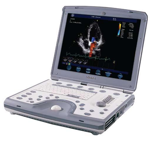
2 Product Description The Vivid i n is a high-performance, battery-operated, ultra-portable diagnostic Ultrasound systems providing exceptional image quality. The Vivid i n is designed for cardiovascular imaging, abdominal, small-parts, perioperative monitoring and 2D ICE imaging. System Architecture The Vivid i n is based on GE s TruScan Architecture, which is common to all GE Ultrasound systems, EchoPAC PC Workstation, software and network solutions. It features a software-driven PC-based platform, raw data storage with advanced post-processing capabilities, complete connectivity and compatibility with the GE family of Cardiovascular Ultrasound Systems. Innovative tools offer advance connectivity, remote monitoring and consultation for improved productivity and standard of care anywhere. Advanced, energy-efficient power management designed for cool operation provides scanning with rechargeable battery power for more than one hour. Standby mode with battery allows fast boot-up anywhere. Data Acquisition Programmable system architecture Application-Specific Channel Architecture: the Vivid i n employs a flexible digital beam-former architecture capable of using up to 1024 channels depending on specific application requirements Application-specific digital beam forming algorithm for each mode Supports Phased Array, Linear and Curved Array, TEE, 2D-ICE and non-imaging Pencil transducers Receive focusing, aperture, apodization and frequency response are all continuously variable as a function of depth Wide aperture mode in convex and linear array probes Data Processing Echo data processing of phase, amplitude and frequency Easily upgradeable for future expansions Digital raw data replay allows for image post processing and un-compromised offline measurement and analysis Display Screen High-resolution, flat 15-inch TFT LCD screen Scanner software supports display resolution of 800 x 600 pixel Screen can be closed or tilted at angles ranging between Wide-angle visibility Digital brightness, contrast and blue-tint adjustment for optimal viewing in different ambient light conditions Closing down the screen will automatically place system into standby mode allowing fast boot-up upon screen opening Display Formats Instant-review screen displays 12 simultaneous loops/images for a quick study review Scanplane position indicator and probe temperature are displayed with all multi-plane TEE probes Image orientation marker Selectable display configuration of duplex and triplex modes: side-by-side or top-bottom, during live, digital replay and clipboard image recall Single, dual and quad-screen view Split screen view Display Annotations Mechanical Index (MI) Thermal index: application dependent Patient name/id and additional patient information Hospital name Time/date Trackball-driven annotation arrows Scanning parameters Application Probe name Stress protocol parameters Active mode display Parameter annotation follow ASE standard Multi-language support for user interface, reports and help manual Display size: 1600 x 1200 pixels with 260,000 simultaneous colors available 2
3 Tissue Imaging General Variable transmit frequencies for resolution/ penetration optimization Display zoom with zoom area control Variable contour filtering for edge enhancement Variable dynamic range and transmit power settings Depth range up to 30 cm probe specific Selectable grayscale parameters: gain, reject, gray-maps, DDP and compress can be adjusted in live, digital replay and image clipboard recall Automatically calculated TGC curves require minimal operator interaction Selectable Automatic Tissue Optimization (ATO) of the real-time, 2D-mode image "Smart Depth" for 2D (option) automatically optimizing transmit pattern parameters according to scan-depth setting 2D-mode Sector tilt and width control Coded octave imaging: second-generation harmonic tissue imaging providing improved lateral and contrast resolution over conventional imaging; features reduced noise and improved wall definition; COI gives improved axial resolution without sacrificing frame rate, making it the tissue modality of choice for all patient groups Confocal imaging: allows for multiple transmit focal zones over range of view and a high-vector density probes dependent and user adjustable Expanded cardiology performance on the 3S-RS probe, including five levels of harmonics and ultra-high frame rates Harmonic tissue imaging on all linear and convex probes Ultra-Definition Clarity setting for improved cardiac IQ Adaptive reject for improved cardiac IQ Ultra-Definition Speckle Reduction Imaging (UD-SRI): performs speckle suppression on Vascular 2D images user can control the amount of speckle suppression and the amount of image smoothing to be retained Coded Phase Inversion (CPI) for improved contrast resolution Variable image width: a reduction either increases frame rate or increases the number of focal zones while maintaining the frame rate application dependent, on linear/convex array probes Multiple-angle compound imaging, multiple co-planar images from different angles combined into a single image in real-time improving border definition, contrast resolution and reducing angular dependence of border or edge (on linear probe) Dual focus: offers additional focal zone for added spatial and contrast resolution from heart base up to apical areas (cardiology application only) L/R and up/down invert in live, digital replay or image clipboard recall Digital replay for retrospective review or automatic looping of images allowing for adjustment of parameters such as gain, compression, reject, anatomical M-mode, persistence and replay speed Data Dependent Processing (DDP) performs temporal processing, which reduces random noise but leaves motion of significant tissue structures largely unaffected, can be adjusted even in digital replay Different gray-maps and colorized 2D-mode user selectable in real-time or in digital replay M-mode Trackball-steerable M-mode line available with all imaging probes max steering angle is probe dependent Simultaneous real-time 2D- and M-mode M-mode PRF 1 khz: all image data acquired are combined to give high-quality recording regardless of display scroll speed Digital replay for retrospective review of spectral data Several top-bottom formats, side-by-side format and time-motion only format can be adjusted in live or digital replay Selectable horizontal scroll speed: 1, 2, 3, 4, 6, 8, 12, 16 seconds across display Horizontal scroll can be adjusted in live or digital replay 3
4 Anatomical M-mode Any plane M-mode display derived from 2D cine loop M-mode cursor can be adjusted at any plane Can be activated from real-time scan, digital replay or image clipboard recall Anatomical color M-mode available in real-time scan, digital replay or image clipboard recall Measurement and analysis capability Anatomical tissue velocity M-mode (requires I 2 option) Color Doppler General Steerable color Doppler available with all imaging probes max steering angle is probe dependent Trackball-controlled ROI Removal of color map from the tissue during digital replay Digital replay for retrospective review of color or color M-mode data allowing for adjustment of parameters, such as color/tissue priority and color gain, even on stored data PRF settings: user selectable Advanced regression wall filter gives efficient suppression of wall clutter For each encoding principle, multiple-color maps can be selected in live and digital replay including variance maps More than 65,000 simultaneous colors processed providing smooth display, 2D color maps containing a multitude of color hues Simultaneous display of grayscale 2D and 2D with color flow in live or in digital replay Color invert: user selectable in live and digital replay Variable color baseline: user selectable in live and digital replay Multivariate color priority function gives reliable delineation of disturbed flows even across bright areas of the 2D-mode image Color Doppler frequency can be changed independently from 2D for optimal flow Color Doppler Imaging Digital signal processing power maintains high frame rates with large ROIs even for very low PRF settings Variable ROI size in width and depth 4 User-selectable radial and lateral averaging for reduction of statistical uncertainty in the color velocity and variance estimates Data Dependent Processing (DDP) performs temporal processing and display smoothing with reduced possibility for loss of transient events of hemodynamic significance Digital replay for retrospective review or automatic looping of color images allowing for adjustment of parameters such as DDP, baseline shift, color maps, color/tissue priority and color gain even on frozen/recalled data Application-dependent multivariate motion discriminator reduces flash artifacts "Smart depth" for color (option) automatically adjusts transmit pattern parameters according to depth of color ROI Color Angio (Color Intensity Imaging) Angle-independent mode for visualization of small vessels with increased sensitivity compared to standard color flow Color M-mode Variable ROI length and position user selectable User-selectable radial averaging for reduction of statistical uncertainty in the color velocity and variance estimates Selectable horizontal scroll speed 1, 2, 3, 4, 6, 8, 12, 16 seconds across display can be adjusted during live, digital replay or image clipboard recall Real-time 2D image while in color M-mode Same controls and functions available as in standard 2D color Doppler Anatomical Color M-mode Vingmed-patented, any plane color M-mode display derived from color Doppler cine loop Available in real-time scan, digital replay or image clipboard recall Also applicable to Tissue Velocity Imaging (option) Measurement and analysis capability B-Flow (option) B-Flow is a new digital imaging technique that provides real-time visualization of vascular hemodynamics by directly visualizing blood reflectors and presenting this information in a grayscale display Use of GE-patented techniques to boost blood echoes, and to preferentially suppress non-moving tissue signals B-Flow is available for most vascular and shared service applications
5 Blood Flow Imaging (BFI) (option) Combines color Doppler with grayscale speckle imaging Allows better delineation of blood flow without bleeding into tissue or vessel wall Spectral Doppler General Operates in PW, HPRF and CW modes Trackball-steerable Doppler available with all imaging probes max steering angle is probe dependent Selectable Doppler optimization Real-time duplex or triplex operation in PW Doppler mode for all velocity settings Frame rate control for optimized use of acquisition power between spectrum, 2D and color Doppler modes in duplex or triplex modes Spectral analysis with an equivalent DFT rate of 0.2 ms Automatic Spectrum Optimization (ASO) provides a single press, automatic, real-time optimization of PW or CW spectrum scale and baseline display Dynamic gain compensation for display of flows with varying signal strengths over the cardiac cycle Dynamic reject gives consistent suppression of background user selectable in real-time, digital replay or image clipboard recall Digital replay for retrospective review of spectral Doppler data Several top-bottom formats, side-by-side format and time-motion only format can be adjusted in live or digital replay Selectable horizontal scroll speed: 1, 2, 3, 4, 6, 8, 12, 16 seconds across display can be adjusted in live or digital replay Adjustable spectral Doppler display parameters: gain, reject, compress, color maps can be adjusted in live or digital replay Adjustable baseline and velocity scale Wall filters with a range of Hz (velocity scale dependent) Angle correction with automatic adjustment of velocity scale in live, digital replay and image clipboard recall Stereo speakers mounted in the front panel Display annotations of frequency, mode, scales, Nyquist limit, wall filter setting, angle correction and acoustic power indices PW/HPRF Doppler Automatic HPRF Doppler maintains its sensitivity even for shallow depths and with the highest PRFs Digital velocity tracking Doppler employs processing in range and time for high-quality spectral displays User-adjustable baseline shift in PW live, digital replay and image clipboard recall Adjustable sample volume size of 1-15 mm (probe dependent) Maximum sample volume depth 30 cm Tissue Doppler Imaging Myocardial PW Doppler provides real-time Doppler spectral information for specified myocardial motion allowing for instantaneous tissue velocity measurement CW Doppler Highly sensitive steerable CW available with all phased array probes Tissue Velocity Imaging and Tissue Tracking (option) Tissue Velocity Imaging (option, requires the i^2 option) Myocardial Doppler imaging with color overlay on tissue image Tissue Doppler data can be acquired in background during regular 2D imaging Segmental wall motion analysis can be obtained with use of anatomical M-mode from digital replay or image clipboard recall Tissue color overlay can be removed to show just the 2D image, still retaining the tissue velocity information Quantitative profiles (Q-Analysis) can be derived on data transferred to EPPC workstation Tissue Tracking (option, requires the i^2 option) Real-time display of the time integral of TVI for quantitative display of myocardial systolic displacement Myocardial displacement is calculated and displayed as a color-coded overlay on the grayscale and M-mode image different colors represent different displacement ranges 5
6 Tissue Synchronization Imaging TSI (option, requires the TVI/TT option) Parametric imaging which gives information about synchronicity of myocardial motion Delayed myocardial segments produce red overlay whereas segments moving in normal rhythm are green Available in live scanning as well as an offline calculation derived from TVI data Cine Memory High-fidelity loops and images may be reviewed by scrolling or by running cine loops TruScan architecture offers broad post-processing capabilities of recalled images and loops allowing manipulation of parameters such as gain, baseline, color maps, sweep speeds, audio gain and cine speed Cine memory contains up to 180,000 frames (with default image settings: 2,500 to 5,600 frames) Image Clipboard for thumbnail storage and review of saved images and loops Trackball-controlled cine review Fast Power-Up Standby mode to support fast boot-up Battery power backup allows continuous scanning during power interruptions Physiological Traces Integrated ECG or external ECG lead input High-resolution display of the ECG trace User-adjustable gain/position control over ECG trace User pre-settable gain/position control of ECG Automatic QRS complex detection User Interface Operator Keyboard Easy-to-learn user interface with intelligent keyboard Keyboard with application-specific push buttons for primary controls Interactive back-lighting of application-specific push buttons Full-size, alphanumeric keyboard with adjustable backlighting Application-specific secondary controls available through slide bars operated by a four-way rocker Slide pot TGC curve with six pots Overall gain for 2D mode and active mode on same rotary Digital harvesting of images and loops into image clipboard Patient browser screen for registration of demographic data and quick review of image clipboard contents Fully programmable user presets for probe/application default settings Support for 12 international (European) keyboard character sets (ISO 8859) Integrated speakers Analysis Program Personalized measurement protocols allow individual set and order of measurement and analysis items Measurements can be labeled seamlessly by using protocols or post assignments Bodymark icons for location and position of probe Cardiac calculation package including extensive measurements and display of multiple repeated measurements Vascular measurements package Measurements assignable to protocol capability Parameter annotation follow ASE standard Measurements assignable to report generator Doppler auto trace function with automatic calculations in both live and digital replay Seamless data storage and report creation Measurements are summarized in worksheets individual results can be edited or deleted User-assignable parameters Report templates can be customized on board ASE-based default text modules (English) user customizable Image view during reporting Ability to export report in PDF format Generate report templates by the Report Designer or import from EchoPAC PC Insite -II capability 6
7 Stress Echo (option) Stress package with memory buffer offers pharmaceutical, exercise and bicycle stress exam protocols with user-configurable templates and shuffle mode Smart Stress function with the ability to save over 17 imaging parameters from each imaging plane these imaging parameters are recalled at each stress level, thereby requiring no system adjustments Advanced and flexible stress-echo examination capabilities Image acquisition, review, wall-segment scoring and reporting Treadmill Stress-exercise with more than 120 seconds of raw data continuous capture Possibility of extensive post-processing of images under review Reference scan display during acquisition for stress level comparison (dual screen) Wall motion scoring (bulls-eye and segmental) Show reference for comparing resting images to each stress level Template Editor to customize the number of stress levels, number of views, number of heart cycles and systolic or full-cycle capture Advanced Options Contrast Imaging All use of contrast agents should be used as described on the label by the contrast agent manufacturers. LVO Contrast (option)* LV Contrast (available on probes: 3S-RS, 5S-RS, 6T-RS and 6Tc-RS) enhances delineation of the LV border in combination with ultrasound contrast agents. The new implementation of GE s Coded Phase Inversion (CPI) provides high-resolution detection of contrast in the LV cavity and excellent suppression of myocardial tissue signals. Vascular/Abdominal Contrast (option)** Vascular Contrast (8L-RS & 12L-RS probes): Coded Phase Inversion enables excellent detection and resolution of vascular contrast imaging Abdominal Contrast (4C-RS probe): using Coded Phase Inversion, optimized for abdominal contrast imaging * Harmonic imaging for supporting contrast agent imaging was developed by Schering. ** GE Healthcare s Vivid i n is designed for compatibility with commercially available contrast agents. Because the availability of these agents is subject to government regulation and approval, product features intended for use with these agents may not be commercially marketed nor made available before the contrast agent is approved for use. Advanced contrast features are only enabled on systems for delivery in countries or regions where the agents are approved for use or for investigational or research use. IMT Measurement Program (option) Automatic measurements (patent pending) of carotid artery Intima-Media Thickness (IMT) on any acquired frame On-board IMT package provides non-interrupted workflow fully integrated with M&A, worksheet, archiving and reporting functions Robust algorithm provides quick, reliable measurements, which can be stored to the on-board archive for review and reporting IMT measurement can be made from frozen images or images retrieved from archive IMT package supports measurements of different regions of the intima in the carotid vessel (e.g., Lt./Rt./CCA/ICA etc.) Frame for IMT measurement can be selected in relation to the ECG waveform 7
8 Wideband Probes Electronic selection between one solid-state connector and one stand-alone Doppler connector probe frequency range catalog # Phased Array Sector Probes 3S-RS MHz H4000PD 5S-RS MHz H4000PC 6S-RS MHz H45021RP 7S-RS MHz H4000PE 10S-RS MHz H4000PF Linear Array Probes 8L-RS MHz H40402LT 12L-RS MHz H40402LY Convex Array (Curved) Probes 4C-RS MHz H4000SR 8C-RS MHz H40402LS Intra-operative Probes i12l-rs MHz H40402LW Doppler Pencil Probes 2D(P2D)-RS 2.0 MHz H45021C 6D(P6D)-RS 6.0 MHz H45021CA Multiplane Transesophageal Phased Array Probes 6T-RS MHz H45531MZ 6Tc-RS MHz H45551ZE 9T-RS MHz H45531YM Intra-Cardiac ICE probes ACUSON AcuNav 10F ICE Probe Connector SwiftLink ICE probe connector, allows use of the ACUSON AcuNav 10F ICE probe on Vivid i n TEE Probe Adaptor Allow to use multi-plane TEE RS probe on other Vivid systems MHz Distributed by BWI for Siemens Supported Applications (probe dependent) Adults Cardiac Pediatrics Cardiac Vascular Pediatric General Neonatal Cephalic H45021YE H45021KP Fetal heart Abdomen Musculoskeletal including Superficial Small Parts Breast Nerve Imaging Intraoperative Intracardiac and Intraluminal Echocardiography (ICE) Biopsy Bracket Support (option) On-screen biopsy Guide-Line, guide-zone and depth measure for Civco multi-angle biopsy bracket, supporting probe models - 3S-RS - 4C-RS - 8L-RS - 12L-RS Image Management and Archiving Raw data workflow: ultimate workflow with instant access data management DICOM 3.0 Image Format: DICOM incorporates raw image data information with all its data management flexibility into the image communication standard DICOM 2D, CFM and TVI data at maximum frame rate may be reviewed by scrolling or by running cine loops Image clipboard for stamp-sized storage and review of stored images and loops Built-in patient archive with images/loops, patient information, measurements and report Configurable HTML-based report function Report template designer package Internal archive data can be exported to removable image storage through DVD/CD-RW, Magnet-Optical Disk and DICOM Media Internal hard disk: for storing programs, application defaults, ultrasound images and patient archive Over 80 Gbyte disk space for exam archive storage All data storage is based on ultrasound raw data allowing to change gain, baseline, color maps, sweep speeds, etc. for recalled images and loops Transcranial (adult cephalic) 8
9 Raw-Data, DICOM, AVI, MPEG and JPEG export DVD writer (supports CD-R and DVD-R) Excel Export Allows export of all archived measurement and textual patient information in standard Microsoft Excel files DICOM Network Connectivity (option) Ethernet network connection DICOM support Storage to DICOM server DICOM structured report SCU Storage commitment Performed procedure step Verify: provides verification of an active connection between the scanner and another DICOM device Modality Worklist (option) Modality worklist: gives access to a list of patients from a worklist server (usually HIS) DICOM Print (option) DICOM Media Support DICOM media: read/write images on DICOM format EchoPAC Connectivity Connectivity and image analysis capability of Vivid i n from EchoPAC PC EchoPAC PC allows instant access to ultrasound raw data provided by the system Comprehensive review, analysis and post-processing capabilities on EchoPAC PC Advanced quantitative analysis and post-processing capabilities Q-Analysis on raw data from Vivid i n on EchoPAC PC Three user levels help organizing data security requirements Virtual Printer (option) Provides the ability to send Print commands to any of two printers even when not connected to a printer upon re-connection of printer, the system automatically produces hard copies from print images saved in chronological succession on disk MPEGvue (option) Using MPEGvue, exams may be stored onto removable media or on remote networked system together with integrated MPEGvue player for viewing on standard PC Smart feature allows transparent transmission of images via using resident Outlook client Patient management utility on standard PC provides ability to organize the exams on different sub-directories on the user s hard disk evue (option) Allows interactive viewing of images, loops or full exams from remote location on any PC, using LAN or wireless LAN Peripherals (options) Super VHS VCR (requires video scan converter) USB black and white video printer with control from system panel USB color video printer with control from system panel USB inkjet printer MO 5.25" drive Secondary DVD/CD-RW USB flash memory card Wireless network interface Accessories (options) Interface cable for External ECG Splash-proof protective keyboard cover Replacement battery External battery charger Replacement hard disk Safety lock Video scan converter GoPac portable carrying case or RollPac on wheels ECG adapter for DIN-type pediatrics electrode leads SafeLock Cart (option) Probe and gel holder Hand rest and handles Anti-theft locking device Four swivel wheels front wheel breaks Two peripheral shelves 9
10 Inputs and Outputs SVGA video out Connectors: - USB-2 (to support CD-RW, video printers, MOD, USB flash-cards, etc.) - LAN Ethernet - PCMCIA (wireless LAN card) - DC power input Dimensions and Weight Depth: 313 mm (12.4 in) Width: 358 mm (14.2 in) Height: 59 mm (2.3 in) Weight: approximately 5 kg (11 lb) without battery Electrical Power Battery or mains-line operation Input rating: VAC/ A Frequency: 50/60 Hz Safety Built to meet the requirements of: IEC/EN/UL (1988) Class I, Type BF (electrical safety) EN : A1(2006) IEC : 2005 IEC/EN (2001) (Ultrasound) IEC/EN (2001) (ECG CF Selected clauses) The European Medical Devices Directive (MDD) 93/42/EEC (CE Mark) CAN/CSA-C22.2 No A1(2006) Virus Protection To minimize virus vulnerability, Vivid i n is configured with a minimal set of open ports and with all network services, not actively used by the system, closed down. This significantly reduces the risk of a virus attack on Vivid i n. GE is continuously judging the need for additional actions to reduce vulnerability of equipment. This includes vulnerability scanning of our products and evaluation of security patches for the third-party technology used. Microsoft and other security patches that address serious issues with Vivid i n will be made available to customers after GE verification of those patches. 10
11 2008 General Electric Company All rights reserved. General Electric Company reserves the right to make changes in specifications and features shown herein, or discontinue the product described at any time without notice or obligation. Contact your GE representative for the most current information. GE, GE Monogram, Vivid and EchoPAC are trademarks of General Electric Company. ACUSON, AcuNav and Swiftlink are trademarks of Siemens Medical Solutions USA, Inc. General Electric Company, doing business as GE Healthcare. Healthcare Re-imagined GE is dedicated to helping you transform healthcare delivery by driving critical breakthroughs in biology and technology. Our expertise in medical imaging and information technologies, medical diagnostics, patient monitoring systems, drug discovery, and biopharmaceutical manufacturing technologies is enabling healthcare professionals around the world to discover new ways to predict, diagnose and treat disease earlier. We call this model of care Early Health. The goal: to help clinicians detect disease earlier, access more information and intervene earlier with more targeted treatments, so they can help their patients live their lives to the fullest. Re-think, Re-discover, Re-invent, Re-imagine. GE Healthcare 3000 North Grandview Waukesha, WI U.S.A.
Vivid i Ultra-Portable Echo System
 Vivid i Ultra-Portable Echo System Product Description The Vivid i is a high-performance, battery-operated, ultra-portable diagnostic Ultrasound systems providing premium image quality. The Vivid i is
Product Description. Display Screen. System Architecture. Display Formats. Data Acquisition. Display Annotations. Data Processing
 Vivid * i BT12 Product Description The Vivid i is a high-performance, battery-operated, ultra-portable diagnostic Ultrasound systems providing exceptional image quality. The Vivid i is designed for cardiovascular
Vivid 7 Pro Breakthrough 2008 Release
 Vivid 7 Pro Breakthrough 2008 Release Product Description The Vivid 7 Pro is a complete digital color flow Doppler ultrasound system providing a wide range of premium performance applications in cardiac,
Vivid S5. Cardiovascular ultrasound system. GE Healthcare. Davis Medical
 GE Healthcare Vivid S5 Cardiovascular ultrasound system Versatility. It s a new design concept one that leverages our miniaturization expertise gained from the Vivid i and our performance expertise of
Quick Reference Guide
 siemens.com/nx3 Quick Reference Guide ACUSON NX3 Series Contents 2 System Overview 3 Getting Started 8 2D Mode and M-mode 12 Color and Spectral Doppler 24 Measurements and Calculations 38 Text, Arrows
GE Healthcare LOGIQ P3. Staying ahead of the curve
 GE Healthcare LOGIQ P3 Staying ahead of the curve Ultrasound Expertise. Enhanced. GE is a trusted partner of many healthcare providers around the world. GE s TruScan technology, incorporating SmartScan,
DC-6. Diagnostic Ultrasound System
 DC-6 Diagnostic Ultrasound System DC-6 is a general purpose color Doppler ultrasound system aiming at most clinical areas both in exam and research with various transducers and multi software packages
GE Healthcare. Vivid S5. Cardiovascular ultrasound system
 GE Healthcare Vivid S5 Cardiovascular ultrasound system Versatility. It s a new design concept one that leverages our miniaturization expertise gained from the Vivid i and our performance expertise of
M5 Diagnostic Ultrasound System
 V0807 M5 Diagnostic Ultrasound System Mindray s ultrasound family is now introducing a new member, M5 hand-carried color Doppler system. M5, coming in a laptop size with comprehensive ergonomic design,
DC-6 Expert. Diagnostic Ultrasound System
 DC-6 Expert Diagnostic Ultrasound System MINDRAY has newly released DC-6 Expert, a general purpose color Doppler ultrasound system with full ergonomic designs, supplying more accessible exams, higher imaging
Diagnostic Ultrasound System. Operation Note
 M5 Diagnostic Ultrasound System Table of Contents System Introduction...3 Control Panel...4 Control Panel...5 Control Panel...6 Control Panel...7 Control Panel...8 Control Panel...9 Power ON / OFF the
Architecture of Quality Imaging Mary K. Henne, MS, CNMT, RDMS, RVT Ultrasound Education Specialist GE Healthcare
 Architecture of Quality Imaging Mary K. Henne, MS, CNMT, RDMS, RVT Ultrasound Education Specialist GE Healthcare 2 DOC1292532 Architecture of Quality Imaging Agile Acoustic Architecture E-Series and XDclear
S S S2 Operation Manual
 PREPARATION 1. How to create and input patient data? In the MAIN INTERFACE, press the key, to enter into the patient exam list interface. Then, click New patient item to create new patient files. Patient
4 Working With Scan Modes
 4 Working With Scan Modes Scan Modes Overview All of the information in this chapter pertains to live imaging. Many of the controls and functions change when you freeze the scan. For information on using
A COMPACT SYSTEM WITH ADVANCED PERFORMANCE
 Samsung Medison is a global leading medical devices company. Founded in 1985, the company sells cutting-edge diagnostic ultrasound devices around the world in various medical fields. The company has attracted
M7Vet Diagnostic Ultrasound System. Operation Note
 M7Vet Diagnostic Ultrasound System Operation Note Table of Contents Table of Contents... i 1 System Introduction... 1 2 Control Panel... 2 3 Icons on the Screens... 6 4 Power ON/ OFF... 7 5 Enter or Search
GE Healthcare. Essential for life. Senographe Essential Full-Field Digital Mammography system
 GE Healthcare Essential for life Senographe Essential Full-Field Digital Mammography system Excellence in FFDM is a process. An ongoing quest, fueled by our continuing breakthroughs in breast cancer detection
Multipurpose Color Ultrasound System IMAGING SYSTEMS
 i3 Multipurpose Color Ultrasound System IMAGING SYSTEMS IMAGING SYSTEMS i3 Color Ultrasound System M The revolutionary i3 provides you outstanding 2D images and fast 4D volume images, while the streamlined
UGEO H60. Performance in Style. Features
 UGEO H60 Performance in Style The UGEO H60 implements superior performance with new design principles of simplicity and lightness. Its 10.1" touchscreen improves usability while its 18.5" LED monitor enhances
M 5. M5 Hand-carried Diagnostic Ultrasound System. Mindray. A practical, durable ultrasound tool for high-quality imaging where and when you need it.
 M 5 Mindray M5 Hand-carried Diagnostic Ultrasound System A practical, durable ultrasound tool for high-quality imaging where and when you need it. Affordable, easy-to-use ultrasound for precision and confidence
Quality Ultrasound Systems
 Quality P11VExpert Medisono offers quality products to the medical imaging market. We offer different solutions tailored to the needs of each patient, offering the best value to every single patient. We
Design Your Performance
 MEDISON has been a leading name in diagnostic ultrasound since its foundation in 1985. As one of the only companies dedicated solely to ultrasound imaging, we have remained at the forefront of research
Discover the new Prestige and experience 3D/4D imaging beyond your imagination.
 3D/4D Beyond Imagination The Prestige ultrasound imaging system represents the pinnacle of more than a decade of technological advancement in 3D/4D ultrasound imaging at MEDISON. Inheriting a tradition
Printed in Japan E318
 Diagnostic Ultrasound System MODEL PROSOUND 6 The specifications, shape and color of this product are subject to change without notice. The standard components and optional items vary depending on the
GE Healthcare. Precision 500D. Digital radiography and fluoroscopy system
 GE Healthcare Precision 500D Digital radiography and fluoroscopy system What do you look for in a classical digital R&F system? Exceptional image quality at low dose, certainly. Increased clinical productivity
Beyond Performance and Value
 Putting the back in ultrasound Beyond Performance and Value Esaote s new ultra-performance MyLab 9 exp ultrasound system is designed to support a full range of shared service diagnostic imaging environments.
5. User-Centered Design Focus - High mobility cart - 19" formate high resolution Flat panel LCD display monitor or more - User adjustable control pane
 A. SPECIFICATIONS : 1. Applications - Abdominal - Small Parts - OB/GYN - Vascular - Breast etc. 2. Imaging Modes - Adaptive Broadband flow imaging or Digital broad bandwith imaging or Advanced Dynamic
Excellent Performance; Ease of Use
 Excellent Performance; Ease of Use High performance with a broad range of applications - the compact ProSound α6 is at your service. Our ProSound series has a well-established reputation in hospitals and
GE Healthcare. Senographe 2000D Full-field digital mammography system
 GE Healthcare Senographe 2000D Full-field digital mammography system Digital has arrived. The Senographe 2000D Full-Field Digital Mammography (FFDM) system gives you a unique competitive advantage. That
Lesson 06: Pulse-echo Imaging and Display Modes. These lessons contain 26 slides plus 15 multiple-choice questions.
 Lesson 06: Pulse-echo Imaging and Display Modes These lessons contain 26 slides plus 15 multiple-choice questions. These lesson were derived from pages 26 through 32 in the textbook: ULTRASOUND IMAGING
SONOACE 6000C MT DIGITAL COLOR. Affordable PC-based Digital CFM Ultrasound System. Specifications.
 Specifications Imaging Modes Gray Scale Focusing PC Monitor Speaker Measurements Image Processing Display Functions Peripheral Devices Support Physical Dimensions Electrical Parameters Probe Types Single
ACUSON X150 Knobology & User Guide
 ACUSON X150 Knobology & User Guide ACUSON X150 CONTROL PANEL Table of Contents The ACUSON X150 knobology and user guide is the clinicians quick reference of system terms, functions and capabilities. This
Optimisation of Image Acquisition Bordeaux 16th November J.S. McGhie W.B. Vletter R. Frowijn No disclosures
 Optimisation of Image Acquisition Bordeaux 16th November 2016 J.S. McGhie W.B. Vletter R. Frowijn No disclosures Image optimisation: The Echo machine It looks difficult to drive an echo machine!! Some
Ultrasound Diagnostic Scanner INSTRUCTION MANUAL
 Ultrasound Diagnostic Scanner HI VISION Avius Operation INSTRUCTION MANUAL Special Notes to Operators and Maintenance Managers Before using this system, be sure to thoroughly read this manual and make
Vevo770 Ultrasound. Quick Operation Guide
 Vevo770 Ultrasound Quick Operation Guide Vevo770 Probe head (707B) Preparation The acoustic membrane is very fragile and sensitive, it must be handled with care. Position the RMV scan head with the fill
Multi-Access Biplane Lab
 Multi-Access Biplane Lab Advanced technolo gies deliver optimized biplane imaging Designed in concert with leading physicians, the Infinix VF-i/BP provides advanced, versatile patient access to meet the
Physics of Ultrasound Ultrasound Imaging and Artifacts รศ.นพ.เดโช จ กราพาน ชก ล สาขาหท ยว ทยา, ภาคว ชาอาย รศาสตร คณะแพทยศาสตร ศ ร ราชพยาบาล
 Physics of Ultrasound Ultrasound Imaging and Artifacts รศ.นพ.เดโช จ กราพาน ชก ล สาขาหท ยว ทยา, ภาคว ชาอาย รศาสตร คณะแพทยศาสตร ศ ร ราชพยาบาล Diagnosis TTE TEE ICE 3D 4D Evaluation of Cardiac Anatomy Hemodynamic
 www.hitachi-aloka.com Revolutionary Performance; Ease of Use The ProSound 6 is the next generation of compact color ultrasound systems, providing unprecedented performance with a broad range of applications.
Get more from your images with Symphony Image Processing
 DIRECT RADIOGRAPHY The user-friendly DelWorks image acquisition and processing software possesses a wide range of tools for a variety of image manipulations. Its user interface simplifies every step of
Lesson 12: Doppler Principles. This lesson contains 50 slides plus 26 multiple-choice questions.
 Lesson 12: Doppler Principles This lesson contains 50 slides plus 26 multiple-choice questions. Accompanying text for the slides in this lesson can be found on pages 59 through 80 in the textbook: DOPPLER
COST EFFECTIVE FLAT PANEL DIGITAL RADIOGRAPHY UPGRADE SOLUTIONS
 COST EFFECTIVE FLAT PANEL DIGITAL RADIOGRAPHY UPGRADE SOLUTIONS DRive is a digital imaging DR hardware & Software solution designed for General Radiography of anatomy. It intended to replace film/screen
Personal Ultrasound The future of ultrasonic scanning is here, today right in your hands. It s portable and affordable giving you the power and
 What the Laptop Did for PCs, MySono201 Does for Ultrasonic Scanning The Personal Digital Ultrasound, Only MySono201 Personal Ultrasound The future of ultrasonic scanning is here, today right in your hands.
Get more from your images with Symphony Image Processing
 DIRECT RADIOGRAPHY The user-friendly DelWorks image acquisition and processing software provides a wide range of tools for a variety of image enhancements. Its user interface simplifies every step of the
LOGIQ e Ultrasound. GE Healthcare. Product Description
 GE Healthcare LOGIQ e Ultrasound Product Description The LOGIQ e combines the high performance of a console system with the portability of a laptop. GE Healthcare s compact system is designed for general
Image Optimization: The Sonographer s Responsibility. Prepared by Cathy Daniels, EdD, RTR, RDMS, RDCS, RVT
 Image Optimization: The Sonographer s Responsibility Prepared by Cathy Daniels, EdD, RTR, RDMS, RDCS, RVT Image Optimization: The Sonographer s Responsibility Cathy Daniels, EdD, RTR, RDMS, RDCS, RVT Disclosure
NextGen LOGIQ e. Ultrasound. GE Healthcare. Product Description
 GE Healthcare NextGen LOGIQ e Ultrasound Product Description The NextGen LOGIQ e combines the high performance of a console system with the portability of a laptop. GE Healthcare s compact system is designed
Model: ESE-6 Portable Color Ultrasound System
 DATA SHEET Model: ESE-6 Portable Color Ultrasound System Ultrasound System Specifications The premium performance of the full functional Portable ESE-6 provides a fast and easy diagnosis by: Ultra-premium
KIZLON. Sentinel House,Harvest Crescent, Ancells Business Park, Fleet Hampshire GU51 2UZ, UK Website:
 KIZLON Sentinel House,Harvest Crescent, Ancells Business Park, Fleet Hampshire GU51 2UZ, UK Email: info@kizlon.com Website: www.kizlon.com Portable Ultrasound Scanners KUS-A100 & A101 Ultrasound Scanners
EVIS EUS ENDOSCOPIC ULTRASOUND CENTER EU-ME2 Dedicated ultrasound processor with versatile functionality.
 EVIS EUS ENDOSCOPIC ULTRASOUND CENTER EU-ME2 Dedicated ultrasound processor with versatile functionality. Advancing the Art of Endosonography The EU-ME2 is a high-quality compact ultrasound processor for
Ultrasound System Specifications
 Ultrasound System Specifications The superb imaging and performance capabilities of VINNO G60 give you the confidence and efficiencies in your daily exam by: World class image quality with unmatched high-frequency
The physics of ultrasound. Dr Graeme Taylor Guy s & St Thomas NHS Trust
 The physics of ultrasound Dr Graeme Taylor Guy s & St Thomas NHS Trust Physics & Instrumentation Modern ultrasound equipment is continually evolving This talk will cover the basics What will be covered?
The Middle East Distributor for AMBISEA Technology Corp. Electro-Medical Product Line
 The Middle East Distributor for AMBISEA Technology Corp. Electro-Medical Product Line AV-9100 Single Channel ECG 1 2 AV-9300 3-Channels ECG 3 4 5 AV-9000B Multi-Parameter Patient Monitor 6 7 8 AV-9000C
The Versatile and Powerful ACLxy. ACLxy
 The Versatile and Powerful ACLxy ACLxy Rolling into a Clinic, Imaging Center and Hospital Near You! COMPUTED RADIOGRAPHY (CR) IS RAPIDLY THE BEGINNING. THE OREX CR SOLUTION DRA- BECOMING A DRIVING FORCE
GE Healthcare. Dash 2500 The standard of excellence for sub-acuity monitoring
 GE Healthcare Dash 2500 The standard of excellence for sub-acuity monitoring The clinical With monitored patients, even small measurement variations can indicate significant changes in your patient s condition.
: PT Philips Indonesia CERTIFICATE OF PRODUCTION SOLE AGENT/ DISTRIBUTOR : NA CERTIFICATE OF DISTRIBUTION (PAK)
") SOLE AGENT/ DISTRIBUTOR : PT Philips Indonesia CERTIFICATE OF PRODUCTION : NA CERTIFICATE OF DISTRIBUTION (PAK) : HK.07.Alkes/IV/625/AK.2.2012 ADDRESS : Jl. Buncit Raya Kav 99-100, Pasar Minggu, Jak-Sel
SOLE AGENT/ DISTRIBUTOR : PT Philips Indonesia CERTIFICATE OF PRODUCTION : NA CERTIFICATE OF DISTRIBUTION (PAK) : HK.07.Alkes/IV/625/AK.2.2012 ADDRESS : Jl. Buncit Raya Kav 99-100, Pasar Minggu, Jak-Sel
Introduction to Ultrasound Physics
 Introduction to Ultrasound Physics Vassilis Sboros Medical Physics and Cardiovascular Sciences University of Edinburgh Transverse waves Water remains in position Disturbance traverse producing more wave
PERFORM Operating Document. Use and Maintenance of G.E. Ultrasound LOGIQ E
 PERFORM Operating Document Use and Maintenance of G.E. Ultrasound LOGIQ E PC-POD-IM-001-v02 Revision History Version Reason for Revision Date 01 New POD 22/May/2013 02 Minor changes only 2.2 Change responsibility
General Imaging Ultrasound in a Whole New Light. Introducing the
 General Imaging Ultrasound in a Whole New Light. Introducing the It s a Tour de Force Redefining innovation through value and performance It s the dawning of a new day in the world of compact ultrasound
DIAGNOSTIC SCANNER HS-1600V. Convex/Linear Ultrasonic System
 HONDA ELECTRONICS CO.,LTD. DIAGNOSTIC SCANNER Convex/Linear Ultrasonic System HS-1600V Our unique technology is the product of our originality and continual pursuance of achieving high quality. It is also
ECOVIEW 9 / ECOVIEW 9 PLUS Digital Radiographic System
 Co., LTD. 3F, Urbanlight B/D, 630, Eonju-ro, Gangnam-gu, Seoul, Korea 135-832 Factory at 621-14, Dochun-dong, Gwangsan-gu, Gwangju, Korea 506-301 TEL. : +82-70-7510-3400 FAX. : +82-70-8630-3420 sales@ecoray.kr
Doppler in Obstetrics: book by K Nicolaides, G Rizzo, K Hecher. Chapter on Doppler ultrasound: principles and practice by Colin Deane
 Doppler in Obstetrics: book by K Nicolaides, G Rizzo, K Hecher Chapter on Doppler ultrasound: principles and practice by Colin Deane INTRODUCTION Competent use of Doppler ultrasound techniques requires
SONOGRAPHIC PHYSICS, INSTRUMENTATION & DOPPLER REVIEW Part 3
 SONOGRAPHIC PHYSICS, INSTRUMENTATION & DOPPLER REVIEW 2012 Part 3 1 Doppler Imaging 2 DOPPLER TRANSDUCER SAME FREQUENCY During Doppler operation, the reflected sound has the same frequency as the transmitted
QUICK GUIDE
 System Basics Chapter QUICK GUIDE 70002256 Rev. 0 (ENG) User Manual(E-CUBE 9) UG20100812 Rev.0 1 Chapter System Basics Copyright and License Reproduction, adaptation, or translation without prior written
2 PLANMECA. PLANMECA ProSensor. ProSensor
 NEW 10 YEAR Warranty Program! Cutting-Edge Image Quality The NEW innovative PlaNmEca Digital Intraoral system sets a new standard in dental X-ray imaging. With a unique combination of high-end patient-centered
ECOVIEW 9 / ECOVIEW 9 PLUS Digital Radiographic System
 ECORAY Co., LTD. 3F, Urbanlight B/D, 630, Eonju-ro, Gangnam-gu, Seoul, Korea 135-832 Factory at 621-14, Dochun-dong, Gwangsan-gu, Gwangju, Korea 506-301 TEL. : +82-70-7510-3400 FAX. : +82-70-8630-3420
Ultrasound System Specifications
 Ultrasound System Specifications Dedicated premium women healthcare ultrasound system VINNO M80 supports you in clinical decision-making and elevates trust in diagnostic confidence by: Premium image quality,
OTC18 OVERHEAD TUBE CRANE SYSTEM
 OTC18 OVERHEAD TUBE CRANE SYSTEM System Overview Clinical Performance Versatile and intuitive, the OTC18M System delivers enhanced patient comfort and optimized workflow. Precisely designed to withstand
Model: ESE-G50 Color Ultrasound System
 DATA SHEET Model: ESE-G50 Color Ultrasound System Ultrasound System Specifications Extremely portable and exceptional performance ESE-G50 meets all your clinical needs by: Unmatched image quality All ranges
Full Digital Ultrasound System UF-870AG
 Option Probe Lineup Phased Array FUT-SA162-5A FUT-1-5PA FUT-3-8PA FUT-3-8TEM Convex Micro Convex FU T- CA602-5A FUT-4-9MC FUT-TVA114-7A FUT-LA385-12A Linear FUT-5-12L50 FUT-PEN2 Pencil FUT-PEN4 FUT-PEN8
FMT18 FLOOR MOUNTED SYSTEM
 mas Time AEC 320 kvp 64 mas 320 ma 320 ma 320 DEN 0.0 mm Cu 17 in X 17 in 72.0 in FMT18 FLOOR MOUNTED SYSTEM with Synchronized Tracking System Overview Clinical Efficiency The FMT18 System was designed
PLD5600A High Frequency Digital Gastrointestinal &DR System(630mA)
") PLD5600A High Frequency Digital Gastrointestinal &DR System(630mA) Application: Full support perspective, gastrointestinal spot film, GI (barium meal, barium enema), orthopedic photography, pediatrics
PLD5600A High Frequency Digital Gastrointestinal &DR System(630mA) Application: Full support perspective, gastrointestinal spot film, GI (barium meal, barium enema), orthopedic photography, pediatrics
Spectrum Analyzer. Spectrum Analyzer. Antenna Panel Inputs. Auxiliary Antenna Inputs. Two models available: 24 GHz and 8 GHz OSCOR
 Whip antenna extension connector Auto Switching (utilizes 5 independent antennas) OSCOR ADVANTAGES FULL 24 GHz COVERAGE Headphone Jack SWEEPS FROM 10 khz TO 24 GHz AT 12.2 khz STEPS IN LESS THAN 1 SECOND
Doppler Ultrasound. Amanda Watson.
 Doppler Ultrasound Amanda Watson amanda.watson1@nhs.net Before we start Why does blood appear black on a B-mode image? B-mode echoes vs. Doppler echoes In B-Mode we are concerned with the position and
ZONARE. z.one pro Ultrasound System. Instructions for Use. (Including Special Procedures interface option)
") ZONARE z.one pro Ultrasound System (Including Special Procedures interface option) Instructions for Use 2014 ZONARE Medical Systems Inc. All rights reserved. Printed in the USA. ZONARE, the ZONARE logo,
ZONARE z.one pro Ultrasound System (Including Special Procedures interface option) Instructions for Use 2014 ZONARE Medical Systems Inc. All rights reserved. Printed in the USA. ZONARE, the ZONARE logo,
FLIR Tools for PC 7/21/2016
 FLIR Tools for PC 7/21/2016 1 2 Tools+ is an upgrade that adds the ability to create Microsoft Word templates and reports, create radiometric panorama images, and record sequences from compatible USB and
The Flash IIP Console is the heart of every FCR system. It s designed to maximize productivity in the busiest environments.
 Choose the FCR system that best fits your practice. The FCR XL-2. Perfect for higher-volume environments. It can process up to 94 images per hour yet it fits right into small exam rooms or offices where
User Manual Veterinary
 Veterinary Acquisition and diagnostic software Doc No.: Rev 1.0.1 Aug 2013 Part No.: CR-FPM-04-022-EN-S 3DISC, FireCR, Quantor and the 3D Cube are trademarks of 3D Imaging & Simulations Corp, South Korea,
GE Healthcare. Discovery MR T. Simply powerful. Powerfully simple.
 GE Healthcare Discovery MR750 3.0T Simply powerful. Powerfully simple. Break free. The breast images you need in only two sequences. A complete liver study in a 15-minute time slot. Routine fmri with shorter
Q5 VET. Compact, Affordable Color Doppler for VET
 Q5 VET Compact, Affordable Color Doppler for VET Key Benefits Ergonomic design for learning easily, using efficiently Professional probes and software for veterinary Abundant image mode: B, 2B,4B,C,PW,M
Ultrasound System Specifications
 Ultrasound System Specifications Advanced women healthcare features and image quality VINNO M50 increases your clinical confidence for all obstetrical, gynecological and breast exams by Innovative, cutting-edge
Nuove tecnologie per ecografia ad ultrasuoni: da 2D a 4D
 DINFO Dipartimento di Ingegneria dell Informazione Department of Information Engineering Nuove tecnologie per ecografia ad ultrasuoni: da 2D a 4D Piero Tortoli Microelectronics Systems Design Lab 1 Introduction
DOCUMENT SCANNER INSTRUCTIONS. Space. Backup. Count Only. New File. Scanner. Feeding Option Manual Auto Semi-Auto
 E FILM F Scanner A Space Count Only New File Feeding Option Manual Auto Semi-Auto Backup DOCUMENT SCANNER INSTRUCTIONS NOTICE q Copyright 2001 by CANON ELECTRONICS INC. All rights reserved. No part of
CR30-ORACLE CR. with Office Viewer. A Revelation in Computed Radiography. Innovations in Digital Imaging. Solution for Quantum Medical Imaging
 CR30-ORACLE CR with Office Viewer A Revelation in Computed Radiography Innovations in Digital Imaging Solution for Quantum Medical Imaging CR30-ORACLE CR with Office Viewer The CR30-ORACLE CR System Package
1 PLANMECA ProSensor. ProSensor Digital Intraoral Systems
 PLANMECA 10-YEAR Warranty Program Digital Intraoral Systems Cutting-Edge Image Quality The Next Evolution The innovative PLANMECA Digital Intraoral System sets a new standard in dental X-ray imaging. With
Invisible sophistication. Visible simplicity. CS Welcome to the simplicity of compact panoramic imaging
 Invisible sophistication. Visible simplicity. CS 8100 Welcome to the simplicity of compact panoramic imaging Introducing the CS 8100 The Carestream Dental Factor Humanized technology We keep our technology
Ultrasound Imaging Ultr Michael Dadd 2007
 Ultrasound Imaging Ultrasound Physics & Instrumentation - Recommended Reading 1. Diagnostic Ultrasound: Principles and Instruments (7th Ed) Frederick W Kremkau W B Saunders Company 2. Applied Physics &
Green ADVANTAGES. Spectrum Analyzer Two models available: 24 GHz and 8 GHz SPECTRUM ANALYZER. Antenna Panel Inputs. Auxiliary Antenna Inputs OSCOR
 Whip antenna extension connector Auto Switching (utilizes 5 independent antennas) Green ADVANTAGES OSCOR FULL 24 GHz COVERAGE Headphone Jack SWEEPS FROM 10 khz TO 24 GHz AT 12.2 khz STEPS IN LESS THAN
Control and confidence all around. Philips EP cockpit people focused solutions for heart rhythm care
 Control and confidence all around Philips EP cockpit people focused solutions for heart rhythm care EP cockpit - brings new innovations EP cockpit simplifies your EP lab 1. Improving your EP lab working
Cutting-edge image quality
 ENGLISH Cutting-edge image quality The innovative Planmeca ProSensor intraoral sensor sets new standards for imaging in dental practice. Planmeca ProSensor is a unique combination of high-end patient-centred
Touch Screen Technology. Classic WAIV. with Touch Screen Technology. Legendary Reputation...Sensitivity to your budget
 Touch Screen Technology Classic WAIV with Touch Screen Technology Legendary Reputation...Sensitivity to your budget Streamline Workflow For Higher Productivity And Patient Throughput. The CRescendo Classic
OEC Brivo 785 Essential
 GE Healthcare OEC Brivo 785 Essential Your essential partner in surgery. The OEC Brivo 785 Essential is not approved for sale in the US Introducing the OEC Brivo 785 Essential For more than three decades,
INTRODUCTION. Strong Performance: High resolution and penetration, achieving precise flaw detection
 Shantou Institute of Ultrasonic Instruments Co., Ltd. Add: 77 Jinsha Road, Shantou, Guangdong 515041, China Tel: 86-754-88250150 Fax: 86-754-88251499 Http://www.siui.com/ndt Product Data CTS-9009 Digital
The Physics of Echo. The Physics of Echo. The Physics of Echo Is there pericardial calcification? 9/30/13
 Basic Ultrasound Physics Kirk Spencer MD Speaker has no disclosures to make Sound Audible range 20Khz Medical ultrasound Megahertz range Advantages of imaging with ultrasound Directed as a beam Tomographic
Performing ultrasound probe quality assurance assessments: A How-to Guide
 Performing ultrasound probe quality assurance assessments: A How-to Guide A comprehensive quality assurance program has the potential to directly contribute to better patient outcomes. Regular testing
Spectrum Analyzer R&S FS300
 Spectrum Analyzer R&S FS300 9 khz to 3 GHz The new product family from Rohde & Schwarz Professional test equipment for laboratory, service and production The R&S FS300 is a highly accurate spectrum analyzer
Iterative Reconstruction in Image Space. Answers for life.
 Iterative Reconstruction in Image Space Answers for life. Iterative Reconstruction in Image Space * (IRIS) * Please note: IRIS is used as an abbreviation for Iterative Reconstruction in Image Space throughout
SmartRAD. Advanced Digital Radiography System
 SmartRAD Advanced Digital Radiography System SmartRAD Expanding The Horizons Of Digital Radiography CMT introduces the SmartRAD Digital Radiography system, featuring an integrated flat panel digital detector
12/26/2017. Alberto Ardon M.D.
 Alberto Ardon M.D. 1 Preparatory Work Ultrasound Physics http://www.nysora.com/mobile/regionalanesthesia/foundations-of-us-guided-nerve-blockstechniques/index.1.html Basic Ultrasound Handling https://www.youtube.com/watch?v=q2otukhrruc
Ultrasound Beamforming and Image Formation. Jeremy J. Dahl
 Ultrasound Beamforming and Image Formation Jeremy J. Dahl Overview Ultrasound Concepts Beamforming Image Formation Absorption and TGC Advanced Beamforming Techniques Synthetic Receive Aperture Parallel
Chairside digital. Imaging Plate System
 Chairside digital Imaging Plate System 2 DIGORA OPTIME The complete digital intraoral Imaging Plate System Speed and performance The DIGORA Optime intraoral digital imaging system is designed to make work