TRINOCULAR ZOOM STEREO MICROSCOPE. Mod. FORINST XTS O1 1
|
|
|
- Hilary Malone
- 5 years ago
- Views:
Transcription

 by means of the special illumination systems supplied with the instruments described below; they can")
1 TRINOCULAR ZOOM STEREO MICROSCOPE Mod. FORINST XTS O1 1 Stereo microscope model XTS O1 is especially recommended for the analysis of graphisms on paper texture. Introduction: Microscopes are instruments that allow the viewing of objects with medium/high magnification. Every microscope is made up essentially of two basic optic units: a lens and an eyepiece. The distance between the eyepiece and the object being observed is invariable in any type of microscope and the object is focused by means of the mechanical adjustment of the optic system being used. The document being examined must be suitably illuminated: from below (transmission illumination) by means of the special illumination systems supplied with the instruments described below; they can also be illuminated laterally or in alignment with the lens (reflection illumination). To increase the magnification, additional lenses can be used or the eyepieces can be substituted with other models that allow greater magnification. 1 REV1.0 [FORINST reserves the right to make changes, technical or other, without notice] 1
2 There are essentially two types of (optic) microscopes: BIOLOGICAL MICROSCOPES and STEREO MICROSCOPES. Biological microscopes have optical systems that achieve extreme magnifications, 20X 1000X and greater. This is why biological microscopes or better for observing microorganisms (cells, fibers, bacteria, which are typically placed on the slides). These microscopes are not suitable for observing opaque or thick bodies. There are monocular biological microscopes (for viewing with one eye only), binoculars (for observing with both eyes) and trinoculars 2. However, the image will not be seen three-dimensionally because there is only one viewing lens. On the contrary stereo microscopes are characterized by a 3D view of what is being observed because they have pairs of lenses and eyepieces that work simultaneously to give a realistic vision of the object being examined. Normally these instruments have magnifications that vary from 10X to 90X but they can reach as much as 180X in the more sophisticated versions (extremely expensive, but necessary in cases where tremendous performances are required). Stereo microscopes are the best for viewing graphisms and paper texture. Why a stereo microscope is the best choice for analyzing graphisms: First and foremost, as mentioned earlier, the stereo microscope is an instrument that allows you to see graphisms in 3D because when observing an exhibit from two slightly different angles, unlike a biological microscope, you obtain the two images needed for stereoscopic vision. Basic notions about stereoscopy Anyone who looks at a computer screen realizes they are looking at images in an unreal scene. This difference occurs because in the three-dimensional world our eyes give us two images that are different from each other. This is possible because our eyes are in two different positions in space, separated from each other by approximately 65 mm, so the brain receives two graphic representations. These are subsequently processed to create one image containing a precise depth perception. When observing physical objects, our vision is stereoscopic because each eye has its own vision of the object. However, when looking at a photograph of the same object, we see an image without any information regarding depth: the image is flat because both eyes are looking at the same information. The principle of three dimensionality Why can humans see objects with three-dimensional vision? Basically, as explained earlier, our eyes are located a short distance from each other and consequently each eye sends the brain a two-dimensional image taken from a point of view that is slightly different from that of the other eye. A simple test is to look at an object very close to you, first with one eye and then with the other: you will macroscopically notice the difference between the images provided by each eye. Though it is the same object, it will be observed from two different vantage points (slightly different perspective angles) and this is the basis of so-called stereoscopic vision. 2 Trinocular microscopes have an additional optical pathwayway to allow image acquisition by means of the camera or digital photo camera) 2
3 How to reproduce reality? To obtain stereo vision, we need to have two images of the same object observed from two different points of view and make sure each eye can see only the image pertaining to it. Subsequently, by analyzing the differences between the two images (by means of the parallax), our brain allows us to perceive the depth of what we are observing. This is why stereo microscopes have two different optical pathwayways. The stereo microscope shows the objects mainly by reflected light and its magnifying power, typically 8X - 50X, is less than that of biological microscopes. In fact with stereo microscopes you cannot see micrometric dimensions 3. Apparently the magnifying factor of the stereo microscope (not very high) may seem like a limitation. On the contrary, the stereo microscope has many advantages (most of which make it unique; at least when used by an expert specialized in the analysis of documents). The main advantages are: a) The stereo microscope operates at a distance from the document to be examined. This is a significant advantage because it allows the operator to easily position the various illuminating systems without touching the exhibits and without needing to fasten the sheets of paper with rigid metal supports that would have disastrous effects on the document itself, i.e. tears or cuts caused by the mechanical parts. b) The stereo microscope is suitable for observing a target that is already visible with the naked eye: a handwritten or typewritten text, an alphanumeric character, initials, a signature, etc. As a result it is extremely user-friendly, even for nonexperts. Quite the opposite, when using a biological microscope you can only see tiny samples that are invisible to the naked eye. Moreover, the small portion of the document to be examined (in order to be focused), must be positioned a few millimeters away from the lens. This requires placing the document on an X-Y table which moves by micrometric increments. Otherwise, the least distraction while sliding the document can cause you to move it a few tenths of a millimeter too far and lose the graphism point being analyzed. c) A strong magnifying power (for analyzing graphisms) is superfluous because an expert graphologist never has to view a single fiber of cellulose in the paper texture. A graphism subjected to such a high magnifying factor entirely loses its meaning. A graphologist specialized in analyzing documents must be able to easily discern: - The level of indentation of the grooves on a manuscript; - The presence of toner microgranules on the sheet to verify whether the document was printed by a laser printer or photocopied and therefore is not a genuine document; - Whether a color printer was used to generate an (apparently) monochromatic document; - Whether the characters have been generated by an inkjet printer or a laser printer (if a laser printer was used, the stereoscopic vision will show the characters in relief ); - Traces of abrasions (suspected forgery of graphisms to alter a date, etc.); - Variations in the hue of the ink pigments, as in the case of a corrections/additions to texts using a different writing instrument on a will, etc. to alter the true content of the pre-existing text) etc. For the sake of completeness, we have provided a concise list of the other types of microscopes on the market. Monocular microscope A microscope having a single optical pathway: the observer looks at the specimen with only one eye. 3 A micrometer corresponds to one thousandth of a millimeter. 3
4 Binocular microscope A microscope having two different optical pathwayways: this allows the observer to use both eyes. Some types of binocular microscopes allow a three-dimensional view of the objects. Ours is this type and it is called a stereoscopic microscope). Trinocular microscope Similar to the binocular microscope, but with the addition of a third optical pathway to allow image acquisition with photo cameras/ cameras, etc. by means of the third mechanical/optical union mentioned above. Inverted microscope A special type of compound microscope, presented around the year 1850, which illuminates the specimen from above and allows you to observe it from below (ergo the name inverted or reversed). Under the specimen is a prism reflecting the beams of the optic tube. Thanks to this device it is possible to observe chemical reactions without allowing the gases or effervescence generated from them to disturb the vision. This is why this type of microscope is also called chemical microscope. It is also suitable for viewing specimens in Petri dishes or flasks for tissue cultures. Light-field microscope In light-field microscopy (by reflected light) the light hits the flat details in the specimen (perpendicular to the optical axis) and is reflected in the lens where it appears light in the image. The contrast is obtained by means of significant variations in terms of absorption and reflection on the planar surface. In other words, the light that hits the nonplanar areas of the surface is deviated and appears dark in the image. This makes it possible to indirectly reconstruct the microstructures of the texture being examined. Specific surface treatments (chemical and mechanical) make it possible to highlight what you want to observe in the analysis (grains, inclusions, etc.). Polarizing microscope This is a compound microscope which casts a beam of polarized light generated by a special prism (see Nicol prism) through the specimen. A second prism, inserted in the eyepiece, serves as an analyzer. These instruments are used for analyzing minerals (crystallography, etc.) to study the crystalline structures. They produce particular images and colorings when polarized light passes through them. Phase contrast microscope The phase contrast microscope exploits the interference between the direct light from the source and the deviated light (diffracted) by the specimen to better highlight the details of the specimen. The ring-shaped device reduces the intensity of the diffused light and at the same time introduces a phase shift of one-fourth of a wavelength between the two parallel incident rays on the specimen. This type of microscope is used (in biology and medicine, etc.) for studying biological tissues. Its main advantage is that it does not require the use of artificial dyes to observe the specimens in vita. Dark-field microscope The dark-field microscope works on the principle of dark-field illumination. This produces the inverse of the image contrast obtained by analysis in a light field. The deviated light is gathered and appears light in the image, while the directly reflected light contributes nothing in forming the image. Even in the case of surfaces having local inclinations, you can obtain a reflection that contributes to the image and produces 4


5 bright spots. As a result, this technique is insensitive in the case of irregular surfaces. However, this kind of microscope makes it possible to observe the color variations of a microstructure which are extremely difficult to discern in the case of light-field analyses. The illumination by reflection on a dark field is obtained by using a ring-shaped mirror that wraps around the path of the light beam in substitution of the semireflecting mirror. Metallographic microscope The metallographic microscope is typically used in the high-precision mechanics industry. This instrument has ultra-high magnifying factors and in the case of opaque objects it cannot view in transparency. On the contrary, metal specimens are observed by reflection. As a result, the optic unit of the microscope does not catch the light traversing the specimen but the light reflected by the specimen. The metallographic microscope shows microstructures that are differentiated mainly by their coloring. The reasons why a stereo microscope having a cantilever stand is better than a regular bench stereo microscope for analyzing graphisms: Stereo microscope with cantilever stand classic stereo bench microscope The ordinary stereo microscope has the advantage of being significantly cheaper than the microscope with a cantilever stand. Normally this economic microscope already has a set of two illuminators (transmitted and reflected light). However, precisely because they are built into the mechanics of the instrument, these illuminators do not allow flexible maneuvering. Moreover the small size of the operating area under the optical unit makes this instrument unsuitable for analyzing a document. Depending on the graphisms to be observed, especially if they are located on the far edges of the exhibit, the sheet must be folded or rotated by 90 or 180. T his problem also occurs when the graphisms are located in the central area of the sheet. On the contrary, these problems do not occur when using a stereo microscope with a cantilever stand. For example it is perfectly possible to inspect an A3 sheet of paper or a thick dossier from which the document to be inspected cannot be removed. Regarding the illumination systems, the stereo microscope with a cantilever stand was conceived deliberately without any accessory of this type to leave the user free to choose the sources of light most appropriate to the needs of the moment. On the following pages we will see a more detailed description of the various illumination systems available. 5

6 The Forinst XTS O1 stereo microscope is made up of the following components: 1. Stand 2. Lenses 3. Micrometric focus knob 4. Macrometric focus knob 5. Ring nut 6. Locking knob 7. Eyepieces 8. Trinocular head Macrometric and micrometric focusing This instrument uses a coaxial mechanism for macrometric and micrometric focusing. Knob 4 adjusts the macrometric focus while knob 3 adjusts the micrometric focus. After inserting the document to be examined on the base of the stand, adjust the macrometric focus knob and then optimize the micrometric focus of the image by using the micrometric adjustment knob. The locking ring nut is for adjusting the friction to prevent the trinocular microscope unit from sinking under its own weight. This prevents an undesired contact between the lenses and the document being examined microscopically. 6
 Illuminator having a dichroic spotlight emitting cold light (white) by means of two")
7 Illumination units supplied with the instrument There are various systems for illuminating graphisms, depending on the characteristics of transparency, opacity, reflectance of the inks. In the photograph above, you can see the stereo microscope complete with the illumination set supplied with it: 1) Illuminator having a dichroic spotlight emitting cold light (white) by means of two metal sheaths having semi-rigid adjustable optic fibers; 2) Ring illuminator emitting diffused light (white) in high-frequency fluorescence; 3) Illuminator emitting diffused light (white) by means of a dichroic spotlight with adjustable intensity and orientation; 4) Small raised glass shelf for easy viewing of documents under light transmitted by optic fiber sheaths as shown in the figure. The most frequently used systems are reflection and diffusion. The former uses light from a source of incandescent (or fluorescent) light. On the contrary, when using transmission lighting, the source of light, completely traverses the graphisms and highlights the areas of dark/light transition, as well as areas having a partial or total absence of inking (e.g. in the grooves generated by a rolling ballpoint pen). 7
 to highlight the depth of the grooves (or the relief of the toner)")
8 Using the technique of transmitted light, it is easy to recognize text generated by a laser printer as opposed to an inkjet printer. The semi-rigid sheaths also make it possible to illuminate the surface of the paper texture, even in sharp angles (nearly grazing) to highlight the depth of the grooves (or the relief of the toner) or determine the presence of the blind grooves, etc. Obviously, the operator must become familiar with the above-mentioned illuminators in order to autonomously decide which one is most suitable depending on what must be highlighted. In this regard, we suggest you read the specific documentation. Note: if the document being examined does not require a source of transmission light, you need not use the glass platform. OPTIONAL ACCESSORIES Object glasses and lenses crease the magnification factor High sensitivity cameras for IR and UV recording Ring illuminator with a flexible sheath having a polarizing filter and mechanical ring nut to allow the filter to be rotated 360 Solid-state ring illuminator (white LED) Battery-operated solid state IR illuminator Additional lenses to create a considerable distance between the microscope and the document to be analyzed UV fluorescent illuminator Solid-state IR ring illuminator Object micrometer on transparent slide (1 cm divided into 100 parts) 8

9 TECHNICAL PERFORMANCES SPECIFICATIONS OF STEREO MICROSCOPE Mod. FORINST XTS O1 DEPENDING ON AVAILABLE OPTICAL SYSTEMS 4 4 Note: only some optical systems indicated on the table are supplied with the stereo microscope. For additional optical systems, contact Forinst of Turin, to tell us your specific application needs. 9


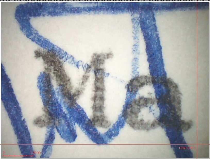
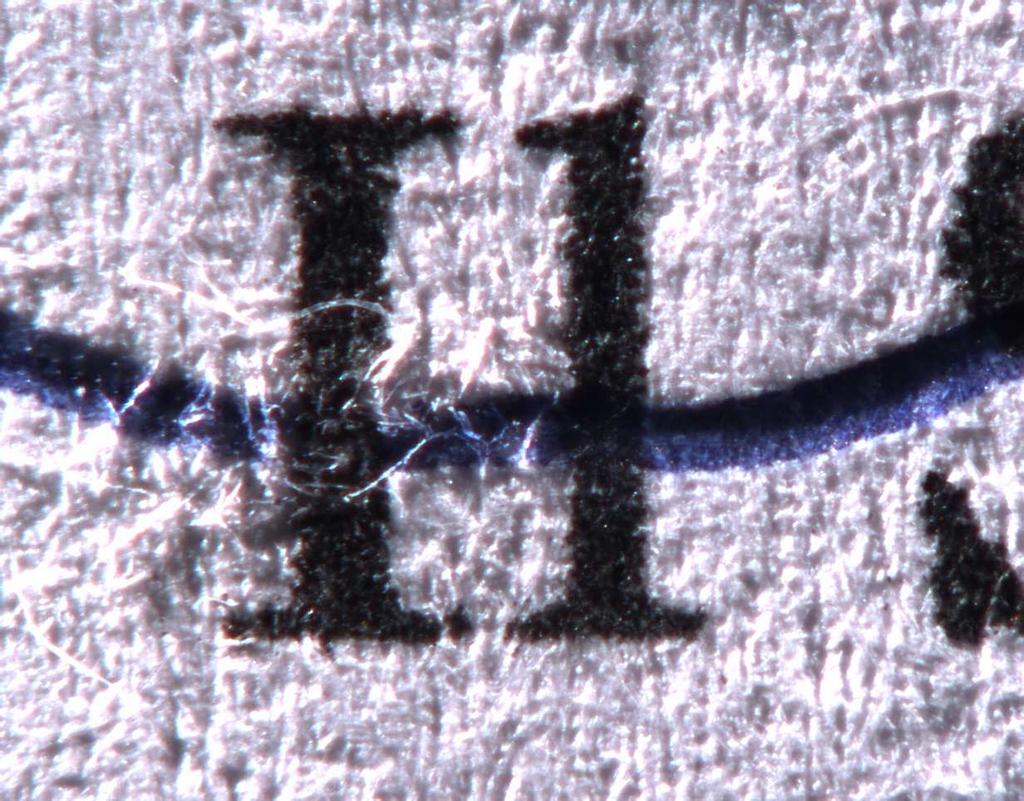
10 Some examples of graphisms viewed by the stereo microscope and later acquired and digitalized by a digital camera 10


11 FORINST FORENSIC INSTRUMENTS TORINO (ITALY) 11
Basics of Light Microscopy and Metallography
 ENGR45: Introduction to Materials Spring 2012 Laboratory 8 Basics of Light Microscopy and Metallography In this exercise you will: gain familiarity with the proper use of a research-grade light microscope
The microscope is useful in making observations and collecting data in scientific experiments. Microscopy involves three basic concepts:
 AP BIOLOGY Chapter 6 NAME DATE Block MICROSCOPE LAB PART I: COMPOUND MICROSCOPE OBJECTIVES: After completing this exercise you should be able to: Demonstrate proper care and use of a compound microscope.
Biology 29 Cell Structure and Function Spring, 2009 Springer LABORATORY 1: THE LIGHT MICROSCOPE
 Biology 29 Cell Structure and Function Spring, 2009 Springer LABORATORY 1: THE LIGHT MICROSCOPE Prior to lab: 1) Read these instructions (p 1-6) 2) Go through the online tutorial, the microscopy pre-lab
Test Review # 8. Physics R: Form TR8.17A. Primary colors of light
 Physics R: Form TR8.17A TEST 8 REVIEW Name Date Period Test Review # 8 Light and Color. Color comes from light, an electromagnetic wave that travels in straight lines in all directions from a light source
LlIGHT REVIEW PART 2 DOWNLOAD, PRINT and submit for 100 points
 WRITE ON SCANTRON WITH NUMBER 2 PENCIL DO NOT WRITE ON THIS TEST LlIGHT REVIEW PART 2 DOWNLOAD, PRINT and submit for 100 points Multiple Choice Identify the choice that best completes the statement or
Match the microscope structures given in the left column with the statements in the right column that identify or describe them.
 49 Prelab for Name Match the microscope structures given in the left column with the statements in the right column that identify or describe them. Key: a. coarse adjustment knob f. turret or nosepiece
Observing Microorganisms through a Microscope LIGHT MICROSCOPY: This type of microscope uses visible light to observe specimens. Compound Light Micros
 PHARMACEUTICAL MICROBIOLOGY JIGAR SHAH INSTITUTE OF PHARMACY NIRMA UNIVERSITY Observing Microorganisms through a Microscope LIGHT MICROSCOPY: This type of microscope uses visible light to observe specimens.
APPLICATIONS FOR TELECENTRIC LIGHTING
 APPLICATIONS FOR TELECENTRIC LIGHTING Telecentric lenses used in combination with telecentric lighting provide the most accurate results for measurement of object shapes and geometries. They make attributes
Introduction. Laboratory Equipment & Supplies. Model 1333PHi Shown (Phase Contrast) (2) Eyepieces (Eyecups installed) Diopter Adjustment Mechanism
 (2) Eyepieces (Eyecups installed) Diopter Adjustment Mechanism") Introduction With the invention of the microscope in the early 17th century, it was made possible to view objects which were too small for the human eye to see. As the microscope evolved, the structure
Introduction With the invention of the microscope in the early 17th century, it was made possible to view objects which were too small for the human eye to see. As the microscope evolved, the structure
ECEN 4606, UNDERGRADUATE OPTICS LAB
 ECEN 4606, UNDERGRADUATE OPTICS LAB Lab 2: Imaging 1 the Telescope Original Version: Prof. McLeod SUMMARY: In this lab you will become familiar with the use of one or more lenses to create images of distant
Basic Principles of the Surgical Microscope. by Charles L. Crain
 Basic Principles of the Surgical Microscope by Charles L. Crain 2006 Charles L. Crain; All Rights Reserved Table of Contents 1. Basic Definition...3 2. Magnification...3 2.1. Illumination/Magnification...3
VISUAL PHYSICS ONLINE DEPTH STUDY: ELECTRON MICROSCOPES
 VISUAL PHYSICS ONLINE DEPTH STUDY: ELECTRON MICROSCOPES Shortly after the experimental confirmation of the wave properties of the electron, it was suggested that the electron could be used to examine objects
MICROSCOPE LAB. Resolving Power How well specimen detail is preserved during the magnifying process.
 AP BIOLOGY Cells ACTIVITY #2 MICROSCOPE LAB OBJECTIVES 1. Demonstrate proper care and use of a compound microscope. 2. Identify the parts of the microscope and describe the function of each part. 3. Compare
Imaging Introduction. September 24, 2010
 Imaging Introduction September 24, 2010 What is a microscope? Merriam-Webster: an optical instrument consisting of a lens or combination of lenses for making enlarged images of minute objects; especially:
Light Microscopy. Upon completion of this lecture, the student should be able to:
 Light Light microscopy is based on the interaction of light and tissue components and can be used to study tissue features. Upon completion of this lecture, the student should be able to: 1- Explain the
Life Science Chapter 2 Study Guide
 Key concepts and definitions Waves and the Electromagnetic Spectrum Wave Energy Medium Mechanical waves Amplitude Wavelength Frequency Speed Properties of Waves (pages 40-41) Trough Crest Hertz Electromagnetic
Person s Optics Test KEY SSSS
 Person s Optics Test KEY SSSS 2017-18 Competitors Names: School Name: All questions are worth one point unless otherwise stated. Show ALL WORK or you may not receive credit. Include correct units whenever
2/4/15. Brightfield Microscopy! It s all about Magnification..! or is it?!
 Brightfield Microscopy It s all about Magnification.. or is it? 1 What actually does go into chosing a microscope Choice depends on what you need the microscope to do. Do you want to magnify stained specimens?
Instruction Manual T Binocular Acromat Research Scope T Trinocular Acromat Research Scope
 Research Scope Instruction Manual T-29031 Binocular Acromat Research Scope T-29041 Trinocular Acromat Research Scope T-29032 Binocular Semi-Plan Research Scope T-29042 Trinocular Semi-Plan Research Scope
Microscopy Techniques that make it easy to see things this small.
 Microscopy Techniques that make it easy to see things this small. What is a Microscope? An instrument for viewing objects that are too small to be seen easily by the naked eye. Dutch spectacle-makers Hans
Test Review # 9. Physics R: Form TR9.15A. Primary colors of light
 Physics R: Form TR9.15A TEST 9 REVIEW Name Date Period Test Review # 9 Light and Color. Color comes from light, an electromagnetic wave that travels in straight lines in all directions from a light source
CALIBRATION OF MICROSCOPE EYEPIECE GRATICULE
 CALIBRATION OF MICROSCOPE EYEPIECE GRATICULE A typical eyepiece graticule looks like this: It is 10mm in length and each mm is divided into 10 parts So each small division = 0.1mm = 100µm The eyepiece
The Nature of Light. Light and Energy
 The Nature of Light Light and Energy - dependent on energy from the sun, directly and indirectly - solar energy intimately associated with existence of life -light absorption: dissipate as heat emitted
The Stereomicroscope CHAPTER 1
 CHAPTER 1 The Stereomicroscope The stereomicroscope is used in most preliminary forensic examinations. This low magnification microscope provides viewing of samples in a manner that is similar to the view
Burton's Microbiology for the Health Sciences
 Burton's Microbiology for the Health Sciences Chapter 2. Viewing the Microbial World Chapter 2 Outline Introduction Using the metric system to express the sizes of microbes Microscopes Simple microscopes
GEOMETRICAL OPTICS Practical 1. Part I. BASIC ELEMENTS AND METHODS FOR CHARACTERIZATION OF OPTICAL SYSTEMS
 GEOMETRICAL OPTICS Practical 1. Part I. BASIC ELEMENTS AND METHODS FOR CHARACTERIZATION OF OPTICAL SYSTEMS Equipment and accessories: an optical bench with a scale, an incandescent lamp, matte, a set of
Section 1: Sound. Sound and Light Section 1
 Sound and Light Section 1 Section 1: Sound Preview Key Ideas Bellringer Properties of Sound Sound Intensity and Decibel Level Musical Instruments Hearing and the Ear The Ear Ultrasound and Sonar Sound
Chapter 25. Optical Instruments
 Chapter 25 Optical Instruments Optical Instruments Analysis generally involves the laws of reflection and refraction Analysis uses the procedures of geometric optics To explain certain phenomena, the wave
Observing Microorganisms through a Microscope
 2016/2/19 PowerPoint Lecture Presentations prepared by Bradley W. Christian, McLennan Community College CHAPTER 3 Observing Microorganisms through a Microscope 1 Figure 3.2 Microscopes and Magnification.
The Care and Use of the Microscope. Lab Exercise #4
 Lab Safety No eating or drinking!!! Long hair must be tied back Clean up your workstation before you leave! Return all materials to the storage sites Clean glassware and wipe down countertops Follow directions
Motorized Axio Observer Start-up instructions
 Start-up instructions 1. If using fluorescence turn on Fluorescent light source. TL light Source (Hal 100) 2. Turn on microscope using switch on lower left side of the microscope. 3. If imaging, turn on
used for low power magnification of a sample image is 3 dimensional
 MICROSCOPES One of the most important inventions in the advancement of Biology 1. Simple Microscopes ie. magnifying glass, stereoscope (dissecting scope) have a single lens or a pair of lenses combined
30 Lenses. Lenses change the paths of light.
 Lenses change the paths of light. A light ray bends as it enters glass and bends again as it leaves. Light passing through glass of a certain shape can form an image that appears larger, smaller, closer,
Reflection! Reflection and Virtual Image!
 1/30/14 Reflection - wave hits non-absorptive surface surface of a smooth water pool - incident vs. reflected wave law of reflection - concept for all electromagnetic waves - wave theory: reflected back
Microscopy. Danil Hammoudi.MD
 Microscopy Danil Hammoudi.MD Care and Handling of the Microscope: A microscope is a delicate piece of equipment and should be treated with care. Use two hands when carrying the microscope. Place one hand
Laser Telemetric System (Metrology)
") Laser Telemetric System (Metrology) Laser telemetric system is a non-contact gauge that measures with a collimated laser beam (Refer Fig. 10.26). It measure at the rate of 150 scans per second. It basically
Laser Telemetric System (Metrology) Laser telemetric system is a non-contact gauge that measures with a collimated laser beam (Refer Fig. 10.26). It measure at the rate of 150 scans per second. It basically
2017 MICROSCOPE REVIEW by Karen L. Lancour RELATIVE SIZE OF MICROBES
 2017 MICROSCOPE REVIEW by Karen L. Lancour RELATIVE SIZE OF MICROBES 1000 millimeters (mm) = 1 meter (m) 1000 micrometers (µm or mcm) = 1 millimeter (mm) 1000 nanometers (nm) = 1 micrometer (mcm) Size
Manual for BMS E1 eplan series, compound microscope
 Manual for BMS E1 eplan series, compound microscope The compound microscope allows it to study, at cell level, structures of textures of botanical and zoological nature. (e.g. slides of roots, leaves and
microscopy A great online resource Molecular Expressions, a Microscope Primer Partha Roy
 Fundamentals of optical microscopy A great online resource Molecular Expressions, a Microscope Primer http://micro.magnet.fsu.edu/primer/index.html Partha Roy 1 Why microscopy Topics Functions of a microscope
Marine Invertebrate Zoology Microscope Introduction
 Marine Invertebrate Zoology Microscope Introduction Introduction A laboratory tool that has become almost synonymous with biology is the microscope. As an extension of your eyes, the microscope is one
2018 MICROSCOPE REVIEW by Karen L. Lancour RELATIVE SIZE OF MICROBES
 2018 MICROSCOPE REVIEW by Karen L. Lancour RELATIVE SIZE OF MICROBES 1000 millimeters (mm) = 1 meter (m) 1000 micrometers (µm or mcm) = 1 millimeter (mm) 1000 nanometers (nm) = 1 micrometer (mcm) Size
LOS 1 LASER OPTICS SET
 LOS 1 LASER OPTICS SET Contents 1 Introduction 3 2 Light interference 5 2.1 Light interference on a thin glass plate 6 2.2 Michelson s interferometer 7 3 Light diffraction 13 3.1 Light diffraction on a
Zoom Stereo Microscope NYMCS-360 Instruction Manual
 Zoom Stereo Microscope NYMCS-60 Instruction Manual This manual is written for stereo microscope NYMCS-60. To ensure the safety, obtain optimum performance and to familiarize yourself fully with the use
Chapter 23 Study Questions Name: Class:
 Chapter 23 Study Questions Name: Class: Multiple Choice Identify the letter of the choice that best completes the statement or answers the question. 1. When you look at yourself in a plane mirror, you
Geometric Optics. Objective: To study the basics of geometric optics and to observe the function of some simple and compound optical devices.
 Geometric Optics Objective: To study the basics of geometric optics and to observe the function of some simple and compound optical devices. Apparatus: Pasco optical bench, mounted lenses (f= +100mm, +200mm,
ECEN 4606, UNDERGRADUATE OPTICS LAB
 ECEN 4606, UNDERGRADUATE OPTICS LAB Lab 3: Imaging 2 the Microscope Original Version: Professor McLeod SUMMARY: In this lab you will become familiar with the use of one or more lenses to create highly
A DIVISION OF FORENSIC TECHNOLOGY. UCM pia The Universal Comparison Macroscope for Forensic Investigations
 A DIVISION OF FORENSIC TECHNOLOGY UCM pia-7000 The Universal Comparison Macroscope for Forensic Investigations PROJECTINA UCM - Outstanding optical performance combined with excellent ergonomics Innovations
Figure 3.4 Approximate size of various types of cells. ~10 um. Red Blood Cells = mm 1500 um. Width of penny Pearson Education, Inc.
 Figure 3.4 Approximate size of various types of cells. ~10 um Red Blood Cells 1.5mm 1500 um Width of penny = 1500 Figure 4.3 The limits of resolution (and some representative objects within those ranges)
ML7520 ML7530 DIOPTER ADJUSTMENT RING BINOCULAR BODY, INCLINED 30. (a) Field Iris Control Lever. (c) Filter Slots EYEPIECES, KHW10X
 Field Iris Control Lever. (c) Filter Slots EYEPIECES, KHW10X") JAPAN DIOPTER ADJUSTMENT RING BINOCULAR BODY, INCLINED 30 (a) Field Iris Control Lever (c) Filter Slots EYEPIECES, KHW10X ANALYZER CONTROL LEVER (b) Aperture Iris Control Lever LIGHT SOURCE HOUSING VERTICAL
JAPAN DIOPTER ADJUSTMENT RING BINOCULAR BODY, INCLINED 30 (a) Field Iris Control Lever (c) Filter Slots EYEPIECES, KHW10X ANALYZER CONTROL LEVER (b) Aperture Iris Control Lever LIGHT SOURCE HOUSING VERTICAL
Using Microscopes. Life Science: Molecular
 Using Microscopes Life Science: Molecular Light Microscopy: Instrumentation and Principles A light microscope is so named because it uses visible light to produce a magnified image. Compound light microscopes
Components of the Microscope
 Swift M3 Microscope The Swift M3 is a versatile microscope designed for both microscopic (high magnification, small field of view) and macroscopic (low magnification, large field of view) applications.
Corundum C Axis Device for Sample Preparation Timothy Thomas, M.E., M.S.E.E. GIA Laboratory June 4, 2009
 Abstract Corundum C Axis Device for Sample Preparation Timothy Thomas, M.E., M.S.E.E. GIA Laboratory June 4, 2009 As a part of GIA s on going project to establish a comprehensive corundum database a need
Optics B. Science Olympiad North Regional Tournament at the University of Florida DO NOT WRITE ON THIS BOOKLET. THIS IS AN TEST SET.
 Optics B Science Olympiad North Regional Tournament at the University of Florida 1 DO NOT WRITE ON THIS BOOKLET. THIS IS AN TEST SET. Part I: General Body Knowledge Questions 2 1) (3 PTS) For much of the
OPTICS DIVISION B. School/#: Names:
 OPTICS DIVISION B School/#: Names: Directions: Fill in your response for each question in the space provided. All questions are worth two points. Multiple Choice (2 points each question) 1. Which of the
STRUCTURE OF THE MICROSCOPE
 STRUCTURE OF THE MICROSCOPE Use the word list to label the microscope below: Light Source Coarse adjustment knob Diaphragm Stage Clips Objectives Fine Adjustment Knob Base Stage Stage Clips Arm Revolving
LAB ACTIVITY: USING A MICROSCOPE
 Name: Date: Period: Lab Partner(s): LAB ACTIVITY: USING A MICROSCOPE Objectives: Demonstrate the proper use and care of a compound light microscope and stereomicroscope. Focus the compound light microscope
A DIVISION OF FORENSIC TECHNOLOGY. UCM pia The Universal Comparison Macroscope for Forensic Investigations
 A DIVISION OF FORENSIC TECHNOLOGY UCM pia-7000 The Universal Comparison Macroscope for Forensic Investigations PROJECTINA UCM - Outstanding optical performance combined with excellent ergonomics Innovations
Ocular Lenses. Head. Arm. Objective Lenses. Slide Holder Stage. On / Off Switch. Condenser with Iris Diaphragm. Light Intensity Control
 BIOLOGY 211: HUMAN ANATOMY & PHYSIOLOGY ********************************************************************************************************* USE OF THE LIGHT MICROSCOPE **********************************************************************************************************
1.When an object is sharply focused and the slide is moved towards you, in which direction does the
 image upright or inverted? Name: Date: _ BIOLOGY EXPERIMENT:Class: Using a Compound Light Microscope II: Depth Perception, resolution, field of view MATERIALS: Compound light microscopecolor magazine clipping
MICROSCOPY MICROSCOPE TERMINOLOGY
 1 MICROSCOPY Most of the microorganisms that we talk about in this class are too small to be seen with the naked eye. The instruments we will use to visualize these microbes are microscopes. The laboratory
GRADE 11-LESSON 2 PHENOMENA RELATED TO OPTICS
 REFLECTION OF LIGHT GRADE 11-LESSON 2 PHENOMENA RELATED TO OPTICS 1.i. What is reflection of light?.. ii. What are the laws of reflection? a...... b.... iii. Consider the diagram at the right. Which one
Care and Use of the Compound Light Microscope
 EXERCISE 2 Care and Use of the Compound Light Microscope Time Estimates for Completing This Lab The activities in this laboratory exercise can be completed in 2 to 2.5 hours. Extra time will be required
Applications of Optics
 Nicholas J. Giordano www.cengage.com/physics/giordano Chapter 26 Applications of Optics Marilyn Akins, PhD Broome Community College Applications of Optics Many devices are based on the principles of optics
Match the correct description with the correct term. Write the letter in the space provided.
 Skills Worksheet Directed Reading A Section: Interactions of Light with Matter REFLECTION Write the letter of the correct answer in the space provided. 1. What happens when light travels through a material
Topic 1 - What is Light? 1. Radiation is the type of energy transfer which does not require... A matter B heat C waves D light
 Grade 8 Unit 1 Test Student Class Topic 1 - What is Light? 1. Radiation is the type of energy transfer which does not require... A matter B heat C waves D light 2. Light-producing technologies, such as
Indian Institute of technology Madras Presents NPTEL NATIONAL PROGRAMME ON TECHNOLOGY ENHANCED LEARNING
 Indian Institute of technology Madras Presents NPTEL NATIONAL PROGRAMME ON TECHNOLOGY ENHANCED LEARNING Lecture - 5 Materials Characterization Fundamentals of Optical microscopy Dr. S. Sankaran Associate
Microscope. & Measurements. Do Now
 Do Now Microscope & Measurements How many: 1. Centimeters (cm) in 4 meters (m)? m 2. Decimeters (dm) in 5 meters (m)? dm 3. Centimeters (cm) in 4,000 millimeters (mm) cm 4. Millimeters (mm) in 40 centimeters
PHYSICS. Chapter 35 Lecture FOR SCIENTISTS AND ENGINEERS A STRATEGIC APPROACH 4/E RANDALL D. KNIGHT
 PHYSICS FOR SCIENTISTS AND ENGINEERS A STRATEGIC APPROACH 4/E Chapter 35 Lecture RANDALL D. KNIGHT Chapter 35 Optical Instruments IN THIS CHAPTER, you will learn about some common optical instruments and
Instructional Resources/Materials: Light vocabulary cards printed (class set) Enough for each student (See card sort below)
 Enough for each student (See card sort below)") Grade Level/Course: Grade 7 Life Science Lesson/Unit Plan Name: Light Card Sort Rationale/Lesson Abstract: Light vocabulary building, students identify and share vocabulary meaning. Timeframe: 10 to 20
Grade Level/Course: Grade 7 Life Science Lesson/Unit Plan Name: Light Card Sort Rationale/Lesson Abstract: Light vocabulary building, students identify and share vocabulary meaning. Timeframe: 10 to 20
Teacher s Resource. 2. The student will see the images reversed left to right.
 Teacher s Resource Answer Booklet Reflection of Light With a Plane (Flat) Mirror Trace a Star Page 16 1. The individual students will complete the activity with varying degrees of difficulty. 2. The student
Unit 8: Light and Optics
 Objectives Unit 8: Light and Optics Explain why we see colors as combinations of three primary colors. Explain the dispersion of light by a prism. Understand how lenses and mirrors work. Explain thermal
OPELCO OPtical ELements COrporation LB Objective Series for Biological Use
 LB Objective Series for Biological Use 105 Executive Drive Suite 100 Dulles, VA 20166-9558 Tel: (703) 471-0080 S PLAN APOCHROMAT OBJECTIVES These objectives compensate for three wavelength of chromatic
UCM: The Universal Comparison Macroscope for Forensic Investigations
 UCM: The Universal Comparison Macroscope for Forensic Investigations New: Motorized objective and magnification changer New: Motorized optics adjustment New: LCD display for operating modes New: Live digital
sclera pupil What happens to light that enters the eye?
 Human Vision Textbook pages 202 215 Before You Read Some people can see things clearly from a great distance. Other people can see things clearly only when they are nearby. Why might this be? Write your
INTRODUCTION THIN LENSES. Introduction. given by the paraxial refraction equation derived last lecture: Thin lenses (19.1) = 1. Double-lens systems
 = 1. Double-lens systems") Chapter 9 OPTICAL INSTRUMENTS Introduction Thin lenses Double-lens systems Aberrations Camera Human eye Compound microscope Summary INTRODUCTION Knowledge of geometrical optics, diffraction and interference,
Chapter 9 OPTICAL INSTRUMENTS Introduction Thin lenses Double-lens systems Aberrations Camera Human eye Compound microscope Summary INTRODUCTION Knowledge of geometrical optics, diffraction and interference,
Examination, TEN1, in courses SK2500/SK2501, Physics of Biomedical Microscopy,
 KTH Applied Physics Examination, TEN1, in courses SK2500/SK2501, Physics of Biomedical Microscopy, 2009-06-05, 8-13, FB51 Allowed aids: Compendium Imaging Physics (handed out) Compendium Light Microscopy
Holography. Introduction
 Holography Introduction Holography is the technique of using monochromatic light sources to produce 3D images on photographic film or specially designed plates. In this experiment you will learn about
Chapter 17: Wave Optics. What is Light? The Models of Light 1/11/13
 Chapter 17: Wave Optics Key Terms Wave model Ray model Diffraction Refraction Fringe spacing Diffraction grating Thin-film interference What is Light? Light is the chameleon of the physical world. Under
PHYS 3153 Methods of Experimental Physics II O2. Applications of Interferometry
 Purpose PHYS 3153 Methods of Experimental Physics II O2. Applications of Interferometry In this experiment, you will study the principles and applications of interferometry. Equipment and components PASCO
A DIVISION OF FORENSIC TECHNOLOGY SEE MORE. SOLVE MORE. CLASS-LEADING COMPARISON MICROSCOPES FOR FORENSIC INVESTIGATIONS
 A DIVISION OF FORENSIC TECHNOLOGY SEE MORE. SOLVE MORE. CLASS-LEADING COMPARISON MICROSCOPES FOR FORENSIC INVESTIGATIONS PROJECTINA AND FORENSIC TECHNOLOGY Forensic Technology pioneered automated ballistic
Chapter 29/30. Wave Fronts and Rays. Refraction of Sound. Dispersion in a Prism. Index of Refraction. Refraction and Lenses
 Chapter 29/30 Refraction and Lenses Refraction Refraction the bending of waves as they pass from one medium into another. Caused by a change in the average speed of light. Analogy A car that drives off
Chapter 36. Image Formation
 Chapter 36 Image Formation Image of Formation Images can result when light rays encounter flat or curved surfaces between two media. Images can be formed either by reflection or refraction due to these
ELECTRON MICROSCOPY AN OVERVIEW
 ELECTRON MICROSCOPY AN OVERVIEW Anjali Priya 1, Abhishek Singh 2, Nikhil Anand Srivastava 3 1,2,3 Department of Electrical & Instrumentation, Sant Longowal Institute of Engg. & Technology, Sangrur, India.
The light microscope
 What is a microscope? The microscope is an essential tool in modern biology. It allows us to view structural details of organs, tissue, and cells not visible to the naked eye. The microscope should always
E X P E R I M E N T 12
 E X P E R I M E N T 12 Mirrors and Lenses Produced by the Physics Staff at Collin College Copyright Collin College Physics Department. All Rights Reserved. University Physics II, Exp 12: Mirrors and Lenses
Aberrations of a lens
 Aberrations of a lens 1. What are aberrations? A lens made of a uniform glass with spherical surfaces cannot form perfect images. Spherical aberration is a prominent image defect for a point source on
ECEN. Spectroscopy. Lab 8. copy. constituents HOMEWORK PR. Figure. 1. Layout of. of the
 ECEN 4606 Lab 8 Spectroscopy SUMMARY: ROBLEM 1: Pedrotti 3 12-10. In this lab, you will design, build and test an optical spectrum analyzer and use it for both absorption and emission spectroscopy. The
Systems Biology. Optical Train, Köhler Illumination
 McGill University Life Sciences Complex Imaging Facility Systems Biology Microscopy Workshop Tuesday December 7 th, 2010 Simple Lenses, Transmitted Light Optical Train, Köhler Illumination What Does a
A BRIEF INTRODUCTION TO MICROSCOPY The two key properties of a microscope that allow you to see microbes are resolution and magnification.
 A BRIEF INTRODUCTION TO MICROSCOPY The two key properties of a microscope that allow you to see microbes are resolution and magnification. Magnification refers to the enlargement of the specimen when seen
MICROSCOPY and CELL STRUCTURE
 MICROSCOPY and CELL STRUCTURE Readings: Review pp. 69-71, and Fig. 4.1 on p. 65 in your text (POHS, 5 th ed.). Introduction: Biologists rely on many different types of microscopic techniques to find out
Studying of Reflected Light Optical Laser Microscope Images Using Image Processing Algorithm
 IRAQI JOURNAL OF APPLIED PHYSICS Fatema H. Rajab Al-Nahrain University, College of Engineering, Department of Laser and Optoelectronic Engineering Studying of Reflected Light Optical Laser Microscope Images
Very short introduction to light microscopy and digital imaging
 Very short introduction to light microscopy and digital imaging Hernan G. Garcia August 1, 2005 1 Light Microscopy Basics In this section we will briefly describe the basic principles of operation and
Lenses- Worksheet. (Use a ray box to answer questions 3 to 7)
") Lenses- Worksheet 1. Look at the lenses in front of you and try to distinguish the different types of lenses? Describe each type and record its characteristics. 2. Using the lenses in front of you, look
Lenses- Worksheet 1. Look at the lenses in front of you and try to distinguish the different types of lenses? Describe each type and record its characteristics. 2. Using the lenses in front of you, look
Name: Date: Block: Light Unit Study Guide Matching Match the correct definition to each term. 1. Waves
 Name: Date: Block: Light Unit Study Guide Matching Match the correct definition to each term. 1. Waves 2. Medium 3. Mechanical waves 4. Longitudinal waves 5. Transverse waves 6. Frequency 7. Reflection
Chapter Introduction. Chapter Wrap-Up. and the Eye
 Chapter Introduction Lesson 1 Lesson 2 Lesson 3 Sound Light Chapter Wrap-Up Mirrors, Lenses, and the Eye How do sound and light waves travel and interact with matter? What do you think? Before you begin,
Notes: Light and Optics. Reflection. Refraction. Law of Reflection. Light goes straight 12/13/2012
 Notes: Light and Optics Light goes straight Light travels in a straight line unless it interacts with a medium. The material through which a wave travels is called a medium. Light can be reflected, refracted
Where Image Quality Begins
 Where Image Quality Begins Filters are a Necessity Not an Accessory Inexpensive Insurance Policy for the System The most cost effective way to improve repeatability and stability in any machine vision
Chapter 2 The Study of Microbial Structure: Microscopy and Specimen Preparation
 Chapter 2 The Study of Microbial Structure: Microscopy and Specimen Preparation 1 Lenses and the Bending of Light light is refracted (bent) when passing from one medium to another refractive index a measure
Education in Microscopy and Digital Imaging
 Contact Us Carl Zeiss Education in Microscopy and Digital Imaging ZEISS Home Products Solutions Support Online Shop ZEISS International ZEISS Campus Home Interactive Tutorials Basic Microscopy Spectral
Laboratory Introduction
 Laboratory Introduction There are two basic categories of microscopes: light microscopes and electron microscopes. Light, or optical, microscopes require light waves to provide the illumination while electron