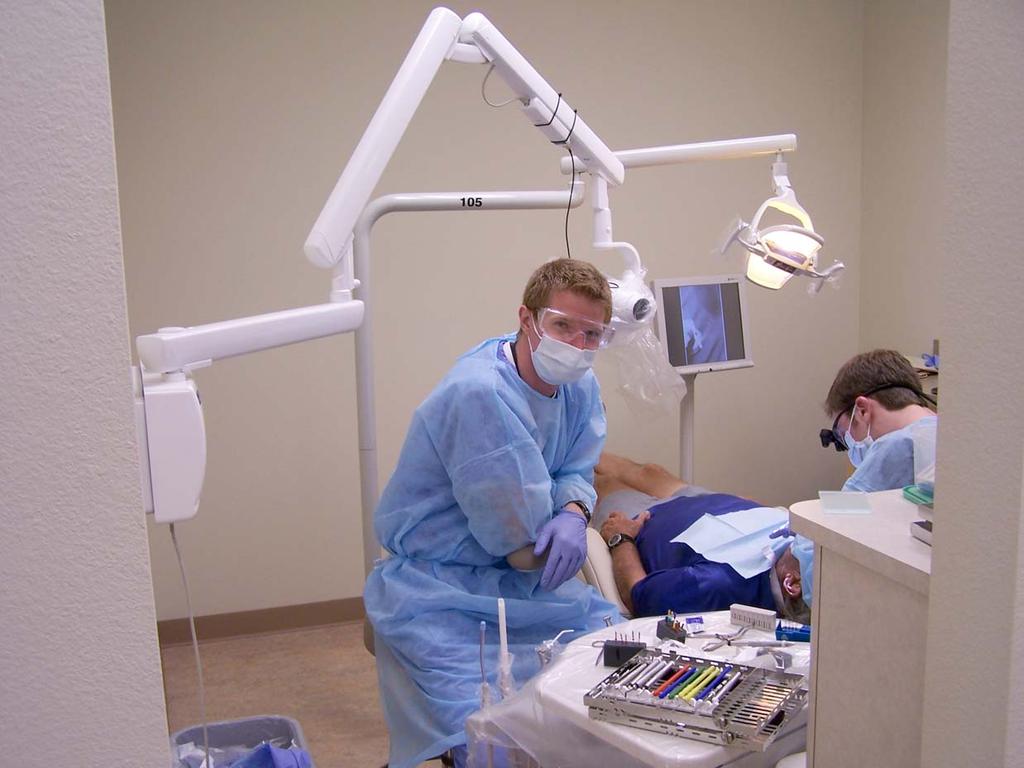
2006 National Primary Oral Health Care Conference Robert A. Cederberg, MA, DDS December 10 14, 2006 Scottsdale, Arizona
|
|
|
- Homer Blankenship
- 5 years ago
- Views:
Transcription


1 Digital Radiography 2006 National Primary Oral Health Care Conference Robert A. Cederberg, MA, DDS December 10 14, 2006 Scottsdale, Arizona
2 Development of Dental Digital Radiography Background, history and development of dental digital radiography Evolution of the technology & systems Conversion from film to digital and options for the office or clinic Advantages and disadvantages Applications and functions Adjunctive techniques

3 Background The development of computed axial tomography (CAT) in 1972 and subsequent advances in digital sensors allowed for dental applications.


4 History of Digital in Dentistry Trophy Radiologie introduced the RVG in 1982 Sensor - CCD (charged-coupled device) with phosphor screen, active area 17mm x 26mm

5 Design and Development First sensors where designed with CCD chip layered with a phosphor screen, latter sensors were developed which were direct exposure.

6 Digital System Components Digital imaging requires a sensor (detector), analog to digital converter (A/D converter), computer or CPU, monitor, printer. Additionally, modem or other (cable, T1, DSL) connection for Teleradiograhpy is important.
7 Steps in Digital Acquisition Electromagnetic energy in the form of x-ray photons strike the detector (CCD electrical charge stored in pixels), (PSP latent image formation) A/D converter converts the electrical charge into a digital format (PSP visible light intensities converted to digital format) A number is assigned to each voltage output ranging from 0 to 256 (2 8 ) or 256 shades of gray in the image Once image is acquired and stored post processing, i.e. image manipulation
8 Enhancements Direct exposure CCDs Early 90 s Photostimuable Phosphor (PSP) technology Early 90 s Complementary Metal Oxide Semiconductor (CMOS) Flat Panel Detectors CBCT (Cone Beam Computed Tomography) Late 90 s software improvements, monitor fidelity improvements.
9 Evolution of Dental X-ray Image Receptors Discovery of x rays in 1895, first image receptors were glass plates celluloid film 1919 single emulsion dental film packets 1924 double emulsion dental film 1940 Ultraspeed film D speed 1980 Ektaspeed film E speed 1982 first intraoral digital sensor

10 Direct Digital Radiography
11 Basic Digital System X-ray source Components Sensor or detector (CCD, CMOS and storage phosphor) Computer or CPU Modem Printer
12 Sensors CCD (Charged Couple Device): silicon chip with an embedded electronic circuit. PSP (Photostimuable Storage Phosphor): plastic plate coated with a photostimuable phosphor layer. CMOS-APS (Complementary Metal Oxide Semiconductor - Active Pixel Sensor): silicon chip with an ADC for each column of pixels.


13 Digital Sensors Optime PSP plates Sizes 0, 1and 2 Trophy Sensor Size 2 Schick Sensors Sizes 0, 1 and 2

14
15 The Physics of CCD Sensors

16 Available Sensors

17 CMOS Sensor

18 The Physics of PSP Sensors
19 Conversion from Film to Digital Probably easiest to slowly transition into digital. Probably best to consider utilizing more than one digital system since no one system will satisfy all of the imaging needs within one office or clinic, i.e. for intraoral imaging, both a PSP and CCD combination.


20 Conversion from Film to Work flow study Digital

21
22

23
24 Conversion from Film to Digital Main advantage no darkroom needed Elimination of the mess of darkroom chemistry, cost and maintenance of film processors Many cities and local municipalities now regulate waste water discharge and require silver collectors to be installed on processors which adds additional mess as well as expense


25 Techniques and Systems Techniques vary only by the type of detector used: either CCD or Storage Phosphor All systems offer the ability to manipulate the image: i.e. image enhancement, colorization, etc.
26 Intraoral CCD Based Systems Product Name CDR CygnusRay MPS Dexis Dixi 2 Dixsy FI iox megapixel MPDx Company Schick Cygnus Technologies ProVision Dental Systems Planmeca Group Villa Sistemi Medicali Fimet Remedent N.V.
27 Intraoral CCD Based Systems Product Name SIDEXIS SIGMA SuniRay TM RVG VistaRay VisualiX USB Company Sirona PaloDex Group Suni Medical Imaging Trophy DÜrr Dental Gendex
28 Intraoral PSP Systems Product Name Company Panorama Xi Orex Combi-Xi Orex DenOptix Gendex - Dentsply Digora Optime PaloDex Group VistaScan DÜrr Dental VistaScan Intra DÜrr Dental
29 Panoramic CCD Based Systems Product Name Company CDRPan Schick Digipan Trophy Dimax2, Proline, Promax Planmeca Group DXIS Signet Orthopantomograph OP100D PaloDex Group Orthoceph OC100D PaloDex Group ORTHOPHOS DS Sirona
30 Panoramic CCD Based Systems Product Name Company Cranex Base X D Cranex Excel D Scanora D Orthoralix 9200 (DDE, DPI) Versaview (5D, SDCP) PaloDex Group PaloDex Group PaloDex Group Gendex - Dentsply Morita
31 Panoramic PSP Based Systems Product Name Combi-Xi DenOptix DenOptix Ceph DEXpan Digora PCT Paxorama Xi VistaScan Company Orex Gendex - Dentsply Gendex - Dentsply Pro Vision Dental PaloDex Group Orex DÜrr Dental


32 Digital X-ray Image? Physical Definition Digital Image Mathematical Definition

33 PIXEL The matrix element of a digital array which identifies a gray level at that point. It can be processed and manipulated, usually expressed in values. Gray Scale O 255 O = Black 255 = White
34 Digital Imaging: Advantages Image enhancement and analysis - may increase diagnostic validity of x-rays of non-optimal density. No-processing problems - darkroom, processor and processing chemistry not required (environmentally friendly). Less exposure errors - density and contrast enhancement. Allows for quantitative evaluation. Lower absorbed doses - 20 to 90% compared to D-speed film. Better patient communication and time saver.
35 Digital Imaging: Disadvantages High cost (Processor/sensor, CPU, monitor, printer) - CCD and CMOS sensors 5 to $7,000 Inflexible cassette for CCD and CMOS sensor - may not allow proper placement in all intraoral locations. Spatial Resolution not as good as film - film 16 to 20 lp/mm, digital 7-13 lp/mm. Printed copy not as good as screen - resolution is lost when image is printed. Not standardized - proprietary hardware and software. Learning curve for staff.

36 PaloDex Group Soredex: Optime


37 Taking The Radiograph Place onto positioning device Use your current x- ray equipment Take x-ray with reduced exposure

38 Digora Optime Hardware Imaging Plate Processing
 Manipulating implant Video")
39 Digital Imaging Software Features Zoom Contrast/Brightness Pseudocolor Rotation Annotation Filters 3D Analysis (Line, Angle) Manipulating implant Video Inversion

40 Negative Conversion

41 Pseudo-Colorization

42 3D Function

43 Measuring Distance and Angle
44 Applications Patient Education Quantifying the image Implant planning Teleradiography Other


45 Patient Education


46 Quantifying the Image All systems provide the ability to measure lengths and angles and to analyze the density gradients throughout the image.

47 Implant Planning Some systems provide for implant overlays which are helpful in assessing potential implant sites.

48 Teleradiography Transmission of digital images to remote sites is a major driving force in the evolution of digital radiography. Remote consultation, Insurance approval.

49 DenOptix QST

50 Digital X-ray Imaging 100% re-usable Same size as film Flexible Thin No wires like CCD sensor Use with existing x-ray equipment Plates

51

52 Preparing the plates To prevent cross contamination, the plates are placed in a disposable plastic barrier

53 Preparing the plates Panometric and Cephalometric are placed in the cassette with screens

54 Taking an x-ray Tear the barriers and drop the imaging plate into the dark box Be careful of light exposure and contamination
55 The DenOptix Scanner Scans all image plate sizes Integrates with practice management software Simple to learn No patient discomfort
 Attach the pan/ceph")
56 Scanning In a semi-darkened area Load the plate on the carousel (can hold up to 29 plates) Attach the pan/ceph plates

57 Scanning Place the loaded carousel into the scanner Close the lid Begin scanning 1 min 12 secs for up to 8 I/O 2 min 30 secs for a Pan 3 min 30 secs for a Ceph
58 Sensor - CCD or CMOS GX-S RFG CDR (Schick) Sidexis Dixi (Planmeca)
59 DC Intraoral Unit

60 Panoramic Unit

61 The source of x-rays, the tube Must have Smallest focal point AC - DC Range of Kv, ma adapted to dental x- rays (digital or not)
62 The detector Film, sensor, imaging plates Image quality parameters are: Quantum Mottle Noise (signal/noise ratio) Resolution Sensitivity or dynamic range All three are important!!
63 Signal/Noise Ratio Depends on the amount of photons that strike the image sensor Noise level: PSP < film < CCD Images with lots of noise are described as grainy

64
65 Resolution X-ray photon detection takes place in layers vs on a surface Resolution is limited by the thickness of the layer, not pixel size Measured by line pairs resolved per mm (lp/mm) Limited by what the eye can detect Film = 20 lp/mm PSP = 7-14 lp/mm (I/O) Sensor = lp/mm (I/O) Diagnostic resolution is the key.

66 Resolution 600 dpi 300 dpi
67 A good image means Good x-ray source Small focal spot, collimator, x-ray beam quality Good positioning Good detector

68

69

70 RINN Sensor Holder

71 Dexis Sensor

72
73

74

75

76

77

78
79

80
81
82 Comparison of Image Receptor Characteristics Contrast resolution Spatial resolution Sensor/receptor latitude Sensor/receptor sensitivity MTF, SNR (NEQ and DQE) Contrast and spatial resolution of the human eye
83 Contrast Resolution The ability to distinguish different densities in the radiographic image. Factors affecting contrast resolution: 1. Attenuation characteristics of the tissues being imaged. 2. Sensor bit depth and noise level. 3. Monitor resolution, bit depth, dot pitch, luminance and display size. 4. Ambient lighting.
84 Contrast Resolution Digital imaging systems are capable of capturing up to 256 different densities, monitors/operating systems are only capable of displaying 242 different densities. The human eye can only distinguish 32 different densities.
85 Spatial Resolution The capacity of the image to display fine detail. *The theoretical limit of resolution is a function of the pixel size of the system.* Image receptor pixel sizes: film 8 microns, high res. CCD 20 microns, PSP 40 microns. Resolution: film = > 20 lp/mm, CCD = 25 lp/mm (theoretical with software enhancements), PSP = 12.5 lp/mm.
86 Sensor Latitude The ability of the sensor to capture a range of x-ray exposures. A desirable quality of a sensor/receptor is the ability to display a full range of densities. Dynamic range of film extends to 4 orders of magnitude (with hot lighting), CCD/CMOS sensors extend to 2 orders of magnitude and PSP sensors extend to 5 orders of magnitude of x-ray exposure.
87 Sensor Sensitivity Sensitivity of a sensor is its ability to respond to small amounts of radiation. Sensitivity of film is directly related to film speed. Sensitivity of digital sensors affected by pixel size and system noise. PSP systems allow dose reductions of about 50% compared to F-speed film. CCD/CMOS less dose reduction than PSP.
88 Modulation Transfer Function (MTF) MTF is a measure of the combined effects of sharpness and resolution. The MTF of E-speed film is superior to the MTF of PSP at high spatial frequencies. The 50% modulation level for PSP is reached at 2.5 lp/mm whereas E-speed film is not reached until 10 lp/mm.
89 Signal-to-Noise Ratio (SNR) NEQ and DQE The ratio of the signal amplitude to the standard deviation of the fluctuations in noise. NEQ noise equivalent quanta DQE detective quantum efficiency At low spatial frequencies PSP systems have superior NEQ to E-speed film across the exposure range. Data suggested that a PSP sensors are able to absorb more quanta (photons) per unit area as compared to film.
90 Signal-to-Noise Ratio (SNR) NEQ and DQE Higher quantum absorption of PSP leads to a superior DQE as compared to E-speed film. Theoretically higher quantum absorption and higher DQE of PSP sensors could provide a more efficient absorption of photons and thereby an image with improved definition.
91 Contrast & Spatial Resolution of the Human Eye In terms of contrast, the displayed image provides up to 8 times the amount of information that the eye can actually see. The minimum contrast threshold (greatest perceptual sensitivity) corresponds to a spatial frequency of approximately 5 lp/cm or about a 1mm wide pixel size. Consequently, the sensor can display approximately 2.5 times the amount of information that the eye can actually perceive.
92 Lesion Recognition Research Studies have looked at both natural carious lesions and simulated lesions and tested the resolving power of film vs digital. Conclusion The ability to recognize incipient caries lesions is NOT dependent on image receptor (film or digital), the system used or the manner in which it is displayed.
93 References Brettle DS, Workman A, et al. The imaging performance of a storage phosphor system for dental radiography, Br. J. Radiol. 69, (1996). Cowen AR, Workman A, Price JS. Physical aspects of photostimuable phosphor computed radiography, Br. J. Radiol. 66, (1993). Wang J, Langer S. A brief review of human perception factors in digital displays for picture archiving and communications systems, J. Digit. Imaging. 10(4), (1997). Cederberg RA, Tidwell E, et al. Endodontic working length assessment comparison of storage phosphor digital imaging and radiographic film. OOOOE. 85, (1998). Cederberg RA, Frederiksen NL, et al. Influence of the digital image display monitor on observer performance. Dentomaxillofac Radiol. 28, (1999).

94 Image Examples From Planmeca s website


95 Planmeca Bite Wing Series left side


96 OpTime Bite Wing Series left side


97 Planmeca image OpTime image
98 Conclusion Storage phosphor technology provides film-like sensors which are easier to place for all intraoral imaging sites. More comfortable for the patient and placement especially for premolar bitewings allows operator to image the distal of the canines. Elapsed time for completion of image is actually less than for some hard-wired systems.
99 Conclusion Portable no need to be tied to a computer. Can be taken in any operatory with a tube head and even with a hand held unit. Widest dynamic range of any sensor allows for potential recovery of diagnostic information in an otherwise underexposed image. Improved DQE theoretically provides better image clarity & definition.
100 Conclusion Some common fallacies concerning the superiority of film and/or CCD/CMOS receptors over PSP receptors: One sensor type will be sufficient for all intraoral imaging applications. Probably only true for film and PSP. Depending on the type of hard-wired sensor there may difficulty obtaining certain images such as molar periapicals and premolar bitewings.
101 Conclusion Some common fallacies concerning the superiority of film and/or CCD/CMOS receptors over PSP receptors: CCD/CMOS systems claims of superior resolutions of 25 lp/mm or greater as compared to PSP. The human eye has the best acuity at pixel sizes approximating 1 mm, 2.5 times the resolving power of most PSP systems. Is it enough to detect decay? Yes! But, image clarity is more important.

102 Digital Subtraction Radiography

103 Cone Beam CT

104

105 Flat Panel Detector Amorphous Silicon Flat Panel Detector
106 Effective Dose Comparison i-cat 20 sec scan 68 usv i-cat 10 sec scan 34 usv Daily background 8 usv Panoramic film usv Panoramic digital usv Full Mouth Series film 150 usv Hitachi MercuRay CBCT 485 usv Medical CT usv

107 Panoramic

108 X-Sectional Imaging


109 Localization


110 Orthodontics

111 Pathology
112 What Does the Future of Digital Radiography Look Like? Evolution and development of existing systems It s here to stay! Further integration of other technologies into existing systems. CBCT will become standard of care for all dental imaging needs. CBCT will become more widely used, price will come down. Perhaps, a CBCT scan and bitewings will someday be the dental exam standard.
RADIOGRAPHY TERMS TO KNOW SELF STUDY DENTALELLE TUTORING
 RADIOGRAPHY TERMS TO KNOW SELF STUDY DENTALELLE TUTORING PLEASE NOTE You DO NOT need to study these for the board exam if this is why you bought our Radiography course, however if you come across any terms
Dental Radiography. One of the problems of dental radiography is having different dimensions than normal.
 The prototype receptor (the recording medium) most commonly used in dental radiography is the radiographic film. However, there are many other new more efficient receptors than the formed one that can
DIGITAL RADIOGRAPHY. Digital radiography is a film-less technology used to record radiographic images.
 DIGITAL RADIOGRAPHY Digital radiography is a film-less technology used to record radiographic images. 1 The purpose of digital imaging is to generate images that can be used in the diagnosis and assessment
ProX Intraoral X-ray. PLANMECA is proud to introduce a new intraoral X-ray unit to its comprehensive collection of imaging products- the ProX.
 The premium intraoral X-ray unit... ProX Intraoral X-ray PLANMECA is proud to introduce a new intraoral X-ray unit to its comprehensive collection of imaging products- the ProX. This advanced unit provides
DIGITAL IMAGING WITHOUT LIMITS. Your Complete Digital X-Ray Solution. Wireless. Flexible. Every image SIZE.
 DIGITAL IMAGING WITHOUT LIMITS. Your Complete Digital X-Ray Solution. Wireless. Flexible. Every image SIZE. C l a s s i c D u o I n t r a o r a l Digital Imaging Without Limits ScanX digital radiography
DIGITAL IMAGING Recognise the importance of quality assurance
 DIGITAL IMAGING Recognise the importance of quality assurance There are two types of digital image receptor both of which capture a two dimensional image of the three dimensional patient. These are Computed
X-Ray/ Digital Imaging
 Chapter 20 X-Ray/ Digital Imaging Carestream - Intraoral Cameras 852 Intraoral X-Rays 855 Digital Sensors 857 Intraoral Imaging Plate System 860 Extraoral Imaging System 862 Intraoral Scanners 868 CBCT
CR Basics and FAQ. Overview. Historical Perspective
 Page: 1 of 6 CR Basics and FAQ Overview Computed Radiography is a term used to describe a system that electronically records a radiographic image. Computed Radiographic systems use unique image receptors
Photostimulable phosphor plates (PSPs)
") DIGITAL IMAGING Digital imaging Photostimulable phosphor plates (PSPs) Indirect digital PSPs are composed of a polyester base with a phosphor layer (europium activated barium fluorohalide) on one side.
DIGITAL IMAGING Digital imaging Photostimulable phosphor plates (PSPs) Indirect digital PSPs are composed of a polyester base with a phosphor layer (europium activated barium fluorohalide) on one side.
AnySensor (1.0 /1.5)
") HQ-20080217-MM-kcg Dental Digital Imaging Radiography System AnySensor (1.0 /1.5) Cutting-edge CMOS Technology CMOS? CCD? CCD CMOS Past - Particular Process -High Quality & High Cost - General Process
HQ-20080217-MM-kcg Dental Digital Imaging Radiography System AnySensor (1.0 /1.5) Cutting-edge CMOS Technology CMOS? CCD? CCD CMOS Past - Particular Process -High Quality & High Cost - General Process
10/3/2012. Study Harder
 This presentation is a professional collaboration of development time prepared by: Rex Christensen Terri Jurkiewicz and Diane Kawamura Study Harder CR detection is inefficient, inferior to film screen
10/26/2015. Study Harder
 This presentation is a professional collaboration of development time prepared by: Rex Christensen Terri Jurkiewicz and Diane Kawamura Study Harder CR detection is inefficient, inferior to film screen
Phosphor Storage Plates 5% 5+ boxes Intraoral. Discounts do not apply to clearance items.
 S15:1 7433 170323 Phosphor Storage Plates We offer a full line of reusable intraoral storage phosphor plates compatible with Air Techniques Scan X, Soredex, and Gendex dental systems. Sizes 0-2 intraoral
Acquisition, Processing and Display
 Acquisition, Processing and Display Terri L. Fauber, R.T. (R)(M) Department of Radiation Sciences School of Allied Health Professions Virginia Commonwealth University Topics Image Characteristics Image
VistaScan Combi View - efficiency without compromise
 VistaScan Combi View - efficiency without compromise The image plate scanner with a touchscreen for all formats COMPRESSED AIR SUCTION IMAGING DENTAL CARE HYGIENE Image plate technology - a With the versatile
Comprehensive product portfolio. FONA, the most exciting new brand in dental. ALL THE ADVANTAGES OF DIGITAL Intraoral Imaging
 Comprehensive product portfolio Treatment centers Instruments Laser Hygiene 3D and Panoramic x-rays Intraoral x-rays Intraoral sensors Phosphor plate scanner FONA, the most exciting new brand in dental
Applied Sciences at Winterthur)
") Comparison of image quality between a digital panorama X-ray unit with a CdTe-CMOS detector and panorama X- ray units with other types of digital detectors Stephan Scheidegger 1 1 Zürcher Hochschule für
Comparison of image quality between a digital panorama X-ray unit with a CdTe-CMOS detector and panorama X- ray units with other types of digital detectors Stephan Scheidegger 1 1 Zürcher Hochschule für
Digital Imaging Considerations Computed Radiography
 Digital Imaging Considerations Digital Radiography Computed Radiography o Cassette based Direct or Indirect Digital Radiography o Cassetteless Computed Radiography 1 CR Image Acquisition Most like conventional
PRODUCT CATALOGUE 20 17
 PRODUCT CATALOGUE 20 17 I-View CMOS Intraoral sensor Trident uses the best of CMOS technology to create I-View: an innovative sensor to get better diagnosis with outstanding images, saving both valuable
Radiology. Radiograph: Is the image of an object made with use of X- ray instead of light.
 Radiology د. اريج Lec. 3 X Ray Films Radiograph: Is the image of an object made with use of X- ray instead of light. Dental x- ray film: Is a recording media on which image of the object was made by exposing
Influence of the Display Monitor on Observer Performance in Detection of Dental Caries
 Received 25 July 2007; Accepted 1 September 2007 Influence of the Display Monitor on Observer Performance in Detection of Dental Caries Farzad Esmaeili 1 Esrafil Balaei 2 Firoz Pouralibaba 3 * Farzaneh
3/31/2011. Objectives. Emory University. Historical Development. Historical Development. Historical Development
 Teaching Radiographic Technique in a Digital Imaging Paradigm Objectives 1. Discuss the historical development of digital imaging. Dawn Couch Moore, M.M.Sc., RT(R) Assistant Professor and Director Emory
Introduction. Chapter 16 Diagnostic Radiology. Primary radiological image. Primary radiological image
 Introduction Chapter 16 Diagnostic Radiology Radiation Dosimetry I Text: H.E Johns and J.R. Cunningham, The physics of radiology, 4 th ed. http://www.utoledo.edu/med/depts/radther In diagnostic radiology
VistaScan Plus Image Plate Scanners The most comprehensive dental scanners
 VistaScan Plus Image Plate Scanners The most comprehensive dental scanners COMPRESSED AIR SUCTION IMAGING DENTAL CARE HYGIENE Make your surgery routines more efficient Intraoral and extraoral images in
SYLLABUS. TITLE: Equipment Operation I. DEPARTMENT: Radiologic Technology
 CODE: RADT 156 INSTITUTE: Health Science TITLE: Equipment Operation I DEPARTMENT: Radiologic Technology COURSE DESCRIPTION: This course covers the principles of equipment operation and maintenance of radiographic
ALL THE ADVANTAGES OF DIGITAL Intraoral Imaging
 ALL THE ADVANTAGES OF DIGITAL Intraoral Imaging FONA Intraoral Imaging PATIENT COMFORT AND EASE OF USE Plate sizes and positioning just as with traditional film allowing ease of use, patient comfort and
Q3D. Speak to a 3D Specialist. CBCT 3D / Panoramic Imaging GENERAL DIMENSIONS. Suni Imaging Product Lines GET.
 GENERAL Q3D Q3D Ceph Exposure Time FOV Voxel Size Focal Spot Target Angle Tube Voltage Tube Current Line Voltage Warranty Panoramic CT 9 to 17 sec 9 to 17 sec 4 to 12 sec 7.7/14.5 sec 7.7/14.5 sec 4 x
Small. Smart. Fast. Digital imaging plate system
 Digital imaging plate system A pioneer with 15 years of experience Summary of benefits. Easy to use Graphical user interface supports easy workflow Image preview Fast and automated Fast image readout Touchless
Visibility of Detail
 Visibility of Detail Radiographic Quality Quality radiographic images represents the, and information is for diagnosis. The of the anatomic structures and the accuracy of their ( ) determine the overall
X-ray Imaging. PHYS Lecture. Carlos Vinhais. Departamento de Física Instituto Superior de Engenharia do Porto
 X-ray Imaging PHYS Lecture Carlos Vinhais Departamento de Física Instituto Superior de Engenharia do Porto cav@isep.ipp.pt Overview Projection Radiography Anode Angle Focal Spot Magnification Blurring
Hardware for High Energy Applications 30 October 2009
 Paper No. 003 09 Hardware for High Energy Applications 30 October 2009 This document was created by the Federal Working Group on Industrial Digital Radiography. Reproduction is authorized. Federal Working
ORTHOPANTOMOGRAPH OP 2D Quality and design
 OP 2D Quality and design OP 2D OP 2D is a digital panoramic X-ray unit that combines distinctive design and reliable quality with all essential tools for standard panoramic imaging needs. OP 2D is part
Digital radiography: Practical advantages of Digital Radiography. Practical Advantages in image quality
 Digital radiography: Digital radiography is set to become the most common form of processing radiographic images in the next 10 years. This is due to a number of practical and image quality issues. Practical
Comparisons of physical imaging pro three kinds of imaging plates used Title photostimulable phosphor systems fo radiography
 Comparisons of physical imaging pro three kinds of imaging plates used Title photostimulable phosphor systems fo radiography Author(s) Nishikawa, K; Ooguro, T; Kuroyanagi Journal Bulletin of Tokyo Dental
Chapters 1-3. Chapter 1: Introduction and applications of photogrammetry Chapter 2: Electro-magnetic radiation. Chapter 3: Basic optics
 Chapters 1-3 Chapter 1: Introduction and applications of photogrammetry Chapter 2: Electro-magnetic radiation Radiation sources Classification of remote sensing systems (passive & active) Electromagnetic
DENTAL LINE. Dental Panoramic - Digital & Film
 DENTAL LINE Dental Panoramic - Digital & Film Evolution comes from experience Rotograph Evo takes the experience of five decades of dedication to X-ray diagnostic imaging to the next level. You can rely
Chapters 1-3. Chapter 1: Introduction and applications of photogrammetry Chapter 2: Electro-magnetic radiation. Chapter 3: Basic optics
 Chapters 1-3 Chapter 1: Introduction and applications of photogrammetry Chapter 2: Electro-magnetic radiation Radiation sources Classification of remote sensing systems (passive & active) Electromagnetic
SECTION I - CHAPTER 1 DIGITAL RADIOGRAPHY: AN OVERVIEW OF THE TEXT. Exam Content Specifications 8/22/2012 RADT 3463 COMPUTERIZED IMAGING
 RADT 3463 - COMPUTERIZED IMAGING Section I: Chapter 1 RADT 3463 Computerized Imaging 1 SECTION I - CHAPTER 1 DIGITAL RADIOGRAPHY: AN OVERVIEW OF THE TEXT RADT 3463 COMPUTERIZED IMAGING Section I: Chapter
Current technology in digital image production (CR/DR and other modalities) Jaroonroj Wongnil 25 Mar 2016
 Jaroonroj Wongnil 25 Mar 2016") Current technology in digital image production (CR/DR and other modalities) Jaroonroj Wongnil 25 Mar 2016 Current technology in digital image production (CR/DR and other modalities) 2/ Overview Digital
Current technology in digital image production (CR/DR and other modalities) Jaroonroj Wongnil 25 Mar 2016 Current technology in digital image production (CR/DR and other modalities) 2/ Overview Digital
Essentials of Digital Imaging
 Essentials of Digital Imaging Module 1 Transcript 2016 ASRT. All rights reserved. Essentials of Digital Imaging Module 1 Fundamentals 1. ASRT Animation 2. Welcome Welcome to Essentials of Digital Imaging
make it easy, with Ray
 Lower dose - Quick scan times - Pulsed X-ray technology - Multiple scan modes 3 Dedicated detectors - Reliable performance - No damage - Long life span Easy upgrade - Ready to upgrade CBCT & Cephalometric
Amorphous Selenium Direct Radiography for Industrial Imaging
 DGZfP Proceedings BB 67-CD Paper 22 Computerized Tomography for Industrial Applications and Image Processing in Radiology March 15-17, 1999, Berlin, Germany Amorphous Selenium Direct Radiography for Industrial
SECTION I - CHAPTER 2 DIGITAL IMAGING PROCESSING CONCEPTS
 RADT 3463 - COMPUTERIZED IMAGING Section I: Chapter 2 RADT 3463 Computerized Imaging 1 SECTION I - CHAPTER 2 DIGITAL IMAGING PROCESSING CONCEPTS RADT 3463 COMPUTERIZED IMAGING Section I: Chapter 2 RADT
Digital Radiography : Flat Panel
 Digital Radiography : Flat Panel Flat panels performances & operation How does it work? - what is a sensor? - ideal sensor Flat panels limits and solutions - offset calibration - gain calibration - non
2017 West Coast Educators Conference Orlando. Projection Geometry. 1. Review hierarchy of image qualities (amplified version):
:") Spatial Resolution in the Digital Age: NOTES Quinn B. Carroll, MEd, RT 2017 West Coast Educators Conference Orlando Projection Geometry 1. Review hierarchy of image qualities (amplified version): a. Maximum
Spatial Resolution in the Digital Age: NOTES Quinn B. Carroll, MEd, RT 2017 West Coast Educators Conference Orlando Projection Geometry 1. Review hierarchy of image qualities (amplified version): a. Maximum
APPENDIX C - PRICING INDEX TO DIR-SDD-1815 Product Line Discounts
 GENDEX GXS-700 Size 1 Sensor GXS70001 $9,995.00 23.50% GENDEX GXS-700 Size 2 Sensor GXS70002 $10,995.00 23.50% GENDEX GXS-700 COMBO (Size 1 and 2) Kit GXS70012 $19,495.00 23.50% GENDEX GXS-700 size 1 positioning
Exposure in Dental Radiology: A Comparison Between Intra-oral, Panoramic and Tomographic Examinations
 Exposure in Dental Radiology: A Comparison Between Intra-oral, Panoramic and Tomographic Examinations S. Baechler 1, P. Monnin 1, A. Aroua 1, J.F. Valley 1, M. Perrier, P. Trueb 3, F.R. Verdun 1 1 University
DELWORKS DR MEDICAL. take the next step
 DELWORKS DR MEDICAL take the next step DELWORKS MEDICAL DR If you are thinking of taking the next step to digital radiography, consider a DelWorks Medical DR Retrofit Package, the easy and affordable way
Digital Radiographic Inspection replacing traditional RT and 3D RT Development
 Digital Radiographic Inspection replacing traditional RT and 3D RT Development Iploca Novel Construction Meeting 27&28 March 2014 Geneva By Jan van der Ent Technical Authority International Contents Introduction
Unit thickness. Unit area. σ = NΔX = ΔI / I 0
 Unit thickness I 0 ΔI I σ = ΔI I 0 NΔX = ΔI / I 0 NΔX Unit area Δx Average probability of reaction with atom for the incident photons at unit area with the thickness of Delta-X Atom number at unit area
CS The Ideal Solution for the Transition to Digital
 CS 7400 The Ideal Solution for the Transition to Digital Larger, high quality images An affordable, cost-effective digital option While many digital radiography systems may require purchasing new equipment,
NDE SOLUTIONS RADIOGRAPHY COURSE OUTLINE
 NDE SOLUTIONS RADIOGRAPHY COURSE OUTLINE 80 Hour Course Length 1.0 NDT Qualification and Introduction (3 Hours) 1.1 NDT Introduction 1.2 NDT Qualification and Certification 1.2.1 NAS 410 1.2.2 SNT-TC-1A
CONTRASTING VIEWS DIGITAL VS CONVENTIONAL RADIOGRAPHY
 Vet Times The website for the veterinary profession https://www.vettimes.co.uk CONTRASTING VIEWS DIGITAL VS CONVENTIONAL RADIOGRAPHY Author : PETRA AGTHE Categories : Vets Date : April 7, 2008 PETRA AGTHE
STUDENT REVIEW QUESTION SET K CR/DR CONTENT AREA
 STUDENT REVIEW QUESTION SET K CR/DR CONTENT AREA RADT 2913 COMPREHENSIVE REVIEW 1 The CR cassette is backed by aluminum that: A. reflects x-rays B. absorbs x-rays C. captures the image D. transmits x-rays
ON THE WAY TO DIGITAL RADIOGRAPHY
 The 14 th International Conference of the Slovenian Society for Non-Destructive Testing»Application of Contemporary Non-Destructive Testing in Engineering«September 4-6, 2017, Bernardin, Slovenia More
Radiology Physics Lectures: Digital Radiography. Digital Radiography. D. J. Hall, Ph.D. x20893
 Digital Radiography D. J. Hall, Ph.D. x20893 djhall@ucsd.edu Background Common Digital Modalities Digital Chest Radiograph - 4096 x 4096 x 12 bit CT - 512 x 512 x 12 bit SPECT - 128 x 128 x 8 bit MRI -
Small Animal Radiographic Techniques and Positioning COPYRIGHTED MATERIAL
 Small Animal Radiographic Techniques and Positioning COPYRIGHTED MATERIAL Section 1 Theory and Equipment 1 Introduction to Digital Imaging Small animal radiography has changed dramatically in the past
PERFORMANCE CHARACTERIZATION OF AMORPHOUS SILICON DIGITAL DETECTOR ARRAYS FOR GAMMA RADIOGRAPHY
 12 th A-PCNDT 2006 Asia-Pacific Conference on NDT, 5 th 10 th Nov 2006, Auckland, New Zealand PERFORMANCE CHARACTERIZATION OF AMORPHOUS SILICON DIGITAL DETECTOR ARRAYS FOR GAMMA RADIOGRAPHY Rajashekar
INNOVATION BY DESIGN. Toshiba A History of Leadership REMOTE CONTROL R/F SYSTEM
 INNOVATION BY DESIGN For over 130 years, Toshiba has led the world in developing technology to improve the quality of life. This Made for Life TM commitment is reflected in our family of leading-edge imaging
Digital Imaging CT & MR
 Digital Imaging CT & MR January 22, 2008 Digital Radiography, CT and MRI generate images in a digital format What is a Digital Image? A digital image is made up of picture elements, pixels row by column
X-RAYS - NO UNAUTHORISED ENTRY
 Licencing of premises Premises Refer Guidelines A radiation warning sign and warning notice, X-RAYS - NO UNAUTHORISED ENTRY must be displayed at all entrances leading to the rooms where x-ray units are
The intraoral X-ray family. dentsplysirona.com
 The intraoral X-ray family dentsplysirona.com 02 I 03 Dentists trust in Dentsply Sirona Imaging systems by Dentsply Sirona are the epitome of quality and supreme diagnostics, providing an array of options
Cutting-edge image quality
 ENGLISH Cutting-edge image quality The innovative Planmeca ProSensor intraoral sensor sets new standards for imaging in dental practice. Planmeca ProSensor is a unique combination of high-end patient-centred
One standard. Two solutions.
 One standard. Two solutions. 6100 KODAK RVG 6100 Digital Radiography System 5100 KODAK RVG 5100 Digital Radiography System 6100 5100 KODAK RVG 6100 Digital Radiography System KODAK RVG 5100 Digital Radiography
Experiences of users in Digital Radiography
 Computed Radiography Products & Applications Experiences of users in Digital Radiography Jimmy Opdekamp May Jimmy 2006Opdekamp Global Product Manager CR Int l Workshop Imaging NDT Chennai, 25-28 April
I AM CONFIDENT SOPIX SERIES TECHNICAL SPECIFICATIONS. A successful X-ray every time with minimal exposure to radiation
 TECHNICAL SPECIFICATIONS Size 1 External dimensions... 25 x 39mm Active surface area...600mm2 (20 x 30mm) Number of pixels... 1.50million SOPIX / SOPIX inside system Technology...CMOS + scintillator+ optic
Planmeca ProX ENGLISH
 Planmeca ProX ENGLISH Planmeca ProX The premium intraoral X-ray unit We re very proud to introduce Planmeca ProX the latest intraoral X-ray unit to feature in our exceptional range of imaging products.
X-ray light valve (XLV): a novel detectors technology for digital mammography*
: a novel detectors technology for digital mammography*") X-ray light valve (XLV): a novel detectors technology for digital mammography* Sorin Marcovici, Vlad Sukhovatkin, Peter Oakham XLV Diagnostics Inc., Thunder Bay, ON P7A 7T1, Canada ABSTRACT A novel method,
X-ray light valve (XLV): a novel detectors technology for digital mammography* Sorin Marcovici, Vlad Sukhovatkin, Peter Oakham XLV Diagnostics Inc., Thunder Bay, ON P7A 7T1, Canada ABSTRACT A novel method,
Digitalise your Practice with Ease, Speed and Comfort. NEW from Gendex!
 NEW from Gendex! Digitalise your Practice with Ease, Speed and Comfort. Cone Beam 3D Imaging Systems Panoramic X-ray Systems Intraoral X-ray Systems Digital Intraoral Sensors Digital X-ray Imaging Plates
Outline ASRT Changes Impact on current curriculum Potential new courses WECM Changes Last update Resources and needs
 Change nd Annual Blinn College 2 nd Educator s Workshop For Radiologic Sciences July 28, 2007 Christi Carter, MSRS, RT(R) Outline ASRT Changes Impact on current curriculum Potential new courses WECM Changes
DIGITAL IMAGE PROCESSING IN X-RAY IMAGING
 DIGITAL IMAGE PROCESSING IN X-RAY IMAGING Shalini Kumari 1, Bachan Prasad 2,Aliya Nasim 3 Department of Electronics And Communication Engineering R.V.S College of Engineering & Technology, Jamshedpur,
IBEX MATERIALS DETECTION TECHNOLOGY
 WHITE PAPER: IBEX MATERIALS DETECTION TECHNOLOGY IBEX Innovations Ltd. Registered in England and Wales: 07208355 Address: Discovery 2, NETPark, William Armstrong Way, Sedgefield, TS21 3FH, UK Patents held
RADIOGRAPHIC EXPOSURE
 RADIOGRAPHIC EXPOSURE Receptor Exposure Receptor Exposure the that interacts with the receptor. Computed Radiography ( ) requires a. Direct Digital Radiography (DR) requires a. Exposure Indicators Exposure
LECTURE 1 The Radiographic Image
 LECTURE 1 The Radiographic Image Prepared by:- KAMARUL AMIN ABDULLAH @ ABU BAKAR UiTM Faculty of Health Sciences Medical Imaging Department 11/23/2011 KAMARUL AMIN (C) 1 Lesson Objectives At the end of
Quality Design Because you pay more and more attention to the look and effectiveness of your practice
 Quality Design Because you pay more and more attention to the look and effectiveness of your practice ACTEON has developed X-Mind unity with: - Uncompromising design - Clean lines - Proven quality - Superior
Digital Knights e l e c t r o n i c s, Inc. 40 Raven Lane, Aliso Viejo, CA Phone (949) Fax (949)
 Fax (949)") Digital Knights e l e c t r o n i c s, Inc. 40 Raven Lane, Aliso Viejo, CA 92656 Phone (949)830-0731 Fax (949)830-3194 DIGITAL RADIOGRAPHY By: Lawrence F. Emmott, DDS Dentistry Today May 1999 Market research
Digital Knights e l e c t r o n i c s, Inc. 40 Raven Lane, Aliso Viejo, CA 92656 Phone (949)830-0731 Fax (949)830-3194 DIGITAL RADIOGRAPHY By: Lawrence F. Emmott, DDS Dentistry Today May 1999 Market research
DALLA LUCE VISIBILE AI RAGGI X: NUOVI RIVELATORI DI IMMAGINI PER RAGGI X A DISCRIMINAZIONE IN ENERGIA ED APPLICAZIONI
 DALLA LUCE VISIBILE AI RAGGI X: NUOVI RIVELATORI DI IMMAGINI PER RAGGI X A DISCRIMINAZIONE IN ENERGIA ED APPLICAZIONI D. Pacella ENEA - Frascati LIMS, Frascati 14-15 ottobre 2015 Come per la fotografia:
P a n o r a m i c a n d C e p h a l o m e t r i c X - r a y s
 AN ALL AROUND PERFECT PICTURE. The perfect combination of image quality, efficiency and design. P a n o r a m i c a n d C e p h a l o m e t r i c X - r a y s Air Techniques experts in digital imaging ProVecta
IMAGING PLATE SYSTEMS BRINGING TOGETHER THE BEST OF DIGITAL AND FILM
 IMAGING PLATE SYSTEMS BRINGING TOGETHER THE BEST OF DIGITAL AND FILM DIGITAL FOR FILM ENTHUSIASTS OUTSTANDING IMAGE QUALITY IN SECONDS Combining a true resolution of up to 17 lp/mm with powerful image
P a n o r a m i c a n d C e p h a l o m e t r i c X - r a y s
 AN ALL AROUND PERFECT PICTURE. The perfect combination of image quality, efficiency and design. P a n o r a m i c a n d C e p h a l o m e t r i c X - r a y s Air Techniques experts in digital imaging ProVecta
Dental Line. Digital Panoramic System. radiology ahead
 Dental Line Digital Panoramic System radiology ahead next generation digital panoramic unit Cutting-edge technology and design Rotograph Prime is the first in a new generation of digital panoramic units,
CXDI-70C WIRELESS SPECIFICATIONS
 CXDI-70C WIRELESS SPECIFICATIONS Purpose Method Sensor Scintillator Pixel Pitch Pixels Image Size A/D Grayscale Wireless Standard Preview Image Access Time High Resolution Image Display Time Cycle Time
Fig.2: Scanner VistaScan for image plates
 RADIOGRAPHIC INSPECTION OF WELDINGS BY DIGITAL SENSORS H. Thiele, H.-J. Friemel RADIS GmbH, Johanniskirchen, Germany Abstract: The newly available digital sensors for radiographic inspection are suitable
Welcome to the smiling world of SOREDEX CRANEX. Novus e. Easy as a breeze. soredex.com
 Welcome to the smiling world of SOREDEX CRANEX Novus e Easy as a breeze soredex.com Benefits at a glance Essential part of your daily patient care Excellent Fast scanning times are convenient for the patient
Veterinary Science Preparatory Training for the Veterinary Assistant. Floron C. Faries, Jr., DVM, MS
 Veterinary Science Preparatory Training for the Veterinary Assistant Floron C. Faries, Jr., DVM, MS Radiology Floron C. Faries, Jr., DVM, MS Objectives Determine the appropriate machine settings for making
Computed Radiography
 BAM Berlin Computed Radiography --INDE 2007, Kalpakkam, India -- Uwe Zscherpel, Uwe Ewert BAM Berlin, Division VIII.3 Requests Requests and and information information to: to: Dr. Dr. U. U. Zscherpel Zscherpel
7/24/2014. Image Quality for the Radiation Oncology Physicist: Review of the Fundamentals and Implementation. Disclosures. Outline
 Image Quality for the Radiation Oncology Physicist: Review of the Fundamentals and Implementation Image Quality Review I: Basics and Image Quality TH-A-16A-1 Thursday 7:30AM - 9:30AM Room: 16A J. Anthony
Redefining Ergonomics
 Samsung Electronics Co., Ltd. inspires the world and shapes the future with transformative ideas and technologies, redefining the worlds of TVs, smartphones, wearable devices, tablets, cameras, digital
Panoura 18S. 3-In-1 Panoramic System. ImageWorks. Autofocus Imaging. Multifocal layer technology
 Panoura 18S 3-In-1 Panoramic System P A N O R A M I C 3 D I M A G I N G C E P H A L O M E T R I C T M J V I E W S I N U S E X A M Autofocus Imaging Multifocal layer technology 4,500 images in a single
Chromatic X-Ray imaging with a fine pitch CdTe sensor coupled to a large area photon counting pixel ASIC
 Chromatic X-Ray imaging with a fine pitch CdTe sensor coupled to a large area photon counting pixel ASIC R. Bellazzini a,b, G. Spandre a*, A. Brez a, M. Minuti a, M. Pinchera a and P. Mozzo b a INFN Pisa
LOW DOSE X-RAY DETECTORS for Dental Applications
 IMAGE SENSORS X-RAY DETECTORS IMAGE PROCESSING CUSTOM SOLUTIONS LOW DOSE X-RAY DETECTORS for Dental Applications COMMITTED TO PEOPLE. DRIVEN BY INNOVATION. IMAGE SENSORS X-RAY DETECTORS IMAGE PROCESSING
COMPUTED RADIOGRAPHY CHAPTER 4 EFFECTIVE USE OF CR
 This presentation is a professional collaboration of development time prepared by: Rex Christensen Terri Jurkiewicz and Diane Kawamura New Technology https://www.youtube.com/watch?v=ptkzznazb 7U COMPUTED
RAD 150 RADIOLOGIC EXPOSURE TECHNIQUE II
 RAD 150 RADIOLOGIC EXPOSURE TECHNIQUE II APPROVED 12/O2/2011 EFFECTIVE SPRING 2013-14 Prefix & Number RAD 150 Course Title: Radiologic Exposure Technique II & Lab Purpose of this submission: New Change/Updated
Moving from film to digital: A study of digital x-ray benefits, challenges and best practices
 Moving from film to digital: A study of digital x-ray benefits, challenges and best practices H.U. Pöhler 1 and N. D Ademo 2 DÜRR NDT GmbH & Co. KG, Höpfigheimer Straße 22, Bietigheim-Bissingen, 74321,
VistaScan Perio Plus image plate scanner sure to give you more time for patients
 VistaScan Perio Plus image plate scanner sure to give you more time for patients COMPreSSeD air SUCtIOn IMaGInG Dental Care HYGIene Effi cient for use in surgeries Up to 8 images in one step taking digital
Course Instructions: Check your for your CE certification of completion (please check your junk/spam folder as well). About SMS CE courses:
. About SMS CE courses:") 2017 Course #4 Self-Study Course Contact Us: Phone 614-292-6737 Toll Free 1-888-476-7678 Fax 614-292-8752 E-mail smsosu@osu.edu Web dentistry.osu.edu/sms The Ohio State University College of Dentistry
2017 Course #4 Self-Study Course Contact Us: Phone 614-292-6737 Toll Free 1-888-476-7678 Fax 614-292-8752 E-mail smsosu@osu.edu Web dentistry.osu.edu/sms The Ohio State University College of Dentistry
Setting up digital imaging department!
 Outline Setting up digital imaging department! From screen/film to digital radiography PACS/Tele radiology Setting up digital department Digital Imaging Napapong Pongnapang, Ph.D. Department of Radiological
Mammography: Physics of Imaging
 Mammography: Physics of Imaging Robert G. Gould, Sc.D. Professor and Vice Chair Department of Radiology and Biomedical Imaging University of California San Francisco, California Mammographic Imaging: Uniqueness
Comparison of computed radiography and filmõscreen combination using a contrast-detail phantom
 JOURNAL OF APPLIED CLINICAL MEDICAL PHYSICS, VOLUME 4, NUMBER 1, WINTER 2003 Comparison of computed radiography and filmõscreen combination using a contrast-detail phantom Z. F. Lu,* E. L. Nickoloff, J.
Veraviewepocs 2D High Speed Panoramic X-Ray Crystal Clear Images with Reduced Radiation
 Diagnostic and Imaging Equipment Treatment Units Handpieces and Instruments Endodontic Systems Laser Equipment Laboratory Devices Veraviewepocs 2D High Speed Panoramic X-Ray Crystal Clear Images with Reduced