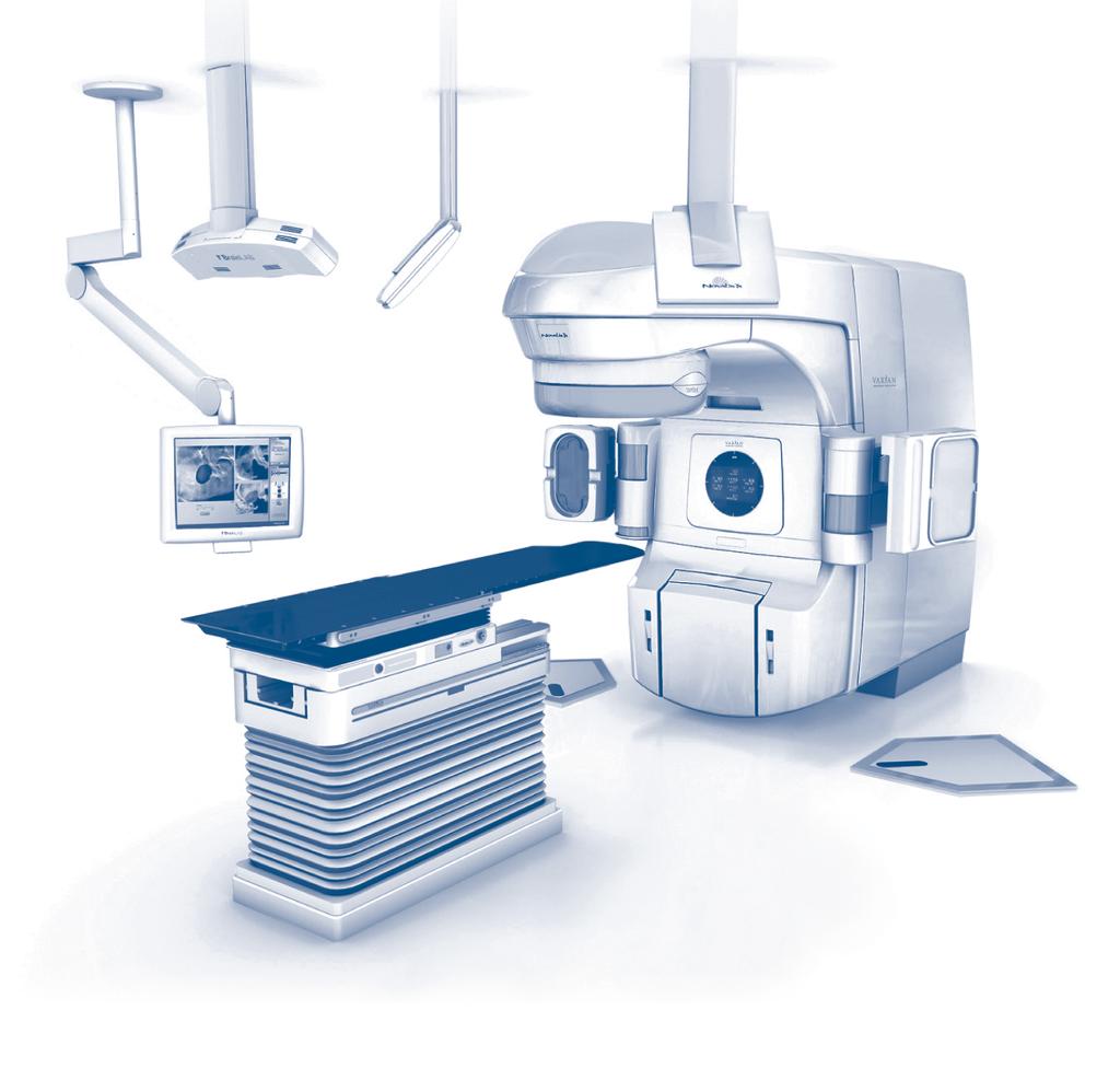
specifications Novalis Tx image-guided radiosurgery linear accelerator
|
|
|
- Peter Chandler
- 5 years ago
- Views:
Transcription
1 specifications Novalis Tx image-guided radiosurgery linear accelerator
2 Specifications Introduction This specification sheet provides information for the Novalis Tx image-guided radiosurgery linear accelerator. 1.0 Photon Beams 1.1 Energy: Three photon beams may be selected in accordance with the specifications and combinations listed in Table 1. Table 1: X-ray Beam Performance X-ray Beam Energy Combinations (MV) SRS Beam Beam I Beam II (BJR 17/BJR 11) / / / /20 Nominal Nominal %Depth Energy (MV) Energy (MV) D max Dose at BJR 17 BJR 11 (cm) 1 10 cm Depth 1 Flatness 2 Symmetry 3 SRS6 4 SRS ± ± 1.0 ±3.0% 2.0% ± ± 1.0 ±2.5% 2.0% ± ± 1.0 ±2.5% 2.0% ± ± 1.0 ±2.5% 2.0% ± ± 1.0 ±2.5% 2.0% ± ± 1.0 ±2.5% 2.0% 1 Depth of ionization applies to 10 x 10 cm 2 field size measured at 100 cm Target-Skin Distance (TSD). 2 Flatness is defined as the maximum variation from the mean dose delivered within the central 80% Full Width Half Maximum (FWHM) region measured at 100 cm TSD at a depth of 10 cm. The mean is the average of the maximum and minimum points within the central 80% FWHM region. The specification of ±2.5% applies to both the radial and transverse axes of all square field sizes from 20 x 20 cm 2 to 40 x 40 cm 2, inclusive. A specification of ±3.0% applies to all square field sizes between 10 x 10 cm 2 and 20 x 20 cm 2, and to 20 MV for all square field sizes larger than 30 x 30 cm 2. For the SRS 6MV beam, a specification of ±3.0% applies to all square field sizes between 10 x 10 cm 2 and 15 x 15 cm 2. 3 Symmetry is defined as the maximum difference between the X-ray dose delivered to any two points which are equidistant and symmetrical about the central axis and within the central 80% FWHM region measured at 100 cm TSD at a depth of 10 cm. This specification applies to the radial and transverse axes of all square field sizes from 10 x 10 cm 2 to 40 x 40 cm 2. For the SRS 6 MV beam, this specification applies to the radial and transverse axes of all square field sizes from 10 x 10 cm 2 to 15 x 15 cm 2. 4 Beam matching between 6 MV Beam I and the SRS 6 MV beam is provided and defined as follows: 4.1 The depth of D max along the central axis in a water phantom at 100 cm TSD is within ±1.5 mm of the average of the two beams. The relative dose at 10 cm depth on the central axis in a water phantom at 100 cm TSD is within ±0.5% of the average of the two beams. 4.2 The dose at any point within the central 80% of the field along the major axes, normalized to the central axis, is within ±1 percentage point of the average of the two beams. This specification applies to beams at 10 cm depth and field dimensions of 10 x 10 cm 2 and above. page 2
3 1.2 Dose Rate: For Beams I and II, the dose rate can be selected in fixed steps of 100 MU/min up to a maximum dose rate of 600 MU/min. For the SRS 6 MV Beam, the dose rate is 1,000 MU/minute. The SRS high dose rate supports efficient delivery of stereotactic radiosurgery, stereotactic radiotherapy, and intensitymodulated radiation therapy (IMRT). Refer to section 12.0 for further information. Photon Energy (BJR17) Photon Dose Rate (MU/min) 6-25 MV 100, 200, 300, 400, 500, 600 SRS 6 MV 1000 An MU is defined for these specifications as one cgy delivered to a tissue-equivalent material at D max and 100 cm SSD, with a 10 x 10 cm 2 field size. 1.3 Maximum Field Intensity at D max : The intensity at the depth of maximum buildup (D max ) does not exceed 109% of the central axis intensity anywhere in the measurement plane of any field size. 1.4 Leakage: The X-ray absorbed dose does not exceed 0.1% of the absorbed dose at the isocenter measured anywhere in the patient plane outside of the maximum useful beam. The neutron dose equivalent (Sievert) does not exceed 0.2% of the X-ray absorbed dose (Gray) at the isocenter. The patient plane is defined as a circular plane with a radius of 2 m, centered on and perpendicular to the axis of the beam at isocenter. The X-ray measurements may be averaged over an area not to exceed 100 cm 2. In all other directions, the X-ray absorbed dose at 1 m from the path of the electrons between the electron gun and the target or electron window does not exceed 0.1% of the absorbed dose at isocenter. 1.5 Collimator Transmission: The X-ray transmission of the upper and lower movable collimator does not exceed 0.5%. 1.6 Spot Size: The electron spot size is less than 3 mm in diameter at the X-ray target. 1.7 Penumbra: The distance between the 20% and 80% isodose lines for a 10 x 10 cm 2 field, measured at a depth of 10 cm with a 100 cm target-skin distance (TSD) along the major axes, measures less than or equal to 9 mm. 1.8 Field Size: The field size is continuously variable from 0.5 x 0.5 cm 2 to 40 x 40 cm 2 as measured at 100 cm TSD. Field sizes larger than 35 x 35 cm 2 are limited to a 49.5 cm diagonal (the diameter of the circle defined by the primary collimator at 100 cm TSD). The field size is defined as the distance along the radial and transverse axes between the points of 50% density on an X-ray film taken at 100 cm TSD with minimum buildup. The SRS 6 MV beam field size is limited to a maximum of 15 x 15 cm Upper and Lower Independent Collimators: Asymmetrical collimation is provided for upper and lower sets of collimators Independent, asymmetrical Upper Collimator travel range: 30 cm (-10 cm to +20 cm relative to central axis) Independent, asymmetrical Lower Collimator travel range: 22 cm (-2 cm to +20 cm relative to central axis) 2.0 Electron Beams 2.1 Novalis Tx offers a range of six (6) electron beams that can be selected in accordance with the specifications and combinations listed in Table 2. The specifications apply to a 15 x 15 cm 2 electron applicator and 100 cm TSD. 2.2 Dose Rate: Electron Dose Rate (MU/min) 100, 200, 300, 400, 500, 600, at 1.6 m (choose either 6 MeV or 9 MeV) A high electron dose rate is available at 6 MeV or 9 MeV electron energy. Refer to section 8.1 for further information. page 3
4 2.3 Field Sizes: A set of five electron applicators can be provided, with selection from 6 sizes: 6 x 6 cm 2, 6 x 10 cm 2, 10 x 10 cm 2, 15 x 15 cm 2, 20 x 20 cm 2, and 25 x 25 cm 2. Field sizes are defined at the isocenter plane, 5 cm from the final field-defining aperture. Hardware is provided to facilitate the fabrication of custom final field defining apertures. Table 2: Electron Beam Performance Electron Energy Groups Nominal Electron Energy (MeV) Group I 4, 6, 9, 12, 15, 18 Group II 6, 9, 12, 15, 18, 22 Group III 4, 6, 9, 12, 16, 20 Depth of Ionization 1 Depth of Dose Value Nominal 90% 80% 50% 30% 85%/2 80% Flatness 3 Symmetry 4 Energy (cm) (cm) (cm) (cm) (cm) 2 (cm) (MU/min) ±7% 2% ±4.5% 2% ±4.5% 2% ±4.5% 2% ±4.5% 2% ±4.5% 2% ±4.5% 2% ±4.5% 2% ±4.5% 2% 1 Depth of Ionization values apply to 15 x 15 cm 2 applicator field size. Electron measurements are made at 100 cm TSD and a nominal 5 cm gap between the bottom of the open field aperture and the water surface. Measurements are defined with a 0.1 cm 3 PTW ionization chamber, or equivalent. 2 D85%/2 is the depth at which flatness and symmetry are specified. Values are defined at 100 cm TSD using a 15 x 15 cm 2 electron applicator field size. No inverse square corrections are assumed. 3 Flatness is defined as the maximum variation from the mean electron ionization within the central 80% FWHM region. The mean is the average of the maximum and minimum points within the central 80% FWHM region. This specification applies to square electron applicator field sizes from 10 x 10 cm 2 to 25 x 25 cm 2 measured on the radial and transverse axes. A specification of ±5% is applied to 6 MeV for 10 x 10 cm 2 applicator field size. The diagonal flatness specification for the above applicator field sizes is ±5%, except 4 MeV. The 4 MeV flatness specification applies only to the radial and transverse axes. 4 Symmetry is defined as the maximum difference between the ionization delivered to any two points that are equidistant and symmetrical about the central axis and within the central 80% FWHM region. This specification applies to the plane normal to the central axis and to square electron applicator field sizes from 10 x 10 cm 2 to 25 x 25 cm 2, except 4 MeV. The 4 MeV specification applies only to the radial and transverse axes. page 4
5 2.4 X-ray Contamination: For nominal energies up to 10 MeV, the X-ray contamination is less than or equal to 2%. For nominal energies greater than 10 MeV, the X-ray contamination is less than or equal to 5%. This specification is defined in water with a 100 cm TSD, at a depth of 10 cm beyond the depth of the 10% isodose line, with a 15 x 15 cm 2 electron applicator. 2.5 Patient Plane Leakage: Electron leakage is less than or equal to 2% of the absorbed dose on central axis. This specification is defined in air, at 100 cm TSD with 1 cm buildup, in an area 4 cm outside the 50% isodose line. 2.6 Applicator Side Plane Leakage: The leakage does not exceed 9% of central axis ionization at D max. This specification is defined along a plane coincident to the side of the electron applicator, measuring 10 cm up from the bottom of an applicator. 3.0 Accelerator System Features 3.1 RF Power Source: Varian s high-efficiency klystron is operated in linear amplifier mode and driven by a solid-state oscillator, with power and frequency automatically locked to required operating levels. 3.2 Electron Gun: The unique triode design of the electron gun allows exact and safe control of electron beam levels in the accelerator. It provides the ability to rapidly and precisely vary output dose rate and turn the beam on or off. This capability is especially important in dynamic dose delivery, where high-speed beam gating and elimination of dark current during beam-off time periods is important. The gun is demountable, resulting in minimum system downtime during replacement. 3.3 Standing Wave Accelerator: The Varian sidecoupled cavity accelerator structure has been developed for optimum use of RF power and narrow output spectrum at the design energy for the guide. Spectrum characteristics, with and without use of an energy switch, have been matched to the transport requirements of the downstream bend magnet to ensure high dose rate capability. 3.4 Patented Non-Contacting Energy Switch: In each of the X-ray treatment modes where this is utilized, the switch functions to change the ratio of electric fields between two sections of the accelerator guide. This is done in such a way as to ensure a tight energy spectrum over a wide range of photon energies, with consequent high output capability and stable operation. 3.5 Solenoid: A full-length magnetic solenoid assures high electron beam transmission through the accelerator structure, resulting in reduced strayradiation and efficient use of RF power. 3.6 Bend Magnet: The patented 270 bend magnet is fully achromatic, with one-to-one imaging for superior transport and control of the beam from the accelerator. The magnet is also equipped with energy slits fixed at ±3%, enabling output beams of consistently high quality and precise dosimetry. 3.7 Radial and Transverse Steering Systems: These systems ensure basic beam alignment in all modes, as well as gantry orientation. Ion chamber sensors, in conjunction with the steering coils and servo electronics, maintain beam symmetry changes to within 2% under all foreseeable conditions. 4.0 Dosimetry System The following specifications apply for both independent dosimetry channels: 4.1 Reproducibility with Energy: Precision of the dosimetry measurement system for each energy is ±1% or ±1 MU, whichever is greater, at a fixed dose rate. 4.2 Dose Calibration Linearity versus Total Dose: The linearity is as follows: 1% for MU 2% for MU 3% for 5-10 MU 4.3 For photon Beams I and II, doses up to 999 MU per field can be delivered. For the SRS 6 MV Beam, doses up to 6,000 MU can be delivered. For all electron beams, doses up to 4,000 MU can be delivered. (Optional) 4.4 Reproducibility of Dose vs. Gantry Angle: The precision of the dosimetry system is ±1.5% at any gantry angle from 0 to 360 degrees. page 5
6 4.5 Reproducibility with Dose vs. Dose Rate: The dose rate dependence of the dosimetry system with variations in dose rate from minimum to maximum is less than ±1% or ±1 MU, whichever is greater. 4.6 Beam-Off Interlocks: The radiation beam automatically terminates in the event of any of the following: Monitor Units 1 complete Monitor Units 2 complete Treatment time complete Radial symmetry exceeds 2% Transverse symmetry exceeds 2% Excess dose rate Excess dose per pulse Excess dose per degree Loss of ion chamber bias voltage Under dose rate 5.0 Beam Matching Option Beam matching of a new Novalis Tx accelerator to existing high-energy Trilogy, Trilogy Tx, Clinac ix linear accelerators, and low- and high-energy Clinac EX accelerators that meet the serial number requirements shown below is available as a purchasable option. If purchased, this option includes on-site demonstration of the matched beams as described below. Fine Beam Matching to existing accelerator systems installed outside a 1-year time frame may be available as a purchasable option (refer to section 5.3). 5.1 Restrictions and Definitions All specifications apply to fields measured in water with the surface 100 cm from the target of the accelerator system D max is the depth at which the maximum dose occurs along the central axis of the beam for a 10 x 10 cm 2 X-ray field R85/2 is one-half the depth where 85% relative ionization occurs on the central axis of an electron field using the 15 x 15 cm 2 applicator Major axes lines orthogonal to the central axis of the beam and perpendicular to the sides of rectangular fields The term average is defined as the average value for the referenced performance specification, calculated using measurements obtained from the new Novalis Tx accelerator and the existing accelerator systems(s) to which it is matched. 5.2 On-Site Demonstration of Matched Beams Fine Photon Beam Matching, per beam Fine Matching of Photon X-Ray Beam Energy: For each X-ray beam of the same nominal energy, the depth of D max along the central axis in water phantom at 100 cm TSD is within ±1.5 mm of the average. For each X-ray beam of the same nominal energy, the relative dose at 10 cm depth on the central axis in a water phantom at 100 cm TSD (normalized to the dose at D max ) is within ±0.5% of the average Fine Matching of Photon X-Ray Beam Flatness: For X-ray beams of the same nominal energy, the maximum dose in the plane normal to the beam axis at a depth of D max in water at 100 cm TSD is within ±1% of the average. For each beam of the same nominal energy, the dose at any point within the central 80% of the in-plane and cross-plane axes, normalized to the central axis, measured at a depth of 10 cm in water at a TSD of 100 cm is within ±2% of the average. This specification applies to X-rays at 10 cm depth and field dimensions greater than 10 x 10 cm 2. page 6
7 5.2.2 Fine Electron Beam Matching, per beam Fine Matching of Electron Beam Energy: For each electron beam of the same nominal energy, the relative ionization values of 90%, 80%, and 50% at 100 cm TSD, are within ±1.0 mm of the average. This specification applies to the 15 x 15 cm 2 applicator Fine Matching of Electron Beam Flatness: For each beam of the same nominal energy, the ionization at any point within the central 80% of the in-plane and cross-plane scans, normalized to the central axis, measured at the depth of D max in water at a TSD of 100 cm is within ± 2% of the average of the measured values at that point. This specification applies to the 25 x 25 cm 2 and 10 x 10 cm 2 applicators. 5.3 Beam Matching to Accelerator Systems Installed Outside a 1-Year Time Frame Fine Beam Matching, including on-site demonstration, of a new Novalis Tx accelerator to existing accelerator systems installed outside a 1-year time frame is available as a purchasable option for accelerator systems that meet the following requirements: Clinac ix, all serial numbers Trilogy, Trilogy Tx, all serial numbers Low-energy Clinac linear accelerators, serial number 244 and higher Clinac 21 series, serial number 865 and higher Clinac 23 series, serial number 144 and higher Silhouette edition Clinac linear accelerators, all serial numbers Fine Beam Matching, including on-site demonstration, of a new Novalis Tx accelerator to existing accelerator systems that do not meet the serial number requirements above may be available as a purchasable option. 6.0 Mechanical Features 6.1 Gantry Rotation Range: ±185 from the vertical Target to Axis Distance: 100 ±0.2 cm Mechanical and radiation isocenter accuracy Novalis Tx not available with Retractable Beam Stopper Requires 52-inch Exact Couch base frame mm radius sphere for gantry and collimator axes mm radius sphere for gantry, collimator, and couch axes Position Indicators (gantry and console) IEC Scale convention (IEC 601 or 1217 compliant) or Varian Scale may be used for position readouts Digital Readouts: Accuracy: ±0.5 Resolution: Gantry Display Only: Enhanced Dynamic Wedge (EDW) beam modulation graphic indicator shows that EDW is enabled in either Y1 or Y2 direction Target to Surface Distance Indicators Optical Distance Indicator: Accuracy: ±0.1 cm at 100 cm, and ±0.5 cm at 70 cm and 156 cm Resolution: 0.5 cm Mechanical Front Pointer: Range: cm Accuracy: ±0.1 cm at 100 cm Resolution: 0.2 cm page 7
8 6.1.6 Isocenter Height (nominal): cm 6.2 Collimator Extended Rotation Range: ± Mechanical Isocenter Accuracy: 0.05 cm radius from isocenter Position Indicators (gantry and console) Digital Readouts: Accuracy: ±0.5 Resolution: Mechanical Scales: Accuracy: ±1.0 Resolution: Field Size Collimation Range: The field size is continuously variable from 0.5 x 0.5 cm 2 to 40 x 40 cm 2 as measured at 100 cm TSD. Field sizes larger than 35 x 35 cm 2 are limited to a 49.5 cm diagonal (the diameter of the circle defined by the primary collimator at 100 cm TSD). The field size is defined as the distance along the radial and transverse axes between the points of 50% density on an X-ray film taken at 100 cm TSD with minimum buildup. The SRS 6 MV beam field size is limited to a maximum of 15 x 15 cm Position Indicators Accuracy: ±0.2 cm Resolution: 0.1 cm Light and X-ray Field Coincidence: The field-defining light coincides to within 1.5 mm of the 50% isodensity line on an X-ray film. This is defined at 100 cm TSD with minimum buildup for any field size. 6.4 Exact Couch with Indexed Immobilization patient positioning The Exact Couch is standard with Novalis Tx. Specifications and standard versus optional accessories for the Exact Couch are provided on Exact Couch Specification, RAD Motion Controls Two Hand Pendants that control the accelerator and Exact Couch Two Couch Side Panels Remote Couch Control Position Indicators Corrective Motions: small translations (in x, y, and z) and small rotation of the couch to fine tune patient setups Planned Motions: large rotations of the couch to sequence between noncoplanar fields and arcs Novalis Tx Dimensions 1260 (126 cm) (49.6") 1070 (107 cm) (42") 552 (55.2 cm) (21.7") 3236 (323.6 cm) (127.4") 32 (3.2 cm) (1.25") 433 (43.3 cm) (17") 2584 (258.4 cm) (102") 1295 (129.5 cm) (51") 1832 (183.2 cm) (72") 2008 (200.8 cm) (79") Illustration not to scale page 8
9 Translation Accuracy: ±0.1 cm Resolution: 0.1 cm Rotation Accuracy: ±0.5 Resolution: BrainLAB ExacTrac 6D Robotic Couch and Imaging Couch Top (Optional) ExacTrac 6D Robotics module from BrainLAB provides correction of patient s longitudinal and lateral rotational misalignments. Max Rotational Angles: pitch ±4.0 if software limit enabled or up to ±6.0 if software limit not enabled; and roll ±2.7 Integrated electrical and mechanical safety system Battery-powered cordless design, including battery charger It requires the BrainLAB ExacTrac X-Ray 6D image-guidance system Imaging Couch Top: Low-density carbon fiber imaging couch top for greater artifact-free X-ray image quality Removable low-density carbon fiber head/neck frameless radiosurgery couch extension Removable connector for BrainLAB stereotactic head ring and mask system Three removable connectors fully compatible with the Civco tabletop fixation system Refer to BrainLAB ExacTrac Specification ETX-TS-E-Sys Oct 2007 for further information and detailed specifications. 6.6 Compact Stand Assembly Single access and through-door viewing of all gas and water system status indicators Imager electronics (PortalVision MV imaging system and On-Board Imager kv imaging system) incorporated in reduced height stand 7.0 Mechanical Accessories Options The following optional accessories are available with Novalis Tx and the Exact Couch: 7.1 Collimator Accessories: Interface Mount Accessory Mount Port Film Graticule 4-Way Wedge Set (four wedges 15, 30, 45, 60 ) Electron Applicators: A set of five electron applicators can be provided, with selection from 6 sizes: 6 x 6 cm 2, 6 x 10 cm 2, 10 x 10 cm 2, 15 x 15 cm 2, 20 x 20 cm 2, and 25 x 25 cm 2. Custom Aperture Fabrication Hardware Mechanical Front Pointer (holder and 4 rods) 7.2 Accessory Spare Parts Kit 8.0 Basic Static Procedures Mode Option 8.1 High Dose Total Skin Electron Mode: Novalis Tx is capable of delivering electron treatments at high dose rates for the purpose of total skin irradiation. The dose rate at 1.6 m is 888 MU/min, corresponding to nominally 2,500 MU at isocenter. This mode is available in 6 MeV or 9 MeV X-ray contamination at calibration point is <1% Symmetry at isocenter is ±2% Integrated dose monitor: 1 to 9,000 MU Exposure time: 0.1 to 99.9 min. 8.2 Total Body Electron (TBE) Mode: Delivers 9,000 MU at isocenter with all normal machine safety and dosimetry interlocks operational, and delivers standard energies at standard dose rate ranges Special TBE accessory tray is provided All beams are calibrated at machine isocenter Integrated dose: 1 to 9,000 MU Exposure time: 0.1 to 99.9 min. page 9
10 8.3 Total Body Irradiation (TBI) Photon X-ray Mode: Delivers 9,000 MU at isocenter with all normal machine safety and dosimetry interlocks operational, and delivers standard energies at standard dose rate ranges Special TBI accessory tray is provided All beams are calibrated at machine isocenter Integrated dose: 1 to 9,000 MU Exposure time: 0.1 to 99.9 min. 9.0 Advanced Static Procedures Mode Auto Field Sequencing (AFS), for use with the 4D Integrated Treatment Console (refer to 4D Integrated Treatment Console Product Brief, RAD 2768 for information and specifications), provides automated delivery of multiple coplanar and non-coplanar fields. With this time saving feature, the accelerator automatically acquires the mode up signal and machine setup information from the 4D Integrated Treatment Console, and then allows the operator to remotely move the gantry, jaws, collimator, and Exact Couch axes between coplanar and noncoplanar treatment fields. This feature eliminates the need to go into the treatment room to alter the machine setup between treatment fields. AFS works in concert with the HD120 multileaf collimator (MLC) to deliver both static and dynamic plans efficiently and smoothly. (Refer to Auto Field Sequencing Specification, RAD 6045.) 10.0 Basic Dynamic Procedures Modes 10.1 Photon Arc Mode and Optional Electron Arc Mode: Novalis Tx is capable of delivering the following dose over a preset gantry rotation of up to 360 degrees or any fraction thereof. MU per degree (MU/DG) is automatically computed based on the preset total dose and the preset arc segment. Photon Beams I and II 0.30 MU to 20 MU per degree SRS 6 MV Beam 0.30 MU to 60 MU per degree All electron beams (optional) 0.30 MU to 20 MU per degree Precision: During Arc treatment, the position of the gantry deviates no more than 0.5 degrees from the desired instantaneous gantry angle, and the dose deviates no more than 0.20 MU from the desired instantaneous total dose, as specified by the user-preset total dose and arc segment. If these tolerances are exceeded, the dose delivery is suspended and the gantry position is targeted to the position dictated by the actual dose delivered. When the gantry is again within 0.5 degrees of the desired position, the treatment will resume. The Dose Position Interlock (DPSN) is asserted if the gantry is not positioned within 0.5 cm of the desired position within 3 seconds. The DPSN will terminate the beam immediately if the position deviates 3.0 degrees or more from the desired position, or the dose delivered exceeds 0.45 MU for dose rates less than 600 MU/min (0.54 MU for dose rate 600 MU/min and 0.90 MU for dose rates greater than 600 MU/min, applies to version 7.8 and above) Arc Dose Rate: The dose rate during a dynamic arc treatment is automatically modulated between zero and the ceiling dose rate selected in Physics Mode Arc Direction: Novalis Tx may be programmed to perform arc therapy in either a clockwise or counterclockwise direction Enhanced Dynamic Wedge (EDW) Mode: EDW can be used with either Beam I or Beam II. EDW utilizes Y-jaws to create wedge shaped dose distributions. Enhanced Dynamic Wedges of 10, 15, 20, 25, 30, 45, and 60 degrees are included, with up to 30 cm (wedge direction) by 40 cm field sizes. (Refer to Enhanced Dynamic Wedge Specification, RAD 1880.) 11.0 Advanced Dynamic Techniques Intensity-modulated radiation therapy (IMRT) and conformal arc therapy are advanced dynamic techniques in which the leaves of the HD120 MLC move during treatment. Refer to HD120 MLC Specification, RAD 9998 for additional information and specifications. page 10
11 11.1 Arc Dynamic MLC allows delivery of MLC fields as a function of gantry arc angle, also known as conformal arc therapy. An MLC shape change every 2 is supported Dose Dynamic MLC allows delivery of MLC fields as a function of percent dose delivered, also known as IMRT. Both dynamic IMRT (i.e., sliding window) and segmental IMRT (i.e., step-and-shoot) techniques are supported. Combinations of the two IMRT techniques also are supported. In addition, Dose Dynamic MLC enables treatment delivery with electronic compensation, in which MLC leaf motion simulates the dosimetric effect of a physical compensator Stereotactic Mode Novalis Tx is capable of delivering stereotactic treatments at high dose rates and with remote couch motion. This mode is available with 6 MV photons. Both cone- and MLC-based treatment delivery are supported. Beam flatness, symmetry, and other specifications can be found in Table Dose Rate: 1,000 MU per minute at D max at 100 cm TSD 12.2 Maximum Dose per Field: 6,000 MU 12.3 Maximum Field Size: 15 x 15 cm Maximum Dose per Degree for Arc Treatments: 60 MU per degree 12.5 Stereotactic Motion Disable Electrical disable for gantry and couch 13.0 Stereotactic Components 13.1 BrainLAB Stereotactic Components Immobilization frame (SRS headring), frameless stereotactic mask system, and accessories Refer to BrainLAB for further information and detailed specifications Conical collimators: Standard Set: Sizes 4.0, 6.0, 7.5, 10, 12.5, and 15 mm Conical aperture to address beam diversion Bayonet mount for quick and safe attachment to collimator mount Quick mounting through use of the accessory tray slot Locks to secure the mount in a fixed position Adjustment of the central axis of collimator Primary anti-scatter collimator of brass Fast mounting of collimators with no additional tools Collimator mount Refer to BrainLAB for further information and detailed specifications iplan stereotactic treatment planning system Refer to BrainLAB for further information and detailed specifications Varian Optical Guidance Components (Optional) Optical Guidance Platform (OGP) with FramelessArray optically guided cranial SRS positioning (Requires the Eclipse treatment planning system. Not available if BrainLAB ExacTrac X-Ray 6D system is present.) 14.0 HD120 MLC High Definition Multileaf Collimator The Varian HD120 MLC is a 120-leaf high-definition collimator that offers 2.5 mm resolution in its central region. Each side of the Varian HD120 MLC is configured with 60 leaves distributed in a 8 cm wide central region with 32 x 2.5 mm leaves, flanked by two 7 cm wide side regions with 14 x 5.0 mm leaves, for a total width of 22 cm. The HD120 MLC operates in static, dynamic, and conformal arc modes. The static mode provides efficient beam shaping for 3D conformal radiation therapy. The dynamic mode enables IMRS with both step-and-shoot and sliding window delivery. In addition, a large field IMRS mode allows treatment delivery in a sequence of carriage positions in a 22 cm x 32 cm field. The conformal arc mode enables conformal arc therapy in which the leaves always conform to the outer boundary of the target as the gantry rotates around the patient. Refer to HD120 MLC Specification, RAD page 11
12 15.0 On-Board Imager Patient Positioning and Target Localization System (Optional) The On-Board Imager provides high-quality kv images in the treatment room for target localization, patient positioning, and motion management. The following clinical capabilities are supported: Acquisition of cone-beam CT scans Online setup correction based on either a kv-kv or kv-mv pair of radiographs Automated and manual alignment of a pair of radiographs to their reference images Acquisition of gated radiographs Online setup correction based on radiopaque markers Pretreatment verification of gated treatment portals using kv fluoroscopy Robotic arms to position and hold the kv source and kv digital detection Remote couch motion to correct patient setups Refer to On-Board Imager Specification, RAD 9502, for information and specifications BrainLAB ExacTrac Patient Positioning and Tracking System (Optional) 16.1 ExacTrac Infrared Optical Tracking System ExacTrac Infrared provides real-time optical tracking of the patient s position and motion / external breathing patterns for initial set-up and during treatment delivery. Interactive control of Robotic Couch Two stereoscopic infrared cameras for patient tracking in three translational and three rotational dimensions In-room software interface via ceiling mounted 17" touch-screen monitor 16.2 ExacTrac X-Ray 6D System The ExacTrac X-Ray 6D utilizes stereoscopic highresolution kv imaging in the treatment room. Two linac-independent kv X-ray units recessed into the treatment room floor and two ceiling-mounted amorphous silicon flat panel detectors are combined with the integrated infrared optical ExacTrac tracking system to monitor and x-ray the patient s position throughout treatment delivery. 6D robotic couch and imaging couch top is included with the Exac Trac X-ray 6D system. The following clinical capabilities are supported: Target localization, patient positioning, and motion management utilizing bony anatomy or implanted fiducials Cranial and extracranial treatment sites Automatic image fusion and setup error determination Frameless radiosurgery setup in x,y,z and all three rotational dimensions (in combination with ExacTrac 6D robotic couch) Snap-Verification: Intra-fraction imaging for detection of PTV misalignments and movements at any point in time during the treatment (beam on/off; any gantry rotation, any couch rotation) Remote access and approval via hospital LAN / WLAN Basis for BrainLAB Adaptive Gating system Refer to BrainLAB ExacTrac Specification ETX-TS-E- Sys 2007 for further information and detailed specifications PortalVision as1000 MV Imaging System The PortalVision as1000 is an MV imaging system that allows for verification of patient setups, treatment portals, and pre-treatment QA. The amorphous silicon detector has an active imaging area of 40 cm x 30 cm with a pixel resolution of 1024 x 768. Image acquisition is supported before, during, and after treatment. Match and Review software is included for image analysis. A motorized, retractable robotic arm is used to position and hold the detector. Refer to PortalVision as1000 Specification, RAD 2553, for information and specifications Portal Dosimetry (Optional) Portal Dosimetry enables use of the PortalVision imager to record the intensity patterns of IMRT fields for pretreatment quality assurance of IMRT planning and delivery. page 12
13 Portal Dosimetry includes integrated image acquisition mode for recording of IMRT fields and image viewing and analysis software. (Use of the image analysis software is optimized when the reference dose image is calculated as dose to amorphous silicon. Currently, only the Eclipse integrated treatment planning system offers this capability.) Refer to PortalVision as1000 Specification, RAD 2553, for additional information and specifications Real-time Position Management (RPM) System (Optional) The RPM respiratory gating system enables passive, real-time monitoring of patient motion for the purpose of intrafraction motion management. Two gating systems are provided. Each system includes an optical tracking camera, external marker block, and RPM respiratory gating system workstation. The RPM respiratory gating system supports gated treatment delivery and image acquisition on the accelerator, gated simulation on compatible simulators (not all simulators are supported), and gated CT acquisition on compatible third-party CT scanners (not all CT scanners are compatible). Depending on the capabilities of the CT scanner, the RPM respiratory gating system supports both retrospective and prospective gating of CT scans. Refer to RPM Respiratory Gating System Specification, RAD 5616 for additional information D Adaptive Gating Module (Optional) The 6D Adaptive Gating Module is based on the ExacTrac X-Ray 6D platform and allows stereoscopic high-resolution imaging of moving targets in real-time throughout treatment delivery. X-ray image acquisition is automatically triggered from the patient s breathing pattern detected by the ExacTrac infrared tracking system. Advanced ExacTrac gating software enables detection of motion pattern changes and automatic adaptation of the patient setup. Refer to BrainLAB for further information and detailed specifications Argus QA Software (Optional) Argus provides powerful software tools for automation of quality assurance data acquisition, data analysis, visual display of data, and reporting. Argus also provides a centralized database for digital storage of data. The Argus QA software provides quality assurance modules for linear accelerators, including static and dynamic MLC using DynaLog file analysis, CT simulators, standard simulators, film processors, and HDR brachytherapy systems Factory Beam Data Set The Factory Beam Data Set is provided in hard copy and ASCII file formats. The data include machine mechanical parameters and representative beam data. The data set is not a substitute for the commissioning process but an aid to speed that process as well as data entry to treatment planning systems. The factory data are representative of the Novalis Tx accelerator manufacturing standard, not the specific machine delivered SmartConnect Remote Access Technology SmartConnect remote access technology connects the Novalis Tx accelerator with Varian Customer Support for expert assistance and online remote analysis. Diagnostic and Morning Checkout Logs can be viewed remotely and transferred to Varian for report generation and trend analysis Treatment Command Center 24.1 The 4D Integrated Treatment Console provides a streamlined front end to the Novalis Tx accelerator. The console integrates use of the accelerator, HD120 MLC, and PortalVision MV imaging system into one application on a single workstation. For image-guided radiosurgery and radiotherapy using kv images, the console is used in combination with the On-Board Imager workstation. The 4D Integrated Treatment Console uses a DICOM RT interface to communicate with the ARIA oncology information system and other information system databases Ergonomic command center configuration places all control modules, monitors, and user interaction devices within easy reach of the operator. Direct access application selection simplifies the workspace by reducing the number of input devices (e.g., keyboard and mouse), while allowing continuous viewing of all applications. page 13
14 25.0 In-Room Display A high-resolution, flat screen, color display monitor is included for in-room display of accelerator parameters and patient-specific information LaserGuard Collision Detection System LaserGuard monitors the MLC collimator face with a plane of infrared light that emanates from a device located within the gantry. Any object that intrudes into this area, called the protection zone, triggers an emergency stop of all accelerator motion. Refer to Auto Field Sequencing with LaserGuard Specification, RAD 6046 for information and specifications Typical Facility Requirements 27.1 Electrical Requirements Typical 60Hz: VAC, line-toline, 3-phase, 4-wire plus ground, 45 KVA load Typical 50Hz: VAC, line-toline, 3-phase, 4-wire plus ground, 45 KVA load Cooling Water Requirements: The cooling water requirements can be satisfied with a one-pass system (domestic supply and waste return) or a closed loop system Ventilation must be sufficient to remove 8 kw from treatment room and 1 kw from control console Compressed Air Requirements: Instrument quality air is required On-Board Imager Power Requirements Input voltage: 400 to 480 Vac (±10%), 3-phase, 4-wire plus ground Input frequency: 50 or 60 Hz (±1%) For comprehensive facilities requirements refer to the On-Board Imager Installation Data Package. For detailed facilities requirements refer to the Novalis Tx Installation Data Package ExacTrac X-Ray 6D Requirements Refer to BrainLAB for further information and detailed facilities requirements. Additional Options Silhouette Edition Novalis Tx is available in a Silhouette edition that fits into an existing vault with a minimum room size of 16 feet (4.9 m) width by 19 feet (5.8 m) length. Treatment Console Area Packaging Compact packaging and cable management of Varianprovided workstations, control modules, and other ancillary devices for easy site preparation and enhanced treatment console area space management. A variety of packaging configurations are available for optimal utilization of the available space. RapidArc Radiotherapy Technology Treatment Delivery RapidArc generates IMRT quality dose distributions in a single optimized arc providing optimized treatment conformity. Dose per degree can be varied per degree of gantry motion (Refer to MLC Dynamic Control Specification, RAD 5610). Laser Alignment System Wall and ceiling lasers CCTV Camera System This two-camera CCTV system is used for monitoring patient activity inside of the treatment room and patient activity from outside the room at the treatment console. Patient Intercom System The Patient Intercom System is used for audio communication with the patient in the treatment room from the treatment console area. Collimator Accessories Electron Arc Applicators and Mold Frames Additional Block Tray sets - Solid or Slotted cm or 1 cm thickness Compensator Mount Upper and Lower Compensator Trays Extended Spare Parts Kit Specifications subject to change without notice. page 14
15
16 Surgical Sciences 3100 Hansen Way Palo Alto, CA tel tel USA Headquarters California Palo Alto, CA Tel: Fax: USA Regional Offices California Corona, CA Tel: Fax: Georgia Marietta, GA Tel: Fax: Illinois Des Plaines, IL Tel: Fax: New Jersey Clark, NJ Tel: Fax: European Headquarters Switzerland International AG Zug, Switzerland Tel: Fax: Austria Gesellschaft m.b.h. Voesendorf, Austria Tel: Fax: Belgium Belgium N.V./S.A. Diegem, Belgium Tel: Fax: Finland Finland Oy Helsinki, Finland Tel: Fax: France France Buc, France Tel: Fax: Germany Deutschland GmbH Darmstadt, Germany Tel: Fax: India India Pvt Ltd. Mumbai, India Tel: Fax: India Pvt Ltd. Chennai, India Tel: Fax: Italy Italia, S.p.A. Cernusco s/n (MI), Italy Tel: Fax: Netherlands Nederland B.V. Houten, Netherlands Tel: Fax: Scandinavia Scandinavia AS Herlev, Denmark Tel: Fax: Spain/Portugal Ibérica, S.L. Madrid, Spain Tel: Fax: UK/Ireland UK Ltd. Crawley, West Sussex, UK Tel: Fax: Asian Headquarters Hong Kong Pacific, Inc. Kowloon, Hong Kong Tel: Fax: China China Ltd. Beijing, P.R. China Tel: Fax: Japan K.K. Chuo-ku, Tokyo, Japan Tel: Fax: Latin American Headquarters Florida Miami, FL USA Tel: Fax: Brazil do Brasil Ltda. São Paulo, Brazil Tel: Fax: Australian Headquarters Australia Australasia Pty Ltd. Sydney, Australia Tel: Fax: Varian,, Clinac, Exact, Indexed Immobilization, On-Board Imager, Silhouette, SmartConnect, and Trilogy are registered trademarks, and Argus, ARIA, DynaLog, Eclipse, Enhanced Dynamic Wedge, FramelessArray, HD120, LaserGuard, Novalis Tx, PortalVision, RapidArc, Real-time Position Management, and Trilogy Tx are trademarks of, Inc. All other trademarks are the property of their respective owners. RAD 10011B 2008, Inc. Printed in USA 11/08
specifications TrueBeam STx System
 specifications TrueBeam STx System TrueBeam STx s The TrueBeam STx system specifications in this document are identified as belonging to two categories, performance specifications and descriptive specifications.
Table 1: Available X-ray Beam Energy Combinations (MV) 6 10/ Yes Yes 6 16/15 6 Yes No 6 23/18 6 Yes No 6 25/20 6 Yes No
 6 10/ Yes Yes 6 16/15 6 Yes No 6 23/18 6 Yes No 6 25/20 6 Yes No") SPECIFICATIONS Introduction This specification sheet provides information for the Trilogy linear accelerators. 1.0 Photon Beams 1.1 Energy: Three photon beams may be selected in accordance with the beam
SPECIFICATIONS Introduction This specification sheet provides information for the Trilogy linear accelerators. 1.0 Photon Beams 1.1 Energy: Three photon beams may be selected in accordance with the beam
specifications The Complete Radiation Oncology Solution
 specifications The Complete Radiation Oncology Solution Specifications This specification sheet provides information for the UNIQUE radiation oncology solution and in particular the UNIQUE linear accelerator.
Welcome to a radically different approach to cancer treatment.
 Welcome to a radically different approach to cancer treatment. Yesterday s boundaries are today s starting points. Introducing the TrueBeam system for radiotherapy and the TrueBeam STx system for radiosurgery.
specifications TrueBeam System
 specifications TrueBeam System TrueBeam System s The TrueBeam system specifications in this document are identified as belonging to two categories, performance specifications and descriptive specifications.
UNIQUE. The Complete Radiation Oncology Solution SPECIFICATIONS
 UNIQUE The Complete Radiation Oncology Solution SPECIFICATIONS UNIQUE SPECIFICATIONS I 2 UNIQUE SPECIFICATIONS I 3 Specifications This specification sheet provides information for the UNIQUE radiation
CHAPTER 2 COMMISSIONING OF KILO-VOLTAGE CONE BEAM COMPUTED TOMOGRAPHY FOR IMAGE-GUIDED RADIOTHERAPY
 14 CHAPTER 2 COMMISSIONING OF KILO-VOLTAGE CONE BEAM COMPUTED TOMOGRAPHY FOR IMAGE-GUIDED RADIOTHERAPY 2.1 INTRODUCTION kv-cbct integrated with linear accelerators as a tool for IGRT, was developed to
8/3/2017. Use of EPIDs for Non-Routine Linac QA. Disclosure. Learning Objectives. Parts of this project received support from Varian Medical System.
 Use of EPIDs for Non-Routine Linac QA Bin Cai PhD Disclosure Parts of this project received support from Varian Medical System. Learning Objectives Learn the recent development of EPID based Non-routine
A positioning QA procedure for 2D/2D (kv/mv) and 3D/3D (CT/CBCT) image matching for radiotherapy patient setup
 and 3D/3D (CT/CBCT) image matching for radiotherapy patient setup") JOURNAL OF APPLIED CLINICAL MEDICAL PHYSICS, VOLUME 10, NUMBER 4, FALL 2009 A positioning QA procedure for 2D/2D (kv/mv) and 3D/3D (CT/CBCT) image matching for radiotherapy patient setup Huaiqun Guan,
JOURNAL OF APPLIED CLINICAL MEDICAL PHYSICS, VOLUME 10, NUMBER 4, FALL 2009 A positioning QA procedure for 2D/2D (kv/mv) and 3D/3D (CT/CBCT) image matching for radiotherapy patient setup Huaiqun Guan,
Development of the Use of Amorphous Silicon (ASi) Electronic Portal Imaging Devices as a Physics Tool for Routine Linear Accelerator QA
 Electronic Portal Imaging Devices as a Physics Tool for Routine Linear Accelerator QA") Development of the Use of Amorphous Silicon (ASi) Electronic Portal Imaging Devices as a Physics Tool for Routine Linear Accelerator QA Gena M.A.H 1, Ahmed L.El-Attar 2, Elbadry M. Zahran 3, Hany El-Gamal
Development of the Use of Amorphous Silicon (ASi) Electronic Portal Imaging Devices as a Physics Tool for Routine Linear Accelerator QA Gena M.A.H 1, Ahmed L.El-Attar 2, Elbadry M. Zahran 3, Hany El-Gamal
The Current State of EPID-Based Linear Accelerator Quality Assurance. Disclosures. Purpose of this First Talk 8/3/2017
 The Current State of EPID-Based Linear Accelerator Quality Assurance Timothy Ritter, PhD, DABR, FAAPM 1 Disclosures Employed by the Veterans Health Administration Faculty appointment with the University
PHYSICS QUESTIONNAIRE FORM
 PHYSICS QUESTIONNAIRE FORM Institution Name: Date: Contact Information (name, address, phone, fax, email): Physicist: Radiation Oncologist: Dosimetrist (if applicable): Study Coordinator (if applicable):
IMRT Delivery System QA. IMRT Dose Delivery. Acceptance testing. Why: specific tests for IMRT? Accuracy of leaf positioning (gaps) MLC Alignment
 MLC Alignment") 1 IMRT Delivery System Q Thomas LoSasso, PhD Memorial Sloan Kettering Cancer Center IMRT Dose Delivery cceptance testing Commissioning Quality assurance Verification Q Why: specific tests for IMRT? 2.
1 IMRT Delivery System Q Thomas LoSasso, PhD Memorial Sloan Kettering Cancer Center IMRT Dose Delivery cceptance testing Commissioning Quality assurance Verification Q Why: specific tests for IMRT? 2.
ISPFILMQATM STATE-OF-THE-ART RADIOTHERAPY VERIFICATION SOFTWARE. Supports all major radiotherapy technologies! FilmQA TM
 FILMQA STATE-OF-THE-ART RADIOTHERAPY VERIFICATION SOFTWARE Supports all major radiotherapy technologies! FilmQA is optimized for use with Gafchromic film products, including EBT2 and RTQA2. FILMQA helps
Monte Carlo study on a new concept of a scanning photon beam system for IMRT
 NUKLEONIKA 2011;56(4):291 297 ORIGINAL PAPER Monte Carlo study on a new concept of a scanning photon beam system for IMRT Anna M. Wysocka-Rabin, Günter H. Hartmann Abstract. Intensity-modulated radiation
Initial setup and subsequent temporal position monitoring using implanted RF transponders
 Initial setup and subsequent temporal position monitoring using implanted RF transponders James Balter, Ph.D. University of Michigan Has financial interest in Calypso Medical Technologies Acknowledgements
SUBCHAPTER 14. THERAPEUTIC INSTALLATIONS
 SUBCHAPTER 14. THERAPEUTIC INSTALLATIONS 7:28-14.1 Scope (a) This subchapter covers therapeutic installations used in the healing arts. These therapeutic installations include x-ray, accelerator and teletherapy
3D Diode Array Commissioning: Building Confidence in 3D QA Technology
 3D Diode Array Commissioning: Building Confidence in 3D QA Technology Caroline Yount, MS CANCER CENTER 3D QA The complex three-dimensional (3D) shapes of intensity modulated radiation therapy (IMRT) dose
MXHF-1500RF is controlled by Digital key panel console that displays KV, ma and mas with APR menu programmed.
 R/F TV X-RAY SYSTEM DIAGNOSTIC RADIOGRAPHIC FLUOROSCOPIC TV SYSTEM MXHF-1500RF SYSTEM OUTLINE Product Data No. 041021-01 MXHF-1500RF is controlled by Digital key panel console that displays KV, ma and
ArcCHECK. The Ultimate 4D QA Solution. Your Most Valuable QA and Dosimetry Tools
 ArcCHECK The Ultimate 4D QA Solution A 4D isotropical cylindrical detector array for arc delivery QA and Dosimetry U.S.Patent No. 8,044,359; 6,125,335 Compatible with: FFF Beams VMAT RapidArc TomoTherapy
COMPREHENSIVE TG-142 IMAGING AND MACHINE QA
 QA SOFTWARE COMPREHENSIVE TG-142 IMAGING AND MACHINE QA Automate the analysis of over thirty TG-142 recommended QA tasks The rapid progress of Radiation Therapy has created the need for Quality Assurance
GafChromic QuiCk Phantom with EBT3P/3+P Film and FilmQA Pro for Radiation Therapy Dosimetry Applications
 GafChromic QuiCk Phantom with EBT3P/3+P Film and FilmQA Pro for Radiation Therapy Dosimetry Applications I. SCOPE The protocol applies to GafChromic EBT3P and EBT3+P films exposed in GafChromic QuiCk Phantom
QUALITY CONTROL PHANTOMS FOR RADIOTHERAPY AND MEDICAL IMAGING
 1 QUALITY CONTROL PHANTOMS FOR RADIOTHERAPY AND MEDICAL IMAGING QualiFormeD Phantoms A selection of test objects facilitating regulatory quality controls in radiation therapy and medical imaging Practical,
TOMOTHERAPY H SERIES TomoH, TomoHD and TomoHDA Systems Technical Specifications
 TOMOTHERAPY H SERIES TomoH, TomoHD and TomoHDA Systems Technical Specifications The TomoTherapy H Series is designed to treat the entire spectrum of radiation therapy patients with enhanced speed, precise
Philips XPER FD10C R7.0.4
 Philips XPER FD10C R7.0.4 Reconditioned 2005 System- Upgraded to R7 in Oct 2010 The Allura Xper FD10 (Ceiling) single-plane cardiovascular system is comprised of a ceiling mounted C-arm stand and digital
TOMOHD. Product Specifications
 TOMOHD Treatment System Product Specifications 1. Complete Treatment System Dimensions Height Width Length Weight 8 ft., 3.25 in. (252.1 cm) 9 ft., 2.50 in. (280.6 cm) 15 ft., 2.25 in. (463.0 cm) 10,000
Isocenter and Field of View Accuracy Measurement Software for Linear Accelerator
 Isocenter and Field of View Accuracy Measurement Software for Linear Accelerator Aleksei E. Zhdanov 1 and Leonid G. Dorosinskiy 1 Ural Federal University named after the first President of Russia B. N.
FMT18 FLOOR MOUNTED SYSTEM
 mas Time AEC 320 kvp 64 mas 320 ma 320 ma 320 DEN 0.0 mm Cu 17 in X 17 in 72.0 in FMT18 FLOOR MOUNTED SYSTEM with Synchronized Tracking System Overview Clinical Efficiency The FMT18 System was designed
Physical and dosimetric aspects of a multileaf collimation system used in the dynamic mode for implementing intensity modulated radiotherapy
 Physical and dosimetric aspects of a multileaf collimation system used in the dynamic mode for implementing intensity modulated radiotherapy Thomas LoSasso, a) Chen-Shou Chui, and C. Clifton Ling Department
IQM Detector Characteristics: Signal reproducibility
 The Integral Quality Monitor (IQM) System is a real-time beam verification system that monitors the accuracy of radiation delivery throughout each patient treatment without any user interaction. IQM continuously
Evaluation of dosimetry parameters of photons and electron beams using a linear ionization chamber array
 Evaluation of dosimetry parameters of photons and electron beams using a linear ionization chamber array José A. Bencomo, * Geoffrey Ibbott, Seungsoo Lee, and Joao A. Borges Department of Radiation Physics.
A diagnostic tool for basic daily quality assurance of a tomotherapy Hi Art machine
 JOURNAL OF APPLIED CLINICAL MEDICAL PHYSICS, VOLUME 10, NUMBER 4, FALL 2009 A diagnostic tool for basic daily quality assurance of a tomotherapy Hi Art machine Iwein Van de Vondel, 1 Koen Tournel, 1 Dirk
OTC18 OVERHEAD TUBE CRANE SYSTEM
 OTC18 OVERHEAD TUBE CRANE SYSTEM System Overview Clinical Performance Versatile and intuitive, the OTC18M System delivers enhanced patient comfort and optimized workflow. Precisely designed to withstand
X3D in Radiation Therapy Procedure Planning. Felix G. Hamza-Lup, Ph.D. Computer Science Armstrong Atlantic State University Savannah, Georgia USA
 X3D in Radiation Therapy Procedure Planning Felix G. Hamza-Lup, Ph.D. Computer Science Armstrong Atlantic State University Savannah, Georgia USA Outline 1. What is radiation therapy? 2. Treatment planning
Commissioning and Calibrating a Linear Accelerator State-of-the-Art in 2010
 Commissioning and Calibrating a Linear Accelerator State-of-the-Art in 2010 Indra J. Das, PhD, FACR Department of Radiation Oncology Indiana University of School of Medicine & Midwest Proton Radiation
Emerging Technology: Real-Time Monitoring of Treatment Delivery EPID Exit Dose QA
 Emerging Technology: Real-Time Monitoring of Treatment Delivery EPID Exit Dose QA Arthur Olch, PhD, FAAPM AAPM Spring Clinical Meeting, March 21, 2017 Or.. What Dose are the Patients Really Getting???
Beam Production, Characteristics and Shaping
 Beam Production, Characteristics and Shaping Dr. Manfred Sassowsky Outline X-ray production 60 Co units Linear Accelerators Beam characteristics Beam shaping Beam Production, Characteristics and Shaping
Installation und Kommissionierung des Viewray MRIdian Linac Hamburg, 28. Mai 2018 Sebastian Klüter
 Installation und Kommissionierung des Viewray MRIdian Linac Hamburg, 28. Mai 2018 Sebastian Klüter MR-guided RT in Heidelberg Funded by the German Research Foundation (DFG) Heidelberg consortium received
Image Quality. HTC Grid High Transmission Cellular Grid provides higher contrast images
 B R E A S T I M A G I N G S O L U T I O N S Setting the benchmark for mammography M-IV Series Innovations in breast imaging The Lorad M-IV Series exemplifies Hologic's commitment to developing advanced
C506-E064. Full digital system. Printed in Japan A-NS
 C506-E064 Full digital system Printed in Japan 6295-08807-30A-NS Full digital system Highest Image Quality in Its Class Comprehensive Full-Digital System FLEXAVISION is a full-digital R/F system equipped
LINEAR ACCELERATOR. Buyer's Guide. Version 1.1
 PRE-OWNED LINEAR ACCELERATOR Buyer's Guide Version 1.1 Pre-Owned Linear Accelerator Buyer's Guide TABLE OF CONTENTS Considerations For Buying A Used Linear Accelerator... 3 Linear Accelerators Overview...
Maximizing clinical outcomes
 Maximizing clinical outcomes Digital Tomosynthesis Dual Energy Subtraction Automated Long Length Imaging Improved image quality at a low dose Xray Xray Patented ISS capture technology promotes high sensitivity
Medical Imaging. Latest generation bucky room. system. radiology ahead
 Medical Imaging Latest generation bucky room system radiology ahead system it grows efficiency, the productivity is on the up Moviplan ic System is the latest-generation X-ray equipment, combining all
ISO Cube Daily QA Package
 ISO Cube Daily QA Package Model 023-05 AFFORDABLE TURNKEY SOLUTION FOR DAILY MACHINE QA POWERED BY AQUILAB 2428 Almeda Avenue Suite 316 Norfolk, Virginia 23513 USA Tel: 757-855-2765 WWW.CIRSINC.COM CAPABILITIES
GAFCHROMIC. Therapy Dosimetry Media Models to
 GAFCHROMIC Therapy Dosimetry Media Models 37-040 to 37-045! Superior uniformity and sensitivity! Dose rate and fractionation independent! Maps dose distribution! Provides quantitative measurements (via
Truly flexible to meet your clinical needs
 Truly flexible to meet your clinical needs 2 Adapting to meet your needs Flexible Fast and responsive Excellent image quality Designed with ergonomic efficiency Equipped with dose management tools 3 Three
- Water resistant. - Large size.
 GAFCHROMIC EBT product brochure GAFCHROMIC EBT FEATURES GAFCHROMIC EBT dosimetry film has been developed specifically to address the needs of the medical physicist and dosimetrist working in the radiotherapy
The evaluation of minimum detectable phantom thickness change using a scanning liquid filled ion chamber EPID dose response
 Iran. J. Radiat. Res., 2005; 3 (1): 3-10 The evaluation of minimum detectable phantom thickness change using a scanning liquid filled ion chamber EPID dose response M. Mohammadi 1,2,3* and E. Bezak 1,2
SPECIFICATION. Kilovoltage X-ray calibration system for protection and diagnostic level dosimetry. Prepared by
 SPECIFICATION Kilovoltage X-ray Prepared by Igor Gomola, Technical Officer, Project ECU6023, Date 2015-Oct-06 Revision Date Status Comments 0.1 2015-Oct-06 Draft Igor Gomola Page 1 of 12 1. Scope This
7/23/2014. Acknowledgements. Implementing a new digital medical accelerator. New Generation of Medical Accelerators
 Implementing a new digital medical accelerator John Wong Johns Hopkins University AAPM, Austin, 2014 Acknowledgements Yin Zhang, Ken Wang, Kai Ding (Commissioning - JHU) Esteban Velarde, Joe Moore (QA
Beam Commissioning and Annual QA Phantoms, Detectors & Accessories. iba-dosimetry.com
 Beam Commissioning and Annual QA Phantoms, Detectors & Accessories iba-dosimetry.com Benefit from efficient and smartly designed beam commissioning & annual QA tools that make your life EASIEST. For MORE
Image Quality. HTC Grid High Transmission Cellular Grid provides higher contrast images
 B R E A S T I M A G I N G S O L U T I O N S Setting the benchmark for mammography M-IV Series Innovations in breast imaging The Lorad M-IV Series exemplifies Hologic s commitment to developing advanced
Mobius3D. Software based IMRT QA
 Mobius3D Software based IMRT QA What is Mobius Medical Systems? Clinical Expertise Software Expertise Nathan Childress, Ph.D., Founder Eli Stevens, Chief Technical Officer Support Expertise Physicists
ArcCHECKTM. The Ultimate 4D QA Solution. Your Most Valuable QA and Dosimetry Tools. VMAT RapidArc TomoTherapy Pinnacle 3 SmartArc Conventional IMRT
 TM The Ultimate 4D QA Solution A 4D isotropical cylindrical detector array for arc delivery QA and Dosimetry U.S.Patent No. 8,044,359 What is? The world s first true 4D detector array The world s first
Sensitivity study of an automated system for daily patient QA using EPID exit dose images
 Received: 27 June 2017 Revised: 8 December 2017 Accepted: 27 January 2018 DOI: 10.1002/acm2.12303 RADIATION ONCOLOGY PHYSICS Sensitivity study of an automated system for daily patient QA using EPID exit
Effect of slit scan imaging techniques on image quality on radiotherapy electronic portal imaging
 The University of Toledo The University of Toledo Digital Repository Theses and Dissertations 2008 Effect of slit scan imaging techniques on image quality on radiotherapy electronic portal imaging Dean
A Generalized Strategy for 3D Dose Verification of IMRT/VMAT Using EPID-measured Transit Images
 A Generalized Strategy for 3D Dose Verification of IMRT/VMAT Using EPID-measured Transit Images Aiping Ding, Bin Han, Lei Wang, Lei Xing Department of Radiation Oncology, Stanford University School of
MOVING LASER SYSTEMS IN RT
 MOVING LASER SYSTEMS IN RT ESSENTIAL FOR PRECISE PATIENT ALIGNMENT NEXT GENERATION LAP IS YOUR IDEAL PARTNER FOR YOUR EXTERNAL LASER SYSTEMS OUR MOVING LASER SYSTEMS ARE RECOMMENDED BY LEADING CT SCANNER
Performance evaluation of the RITG148 + set of TomoTherapy quality assurance tools using RTQA 2 radiochromic film
 JOURNAL OF APPLIED CLINICAL MEDICAL PHYSICS, VOLUME 17, NUMBER 4, 2016 Performance evaluation of the RITG148 + set of TomoTherapy quality assurance tools using RTQA 2 radiochromic film Eric C. Lobb Department
BRANDON RICE UNIVERSITY OF FLORIDA
 METHODS FOR PRODUCING OFF-AXIS RATIO TABLES FROM MINI-MULTILEAF COLLIMATOR SHAPED CIRCULAR FIELDS FOR INPUT INTO A STEREOTACTIC RADIOSURGERY TREATMENT PLANNING SYSTEM By BRANDON RICE A THESIS PRESENTED
A Guide to Radiochromic Film Dosimetry with EBT2 and EBT3
 A Guide to Radiochromic Film Dosimetry with EBT2 and EBT3 David F. Lewis Advanced Materials Group Ashland Specialty Ingredients Spain, April 2014 What is Radiochromic Film? A film that instantly changes
Gantry angle determination during arc IMRT: evaluation of a simple EPID-based technique and two commercial inclinometers
 JOURNAL OF APPLIED CLINICAL MEDICAL PHYSICS, VOLUME 13, NUMBER 6, 2012 Gantry angle determination during arc IMRT: evaluation of a simple EPID-based technique and two commercial inclinometers Pejman Rowshanfarzad,
DOSELAB TOMOTHERAPY TG-148 QA QUICK GUIDE TG-148 RECOMMENDED TESTS 1. V.B.1.C. - Y-JAW DIVERGENCE/BEAM CENTERING
 DOSELAB TOMOTHERAPY TG-148 QA QUICK GUIDE Rev. 1.0 DOSELAB TOMOTHERAPY TG-148 QA QUICK GUIDE DoseLab users may reference the following instructions to perform Tomotherapy Quality Assurance tests as recommended
SRS MapCHECK. SRS Patient QA, No Film. Your Most Valuable QA and Dosimetry Tools
 SRS MapCHECK SRS Patient QA, No Film Your Most Valuable QA and Dosimetry Tools SRS Patient QA, No Film With improvements in targeting and localization, stereotactic treatments have become prevalent. To
Monica Kishore. Medical Physics Graduate Program Duke University. Approved: Jennifer O Daniel, Co-Supervisor. Fang-Fang Yin, Co-Supervisor
 Accuracy of Planar Dosimetry for Volumetric Modulated Arc Therapy Quality Assurance by Monica Kishore Medical Physics Graduate Program Duke University Date: Approved: Jennifer O Daniel, Co-Supervisor Fang-Fang
Peace of Mind. Automated.
 1 Peace of Mind. Automated. Automated and guided beam commissioning Why SMARTSCAN? Get thousands of beam scans done effortlessly. Save your valuable time and manual operations at the water phantom and
CyberKnife Iris Beam QA using Fluence Divergence
 CyberKnife Iris Beam QA using Fluence Divergence Ronald Berg, Ph.D., Jesse McKay, M.S. and Brett Nelson, M.S. Erlanger Medical Center and Logos Systems, Scotts Valley, CA Introduction The CyberKnife radiosurgery
Commissioning an Elekta Versa HD linear accelerator
 JOURNAL OF APPLIED CLINICAL MEDICAL PHYSICS, VOLUME 17, NUMBER 1, 2016 Commissioning an Elekta Versa HD linear accelerator Ganesh Narayanasamy, 1,2 Daniel Saenz, 1 Wilbert Cruz, 1,3 Chul S. Ha, 1 Niko
YSF - 300/DAR i
 C506-E067 Integrated Digital R/F System YSF - 300/DAR - 8000i photo shown with optional I.I. cover A Fully Integrated Digital R/F System Expanding the Clinical Boundaries. With more than 130 years of dedication
A proposed method for linear accelerator photon beam steering using EPID
 Received: 13 January 2018 Revised: 11 May 2018 Accepted: 29 June 2018 DOI: 10.1002/acm2.12419 RADIATION ONCOLOGY PHYSICS A proposed method for linear accelerator photon beam steering using EPID Michael
New spectral benefi ts, proven low dose
 New spectral benefi ts, proven low dose Philips MicroDose mammography SI, technical data sheet Philips MicroDose SI with single-shot spectral imaging is a fullfi eld digital mammography solution that delivers
Dornier Genesis. Urology Imaging System with FPD
 Dornier Genesis Urology Imaging System with FPD Dornier Genesis Dornier Genesis, with 43 cm x 43 cm (17 in. x 17 in.) dynamic flat panel detector, addresses a broad range of applications and is a state-of-the-art
Gantry design and experience at PSI
 Gantry design and experience at PSI Eros Pedroni for the R&D Technology Team Center for Proton Therapy Paul Scherrer Institute Villigen-PSI SWITZERLAND Workshop on Hadron Beam Therapy of Cancer Erice April
Accurate two-dimensional IMRT verification using a back-projection EPID dosimetry method
 Accurate two-dimensional IMRT verification using a back-projection EPID dosimetry method Markus Wendling, Robert J. W. Louwe, a Leah N. McDermott, Jan-Jakob Sonke, Marcel van Herk, and Ben J. Mijnheer
Beam Production, characteristics and shaping
 Kantonsspital Luzern Beam Production, characteristics and shaping Dr. Manfred Sassowsky Cantonal Hospital Lucerne (KSL) Institute for Radio-Oncology 3.9.2007 X-ray production 60 Co units Linear Accelerators
Quality control of Gamma Camera. By Dr/ Ibrahim Elsayed Saad 242 NMT
 Quality control of Gamma Camera By Dr/ Ibrahim Elsayed Saad 242 NMT WHAT IS QUALITY? The quality of a practice is to fulfill the expectations and demands from: Patient Clinicain Your self Quality assurance
The most Comprehensive, Reliable, Economical and Easy to use GAFCHROMIC film based RT QA system Updated Feb 08 BUSINESS UNIT OF ISP
 The most Comprehensive, Reliable, Economical and Easy to use GAFCHROMIC film based RT QA system Updated Feb 08 GAFCHROMIC EBT dosimetry film Designed and optimized for ALL RT procedures Can be cut into
A new approach to measure dwell position inaccuracy in HDR ring applicators quantification and corrective QA
 JOURNAL OF APPLIED CLINICAL MEDICAL PHYSICS, VOLUME 12, NUMBER 1, WINTER 2010 A new approach to measure dwell position inaccuracy in HDR ring applicators quantification and corrective QA Abdul Qadir Jangda,
CHAPTER 6 QUALITY ASSURANCE OF VARIAN ON-BOARD IMAGER
 127 CHAPTER 6 QUALITY ASSURANCE OF VARIAN ON-BOARD IMAGER 6.1 INTRODUCTION Accurate and repeatable setup of patients is a requisite in radiotherapy. In the treatment of head-and-neck tumors, accurate setup
Electronic Brachytherapy Sources. Thomas W. Rusch
 Electronic Brachytherapy Sources Thomas W. Rusch Educational Objectives Understand key elements of ebx source construction & operation Understand the rationale and methods for air kerma strength calibration
Sub-mm accuracy of accelerators: How manufacturers achieve it, how physicists verify it
 Sub-mm accuracy of accelerators: How manufacturers achieve it, how physicists verify it Presented by Ivan A. Brezovich, PhD, at the 2015 Annual meeting of the SEAAPM, Raleigh, NC, April 24, 2015 Purpose
Coherent Laser Measurement and Control Beam Diagnostics
 Coherent Laser Measurement and Control M 2 Propagation Analyzer Measurement and display of CW laser divergence, M 2 (or k) and astigmatism sizes 0.2 mm to 25 mm Wavelengths from 220 nm to 15 µm Determination
China Resources Wandong Medical Equipment Co., Ltd. High Frequency 50kW Digital RF System - HF51-5
 China Resources Wandong Medical Equipment Co., Ltd. High Frequency 50kW Digital RF System - HF51-5 #3, No.9, Jiuxianqiaodong Road, Chaoyang District, Beijing 100015, P.R. China E-mail: international@wandong.com.cn
GE Healthcare. Senographe Crystal The choice is crystal clear
 Senographe Crystal The choice is crystal clear Senographe Crystal The choice is crystal clear. The Senographe* Crystal mammography system makes it easy to transition to full-field digital mammography.
Digital Radiography System
 www.drgem.co.kr GXR-SD SERIES GXR-SD SERIES Digital Radiography System GXR-SD Series www.drgem.co.kr High Performance and Reliability The Optimal System for Digital Solution The GXR-SD Series is a diagnostic
SRS MapCHECK. SRS Patient QA, No Film. Your Most Valuable QA and Dosimetry Tools
 SRS MapCHECK SRS Patient QA, No Film Your Most Valuable QA and Dosimetry Tools SRS Patient QA, No Film As clinics strive to treat with the smallest field possible, the demand for SRS/SBRT QA grows as well.
Comparison of measured Varian Clinac 21EX and TrueBeam accelerator electron field characteristics
 JOURNAL OF APPLIED CLINICAL MEDICAL PHYSICS, VOLUME 16, NUMBER 4, 2015 Comparison of measured Varian Clinac 21EX and TrueBeam accelerator electron field characteristics Samantha A.M. Lloyd, 1a Sergei Zavgorodni,
METALLOGRAPHY EQUIPMENT
 Fully matic Abrasive Cutting Machine - SERVOCUT 602 www.worldoftest.com/fully-automatic-abrasive-cutting-machine-servocut-602 Applications: Fully matic Abrasive Cutting Machine - SERVOCUT 602 is a fully
8/3/2016. The EPID Strikes Back. Novel Applications for Current EPID Technology. Joerg Rottmann, PhD. Disclosures and acknowledgements
 The EPID Strikes Back Joerg Rottmann Brigham and Women s Hospital / Dana-Farber Cancer Institute Harvard Medical School Disclosures and acknowledgements Disclosures Varian MRA grant Acknowledgements Boston
Product Range Electronic Units
 Pyramid Technical Consultants, Inc. 1050 Waltham Street Suite 200 Lexington, MA 02421 TEL: +1 781 402-1700 TEL (UK): +44 1273 492001 FAX: (781) 402-1750 EMAIL: SUPPORT@PTCUSA.COM Product Range Electronic
Cylindrical Ion Chambers
 Cylindrical Ion Chambers Radiation Oncology ON Victoreen Model 550T Series Cylindrical Ion Chambers for use with Model 35040 and Model 560 electrometers Wide range of applications in Diagnostic X-Ray and
The Ultimate 4D QA Solution A 4D isotropic cylindrical detector array for arc delivery QA and Dosimetry.
 The Ultimate 4D QA Solution A 4D isotropic cylindrical detector array for arc delivery QA and Dosimetry. U.S.Patent No. 8,044,359; 6,125,335 Your Most Valuable QA and Dosimetry Tools 2 Y o u r M o s t
DICOM Correction Item
 DICOM Correction Item Correction Number CP- 617 Log Summary: Type of Modification Addition Name of Standard PS 3.3 2006 Rationale for Correction: The motion of modern patient support devices is no longer
SCINTILLATING FIBER DOSIMETER ARRAY
 SCINTILLATING FIBER DOSIMETER ARRAY FIELD OF THE INVENTION [0001] This invention relates generally to the field of dosimetry and, more particularly, to rapid, high-resolution dosimeters for advanced treatment
The VARIAN 250 MeV Superconducting Compact Proton Cyclotron
 The VARIAN 250 MeV Superconducting Compact Proton Cyclotron VARIAN Medical Systems Particle Therapy GmbH Friedrich-Ebert-Str. 1 D-51429 BERGISCH GLADBACH GERMANY OUTLINE 1. Why having a Superconducting
Fluoroscan InSight FD Mini C-Arm Imaging System
 Extremity Imaging Fluoroscan InSight FD Mini C-Arm Imaging System Redefining Mini C-arm Technology More Imaging Options to suit your needs Building on Hologic s tradition of innovation in imaging, the
Beam-Restricting Devices
 Beam-Restricting Devices Three factors contribute to an increase in scatter radiation: Increased kvp Increased Field Size Increased Patient or Body Part Size. X-ray Interactions a some interact with the
Tomosynthesis. Energy Subtraction. Long View Imaging
 Freedom and Flexibility in Imaging Experience a wide range of applications targeted to improve diagnostic capabilities combined with a precise design that facilitates imaging. With unique image processing
SHIMADZU GENERAL RADIOGRAPHIC SYSTEM
 C1-E023B SHIMADZU GENERAL RADIOGRAPHIC SYSTEM SHIMADZU CORPORATION. International Marketing Division 3. Kanda-Nishikicho 1-chome, Chiyoda-ku, Tokyo 101-8448, Japan Phone: 81(3)3219-5641 Fax. 81(3)3219-5710
Volumetric Modulated Arc Therapy. David Shepard Swedish Cancer Institute Seattle, WA
 Volumetric Modulated Arc Therapy David Shepard Swedish Cancer Institute Seattle, WA Disclaimer Our VMAT work has been sponsored in part by Elekta. Outline David Shepard VMAT Basics and VMAT Plan Quality