Understanding Quantra 2.0 User Manual
|
|
|
- Beatrice Chapman
- 6 years ago
- Views:
Transcription


1 BREAST IMAGING SOLUTIONS Understanding Quantra 2.0 User Manual MAN Rev 004
2
3 Understanding Quantra 2.0 User Manual MAN Rev 004
4 Technical Support For support in North America contact: Toll Free: ( CHECKED) Hours: Monday Friday, 6:00 AM 5:00 PM, PT (GMT 8:00) Website: For support in Europe, South America, or Asia, contact your local dealer or distributor. 2012, Hologic, Inc. All rights reserved. Duplication or distribution without written permission is prohibited. Hologic reserves the right to revise this manual. Issued October Protected by one or more of the following U.S. Patents: , , , , , , , , , , , , , , , , , , , , , , , , , , , , , , , , , , , , , , , , , , , , , , , , , , , Hologic, the Hologic logo, Cenova, Dimensions, Quantra, SecurView and Selenia are trademarks or registered trademarks of Hologic in the USA. BI-RADS is a registered trademark of the American College of Radiology. Mammomat is a registered trademark of Siemens Medical Solutions in the USA. Senographe is a trademark of General Electric Company. Hologic Inc. 35 Crosby Drive Bedford, MA USA Tel: Sales: Fax: Hologic N.V. Authorized Representative Leuvensesteenweg 250A 1800 Vilvoorde, Belgium Tel: Fax: For more information about Hologic products and services, visit ii Understanding Quantra 2.0 User Manual MAN Rev 004
5 Contents Chapter 1: Introduction Intended Use Using This Manual Resources Available Warnings and Precautions Overview of Quantra Benefits of Quantra System Requirements... 6 Chapter 2: Image Processing and Supported Views Image Processing Image Acquisition Systems Inputs and Supported Views... 8 Chapter 3: Algorithm Description Quantra Algorithm Structure Volumetric Assessment Area Assessment BI-RADS-Like Scores Combination of Quantra Results Quantra Result Ranges Examples of Quantra Results Temporal Display of Quantra Results Comparison with BI-RADS Categories Atypical Images Performance Testing Index Understanding Quantra 2.0 User Manual MAN Rev 004 iii
6 2012, Hologic, Inc. All rights reserved. Duplication or distribution without written permission is prohibited. Hologic reserves the right to revise this manual. Issued October Protected by one or more of the following U.S. Patents: , , , , , , , , , , , , , , , , , , , , , , , , , , , , , , , , , , , , , , , , , , , , , , , , , , , Hologic, the Hologic logo, Cenova, Dimensions, Quantra, SecurView and Selenia are trademarks or registered trademarks of Hologic in the USA. BI-RADS is a registered trademark of the American College of Radiology. Mammomat is a registered trademark of Siemens Medical Solutions in the USA. Senographe is a trademark of General Electric Company. Hologic Inc. 35 Crosby Drive Bedford, MA USA Tel: Sales: Fax: Hologic N.V. Authorized Representative Leuvensesteenweg 250A 1800 Vilvoorde, Belgium Tel: Fax: For more information about Hologic products and services, visit iv Understanding Quantra 2.0 User Manual MAN Rev 004
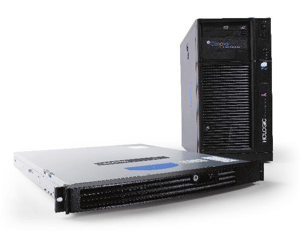
7 Chapter 1: Introduction 1.1. Intended Use 1.1. Intended Use 1.2. Using This Manual 1.3. Resources Available 1.4. Warnings and Precautions 1.5. Overview of Quantra 1.6. Benefits of Quantra 1.7. System Requirements Quantra is a software application used by radiologists to calculate volumetric and area breast densities from two-dimensional digital mammography images. The software is a licensed option with Hologic s Cenova server or any server with comparable functionality (that meets the Quantra data input and output requirements). The information in this manual is intended to serve as a reference for radiologists and clinic personnel who need to understand how Quantra operates and how volumetric assessment can be integrated into their practices. United States federal law restricts this device to use by, or on the order of, a physician. Quantra is a software application intended for use with images acquired using digital breast x-ray systems. Quantra calculates volumetric breast density as a ratio of fibroglandular tissue and total breast volume estimates; and area breast density as a ratio of fibroglandular tissue area and total breast area estimates. It segregates breast density into BI-RADS-like breast composition categories, which may be useful in the reporting of consistent breast composition values as mandated by certain state regulations. Quantra provides these numerical values for each image, breast, and subject, to aid radiologists in the assessment of breast tissue composition. Quantra produces adjunctive information; it is not an interpretive or diagnostic aid. Quantra runs on a Windows platform. Understanding Quantra 2.0 User Manual MAN Rev 004 1
8 Chapter 1: Introduction 1.2. Using This Manual 1.3. Resources Available This manual is organized as follows: Chapter 1: Introduction provides an overview of the Quantra application including features, benefits, and precautions for use. Chapter 2: Image Processing and Supported Views explains how information flows through systems with Quantra, the supported mammography views, and how to manage workflow. Chapter 3: Algorithm Description describes how the Quantra algorithm analyzes mammography images. This manual uses the following conventions to provide technical and safety information of special interest. m WARNING! An instruction that, if not followed, can result in a hazardous condition. m CAUTION: An instruction that, if not followed, can result in damage to the system. m Important: An instruction provided to ensure correct results and optimal performance, or to clarify limitations of the device. m Note: Information provided to clarify a particular step or procedure. In addition to this manual, the following resources are available to assist you. Hologic Member Center: This website provides quick access to electronic (pdf) versions of manuals and training materials for Hologic products. The Member Center is available at no charge to our customers under warranty or Hologic service contract. You can find the Member Center by visiting the Hologic website ( Training: The Hologic Member Center provides training materials for this application. The Hologic Applications team is available to train your staff, should you feel they need additional training. To purchase additional personalized instruction, contact your Hologic Account Manager. 2 Understanding Quantra 2.0 User Manual MAN Rev 004
9 1.4. Warnings and Precautions 1.4. Warnings and Precautions m m Note: For warnings and precautions related to the installation, operation, and maintenance of the Cenova server, refer to the Cenova User Manual. m Important: Please note the following: Quantra is intended to provide adjunctive information; it is not an interpretative or diagnostic aid. The performance of the Quantra software has been evaluated only for images labeled as the four standard screening views: LCC, RCC, LMLO, and RMLO. Results are not reported for: Images that are equivalent or reversed equivalent views (e.g., ML, XCCL, or LM, LMO). Images labeled with the view modifiers M, CV, or S (magnified, cleavage, or spotcompressed views). Digitized images (scanned film images). Images showing breast implants may be processed by the software, although the software has not been designed for that purpose. The software is likely to produce inaccurate Quantra results for patient images with breast implants. Images of partial views of the breast that are not correctly identified as such may be processed by the software, although the software has not been designed for that purpose. The software is unlikely to produce accurate Quantra results for partial view images. m Note: Quantra does not use data compression. Understanding Quantra 2.0 User Manual MAN Rev 004 3
10 Chapter 1: Introduction 1.5. Overview of Quantra Quantra is a software application used to produce assessments of breast composition, both of the breast as a whole and the fibroglandular tissue. Fibroglandular tissue contains a mixture of fibrous connective tissue (stroma) and glandular tissue (epithelial cells), and usually appears brighter than surrounding tissue on a mammogram. The Quantra algorithm first estimates the volume of the imaged portion of the breast, and then separates the breast into portions of fat and portions of fibroglandular tissue. Through arithmetic division the algorithm determines and reports the ratio of fibroglandular tissue as a percentage of total breast volume. The Quantra algorithm estimates two volumes: Volume of fibroglandular tissue in cubic centimeters (cm 3 ) Volume of the breast in cm 3 It then divides the volumes to produce: Volumetric fraction of breast fibroglandular tissue as a percentage From these measurements, Quantra reports scores that compare the results to those of a reference population. Based on partial products from the volumetric assessments, Quantra also calculates the ratio of the area of fibroglandular tissue relative to the total area of the breast in a result known as: Area breast density The Quantra results serve as a convenience to assist the radiologist in assessing the proportion of fibroglandular tissue in the breast. For more information on the individual measures produced by Quantra, see 3.2. Volumetric Assessment and 3.3. Area Assessment. 4 Understanding Quantra 2.0 User Manual MAN Rev 004
11 1.6. Benefits of Quantra 1.6. Benefits of Quantra In recent years, the medical community has shown increasing interest in understanding the relationship between the gross morphology of breast tissue and the risk of developing cancer. Most literature discussing the analysis of breast tissue composition has focused on visual (human) assessments of breast tissue. Currently the most prominent human classification system is the BI-RADS composition scale from the Breast Imaging Reporting and Data System Atlas, Fourth Edition, developed by the American College of Radiology (ACR). BI-RADS provides a standardized breast composition classification system for mammographic studies. The ACR recommends that radiologists practicing in the United States make a visual assessment of breast composition as a part of the reading of a study. The BI-RADS Atlas divides breast composition into the following categories: BI-RADS Composition Description 1 The breast is almost entirely fat (<25% glandular) 2 There are scattered fibroglandular densities (approximately 25 50% glandular) 3 The breast tissue is heterogeneously dense, which could obscure detection of small masses (approximately 51 75% glandular) 4 The breast tissue is extremely dense. This may lower the sensitivity of mammography (>75% glandular) Characterization of breast composition, as described in BI-RADS, depends on the radiologist s assessment of both tissue pattern (the text portion of each description in the table) and density (the numerical range provided with each description). Assessment of breast composition by BI-RADS classification is difficult because tissue pattern in a mammogram may not correlate with density in the same mammogram, and dense tissues may be concentrated in one region of the breast. The radiologist must decide whether density or pattern is the more important factor in assigning a classification in any specific case. This, along with variations in image processing and inter-observer differences, makes composition classification imprecise and unrepeatable. Quantra has been developed in order to provide estimations of breast tissue volumes. Through a proprietary software algorithm, Quantra produces an estimate of fibroglandular tissue volume relative to total breast volume that is not subject to human imprecision. Quantra is not intended as a substitute for BI-RADS composition assessment; rather, it serves as an adjunct technology that can help the radiologist make more consistent breast composition assessments. Understanding Quantra 2.0 User Manual MAN Rev 004 5
12 Chapter 1: Introduction 1.7. System Requirements The table provides the minimum recommended specifications for the server that runs the Quantra application. All specifications are subject to change without notice. Operating System Windows XP Windows 7 Processor Speed 1 GHz 1 GHz Memory (RAM) 1 GB 2 GB HDD Free Disk Space 5 GB 20 GB Optical Drive CD-ROM CD-ROM 6 Understanding Quantra 2.0 User Manual MAN Rev 004
13 Chapter 2: Image Processing and Supported Views 2.1. Image Processing 2.1. Image Processing 2.2. Image Acquisition Systems 2.3. Inputs and Supported Views This chapter explains how information flows through systems with Quantra and the supported mammography views. The Quantra software runs on a server that manages DICOM images and processes the algorithm results. Image and data flows are generally as follows: 1 A Full-Field Digital Mammography (FFDM) system produces two-dimensional digital x-ray images in two forms: DICOM Digital Mammography X-Ray Image For Processing DICOM Digital Mammography X-Ray Image For Presentation 2 The FFDM system sends the For Processing images to the server software and sends the For Presentation images to a review workstation or PACS. 3 The server software receives the For Processing images, groups them by the identified study, and passes the studies to the Quantra software. 4 Quantra analyzes the images, produces results for each study in the form of an.xml file, and outputs the file to the server software. 5 The server software generates results in the form of DICOM Structured Report (SR) or DICOM Secondary Capture Image. 6 For each study, the review workstation displays the Quantra results with the For Presentation images produced by the FFDM system. Radiologists can review the Quantra results at any time as a normal part of the diagnostic reading process. m Note: The appearance of the images on the workstation is dependent upon the acquisition modality and the workstation s display capabilities, and is not affected by the Quantra software Image Acquisition Systems Quantra processes images originating from the following FFDM systems: Hologic Selenia, Hologic Selenia Dimensions (2D) GE Senographe 2000D, GE Senographe DS, and GE Senographe Essential Siemens Mammomat Novation DR Whether images are transmitted directly from the FFDM system or obtained from a PACS, the Quantra software expects to receive For Processing (raw) images rather than For Presentation (processed) images. As many sites do not store raw images, it is important that images in the correct format be available for Quantra if a retrospective research study is planned in the absence of stored Quantra results. Understanding Quantra 2.0 User Manual MAN Rev 004 7
14 Chapter 2: Image Processing and Supported Views 2.3. Inputs and Supported Views Quantra analyzes digital mammography images that conform to the DICOM standard. The Quantra software processes the four mammography screening views: LCC Left Cranio-Caudal LMLO Left Medio-Lateral Oblique RCC Right Cranio-Caudal RMLO Right Medio-Lateral Oblique The server software reads the DICOM header of each received image and groups successive images from a single subject into a study, which then passes to the Quantra algorithm. The following criteria determine which images will be used when reporting results: If a study includes exactly one image for each of the four screening views, then Quantra processes all images. If a study includes multiple images of the same view and laterality (e.g., two RCC views), then the results are derived only from the last image produced by the FFDM device for each of the four screening views. The image-acquisition time is included in the DICOM header for each image. m Note: As an exception to this rule, Quantra processes images with the Implant Displaced DICOM view modifier, even if they are acquired before the implant views. For example, the diagram below shows how images are selected for a study that includes the four screening views, an extra RMLO image, and an RXCCL image. Quantra Image Processing The diagram shows that when Quantra selects images, the algorithm reports results only for the last RMLO image acquired by the FFDM device (along with the other three screening views). In addition, even though the RXCCL image is newer than the RCC image, results for the RXCCL image are not reported because Quantra does not process equivalent views. 8 Understanding Quantra 2.0 User Manual MAN Rev 004
15 Chapter 3: Algorithm Description 3.1. Quantra Algorithm Structure 3.2. Volumetric Assessment 3.3. Area Assessment 3.4. BI-RADS-Like Scores 3.5. Combination of Quantra Results 3.6. Quantra Result Ranges 3.7. Examples of Quantra Results 3.8. Temporal Display of Quantra Results 3.9. Comparison with BI-RADS Categories Atypical Images Performance Testing This chapter describes the Quantra algorithms and results produced when Quantra analyzes mammography images Quantra Algorithm Structure Quantra contains a hierarchy of algorithms that derive estimates of breast density and related information from digital mammography images. Quantra uses the digital mammography image components to perform volumetric assessment estimates and calculation of statistical measures based on the volume estimates (1). Quantra then derives area assessment estimates and calculation of statistical measures based on the area estimates (2). Quantra Algorithm Flow Understanding Quantra 2.0 User Manual MAN Rev 004 9
16 Chapter 3: Algorithm Description 3.2. Volumetric Assessment The Quantra volumetric assessment algorithm is based on a physical model of the x-ray imaging chain that relates breast tissue x-ray attenuation to the digital mammography images provided to the radiologist. Quantra bases its volumetric assessments on published physical parameters for the breast and the imaging system, as well as information about individual x-ray exposures, including: attenuation coefficients for breast tissue 1 x-ray spectra for the target material 2 kvp, mas, and thickness of the imaged tissue Quantra estimates the amount of fibroglandular tissue that an x-ray must have penetrated in order to deposit a measured amount of energy at the detector. It also compensates for the penetration through skin in order to eliminate the impact that skin has on the estimate of fibroglandular tissue volume. Quantra then calculates a height in centimeters of fibroglandular tissue penetrated (Hfg) at each pixel in the image. The algorithm then derives statistical measures that estimate the measures for an individual subject relative to a reference population. In the case of Quantra 2.0, the baseline values for the reference population were derived from a large number of mammograms from multiple institutions across the United States. A comparison of age and BI-RADS density distributions of the reference population with the DMIST study demonstrated that the reference population is representative of the population of approximately 43,000 women as described in the large multicenter American College of Radiology sponsored trial and peer-reviewed publication. 3 Quantra calculates the statistical measures for both the volume of fibroglandular tissue and the volumetric breast density as the number of standard deviations from the mean of the reference population. 1 P. C. Johns and M. J. Yaffe. X-ray characterization of normal and neoplastic breast tissue. Physics in Medicine and Biology, 32: , J. M. Boone, T. R. Fewell, and R. J. Jennings, Molybdenum, rhodium, and tungsten anode spectral models using interpolating polynomials with application to mammography, Med. Phys. 24, E. D. Pisano, C. Gatsonis, E. Hendrick et al. Diagnostic performance of digital versus film mammography for breast-cancer screening. N Engl J Med. 353(17): , 2005 Oct Understanding Quantra 2.0 User Manual MAN Rev 004
17 3.2. Volumetric Assessment Fibroglandular Tissue Volume (Vfg) After Quantra completes its analysis on a pixel-by-pixel basis inside the breast (excluding the pectoral muscle), it aggregates the Hfg heights for each pixel value into the volume of fibroglandular tissue, given in cubic centimeters (cm 3 ). Total Breast Volume (Vb) Assessment of Fibroglandular Tissue Volume Through a similar process, Quantra considers the entire imaged breast outline, including those portions of the breast that were not compressed. In the following diagram, note the difference between the compressed thickness H and the thickness of the breast in the uncompressed region Hu. Quantra compensates for such uncompressed regions in its estimations of breast volume. Volumetric Breast Density (Vbd) Assessment of Total Breast Volume Quantra divides the estimated fibroglandular tissue volume by the estimated breast volume to determine the volumetric percentage of fibroglandular tissue in the breast. m Note: Quantra volumetric breast density (Vbd) is different from traditional human viewing of mammograms in that its measurements are based on estimates of breast tissue volumes, rather than on human estimates of areas. As a result, the volumes produced tend to be lower than one might determine from visual inspection. Volume of Fibroglandular Tissue Score (Vfg-score) The Vfg-score indicates how far the subject Vfg value is from the mean Vfg value of the reference population, measured in standard deviations. The score is positive if the Vfg value is greater than the mean, and negative if it is less than the mean. Quantra reports the score for each image, breast, and subject. Volumetric Breast Density Score (Vbd-score) The Vbd-score indicates how far the subject Vbd value is from the mean Vbd value of the reference population, measured in standard deviations. The score is positive if the Vbd value is greater than the mean, and negative if it is less than the mean. Quantra reports the score for each image, breast, and subject. Understanding Quantra 2.0 User Manual MAN Rev
18 Chapter 3: Algorithm Description 3.3. Area Assessment The Quantra area assessment algorithm operates on partial products from the Quantra volumetric assessment algorithm described above. It selects pixels (based on the Hfg values calculated in the volumetric assessment algorithm) that can be associated as representing significant fibroglandular tissue. The area of the selected dense pixels is the basis for the area assessments. Area Breast Density (Abd) 3.4. BI-RADS-Like Scores Quantra calculates the area breast density as the ratio of the area of the pixels selected as dense divided by the total area of the breast, derived from a standard mammographic breast segmentation method. When in view, Quantra excludes the pectoralis muscle from the estimate of the total breast area used in the Abd calculation. Fractional Quantized Density (q_abd) Quantra maps the estimated volumetric breast density into q_abd, an estimate of overall breast composition relative to the reference population. The q_abd value is a continuous measure of breast composition, ranging from 0.5 for fatty breasts with very low breast density to 4.5 for extremely dense breasts with very high volumetric breast density. Quantized Density (Q_abd) Q_abd is derived by rounding off the q_abd value. It provides an estimate of overall breast composition that is analogous to the four-point breast composition scale of the BI-RADS Atlas 4.0 used by radiologists in many countries to report breast composition. The Q_abd value is an integer in the range Understanding Quantra 2.0 User Manual MAN Rev 004
19 3.5. Combination of Quantra Results 3.5. Combination of Quantra Results Quantra produces three different levels of results. It first calculates the individual Per- Image parameters for each supported image view. Once it completes the Per-Image calculations, it aggregates the results first into Per-Breast results, and further into Per- Subject results. Per-Image Results The server software assesses each received image to see if the image is suitable for Quantra. This excludes images such as spot compressions, magnification views, partial views and those with implants in view. Quantra processes each of the accepted images and calculates Per-Image results for each of the parameters described previously. Per-Breast Results After calculating the Per-Image results, Quantra combines the results into Per-Breast results. Quantra combines results from the orthogonal views (e.g., LCC and LMLO) as follows, in this order: Measures Vfg, Vb Vbd Abd, Vbd-score, Vfgscore, q_abd Q_abd Method Take maximum Per-Image values from CC and MLO views Divide Per-Breast result of Vfg by Per-Breast result of Vb Average Per-Image values from CC and MLO views Round-off Per-Breast q_abd value Per-Subject Results Quantra combines the left and right Per-Breast results to produce Per-Subject results as follows, in this order: Measures Vfg, Vb Vbd Abd Vbd-score, Vfg-score, q_abd, Q_abd Method Sum Per-Breast values from L and R breasts Divide Per-Subject result of Vfg by Per-subject result of Vb Average Per-Breast values from L and R breasts Take maximum Per-Breast values from L and R breasts For unilateral studies, Quantra reports the Per-Breast values. Understanding Quantra 2.0 User Manual MAN Rev
20 Chapter 3: Algorithm Description 3.6. Quantra Result Ranges Measure Description The following table provides the ranges for the results produced by Quantra. Vfg Fibroglandular Tissue Volume 0 to size of breast Vb Total Breast Volume 0 to size of breast Nominal Range Units Notes cm 3 Normally much less than size of breast Vbd Volumetric Breast Density Percent (%) Normally less than 50% even for very dense breast since this is a volumetric measurement Abd Area Breast Density % Normally higher than Vbd due to Area vs. Volume characteristics Vbd-score Vfg-score Q_abd Volumetric Breast Density Score how far the patient s Vbd value is from the mean Vbd of the reference population Volume of Fibroglandular Tissue Score how far the patient s Vfg value is from the mean Vfg of the reference population Quantized Density rounding of q_abd such that the integral Q- abd values have the same BI-RADS distribution as a reference population q_abd Fractional Quantized Density mapping of volumetric breast density relative to the reference population cm 3 3 to +3 Number of Standard Deviations from the mean 3 to +3 Number of Standard Deviations from the mean 1 to 4 Unit-less 0.5 and 4.5 Unit-less 99.73% of the data will lie within 3 standard deviations from the mean 99.73% of the data will lie within 3 standard deviations from the mean Continuous mapping 14 Understanding Quantra 2.0 User Manual MAN Rev 004

21 3.7. Examples of Quantra Results 3.7. Examples of Quantra Results This section provides examples of Quantra results. These examples show how the Hologic SecurView diagnostic review workstation displays Quantra results when it receives results in DICOM SR format. m Note: The display of Quantra results varies depending upon how they are implemented on the diagnostic review workstation. Quantra Per-Image Results Quantra Per-Breast Results Understanding Quantra 2.0 User Manual MAN Rev
.")
22 Chapter 3: Algorithm Description Quantra Per-Subject Results SecurView workstations can also display results in DICOM Secondary Capture Image format, as is shown in the following example: Quantra Results in DICOM SC Image Format For some studies, Quantra may not be able to report results: For incomplete studies, Quantra displays an empty cell in the appropriate column(s). For images that cannot be processed, Quantra displays a dash ( ). m Important: Presentation of results depends upon how the Quantra output is configured to appear on the review workstation. Results may appear differently depending upon the software versions of the Quantra algorithm and the review workstation. 16 Understanding Quantra 2.0 User Manual MAN Rev 004

23 3.8. Temporal Display of Quantra Results 3.8. Temporal Display of Quantra Results Hologic SecurView has the ability to display Quantra results from multiple DICOM SR objects for the same subject. This allows the radiologist to view temporal differences in Quantra assessments. The studies are presented from latest (left) to earliest (right) as shown in this example from the SecurView diagnostic review workstation: Quantra Temporal Display on SecurView In order to use the temporal display feature, the Quantra DICOM SR object from the previous exam must be retrievable from the PACS. Understanding Quantra 2.0 User Manual MAN Rev


 18 Understanding")
24 Chapter 3: Algorithm Description 3.9. Comparison with BI-RADS Categories This section shows images of a typical case for each BI-RADS category followed by the Quantra results for each case as they appear on Hologic s SecurView workstation. BI-RADS 1: <25% Glandular (mostly fat) 18 Understanding Quantra 2.0 User Manual MAN Rev 004


25 3.9. Comparison with BI-RADS Categories BI-RADS 2: 25 50% Glandular (scattered fibroglandular densities) Understanding Quantra 2.0 User Manual MAN Rev


26 Chapter 3: Algorithm Description BI-RADS 3: 51 75% Glandular (heterogeneously dense) 20 Understanding Quantra 2.0 User Manual MAN Rev 004


27 3.9. Comparison with BI-RADS Categories BI-RADS 4: >75% Glandular (extremely dense) Understanding Quantra 2.0 User Manual MAN Rev
28 Chapter 3: Algorithm Description Atypical Images Some atypical images can affect Quantra results. The following table provides explanations and recommendations for these situations: Observation Small object: An image of the breast contains a small manmade object, such as a BB. Large object: An image of the breast contains a large manmade object, such as a paddle. Skin fold: An image of the breast contains a skin fold within the compressed region of the breast. Dense breasts: An image is of an extraordinarily dense breast with little visible fat. Explanations, Recommendations, and Notes Explanation: The object creates an air gap that may cause the algorithm to misjudge the breast thickness. In such instances Quantra may overestimate the volume of fibroglandular tissue. Recommendation: Since women s breasts tend to be grossly symmetrical, consider using the values from the contralateral breast as substitute values. m Note: Objects with very small dimensions (such as J-wires) or those completely contained within the breast (such as biopsy markers and surgical staples) will not cause air gaps and therefore not cause thickness calibration problems. Explanation: Quantra is designed to run on standard screening views. However, some small paddles imaged on large detectors may cause Quantra adjustment errors, especially if the paddle edge lies over breast tissue. Because the content of such images is so varied, it is impossible to predict whether Quantra results will be less reliable. Recommendation: Since women s breasts tend to be grossly symmetrical, consider using the values from the contralateral breast as substitute values. m Note: Diagnostic views that are not described accurately in the DICOM header may not produce reliable Quantra results. Explanation: A skin fold can contain air and may cause the algorithm to misjudge the breast thickness. In such instances Quantra may overestimate the volume of fibroglandular tissue. Recommendation: Since women s breasts tend to be grossly symmetrical, consider using the values from the contralateral breast as substitute values. Explanation: Quantra relies on the detection of fat for a portion of its internal adjustments. Quantra may underestimate the volume of fibroglandular tissue in such instances. Recommendation: Validate with visual assessment. 22 Understanding Quantra 2.0 User Manual MAN Rev 004
29 3.11. Performance Testing Performance Testing Quantra performance was tested against a database of 263 cases, each with a BI-RADS density assessment from 15 different radiologists. The mode (the most frequent value) of the 15 radiologists readings for each case was used as the truth, which was then compared to the measured Quantra values for Abd (area breast density), Vbd (volumetric breast density) and q_abd (fractional quantized volumetric density) values. The following box plots show the results of these comparisons for the Abd and Vbd values. Abd Values versus the Mode of 15 Radiologists Scores Vbd Values versus the Mode of 15 Radiologists Scores As shown in the Vbd plot, the BI-RADS 1 readings included some fibroglandular tissue even in predominately fatty breasts (as there will always be some volumetric measurable fibroglandular tissue present). This phenomenon is not seen in the Abd plot because this small amount of fibroglandular tissue typically falls below the threshold for Abd inclusion. Understanding Quantra 2.0 User Manual MAN Rev
30 Chapter 3: Algorithm Description The following figure presents a scatter plot of q_abd versus the mean BI-RADS values of the 15 radiologists. The Pearson s Correlation Coefficient (PCC) of the two continuous variables is q-abd Values versus the Mean of 15 Radiologists Scores The Vbd-score and Vfg-score measures were validated by correlating the CC / MLO values of the same breast and right and left breasts of the same patient. The following table shows the PCC values for each score across the Hologic, GE and Siemens FFDMs. Measure Type of Correlation Hologic PCC n=5358 GE PCC n=2417 Siemens PCC n=161 Vfg-score CC / MLO L / R Vbd-score CC / MLO L / R Understanding Quantra 2.0 User Manual MAN Rev 004
31 Index A acquisition workstations for Quantra, 7 area assessments, 12 atypical images for Quantra, B BI-RADS, 5, 12 comparison with Quantra, C cleavage views, 3 customer support resources, 2 D DICOM, 7 header, 8 F FFDM systems for Quantra, 7 G GE Healthcare Senographe system, 7, 24 I image processing by Quantra, 7 image specifications for Quantra, 8 implants, breast with Quantra, 3, 13 M magnified views, 3 P partial view images with Quantra, 3 performance testing, algorithm, precautions for Quantra, 3 Q Quantra algorithm description, 9 24 benefits of, 5 image specifications, 8 intended use, 1 overview of, 4 results, selecting views for processing, 8 R requirements, hardware, 6 results, Quantra, area assessments, 12 combined, 13 output format, 7 ranges of, 14 SecurView workstation examples, temporal, 17 volumetric assessments, review workstations for Quantra, 7, S screening views with Quantra, 8 SecurView diagnostic review workstation, 15 17, 17 Selenia FFDM system, 7, 24 server specifications, 6 Siemens AG Mammomat Novation system, 7, 24 spot-compressed views, 3 T temporal results for Quantra, 17 training, 2 V view modifiers with Quantra, 3 views, supported for Quantra, 8 volumetric assessments, W warnings for Quantra, 3 workflow, clinical with Quantra, 7 Understanding Quantra 2.0 User Manual MAN Rev
32 26 Understanding Quantra 2.0 User Manual MAN Rev 004
33
34 At Hologic, we turn passion into action, and action into change. Hologic is defi ning the standard of care in women s health. Our technologies help doctors see better, know sooner, reach further and touch more lives. BREAST IMAGING SOLUTIONS INTERVENTIONAL BREAST SOLUTIONS BONE HEALTH PRENATAL HEALTH GYNECOLOGIC HEALTH MOLECULAR DIAGNOSTICS info@hologic.com North America / Latin America 35 Crosby Drive Bedford, MA USA Europe Everest (Cross Point) Leuvensesteenweg 250A 1800 Vilvoorde Belgium Asia Pacific 7th Floor, Biotech Centre 2 No. 11 Science Park West Avenue Hong Kong Science Park Shatin, New Territories Hong Kong Australia / New Zealand Suite 402, Level 4 2 Lyon Park Road Macquarie Park NSW 2113 Australia
Image Quality. HTC Grid High Transmission Cellular Grid provides higher contrast images
 B R E A S T I M A G I N G S O L U T I O N S Setting the benchmark for mammography M-IV Series Innovations in breast imaging The Lorad M-IV Series exemplifies Hologic s commitment to developing advanced
Image Quality. HTC Grid High Transmission Cellular Grid provides higher contrast images
 B R E A S T I M A G I N G S O L U T I O N S Setting the benchmark for mammography M-IV Series Innovations in breast imaging The Lorad M-IV Series exemplifies Hologic's commitment to developing advanced
GE Healthcare. Senographe 2000D Full-field digital mammography system
 GE Healthcare Senographe 2000D Full-field digital mammography system Digital has arrived. The Senographe 2000D Full-Field Digital Mammography (FFDM) system gives you a unique competitive advantage. That
Acceptance Testing of a Digital Breast Tomosynthesis Unit
 Acceptance Testing of a Digital Breast Tomosynthesis Unit 2012 AAPM Spring Clinical Meeting Jessica Clements, M.S., DABR Objectives Review of technology and clinical advantages Acceptance Testing Procedures
Patient-Assisted Compression Impact on Image Quality and Workflow
 Patient-Assisted Compression Impact on Image Quality and Workflow Senographe Pristina In 2017, GE Healthcare s Senographe Pristina ( Pristina ) was approved by the FDA using the standard technologist-controlled
Fluoroscan InSight FD Mini C-Arm Imaging System
 Extremity Imaging Fluoroscan InSight FD Mini C-Arm Imaging System Redefining Mini C-arm Technology More Imaging Options to suit your needs Building on Hologic s tradition of innovation in imaging, the
THE ART OF THE IMAGE: IDENTIFICATION AND REMEDIATION OF IMAGE ARTIFACTS IN MAMMOGRAPHY
 THE ART OF THE IMAGE: IDENTIFICATION AND REMEDIATION OF IMAGE ARTIFACTS IN MAMMOGRAPHY William Geiser, MS DABR Senior Medical Physicist MD Anderson Cancer Center Houston, Texas wgeiser@mdanderson.org INTRODUCTION
New spectral benefi ts, proven low dose
 New spectral benefi ts, proven low dose Philips MicroDose mammography SI, technical data sheet Philips MicroDose SI with single-shot spectral imaging is a fullfi eld digital mammography solution that delivers
Cenova 2.1 DICOM Conformance Statement
 BREAST IMAGING SOLUTIONS DICOM Standard Interface Cenova 2.1 DICOM Conformance Statement MAN-02008 Rev 001 Cenova 2.1 DICOM Conformance Statement MAN-02008 Rev 001 Technical Support For support in North
Breast Tomosynthesis. Bob Liu, Ph.D. Department of Radiology Massachusetts General Hospital And Harvard Medical School
 Breast Tomosynthesis Bob Liu, Ph.D. Department of Radiology Massachusetts General Hospital And Harvard Medical School Outline Physics aspects of breast tomosynthesis Quality control of breast tomosynthesis
Mammography: Physics of Imaging
 Mammography: Physics of Imaging Robert G. Gould, Sc.D. Professor and Vice Chair Department of Radiology and Biomedical Imaging University of California San Francisco, California Mammographic Imaging: Uniqueness
GE Healthcare. Essential for life. Senographe Essential Full-Field Digital Mammography system
 GE Healthcare Essential for life Senographe Essential Full-Field Digital Mammography system Excellence in FFDM is a process. An ongoing quest, fueled by our continuing breakthroughs in breast cancer detection
Practical Aspects of Medical Physics Surveys of Mammography Equipment and Facilities
 Practical Aspects of Medical Physics Surveys of Mammography Equipment and Facilities Melissa Martin, M.S., FAAPM, FACR, FACMP AAPM Annual Meeting - Philadelphia July 19, 2010 MO-B-204C-1 Educational Objectives
Imaging Technique Optimization of Tungsten Anode FFDM System
 Imaging Technique Optimization of Tungsten Anode FFDM System Biao Chen a*, Andrew P. Smith b, Zhenxue Jing a, Elena Ingal a a Hologic, Inc. 600 Technology Drive, DE 1970 b Hologic, Inc. 35 Crosby Drive,
Quality Control of Full Field Digital Mammography Units
 Quality Control of Full Field Digital Mammography Units Melissa C. Martin, M.S., FACMP, FACR, FAAPM Melissa@TherapyPhysics.com 310-612-8127 ACMP Annual Meeting Virginia Beach, VA May 2, 2009 History of
Performance and care. all in one
 Performance and care all in one INNOVATION IS WHAT DRIVES US THINKING ABOUT THE FUTURE Preventive diagnostics remains an essential weapon in defeating breast cancer. Metaltronica s forward-thinking design
KODAK DIRECTVIEW CR Mammography Feature User s Guide
 KODAK DIRECTVIEW CR Mammography Feature User s Guide 17 September 2010 9G3741 Version 1.0 Carestream Health, Inc. 150 Verona Street Rochester, NY 14608 CARESTREAM, DIRECTVIEW, and DRYVIEW are trademarks
GE Healthcare. Performa. High-performance breast imaging
 GE Healthcare Performa High-performance breast imaging Moving mammography forward. And patients faster. GE Healthcare s unparalleled leadership across mammography begins with a deep understanding of breast
Digital Breast Tomosynthesis
 Digital Breast Tomosynthesis OLIVE PEART MS, RT(R) (M) HTTP://WWW.OPEART.COM 2D Mammography Not 100% effective Limited by tissue superimposition Overlapping tissue can mask tumors False negative Overlapping
Filtering for More Accurate Dense Tissue Segmentation in Digitized Mammograms
 Filtering for More Accurate Dense Tissue Segmentation in Digitized Mammograms Mario Mustra, Mislav Grgic University of Zagreb, Faculty of Electrical Engineering and Computing, Unska 3, 10000 Zagreb, Croatia
Mammography Solution. AMULET Innovality. The new leader in the AMULET series. Tomosynthesis, 3D mammography and biopsy are all available.
 Mammography Solution AMULET Innovality The new leader in the AMULET series. Tomosynthesis, 3D mammography and biopsy are all available. FUJIFILM supports the Pink Ribbon Campaign for early detection of
Surveying and QC of Stereotactic Breast Biopsy Units for ACR Accreditation
 Surveying and QC of Stereotactic Breast Biopsy Units for ACR Accreditation AAPM Annual Clinical Meeting Indianapolis, IN August 5, 2013 Learning Objectives Become familiar with the recommendations and
Collimation Assessment Using GAFCHROMIC XR-M2
 Collimation Assessment Using GAFCHROMIC XR-M2 I. Introduction A method of collimation assessment for GE Senographe full-field digital mammography (FFDM) systems is described that uses a self-developing
GE Healthcare. Senographe Crystal The choice is crystal clear
 Senographe Crystal The choice is crystal clear Senographe Crystal The choice is crystal clear. The Senographe* Crystal mammography system makes it easy to transition to full-field digital mammography.
Mammography is a radiographic procedure specially designed for detecting breast pathology Approximately 1 woman in 8 will develop breast cancer over
 Mammography is a radiographic procedure specially designed for detecting breast pathology Approximately 1 woman in 8 will develop breast cancer over a lifetime Breast cancer screening programs rely on
Published text: Institute of Cancer Research Repository Please direct all s to:
 This is an author produced version of an article that appears in: MEDICAL PHYSICS The internet address for this paper is: https://publications.icr.ac.uk/1316/ Copyright information: http://www.aip.org/pubservs/web_posting_guidelines.html
Predicted image quality of a CMOS APS X-ray detector across a range of mammographic beam qualities
 Journal of Physics: Conference Series PAPER OPEN ACCESS Predicted image quality of a CMOS APS X-ray detector across a range of mammographic beam qualities Recent citations - Resolution Properties of a
The Versatile and Powerful ACLxy. ACLxy
 The Versatile and Powerful ACLxy ACLxy Rolling into a Clinic, Imaging Center and Hospital Near You! COMPUTED RADIOGRAPHY (CR) IS RAPIDLY THE BEGINNING. THE OREX CR SOLUTION DRA- BECOMING A DRIVING FORCE
Digital Imaging started in the 1972 with Digital subtraction angiography Clinical digital imaging was employed from the 1980 ~ 37 years ago Amount of
 Digital Imaging started in the 1972 with Digital subtraction angiography Clinical digital imaging was employed from the 1980 ~ 37 years ago Amount of radiation to the population due to Medical Imaging
4/19/2016. Quality Control Activities for the RadiologicTechnologist. Objectives. 3D Tomosynthesis QC differences
 Quality Control Activities for the RadiologicTechnologist Quality Control Tests 2D QC Tomosynthesis QC DICOM Printer Quality Control Weekly Detector Flat Field Calibration Weekl Artifact Evaluation Weekly
MAMMOGRAPHY - HIGH LEVEL TROUBLESHOOTING
 MAMMOGRAPHY - HIGH LEVEL TROUBLESHOOTING Maynard High New York Medical College SS2001-M.High 1 Objectives: Review MQSA and ACR annual QC tests as opportunities for troubleshooting before a significant
2 nd generation TOMOSYNTHESIS
 2 nd generation TOMOSYNTHESIS 2 nd generation DBT true innovation in breast imaging synthesis graphy Combo mode Stereotactic Biopsy Works in progress: Advanced Technology, simplicity and ergonomics Raffaello
TECHNICAL DATA. GIOTTO IMAGE SDL/W is pre-arranged for Full Field Digital Biopsy examination with the patient in prone position.
 Ver. 01/06/07 TECHNICAL DATA GIOTTO IMAGE SDL/W LOW DOSE, FULL FIELD DIGITAL MAMMOGRAPHY UNIT USING AMORPHOUS SELENIUM (a-se) TECHNOLOGY DETECTOR (pre-arranged for stereotactic biopsy with the same digital
IHE Radiology Technical Framework Supplement. Stereotactic Mammography Image (SMI) Trial Implementation
 Trial Implementation") Integrating the Healthcare Enterprise 5 IHE Radiology Technical Framework Supplement 10 Stereotactic Mammography Image (SMI) 15 Trial Implementation 20 25 Date: June 11, 2013 Author: IHE Radiology Technical
Integrating the Healthcare Enterprise 5 IHE Radiology Technical Framework Supplement 10 Stereotactic Mammography Image (SMI) 15 Trial Implementation 20 25 Date: June 11, 2013 Author: IHE Radiology Technical
Breast Imaging Basics: Module 10 Digital Mammography
 Module 10 Transcript For educational and institutional use. This test bank is licensed for noncommercial, educational inhouse or online educational course use only in educational and corporate institutions.
Introduction. Digital Mammography QA: Comparing the Manufacturers Recommendations. What is QC and why is it important? Review & compare QC tests
 Slide 1 Digital Mammography QA: Comparing the Manufacturers Recommendations Eric A. Berns, Ph.D. Slide 2 Introduction What is QC and why is it important? Review & compare QC tests Key take home points
Effect of pressure, temperature and humidity in air on photon fluence and air kerma values at low photon energies
 ARTICLE IN PRESS Radiation Physics and Chemistry 68 (2003) 707 720 Effect of pressure, temperature and humidity in air on photon fluence and air kerma values at low photon energies M. Assiamah, D. Mavunda,
SECTION I - CHAPTER 1 DIGITAL RADIOGRAPHY: AN OVERVIEW OF THE TEXT. Exam Content Specifications 8/22/2012 RADT 3463 COMPUTERIZED IMAGING
 RADT 3463 - COMPUTERIZED IMAGING Section I: Chapter 1 RADT 3463 Computerized Imaging 1 SECTION I - CHAPTER 1 DIGITAL RADIOGRAPHY: AN OVERVIEW OF THE TEXT RADT 3463 COMPUTERIZED IMAGING Section I: Chapter
STEREOTACTIC BREAST BIOPSY EQUIPMENT SURVEYS
 STEREOTACTIC BREAST BIOPSY EQUIPMENT SURVEYS JAMES A. TOMLINSON, M.S. Diagnostic Radiological Physicist American Board of Radiology Certified Medical Physics Consultants, Inc. Bio 28 yrs experience 100%
8/2/2017. Radiologist Responsibilities. Radiologist Responsibilities. Medical Physicist Mammography Equipment Evaluation and Annual Survey
 Implementation of the 2016 ACR Digital Mammography QC Manual Medical Physicist Mammography Equipment Evaluation and Annual Survey Eric A Berns, PhD, FACR Radiologist Responsibilities Radiologist Responsibilities
Optimization of Digital Mammography Resolution Using Magnification Technique in Computed Radiography 1
 Optimization of Digital Mammography Resolution Using Magnification Technique in Computed Radiography 1 Gham Hur, M.D., Yoon Joon Hwang, M.D., Soon Joo Cha, M.D., Su Young Kim, M.D., Yong Hoon Kim, M.D.
FFDM in the Field: Physicist's Role in the QC of Mammography Laser Printers May Carl R. Keener, Ph.D., DABMP, DABR
 FFDM in the Field: Physicist's Role in the QC of Mammography Laser Printers May 2010 Carl R. Keener, Ph.D., DABMP, DABR keener@marpinc.com MARP Medical & Radiation Physics, Inc. Physicist's Role in the
Differentiation of Malignant and Benign Masses on Mammograms Using Radial Local Ternary Pattern
 Differentiation of Malignant and Benign Masses on Mammograms Using Radial Local Ternary Pattern Chisako Muramatsu 1, Min Zhang 1, Takeshi Hara 1, Tokiko Endo 2,3, and Hiroshi Fujita 1 1 Department of Intelligent
Positive Pixel Count Algorithm. User s Guide
 Positive Pixel Count Algorithm User s Guide Copyright 2004, 2006 2008 Aperio Technologies, Inc. Part Number/Revision: MAN 0024, Revision B Date: December 9, 2008 This document applies to software versions
Investigation of the line-pair pattern method for evaluating mammographic focal spot performance
 Investigation of the line-pair pattern method for evaluating mammographic focal spot performance Mitchell M. Goodsitt, a) Heang-Ping Chan, and Bob Liu Department of Radiology, University of Michigan, Ann
Digital radiography (DR) post processing techniques for pediatric radiology
 post processing techniques for pediatric radiology") Digital radiography (DR) post processing techniques for pediatric radiology St Jude Children s Research Hospital Samuel Brady, MS PhD DABR samuel.brady@stjude.org Purpose Review common issues and solutions
Digital radiography (DR) post processing techniques for pediatric radiology St Jude Children s Research Hospital Samuel Brady, MS PhD DABR samuel.brady@stjude.org Purpose Review common issues and solutions
The Evaluation of Collimator Alignment of Diagnostic X-ray Tube Using Computed Radiography System
 The Evaluation of Collimator Alignment of Diagnostic X-ray Tube Using Computed Radiography System The Evaluation of Collimator Alignment of Diagnostic X-ray Tube Using Computed Radiography System Manus
COMPUTER-AIDED DETECTION OF CLUSTERED CALCIFICATION USING IMAGE MORPHOLOGY
 COMPUTER-AIDED DETECTION OF CLUSTERED CALCIFICATION USING IMAGE MORPHOLOGY Ariya Namvong Department of Information and Communication Technology, Rajamangala University of Technology Isan, Nakhon Ratchasima,
DICOM Correction Item
 DICOM Correction Item Correction Number CP-564 Log Summary: Type of Modification Correction Name of Standard PS 3.3, PS 3.6, PS 3.17 2004 Rationale for Correction A mammography CAD system often prefers
Solutions Page Content ImagePilot. Primary keywords: Digital radiography Computed radiography Image viewing and storage
 Solutions Page Content Primary keywords: Digital radiography Computed radiography Image viewing and storage Solution Description For facilities with medium volume imaging, Konica Minolta s original all-in-one
GE Healthcare. Senographe Crystal The choice is crystal clear
 Senographe Crystal The choice is crystal clear Senographe Crystal The choice is crystal clear. The Senographe* Crystal mammography system makes it easy to transition to full-field digital mammography.
ADVANCED MEDICAL SYSTEMS PTE LTD Singapore Malaysia India Australia
 Innovative design is combined with cutting-edge technology to yield a definitive diagnosis and never before seen ergonomics GIOTTO CLASS is the result of 25 years of experience in the research and development
IBEX MATERIALS DETECTION TECHNOLOGY
 WHITE PAPER: IBEX MATERIALS DETECTION TECHNOLOGY IBEX Innovations Ltd. Registered in England and Wales: 07208355 Address: Discovery 2, NETPark, William Armstrong Way, Sedgefield, TS21 3FH, UK Patents held
Quality Control for Stereotactic Breast Biopsy. Robert J. Pizzutiello, Jr., F.A.C.M.P. Upstate Medical Physics, Inc
 Quality Control for Stereotactic Breast Biopsy Robert J. Pizzutiello, Jr., F.A.C.M.P. Upstate Medical Physics, Inc. 716-924-0350 Methods of Imaging Guided Breast Biopsy Ultrasound guided, hand-held needle
Detection of Breast Masses in Digital Mammograms using Multiple Concentric Layers
 Detection of Breast Masses in Digital Mammograms using Multiple Concentric Layers Chethan K Dr. Krishna A N 4 th Sem, M.Tech Associate Professor Dept of CSE, SJBIT Dept of CSE, SJBIT Bangalore-60 Bangalore-60
Initial Survey Results on the Accuracy of Dose Data in the DICOM Objects
 Initial Survey Results on the Accuracy of Dose Data in the DICOM Objects Renato Padovani, Annalisa Trianni University Hospital S. Maria della Misericordia Udine, Italy INTRODUCTION The EC MED has forced
Pseudocolour Image Processing in Digital Mammography
 Pseudocolour Image Processing in Digital Mammography M.Y. Sanavullah 1 and R. Samson Ravindran 2 1 Principal and Research Guide, VMKV Engineering College, V.M.R.F Deemed University, Salem, TN 2 Research
IBEX TECHNOLOGY APPLIED TO DIGITAL RADIOGRAPHY
 WHITE PAPER: IBEX TECHNOLOGY APPLIED TO DIGITAL RADIOGRAPHY IBEX Innovations Ltd. Registered in England and Wales: 07208355 Address: Discovery 2, NETPark, William Armstrong Way, Sedgefield, UK Patents:
DICOM Conformance. DICOM Detailed Specification for Diagnostic Labs and Radiology Center Connectivity
 DICOM Detailed Specification for Diagnostic Labs and Radiology Center Connectivity Authored by Global Engineering Team, Health Gorilla April 10, 2014 Table of Contents About Health Gorilla s Online Healthcare
Yinsheng Li 1, Peter Bannas 2, M.D., Perry Pickhardt M.D. 2, Meghan Lubner M.D. 2, Ke Li Ph.D. 1,2, and Guang-Hong Chen Ph.D. 1,2
 Yinsheng Li 1, Peter Bannas 2, M.D., Perry Pickhardt M.D. 2, Meghan Lubner M.D. 2, Ke Li Ph.D. 1,2, and Guang-Hong Chen Ph.D. 1,2 1. Department of Medical Physics, University of Wisconsin-Madison 2. Department
COST EFFECTIVE FLAT PANEL DIGITAL RADIOGRAPHY UPGRADE SOLUTIONS
 COST EFFECTIVE FLAT PANEL DIGITAL RADIOGRAPHY UPGRADE SOLUTIONS DRive is a digital imaging DR hardware & Software solution designed for General Radiography of anatomy. It intended to replace film/screen
Things you may want to know
 Things you may want to know 1 / 9 Last updated 18-Nov-05 Why medical monitors are special? Unlike commercial displays, Medical displays are used in the hospital and they are displaying contain life critical
Introduction. Chapter 16 Diagnostic Radiology. Primary radiological image. Primary radiological image
 Introduction Chapter 16 Diagnostic Radiology Radiation Dosimetry I Text: H.E Johns and J.R. Cunningham, The physics of radiology, 4 th ed. http://www.utoledo.edu/med/depts/radther In diagnostic radiology
Image Display and Perception
 Image Display and Perception J. Anthony Seibert, Ph.D. Department of Radiology UC Davis Medical Center Sacramento, California, USA Image acquisition, display, & interpretation X-rays kvp mas Tube filtration
TORNIER BLUEPRINT. 3D Planning + PSI SCAN PROTOCOL
 TORNIER BLUEPRINT 3D Planning + PSI SCAN PROTOCOL Contents 3 Introduction 3 Patient preparation 3 Scanning instructions 4 Image instructions 5 Scanning parameters 6 Technical instructions 2 BLUEPRINT 3D
SECTION I - CHAPTER 2 DIGITAL IMAGING PROCESSING CONCEPTS
 RADT 3463 - COMPUTERIZED IMAGING Section I: Chapter 2 RADT 3463 Computerized Imaging 1 SECTION I - CHAPTER 2 DIGITAL IMAGING PROCESSING CONCEPTS RADT 3463 COMPUTERIZED IMAGING Section I: Chapter 2 RADT
DELWORKS DR MEDICAL. take the next step
 DELWORKS DR MEDICAL take the next step DELWORKS MEDICAL DR If you are thinking of taking the next step to digital radiography, consider a DelWorks Medical DR Retrofit Package, the easy and affordable way
MAN Revision 002. InSight Mini C-arm Imaging System Technical Reference Manual
 MAN-00668 Revision 002 InSight Mini C-arm Imaging System Technical Reference Manual April 2007 The information contained in this Manual is confidential and proprietary to Hologic, Inc. This information
Quality control for digital mammography: Part II recommendations from the ACRIN DMIST trial
 Quality control for digital mammography: Part II recommendations from the ACRIN DMIST trial Martin J. Yaffe, Aili K. Bloomquist, Gordon E. Mawdsley, Etta D. Pisano, R. Edward Hendrick, Laurie L. Fajardo,
Digital Quality Control Phantom for X-ray Mammography Image
 Digital Quality Control Phantom for X-ray Mammography Image Yuichi Nagai, Atsuko Takada National: Cancer Hospital East Department of Radiology Collaborated by Nobuyuki Niwa : Kyoto Kagaku Co., Ltd. Introduction
Improved Tomosynthesis Reconstruction using Super-resolution and Iterative Techniques
 Improved Tomosynthesis Reconstruction using Super-resolution and Iterative Techniques Wataru FUKUDA* Junya MORITA* and Masahiko YAMADA* Abstract Tomosynthesis is a three-dimensional imaging technology
DICOM Conformance Statement
 DICOM Conformance Statement Application Annex: 3D Roadmap R1.1.5 Koninklijke Philips N.V. 2015 All rights are reserved. Document Number: ICAP-PF.0015381 Issued by: Philips Medical Systems Nederland BV,
SmartRAD. Advanced Digital Radiography System
 SmartRAD Advanced Digital Radiography System SmartRAD Expanding The Horizons Of Digital Radiography CMT introduces the SmartRAD Digital Radiography system, featuring an integrated flat panel digital detector
DICOM Conformance Statement
 DICOM Conformance Statement Application Annex: Stentboost R4.2.5 Koninklijke Philips N.V. 2015 All rights are reserved. Document Number: ICAP-PF.0015387 Issued by: Philips Medical Systems Nederland BV,
Tables and Figures. Germination rates were significantly higher after 24 h in running water than in controls (Fig. 4).
.") Tables and Figures Text: contrary to what you may have heard, not all analyses or results warrant a Table or Figure. Some simple results are best stated in a single sentence, with data summarized parenthetically:
Tables and Figures Text: contrary to what you may have heard, not all analyses or results warrant a Table or Figure. Some simple results are best stated in a single sentence, with data summarized parenthetically:
Medical Imaging. X-rays, CT/CAT scans, Ultrasound, Magnetic Resonance Imaging
 Medical Imaging X-rays, CT/CAT scans, Ultrasound, Magnetic Resonance Imaging From: Physics for the IB Diploma Coursebook 6th Edition by Tsokos, Hoeben and Headlee And Higher Level Physics 2 nd Edition
Abstract. ZEIGLER, GARY BOYCE, II. Direct Detection of Microcalcification Pairs in Simulated
 Abstract ZEIGLER, GARY BOYCE, II. Direct Detection of Microcalcification Pairs in Simulated Digital Mammograms. (Under the Direction of Professor Kuruvilla Verghese.) Using the MCMIS (Monte Carlo for Mammography
AN APPROXIMATION-WEIGHTED DETAIL CONTRAST ENHANCEMENT FILTER FOR LESION DETECTION ON MAMMOGRAMS
 AN APPROXIMATION-WEIGHTED DETAIL CONTRAST ENHANCEMENT FILTER FOR LESION DETECTION ON MAMMOGRAMS Zhuangzhi Yan, Xuan He, Shupeng Liu, and Donghui Lu Department of Biomedical Engineering, Shanghai University,
ISO Cube Daily QA Package
 ISO Cube Daily QA Package Model 023-05 AFFORDABLE TURNKEY SOLUTION FOR DAILY MACHINE QA POWERED BY AQUILAB 2428 Almeda Avenue Suite 316 Norfolk, Virginia 23513 USA Tel: 757-855-2765 WWW.CIRSINC.COM CAPABILITIES
Maximizing clinical outcomes
 Maximizing clinical outcomes Digital Tomosynthesis Dual Energy Subtraction Automated Long Length Imaging Improved image quality at a low dose Xray Xray Patented ISS capture technology promotes high sensitivity
Aspire HD. Program Manual. 2nd Edition - October 2012
 Quality Control 1 Aspire HD Quality Control Program Manual 2nd Edition - October 2012 Overview Installation of FDR Mammography QC Program Weekly Test 2 3 4 Quarterly Test 5 Semi-annual Test 6 Annual Test
Grubstr- 6-8 D Vörstetten Tel.: +49(0) Fax: +49(0) Steinhart Medizinsysteme GmbH
 Fax: +49(0) Steinhart Medizinsysteme GmbH") Hipax Diagnostic Workstation Table of Contents 1. PRODUCT DESCRIPTION... 2 2. HIPAX DW MODULES... 2 2.1 DW BASE MODULE... 2 2.1.1 DW BASE MODULE STANDARD (05-010)... 2 2.1.2 DW BASE MODULE LIGHT (05-020)...
Hipax Diagnostic Workstation Table of Contents 1. PRODUCT DESCRIPTION... 2 2. HIPAX DW MODULES... 2 2.1 DW BASE MODULE... 2 2.1.1 DW BASE MODULE STANDARD (05-010)... 2 2.1.2 DW BASE MODULE LIGHT (05-020)...
DR _ solutions. We understand that customers don t need just products, they want. solutions
 DR _ solutions We understand that customers don t need just products, they want solutions index company profile 1974-2005 2006-2007 - 2008 ITALRAY Srl was founded in 1974 as the production branch of Marzocchi
Testing a wavelet based noise reduction method using computersimulated
 Testing a wavelet based noise reduction method using computersimulated mammograms Christoph Hoeschen 1, Oleg Tischenko 1, David R Dance 2, Roger A Hunt 2, Andrew DA Maidment 3, Predrag R Bakic 3 1 GSF-
A Study On Preprocessing A Mammogram Image Using Adaptive Median Filter
 A Study On Preprocessing A Mammogram Image Using Adaptive Median Filter Dr.K.Meenakshi Sundaram 1, D.Sasikala 2, P.Aarthi Rani 3 Associate Professor, Department of Computer Science, Erode Arts and Science
ACPSEM Position Paper RECOMMENDATIONS FOR A DIGITAL MAMMOGRAPHY QUALITY ASSURANCE PROGRAM V4.0
 Heggie et al ACPSEM Position Paper: Digital Mammography V4.0 ACPSEM Position Paper RECOMMENDATIONS FOR A DIGITAL MAMMOGRAPHY QUALITY ASSURANCE PROGRAM V4.0 JCP Heggie 1, P Barnes 2, L Cartwright 3, J Diffey
Chairside digital. Imaging Plate System
 Chairside digital Imaging Plate System 2 DIGORA OPTIME The complete digital intraoral Imaging Plate System Speed and performance The DIGORA Optime intraoral digital imaging system is designed to make work
Tailoring automatic exposure control toward constant detectability in digital mammography
 Tailoring automatic exposure control toward constant detectability in digital mammography Elena Salvagnini a) Department of Imaging and Pathology, Medical Physics and Quality Assessment, KUL, Herestraat
Clinical and management aspects of digital imaging and PACS
 Clinical and management aspects of digital imaging and PACS พญ.จามร เช อเพชระโสภณ นายกร งส ว ทยาสมาคมแห งประเทศไทย chamareec@gmail.com www.radiologythailand.org Digital imaging Abbreviation and terminology
Observer Performance of Reduced X-Ray Images on Liquid Crystal Displays
 Original Paper Forma, 29, S45 S51, 2014 Observer Performance of Reduced X-Ray Images on Liquid Crystal Displays Akiko Ihori 1, Chihiro Kataoka 2, Daigo Yokoyama 2, Naotoshi Fujita 3, Naruomi Yasuda 4,
RAD 150 RADIOLOGIC EXPOSURE TECHNIQUE II
 RAD 150 RADIOLOGIC EXPOSURE TECHNIQUE II APPROVED 12/O2/2011 EFFECTIVE SPRING 2013-14 Prefix & Number RAD 150 Course Title: Radiologic Exposure Technique II & Lab Purpose of this submission: New Change/Updated
DICOM Conformance Statement
 DICOM Conformance Statement Application Annex: MR-CT Roadmap R1.1 On Interventional Workspot R1.4 Koninklijke Philips N.V. 2017 All rights are reserved. ICAP-T-030001.09b Corresponds to ICAP-W-030001.02
Cutting-edge image quality
 ENGLISH Cutting-edge image quality The innovative Planmeca ProSensor intraoral sensor sets new standards for imaging in dental practice. Planmeca ProSensor is a unique combination of high-end patient-centred
Comparison between film-screen and computed radiography systems in Brazilian mammography
 X Congreso Regional Latinoamericano IRPA de Protección y Seguridad Radiológica Radioprotección: Nuevos Desafíos para un Mundo en Evolución Buenos Aires, al 7 de abril, 05 SOCIEDAD ARGENTINA DE RADIOPROTECCIÓN
Fluke199XRAY. Users Manual. Medical ScopeMeter
 Fluke199XRAY Medical ScopeMeter Users Manual 4822 872 30791 August 2006 2006 Fluke Corporation, All rights reserved. All product names are trademarks of their respective companies. Table of Contents Title
A Practical Overview of the Clinical and Operational Impact of Computed Radiography(CR) Implementations. Shirley Weddle, RT(R)(M), CIIP, BBA
 Implementations. Shirley Weddle, RT(R)(M), CIIP, BBA") A Practical Overview of the Clinical and Operational Impact of Computed Radiography(CR) Implementations Shirley Weddle, RT(R)(M), CIIP, BBA OBJECTIVES Define Computed Radiography (CR) Discuss CR vendor
A Practical Overview of the Clinical and Operational Impact of Computed Radiography(CR) Implementations Shirley Weddle, RT(R)(M), CIIP, BBA OBJECTIVES Define Computed Radiography (CR) Discuss CR vendor
Seminar 8. Radiology S8 1
 Seminar 8 Radiology Medical imaging. X-ray image formation. Energizing and controlling the X-ray tube. Image detectors. The acquisition of analog and digital images. Digital image processing. Selected
Test Equipment for Radiology and CT Quality Control Contents
 Test Equipment for Radiology and CT Quality Control Contents Quality Control Testing...2 Photometers for Digital Clinical Display QC...3 Primary Workstations...3 Secondary Workstations...3 Testing of workstations...3
Quality control of Gamma Camera. By Dr/ Ibrahim Elsayed Saad 242 NMT
 Quality control of Gamma Camera By Dr/ Ibrahim Elsayed Saad 242 NMT WHAT IS QUALITY? The quality of a practice is to fulfill the expectations and demands from: Patient Clinicain Your self Quality assurance
Optimized CT metal artifact reduction using the Metal Deletion Technique (MDT)
") Optimized CT metal artifact reduction using the Metal Deletion Technique (MDT) F Edward Boas, Roland Bammer, and Dominik Fleischmann Extended abstract for RSNA 2012 Purpose CT metal streak artifacts are
Optimized CT metal artifact reduction using the Metal Deletion Technique (MDT) F Edward Boas, Roland Bammer, and Dominik Fleischmann Extended abstract for RSNA 2012 Purpose CT metal streak artifacts are