Novel Endoscopic Imaging Techniques
|
|
|
- Jasmin Snow
- 5 years ago
- Views:
Transcription
1

2 ENDOSCOPY nd Falk Symposium, Berlin May 4-5, 2006 Novel Endoscopic Imaging Techniques Paul Fockens, MD PhD Academic Medical Center, University of Amsterdam
3 Novel Endoscopic Imaging Techniques Current optimal techniques High-resolution and magnification endoscopy Chromo-endoscopy New techniques Narrow Band Imaging Autofluorescence Imaging Endo-microscopy
4 Novel Endoscopic Imaging Techniques Current optimal techniques High-resolution and magnification endoscopy Chromo-endoscopy New techniques Narrow Band Imaging Autofluorescence Imaging Endo-microscopy
5 Optical Magnification (1,5-150 x) + (> pixels) High resolution
6 Chromo-endoscopy Contrast stains Indigo-carmine (0,1 0,4%) Absorptive stains Cresyl violet Methylene blue (0,1%) Spraying catheter useful, not crucial Goal Delineate exact borders Surface analysis (zoom)


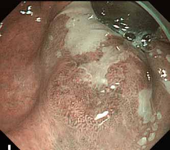
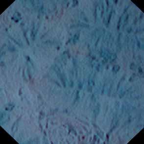
7 Chromo-endoscopy Delineate borders Carefull non-magnified endoscopy Use contrast dye at any discoloration etc.
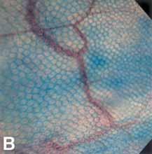
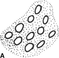
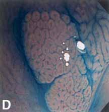
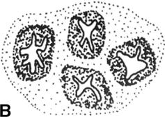
8 Chromo-endoscopy Surface analysis Kudo Type I Kudo Type II
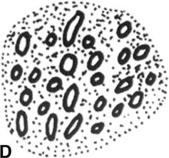
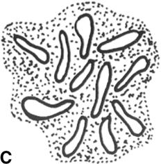
9 Chromo-endoscopy Surface analysis Kudo Type IIIL Kudo Type IIIS
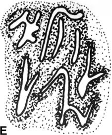
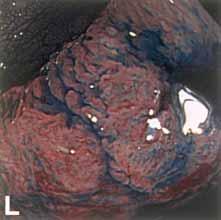
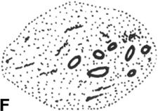
10 Chromo-endoscopy Surface analysis Kudo Type IV Kudo Type V

11 Detection of dysplasia in UC Chromoendoscopy vs. standard 4q biopsies Kiesslich et al, Gastro 2003
12 Novel Endoscopic Imaging Techniques Current optimal techniques High-resolution and magnification endoscopy Chromo-endoscopy New techniques Narrow Band Imaging Autofluorescence Imaging Endo-microscopy

13 Narrow Band Imaging (NBI) Conventional Filter B G R NBI Filter B G R

 imaging")
14 Narrow Band Imaging (NBI) Conventional Filter B G R B B G R NBI Filter G R Band-pass ranges of RGB are narrowed Relatively more contribution of blue light Result: more superficial (=mucosal) imaging


15 Narrow Band Imaging Standard Modus CCD CV-180 NBI- Filter Standard Modus Mucosa CLV-180


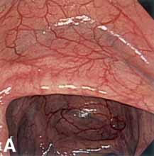
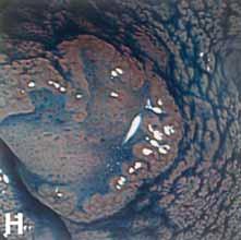
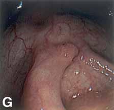
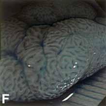
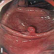
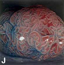
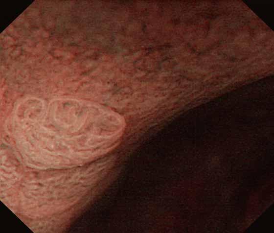
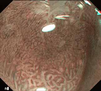
16 Narrow Band Imaging Colon


17 Narrow Band Imaging Colon


18 Narrow Band Imaging Colon

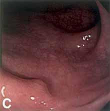
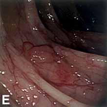
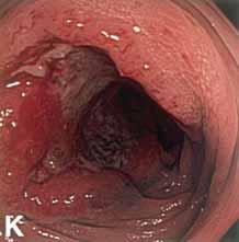
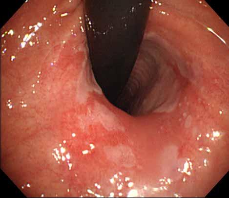
19 NBI Flat Adenoma in UC
20 Narrow Band Imaging UC surveillance Comparative study in UC-surveillance 2 colonoscopies 6-8 wks apart NBI with targetted biopsies Standard colonoscopy with 4 biopsies / 10cm Different endoscopist 08/04: 44 patients included Dekker et al, DDW New Orleans 2004
21 Narrow Band Imaging UC surveillance First colo Feasible Time (min) # biopsies Conventional (n=44) (25-90) 38 (20-68) NBI (n=44) (20-90) 4 (0-26) Dekker et al, DDW New Orleans 2004
22 Narrow Band Imaging UC surveillance Number of endoscopies with abnormalities Dysplastic lesions Random biopsies with dysplasia Total number of patiens with dysplasia Conventional 14 9/14 3/43 12 NBI 15 9/15 n.a. 9 Dekker et al, DDW New Orleans 2004
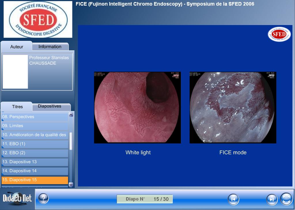
23 Fujinon Intelligent Chromo Endoscopy FICE = post-processing processing technique No scientific data available PubMed negative DDW2006 abstracts negative
24
25
26 Novel Endoscopic Imaging Techniques Current optimal techniques High-resolution and magnification endoscopy Chromo-endoscopy New techniques Narrow Band Imaging Autofluorescence Imaging Endo-microscopy
27 Fluorescence Imaging Excitation light (short wavelength) Fluorescent light (longer wavelength) Tissue fluorophores

28 AFI Image Monochromatic CCD Barrier Filter Mucosa Xenon lamp Rotary Filter
29
30
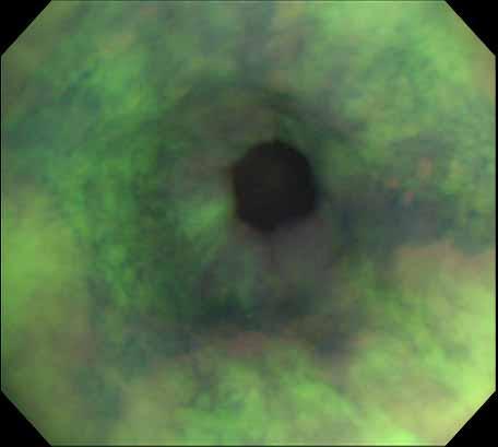
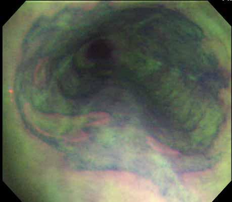
31 AFI feasibility study in 60 pts 21 pts had detectable lesions with HGIN 14 pts: abnormalities with WLE & AFI 7 pts: lesions detected with AFI but not with WLE 6 pts: 11 additional lesions with AFI Kara et al, GIEndoscopy 2005
32 False positive rate of AFI AFI Positive HGD/ EC 40 LGD 8 IN D 5 NDBE 28 Tota l 81 False positive rate: ~ 50% Kara et al, GIEndoscopy 2005
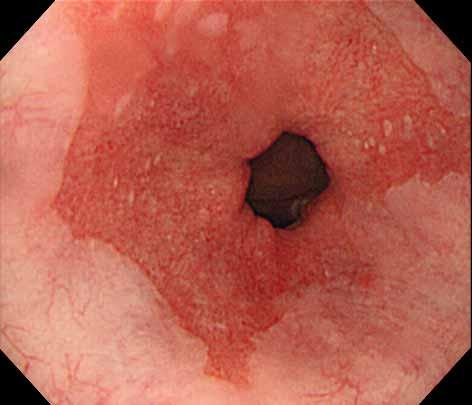
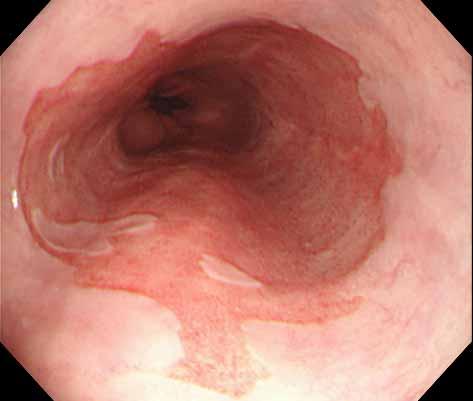
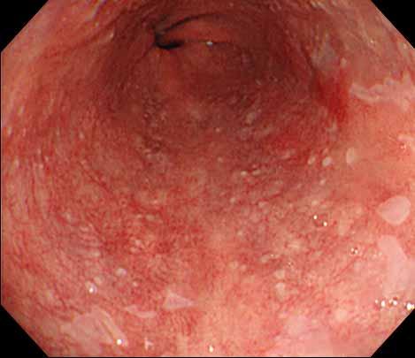
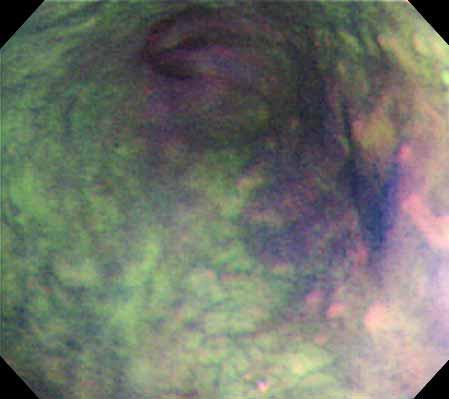
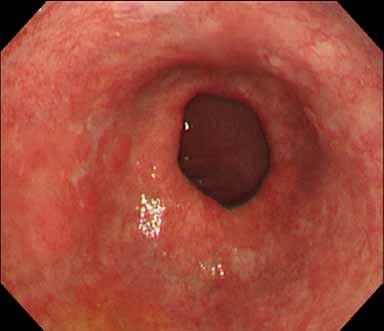
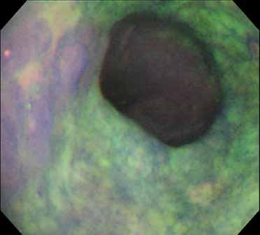
33 Endoscopic Tri-Modal Imaging ETMI (WLE,, AFI en NBI)
34 Novel Endoscopic Imaging Techniques Current optimal techniques High-resolution and magnification endoscopy Chromo-endoscopy New techniques Narrow Band Imaging Autofluorescence Imaging Endo-microscopy
 Joint venture:")

35 Confocal Laser Endoscopy (Pentax) Joint venture: Pentax, Japan & Optiscan, Australia Images provided by Dr Kiesslich, Mainz

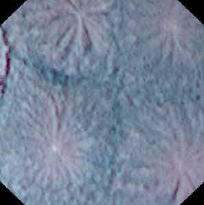
36 Normal Rectum Images provided by Dr Kiesslich, Mainz DDW 2004

37 Adenoma; HGD Images provided by Dr Kiesslich, Mainz - Gastro 2004


38 Endo-Cytoscopy System (Olympus) Probe-type videoscope system with ultra high magnification Compatible to 3.7mm working channel Very high resolution: 1.7 or 4.2 µm Working depth: less than 30 µm Absorptive stain necessary
39 Endo-Cytoscopy System (Olympus)
40 Novel Endoscopic Imaging Techniques New techniques are necessary We miss flat + depressed lesions and dysplasia in UC Chromo-endoscopy is good & easy but underused Indigo-carmine should always be available NBI is now commercially available Olympus 180 series: High resolution (HDTV) + NBI AFI is very attractive as a red-flag technique All new techniques need scientific evaluation
41
Real-Time in vivo Observation of Cells and Nuclei Opens New Possibilities for Diagnostic Endoscopy
 Beyond Imagination Introducing Endocyto, Olympus has broken a new ground in endoscopy. Ultra-high magnification with up to 520x magnification ratio enables observation on microscopic level and helps to
Fuji Intelligent Chromo Endoscopy
 Fuji Intelligent Chromo Endoscopy The next generation of endoscopic diagnosis has arrived with Fujinon's new EPX-4400 video processor. F.I.C.E. (FUJI Intelligent Chromo Endoscopy, ) installed in the EPX-4400,
Quantitative analysis and development of a computer-aided system for identification of
 Quantitative analysis and development of a computer-aided system for identification of regular pit patterns of colorectal lesions Yoshito Takemura, MD, 1 Shigeto Yoshida, MD, 2 Shinji Tanaka, MD, 2 5 Keiichi
Towards Optical Biopsies with an Integrated Fibered Confocal Fluorescence Microscope
 Towards Optical Biopsies with an Integrated Fibered Confocal Fluorescence Microscope Georges Le Goualher, Aymeric Perchant, Magalie Genet, Charlotte Cavé, Bertrand Viellerobe, Fredéric Berier, Benjamin
EXpERIENCE THE power Of LIGHT
 EXpERIENCE THE power Of LIGHT GASTROENTEROLOGY MAKING YOUR DAILY WORK EaSIER ENDOSCOpES processors preparation & HYGIENICS ULTRa- SONOGRapHY ENDOSCOpY SYSTEM fujifilm is a pioneer in diagnostic imaging
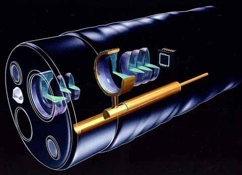
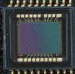
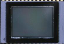
Paul Musto. Endoscopes and their construction
 Paul Musto Endoscopes and their construction Types of Endoscopes Flexible instruments Rigid instruments Fibre optic imaging systems CCD imaging systems Medical Endoscopes Industrial Endoscopes History
Magnified Examination of Small Colorectal Polyps Using a Prototype Electronic Endoscope: Preliminary Experience
 Diagnostic and Therapeutic Endoscopy, Vol. 6, pp. 77-82 Reprints available directly from the publisher Photocopying permitted by license only (C) 2000 OPA (Overseas Publishers Association) N.V. Published
2017 ADVANCING DEEPER INSIGHTS IN ENDOSCOPY
 2017 ADVANCING DEEPER INSIGHTS IN ENDOSCOPY CONTENT About Fujifilm 4 5 Technologies 6 13 ELUXEO 700 series 14 19 600 Series 20 23 580 Series 24 27 Double Balloon Endoscopy System 28 33 530 Series 34 39
Advancing the Art of Endoscopy
 Advancing the Art of Endoscopy Advancing the Art of Endoscopy with an array of opto-digital innovations. OLYMPUS technology continues to advance the art of endoscopy. As the world leader in endoscopy,
Single balloon Enteroscope system SIF-Q180 / OBCU
 Single Balloon Enteroscope System Single balloon Enteroscope system SIF-Q180 / OBCU There s only one answer to your requirements: the Single Balloon System from Olympus Despite the rapid technological
EPX-3500 HD FUJIFILM High-end Electronic Video Endoscopy System
 New EPX-3500 HD FUJIFILM High-end Electronic Video Endoscopy System FUJIFILM Asia Pacific Pte. Ltd. 10 New Industrial Road, FUJIFILM Building, Singapore 536201 http://www.fujifilm.com.sg High-definition
In-Vivo IMAGING SYSTEMS. A complete line of high resolution optical & X-ray systems for pre-clinical imaging
 In-Vivo IMAGING SYSTEMS A complete line of high resolution optical & X-ray systems for pre-clinical imaging In-Vivo Imaging Systems Carestream is a strong, successful, multi-billion dollar, international
In vivo white light and contrastenhanced
 In vivo white light and contrastenhanced vital-dye fluorescence imaging of Barrett s-related neoplasia in a single-endoscopic insertion Yubo Tang Jennifer Carns Alexandros D. Polydorides Sharmila Anandasabapathy
Invitation for a walk through microscopy. Sebastian Schuchmann Jörg Rösner
 Invitation for a walk through microscopy Sebastian Schuchmann Jörg Rösner joerg.roesner@charite.de Techniques in microscopy Conventional (light) microscopy bright & dark field, phase & interference contrast
Endoscopy Equipment. D. J. McMahon rev cewood
 Endoscopy Equipment D. J. McMahon 150512 rev cewood 2018-01-29 Key Points Medical Video 2 - Endoscopy: Know the proper installation and hazards associated with xenon lamps Know what an insufflator is and
Examination, TEN1, in courses SK2500/SK2501, Physics of Biomedical Microscopy,
 KTH Applied Physics Examination, TEN1, in courses SK2500/SK2501, Physics of Biomedical Microscopy, 2009-06-05, 8-13, FB51 Allowed aids: Compendium Imaging Physics (handed out) Compendium Light Microscopy
At Fujinon we are committed to delivering innovative endoscopic solutions exactly as envisioned by our customers. Our latest achievement, the Fujinon
 At Fujinon we are committed to delivering innovative endoscopic solutions exactly as envisioned by our customers. Our latest achievement, the Fujinon 4400, is a crowning example of the most advanced optical
Medical Video 2. Endoscopy Equipment. Bronchoscopy. Colonoscopy. Gastroscopy. Key Points Medical Video 2 - Endoscopy:
 Medical Video 2 Endoscopy Equipment D. J. McMahon 150512 rev cewood 2018-01-29 Key Points Medical Video 2 - Endoscopy: Know the proper installation and hazards associated with xenon lamps Know what an
Optical coherence tomography
 Optical coherence tomography Peter E. Andersen Optics and Plasma Research Department Risø National Laboratory E-mail peter.andersen@risoe.dk Outline Part I: Introduction to optical coherence tomography
Basics of confocal imaging (part I)
") Basics of confocal imaging (part I) Swiss Institute of Technology (EPFL) Faculty of Life Sciences Head of BIOIMAGING AND OPTICS BIOP arne.seitz@epfl.ch Lateral resolution BioImaging &Optics Platform Light
Basics of confocal imaging (part I) Swiss Institute of Technology (EPFL) Faculty of Life Sciences Head of BIOIMAGING AND OPTICS BIOP arne.seitz@epfl.ch Lateral resolution BioImaging &Optics Platform Light
OPTIVISTA TM EPK-i7010
 OPTIVIST TM EPK-i7010 unique combination of optical and digital enhancement in one endoscopy processor Courtesy of Dr. Federico uffoli, Cremona Hospital, Italy PENTX MEDICL OPTIVIST TM EPK-i7010 Enlighten
The Compound Microscope. Brightfield: Köhler Illumination
 Outline History of Microscopy The Magnifying Glass The Compound Microscope Brightfield: Köhler Illumination Microscopy µικροσ (mikros): small σκοπειν (skopein): to observe History of Microscopy Well :
Lumencor White Paper: Solid state light engines for bioanalytical instruments and biomedical devices
 lumencor Lumencor White Paper: Solid state light engines for bioanalytical instruments and biomedical devices Prepared by: Claudia B. Jaffe, Ph.D., Vice President, Business Development and Steven M. Jaffe,
Guidebook Endoscopes 2014/2015. New horizons in Endoscopy
 Guidebook Endoscopes 2014/2015 New horizons in Endoscopy 600 Series endoscopes name Size of image Gastroscopes EG-600WR 2 100 140 9.2 9.3 2.8 210 /90 100 /100 1,100 130 % EG-600ZW Colonoscopes WD: 3-100
a) How big will that physical image of the cells be your camera sensor?
 How big will that physical image of the cells be your camera sensor?") 1. Consider a regular wide-field microscope set up with a 60x, NA = 1.4 objective and a monochromatic digital camera with 8 um pixels, properly positioned in the primary image plane. This microscope is
1. Consider a regular wide-field microscope set up with a 60x, NA = 1.4 objective and a monochromatic digital camera with 8 um pixels, properly positioned in the primary image plane. This microscope is
Advancing the Art of Endoscopy
 Advancing the Art of Endoscopy Advancing the Art of Endoscopy with an array of opto-digital innovations. OLYMPUS technology continues to advance the art of endoscopy. As the world leader in endoscopy,
Products - Microarray Scanners - Laser Scanners - InnoScan 900 Series and MAPIX Software
 Products - Microarray Scanners - Laser Scanners - InnoScan 900 Series and MAPIX Software Arrayit offers the world s only next generation microarray scanning technology, with proprietary rotary motion control,
CANCER INSTITUTE (WIA) CANAL BANK ROAD, ADYAR, CHENNAI
 CANAL BANK ROAD, ADYAR, CHENNAI") Minutes of Pre-bid Conference CANCER INSTITUTE (WIA) Procurement of : ENDOSCOPIC ULTRASOUND BIDREFERENCE : CI: SCI:NP/SOG/P-I/2016-17/01 DATED 15/12/2016 Venue : Board Room, Lion Cancer Support Center,
Minutes of Pre-bid Conference CANCER INSTITUTE (WIA) Procurement of : ENDOSCOPIC ULTRASOUND BIDREFERENCE : CI: SCI:NP/SOG/P-I/2016-17/01 DATED 15/12/2016 Venue : Board Room, Lion Cancer Support Center,
Physics limited resolution of videoscopes Pushing the limits of resolution and why optics know-how is now critical
 Physics limited resolution of videoscopes Pushing the limits of resolution and why optics know-how is now critical Frank Lafleur Product Manager Feb 2, 2017 How does Olympus lead in the world of optics
Guidebook Endoscopes
 2016 Guidebook Endoscopes 2 3 600 series oscopes name Working length Gastroscopes EG-600WR 2 100 140 9.2 9.3 2.8 210 /90 100 /100 1,100 130 % Water jet EG-600ZW WD: 3-100 TL: 1.5-2.5 WD: 140 TL: 56 9.9
X-Rays and endoscopes
 X-Rays and endoscopes 1 What are X-rays? X-ray refers to electromagnetic radiation with a wavelength between 0.01nm - 10nm. increasing wavelength visible light ultraviolet x-ray increasing energy X-rays
ATLAS. Atlas of Spectral Endoscopic Images
 Atlas of Spectral Endoscopic Images ATLAS Atlas of Spectral Endoscopic Images Academic Research Report for Health Care Professionals Supervised by Yoichi Miyake, Research Center for Frontier Medical Engineering,
(Quantitative Imaging for) Colocalisation Analysis
 Colocalisation Analysis") (Quantitative Imaging for) Colocalisation Analysis or Why Colour Merge / Overlay Images are EVIL! Special course for DIGS-BB PhD program What is an Image anyway..? An image is a representation of reality
(Quantitative Imaging for) Colocalisation Analysis or Why Colour Merge / Overlay Images are EVIL! Special course for DIGS-BB PhD program What is an Image anyway..? An image is a representation of reality
You won t be able to measure the incident power precisely. The readout of the power would be lower than the real incident power.
 1. a) Given the transfer function of a detector (below), label and describe these terms: i. dynamic range ii. linear dynamic range iii. sensitivity iv. responsivity b) Imagine you are using an optical
TECHNICAL DATA. OPTIV CLASSIC 322 Version 3/2013
 TECHNICAL DATA OPTIV CLASSIC 322 Version 3/2013 Technical Data Product description The Optiv Classic 322 combines optical and tactile measurement in one system (optional touchtrigger probe). The system
The Sussex Declaration
 THE BRITISH LIBRARY The Sussex Declaration Technical report for Add Mss 8981 Duffy, Christina 11/1/2017 Technical report of scientific analysis held at the British Library 1-3 August 2017 of Add Mss 8981
Bandpass Edge Dichroic Notch & More
 Edmund Optics BROCHURE Filters COPYRIGHT 217 EDMUND OPTICS, INC. ALL RIGHTS RESERVED 1/17 Bandpass Edge Dichroic Notch & More Contact us for a Stock or Custom Quote Today! USA: +1-856-547-3488 EUROPE:
Things to check before start-up.
 Byeong Cha Page 1 11/24/2009 Manual for Leica SP2 Confocal Microscope Enter you name, the date, the time, and the account number in the user log book. Things to check before start-up. Make sure that your
Ratio Imaging. Dividing one image by another to detect changing conditions. Images collected at different times, wavelengths, polarities, etc
 Ratio Imaging Dividing one image by another to detect changing conditions Images collected at different times, wavelengths, polarities, etc Most common use of ratio imaging is to provide a quick spectral
Compound Light Microscopy. Microscopy. Things to remember... 1/22/2017. This is what we use in the laboratory
 Compound Light Microscopy This is what we use in the laboratory Microscopy Chapter 3 BIO 440 A series of finely ground lenses is used to form a magnified image Specimen is illuminated with visible light
User manual for Olympus SD-OSR spinning disk confocal microscope
 User manual for Olympus SD-OSR spinning disk confocal microscope Ved Prakash, PhD. Research imaging specialist Imaging & histology core University of Texas, Dallas ved.prakash@utdallas.edu Once you open
3D light microscopy techniques
 3D light microscopy techniques The image of a point is a 3D feature In-focus image Out-of-focus image The image of a point is not a point Point Spread Function (PSF) 1D imaging 2D imaging 3D imaging Resolution
LSM 710 Confocal Microscope Standard Operation Protocol
 LSM 710 Confocal Microscope Standard Operation Protocol Basic Operation Turning on the system 1. Switch on Main power switch 2. Switch on System / PC power button 3. Switch on Components power button 4.
A Compact, High-Performance Ultrasound System That Enables Endoscopic Ultrasonography With Convenient Ultrasonic Probes. Endoscopic Ultrasound Center
 A Compact, High-Performance Ultrasound System That Enables Endoscopic Ultrasonography With Convenient Ultrasonic Probes Endoscopic Ultrasound Center EU-M30S Make Ultrasonography A Part Of Your Routine
Non-Descanned FLIM Detection in Multiphoton Microscopes
 Non-Descanned FLIM Detection in Multiphoton Microscopes Abstract. Multiphoton microscopes use a femtosecond NIR laser to excite fluorescence in the sample. Excitation is performed via a multi-photon absorption
MIRAX SCAN The new way of looking at pathology
 Microscopy from Carl Zeiss MIRAX SCAN The new way of looking at pathology Greater reliability. Greater efficiency. Plus points for your diagnostics Better. More efficient. Quality as a factor for success
technology meets pathology Institute of Pathology, Charité Universitätsmedizin Berlin, Berlin, Germany 3 Overview
 ASSESSMENT OF TECHNICAL PARAMETERS A. Alekseychuk 1, N. Zerbe 2, Y. Yagi 3 1 Computer Vision and Remote Sensing, TU Berlin, Berlin, Germany 2 Institute of Pathology, Charité Universitätsmedizin Berlin,
Microscope anatomy, image formation and resolution
 Microscope anatomy, image formation and resolution Ian Dobbie Buy this book for your lab: D.B. Murphy, "Fundamentals of light microscopy and electronic imaging", ISBN 0-471-25391-X Visit these websites:
Fibered confocal spectroscopy and multicolor imaging system for in vivo fluorescence analysis
 Fibered confocal spectroscopy and multicolor imaging system for in vivo fluorescence analysis Florence Jean, Genevieve Bourg-Heckly Universite Pierre et Marie Curie, Laboratoire de Biophysique Moleculaire
3D light microscopy techniques
 3D light microscopy techniques The image of a point is a 3D feature In-focus image Out-of-focus image The image of a point is not a point Point Spread Function (PSF) 1D imaging 1 1 2! NA = 0.5! NA 2D imaging
 07 Setting Place a specimen, and select a fluorescence dye. The FV10i automatically selects the most suitable imaging conditions based on the fluorescence dye selection. Set Image mapping menu Just click
INTRODUCTION TO MICROSCOPY. Urs Ziegler THE PROBLEM
 INTRODUCTION TO MICROSCOPY Urs Ziegler ziegler@zmb.uzh.ch THE PROBLEM 1 ORGANISMS ARE LARGE LIGHT AND ELECTRONS: ELECTROMAGNETIC WAVES v = Wavelength ( ) Speed (v) Frequency ( ) Amplitude (A) Propagation
Nature Methods: doi: /nmeth Supplementary Figure 1. Schematic of 2P-ISIM AO optical setup.
 Supplementary Figure 1 Schematic of 2P-ISIM AO optical setup. Excitation from a femtosecond laser is passed through intensity control and shuttering optics (1/2 λ wave plate, polarizing beam splitting
Resolution. Diffraction from apertures limits resolution. Rayleigh criterion θ Rayleigh = 1.22 λ/d 1 peak at 2 nd minimum. θ f D
 Microscopy Outline 1. Resolution and Simple Optical Microscope 2. Contrast enhancement: Dark field, Fluorescence (Chelsea & Peter), Phase Contrast, DIC 3. Newer Methods: Scanning Tunneling microscopy (STM),
Fluorescence Microscopy Light Sources
 Kavita Aswani, 1 Tushare Jinadasa, 2 and Claire M. Brown 2,3 * 1 Life Sciences Division, Lumen Dynamics, 2260 Argentia Rd., Mississauga, Ontario, L5N 6H7, Canada 2 Department of Physiology, McGill University,
2016 ADVANCING DEEPER INSIGHTS IN ENDOSCOPY
 2016 ADVANCING DEEPER INSIGHTS IN ENDOSCOPY CONTENT The Company 4 5 Technologies 6 11 600 Series 12 17 580 Series 18 21 Double Balloon Endoscopy System 22 27 590 & 530 Series 28 35 EPX Video-Processors
Ayuekanbe Atagabe. Physics 464(applied Optics) Winter Project Report. Fiber Optics in Medicine. March 11, 2003
 Winter Project Report. Fiber Optics in Medicine. March 11, 2003") Ayuekanbe Atagabe Physics 464(applied Optics) Winter 2003 Project Report Fiber Optics in Medicine March 11, 2003 Abstract: Fiber optics have become very important in medical diagnoses in this modern era
Ayuekanbe Atagabe Physics 464(applied Optics) Winter 2003 Project Report Fiber Optics in Medicine March 11, 2003 Abstract: Fiber optics have become very important in medical diagnoses in this modern era
Practical work no. 3: Confocal Live Cell Microscopy
 Practical work no. 3: Confocal Live Cell Microscopy Course Instructor: Mikko Liljeström (MIU) 1 Background Confocal microscopy: The main idea behind confocality is that it suppresses the signal outside
Operating Instructions for Zeiss LSM 510
 Operating Instructions for Zeiss LSM 510 Location: GNL 6.312q (BSL3) Questions? Contact: Maxim Ivannikov, maivanni@utmb.edu 1 Attend A Complementary Training Before Using The Microscope All future users
TECHNICAL DATA OPTIV CLASSIC 432
 TECHNICAL DATA OPTIV CLASSIC 432 Technical Data Product description The Optiv Classic 432 combines optical and tactile measurement in one system (optional touchtrigger probe). The system supports multi-sensor
Lecture 23 MNS 102: Techniques for Materials and Nano Sciences
 Lecture 23 MNS 102: Techniques for Materials and Nano Sciences Reference: #1 C. R. Brundle, C. A. Evans, S. Wilson, "Encyclopedia of Materials Characterization", Butterworth-Heinemann, Toronto (1992),
Observing Microorganisms through a Microscope LIGHT MICROSCOPY: This type of microscope uses visible light to observe specimens. Compound Light Micros
 PHARMACEUTICAL MICROBIOLOGY JIGAR SHAH INSTITUTE OF PHARMACY NIRMA UNIVERSITY Observing Microorganisms through a Microscope LIGHT MICROSCOPY: This type of microscope uses visible light to observe specimens.
Boulevard du Temple Daguerrotype (Paris,1838) a busy street? Nyquist sampling for movement
 a busy street? Nyquist sampling for movement") Boulevard du Temple Daguerrotype (Paris,1838) a busy street? Nyquist sampling for movement CONFOCAL MICROSCOPY BioVis Uppsala, 2017 Jeremy Adler Matyas Molnar Dirk Pacholsky Widefield & Confocal Microscopy
Boulevard du Temple Daguerrotype (Paris,1838) a busy street? Nyquist sampling for movement CONFOCAL MICROSCOPY BioVis Uppsala, 2017 Jeremy Adler Matyas Molnar Dirk Pacholsky Widefield & Confocal Microscopy
Zeiss Axiovert 135 Fluorescence Microscope Quick Guide / Operations Manual (v. 1.0 February 09)
") University of Chicago Integrated Light Microscopy Core Dr. Vytas Bindokas, Director http://digital.bsd.uchicago.edu By: Christine Labno, Assistant Director Room: AB-129 Phone: 4-9040 Zeiss Axiovert 135
University of Chicago Integrated Light Microscopy Core Dr. Vytas Bindokas, Director http://digital.bsd.uchicago.edu By: Christine Labno, Assistant Director Room: AB-129 Phone: 4-9040 Zeiss Axiovert 135
Study on the Binder Distribution related to Drying
 International Symposium on Computers & Informatics (ISCI 2015) Study on the Binder Distribution related to Drying Ying Li 1,a, Qinming Wang 1, Wenjuan Gu 1 and Banggui He 1 1 Faculty of Mechanical and
Miscellaneous Accessories and technical equipment.
 Miscellaneous Accessories and technical equipment. Accessories Accessories. Clever equipment for smart endoscopy. MICRO-TECH has a close cooperation with the company Medgic a member of the MICRO-TECH Group.
Administrative details:
 Administrative details: Anything from your side? www.photonics.ethz.ch 1 What are we actually doing here? Optical imaging: Focusing by a lens Angular spectrum Paraxial approximation Gaussian beams Method
Quantitative, spectrally- resolved intraoperative
 1 Quantitative, spectrally- resolved intraoperative fluorescence imaging Pablo A. Valdés 1,2,3, Frederic Leblond 1, Valerie L. Jacobs 2, Brian C. Wilson 5, Keith D. Paulsen 1,2,4, & David W. Roberts 2,3,4
GRINTECH GmbH. product information.
 GRINTECH GmbH product information www.grintech.de GRIN rod lenses Gradient index lenses for fiber coupling and beam shaping of laser diodes z l d s f Order example: GT-LFRL-100-025-50-CC (670) Design wavelength
Bio 407. Applied microscopy. Introduction into light microscopy. José María Mateos. Center for Microscopy and Image Analysis
 Center for Microscopy and Image Analysis Bio 407 Applied Introduction into light José María Mateos Fundamentals of light Compound microscope Microscope composed of an objective and an additional lens (eyepiece,
Supplemental Figure 1: Histogram of 63x Objective Lens z axis Calculated Resolutions. Results from the MetroloJ z axis fits for 5 beads from each
 Supplemental Figure 1: Histogram of 63x Objective Lens z axis Calculated Resolutions. Results from the MetroloJ z axis fits for 5 beads from each lens with a 1 Airy unit pinhole setting. Many water lenses
Designed to improve physician productivity and patient care.
 Designed to improve physician productivity and patient care. Fujinon s most advanced 400 Video Bronchoscopes are designed for physician productivity and patient care. Advanced miniature chip technology
The Nature of Light. Light and Energy
 The Nature of Light Light and Energy - dependent on energy from the sun, directly and indirectly - solar energy intimately associated with existence of life -light absorption: dissipate as heat emitted
AxioCam HR Success Through Performance
 Microscopy from Carl Zeiss AxioCam HR Success Through Performance The high-resolution camera for digital documentation Superior performance for research and routine work brilliant quality documentation
Bi Imaging. Multicolor Imaging: The Important Question of Co-Localization. Anna Smallcombe Bio-Rad Laboratories, Hemel Hempstead, UK
 Multicolor Imaging: The Important Question of Co-Localization Anna Smallcombe Bio-Rad Laboratories, Hemel Hempstead, UK The use of specific fluorescent probes, combined with confocal or multiphoton microscopy
picoemerald Tunable Two-Color ps Light Source Microscopy & Spectroscopy CARS SRS
 picoemerald Tunable Two-Color ps Light Source Microscopy & Spectroscopy CARS SRS 1 picoemerald Two Colors in One Box Microscopy and Spectroscopy with a Tunable Two-Color Source CARS and SRS microscopy
OPTICAL PRINCIPLES OF MICROSCOPY. Interuniversity Course 28 December 2003 Aryeh M. Weiss Bar Ilan University
 OPTICAL PRINCIPLES OF MICROSCOPY Interuniversity Course 28 December 2003 Aryeh M. Weiss Bar Ilan University FOREWORD This slide set was originally presented at the ISM Workshop on Theoretical and Experimental
T:GENIUS GEL IMAGING AT A TOUCH
 T:GENIUS GEL IMAGING AT A TOUCH The T:Genius is an integrated system for DNA and protein analysis and gel documentation. Based on the successful Syngene gel documentation range, the T:Genius features an
Systematic Workflow via Intuitive GUI. Easy operation accomplishes your goals faster than ever.
 Systematic Workflow via Intuitive GUI Easy operation accomplishes your goals faster than ever. 16 With the LEXT OLS4100, observation or measurement begins immediately once the sample is placed on the stage.
STORM/ PALM ANSWER KEY
 STORM/ PALM ANSWER KEY Phys598BP Spring 2016 University of Illinois at Urbana-Champaign Questions for Lab Report 1. How do you define a resolution in STORM imaging? If you are given a STORM setup, how
Final Exam, 150 points PMB 185: Techniques in Light Microscopy
 Final Exam, 150 points Name PMB 185: Techniques in Light Microscopy Point value is in parentheses at the end of each question. Note: GFP = green fluorescent protein ; CFP = cyan fluorescent protein ; YFP
Operation Guide for the Leica SP2 Confocal Microscope Bio-Imaging Facility Hunter College October 2009
 Operation Guide for the Leica SP2 Confocal Microscope Bio-Imaging Facility Hunter College October 2009 Introduction of Fluoresence Confocal Microscopy The first confocal microscope was invented by Princeton
Femtosecond laser microfabrication in. Prof. Dr. Cleber R. Mendonca
 Femtosecond laser microfabrication in polymers Prof. Dr. Cleber R. Mendonca laser microfabrication focus laser beam on material s surface laser microfabrication laser microfabrication laser microfabrication
Confocal Imaging Through Scattering Media with a Volume Holographic Filter
 Confocal Imaging Through Scattering Media with a Volume Holographic Filter Michal Balberg +, George Barbastathis*, Sergio Fantini % and David J. Brady University of Illinois at Urbana-Champaign, Urbana,
OPTIV CLASSIC 321 GL TECHNICAL DATA
 OPTIV CLASSIC 321 GL TECHNICAL DATA TECHNICAL DATA Product description The Optiv Classic 321 GL offers an innovative design for non-contact measurement. The benchtop video-based measuring machine is equipped
Confocal Microscopy and Related Techniques
 Confocal Microscopy and Related Techniques Chau-Hwang Lee Associate Research Fellow Research Center for Applied Sciences, Academia Sinica 128 Sec. 2, Academia Rd., Nankang, Taipei 11529, Taiwan E-mail:
LSM 780 Confocal Microscope Standard Operation Protocol
 LSM 780 Confocal Microscope Standard Operation Protocol Basic Operation Turning on the system 1. Sign on log sheet according to Actual start time 2. Check Compressed Air supply for the air table 3. Switch
Figure 1. Relative intensity of solar energy of different wavelength at the earth's surface.
 Spectrum of light from the sun: Fig.1 Figure 1. Relative intensity of solar energy of different wavelength at the earth's surface. Properties of light 1-The speed of light changes when it goes from one
Εισαγωγική στην Οπτική Απεικόνιση
 Εισαγωγική στην Οπτική Απεικόνιση Δημήτριος Τζεράνης, Ph.D. Εμβιομηχανική και Βιοϊατρική Τεχνολογία Τμήμα Μηχανολόγων Μηχανικών Ε.Μ.Π. Χειμερινό Εξάμηνο 2015 Light: A type of EM Radiation EM radiation:
for courses SK2500 & SK2501, Physics of Biomedical Microscopy, Physics of Biomedical Microscopy, Extended Course Kjell Carlsson
 ? Biomedical & X-ray Physics Kjell Carlsson Problems (and solutions) for courses SK500 & SK501, Physics of Biomedical Microscopy, Physics of Biomedical Microscopy, Extended Course by Kjell Carlsson Physics
FLUORESCENCE MICROSCOPY. Matyas Molnar and Dirk Pacholsky
 FLUORESCENCE MICROSCOPY Matyas Molnar and Dirk Pacholsky 1 The human eye perceives app. 400-700 nm; best at around 500 nm (green) Has a general resolution down to150-300 μm (human hair: 40-250 μm) We need
High-sensitivity. optical molecular imaging and high-resolution digital X-ray. In-Vivo Imaging Systems
 High-sensitivity optical molecular imaging and high-resolution digital X-ray In-Vivo Imaging Systems In vivo imaging solutions available in several packages Carestream Molecular Imaging offers a selection
FLUORESCENCE MICROSCOPY
 FLUORESCENCE MICROSCOPY Methods for Cell Analysis Course BioVis Uppsala, 2015 Matyas Molnar and Dirk Pacholsky 1 Information This lecture contains images and information from the following internet homepages
Advanced Optical Microscopy
 Nanosystems I - Seminar TU München 8th December 2008 1 Introduction to Classical Optical Microscopy Denitions in Optical Microscopy Contrast and Contrast Enhancement 1 Introduction to Classical Optical
Last updated: May 2014 Y.DeGraaf
 FLINDERS MICROSCOPY BIOMEDICAL SERVICES AVAILABLE MICROSCOPES AND SPECIFICATIONS & INFORMATION REGARDING TRAINING FOR NEW USERS Last updated: May 2014 Y.DeGraaf If you have new staff or students (Honours/Masters
MINING COLONOSCOPY VIDEOS TO MEASURE QUALITY OF COLONOSCOPIC PROCEDURES
 MINING COLONOSCOPY VIDEOS TO MEASURE QUALITY OF COLONOSCOPIC PROCEDURES Danyu Liu a, Yu Cao a, Wallapak Tavanapong a, Johnny Wong a, JungHwan Oh b, and Piet C. de Groen c a Department of Computer Science,
contents TABLE OF The SECOM platform Applications - sections Applications - whole cells Features Integrated workflow Automated overlay
 S E C O M TABLE OF contents The SECOM platform 4 Applications - sections 5 Applications - whole cells 8 Features 9 Integrated workflow 12 Automated overlay ODEMIS - integrated software Specifications 13
1.The Problem LIGHT-LEVEL LEVEL IMAGING. light-level level Cameras. 3. Solutions. 2. Low-light LOW-LIGHT
 LOW-LIGHT LIGHT-LEVEL LEVEL IMAGING 1.The Problem 2. Low-light light-level level Cameras 3. Solutions How Much Light? I. Illumination system: 75 W Xenon Arc (~1mW/nm in visible) 490/10 nm exciter filter
TCSPC at Wavelengths from 900 nm to 1700 nm
 TCSPC at Wavelengths from 900 nm to 1700 nm We describe picosecond time-resolved optical signal recording in the spectral range from 900 nm to 1700 nm. The system consists of an id Quantique id220 InGaAs
Why and How? Daniel Gitler Dept. of Physiology Ben-Gurion University of the Negev. Microscopy course, Michmoret Dec 2005
 Why and How? Daniel Gitler Dept. of Physiology Ben-Gurion University of the Negev Why use confocal microscopy? Principles of the laser scanning confocal microscope. Image resolution. Manipulating the