X-Rays and endoscopes
|
|
|
- Caroline Lawrence
- 5 years ago
- Views:
Transcription
1 X-Rays and endoscopes 1
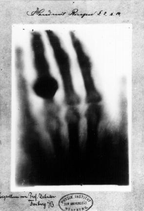
2 What are X-rays? X-ray refers to electromagnetic radiation with a wavelength between 0.01nm - 10nm. increasing wavelength visible light ultraviolet x-ray increasing energy X-rays are used in imaging because their energy is sufficiently high to penetrate human tissue. They were first studied systematically in 1895 by Wilhelm Röntgen (he later received a Nobel prize). 2

3 3
4 Making X-rays X-rays are generated using similar principles to a discharge tube. Electrons travel inside an evacuated tube and strike a tungsten anode. X-rays are released during this collision. Heated filament Electron beam Metal target (tungsten) Evacuated chamber Anode mounting (copper) Coolant circulates here X-rays Window Very high potential difference Jacaranda Physics 2, 3rd Ed. p345 4
5 continued... Only a very small percentage (~1%) of the energy reaching the anode is released as x-ray radiation. The rest is released as heat. This necessitates cooling systems. Tungsten is used as the anode target because of its high melting point (3400 C). Sometimes the target rotates quickly to spread the heat. The angle of anode is used to direct x-rays out of the device. Very high voltages (25kV - 250kV). 5
6 Releasing X-ray photons There are two main mechanisms by which x-ray photons are released in the anode: Braking (Bremsstrahlung) radiation. The electron slows as it strikes the target, and the lost kinetic energy is released in a photon of X-ray radiation. This produces a spectrum of energies. X-ray fluorescence. If an electron strikes a target atom, it may excite (and liberate) a bound electron. As other electrons drop down to fill its place, X-ray radiation of specific energy is released. 6
7 Characteristic wavelengths Relative intensity Wavelengths due to Bremsstrahlung radiation E max Photon energy (kev) Typical output of X-ray generator. The spikes are due to fluorescence and the spectrum due to braking radiation. Jacaranda Physics 2, 3rd Ed. p345 7
8 Questions 1. Calculate the photon energy of the following X-ray wavelengths: a) 10nm b) 0.1nm c) 0.01nm 2. Convert these energies to electron volts (1eV = 1.602x10⁻¹⁹ J). 3. Ultrasound strikes a fat-soft tissue then a soft tissuebone interface. Calculate the fraction of original wave intensity which returns to the detector. 8

9 Soft and hard X-rays We classify X-rays as hard or soft depending on their penetrating ability. The boundary between the two categories is roughly 10keV (~0.1nm). Hard X-rays are required for imaging. We try to eliminate soft X- rays because they expose patients to unnecessary radiation. 9
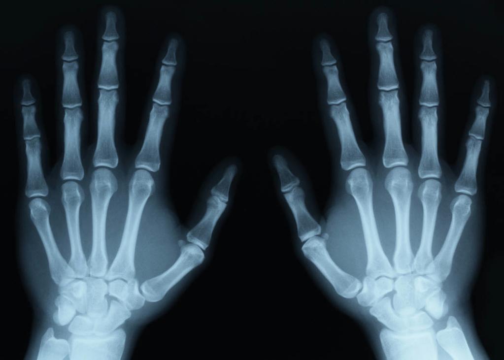
10 Imaging with X-rays The X-rays pass through the body and are absorbed by different structure in different amounts (eg. bones absorb more than muscle). A detector on the other side of the body captures the remaining radiation, which is made into an image. Photographic film was the earliest detector. The spacing of detectors determines maximum image resolution. Usually around 30keV provides the best contrast between different tissue. 10
11 11


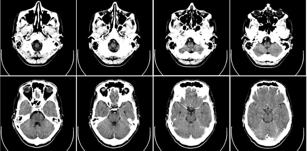
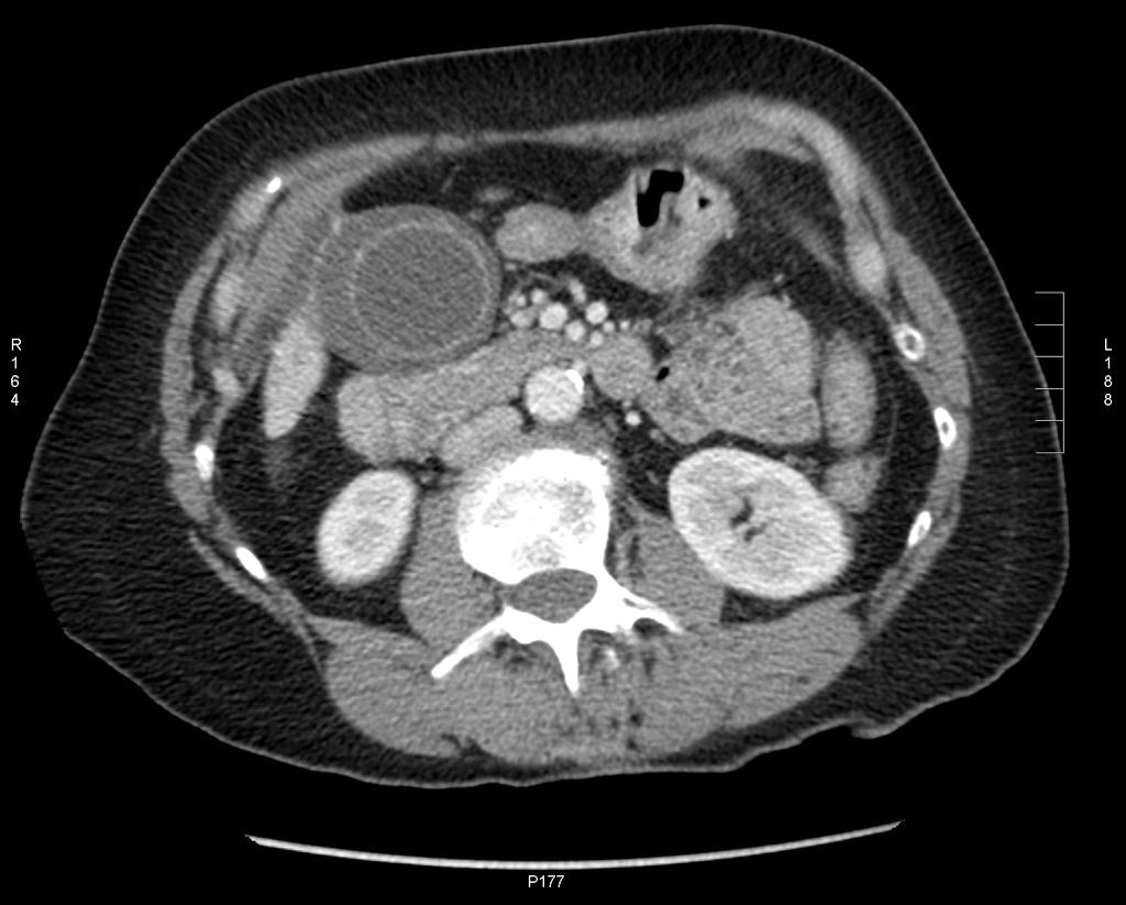
12 CAT scans CAT (computer axial tomography) scans are produced from multiple X-rays taken through a variety of angles. An X-ray source (and array of detectors) rotate around a patient taking multiple snapshots. A computer combines the snapshots into one image 12

13 13
14 14
15 15
16 X-ray vs CAT CAT scans are more expensive and expose the patient to higher levels of radiation but are preferable to X-rays in a number of situations: They have a much higher resolution (so they can show fine detail). They can image soft tissue much better than X- rays (and so can see different organs etc). It is possible to see behind bone (eg. the skull) because of the way that multiple scans are used. 16
17 Homework 1. Print one X-ray of a bone fracture and one X-ray of something else. Discuss the contrast and resolution of each. 2. Find and print a CAT scan and X-ray of the same part of the body. Compare the two images as diagnostic tools. 17
18 Endoscopes An endoscope is a way of imaging the human body with visible light (EM radiation). A small probe is inserted through a body opening: Light travels to and from the probe via fibre optic systems. The incoming light illuminates the region in focus while the returning light carries an image to the operator. Air/water nozzle Biopsy/suction channel Objective lens Illumination lenses Jacaranda Physics 2, 3rd Ed. p345 18


19 Fibre optics Fibre optics work on the principle of total internal reflection. Each strand has a glass core surrounded by a cladding with lower refractive index. total internal reflection 19
20 Coherent bundles A fibre optic bundle (a group of fibre optic strands) can be coherent or incoherent: In coherent bundles each strand is the same length and has the same orientation at both ends of the cable. They are more expensive. Incoherent bundles do not have the same orientation at both ends, and each strand is not necessarily the same length. They are cheaper. Each has a purpose in endoscope technology. 20


 Through non-coherent")


21 (a) Through coherent bundles, the parts of the image are transmitted in correct relative positions. Light reflected from object (b) Through non-coherent bundles, light is transmitted but image has lost its shape as parts are not in correct positions. Coherent bundles must be used to carry the image from the probe, but incoherent bundles are fine for the incoming light to the probe. Jacaranda Physics 2, 3rd Ed. p345 21
22 Putting it together Endoscope inserted into body Light travels down incoherent bundles, illuminates body Light (image) travels back through coherent bundles realtime viewing video camera still camera 22
23 NAME OF PROCEDURE PLACE OF INSERTION OF ENDOSCOPE PURPOSE OF PROCEDURE Arthroscopy Through skin near joint To examine joints and carry out repairs such as removal of torn cartilage Bronchoscopy Through bronchial tubes To examine trachea and lungs to show problems such as inflammation, bronchitis, cancer and tuberculosis Colonoscopy Through the anus To detect problems such as polyps, tumours, ulceration and inflammation in the colon and large intestine Colposcopy Through the vagina To look for problems such as inflammation and cancer in the vagina and cervix (in females) Cytoscopy Through the urinary tract To examine the bladder, urethra and opening of the prostate gland (in males) Endoscope biopsy Through a natural opening or through an incision To remove specimens of tissue for examination and analysis by a pathologist Gastroscopy Through the mouth To look for the source of problems such as bleeding from the lining of the oesophagus, stomach and duodenum Laparoscopy Through an incision in the abdominal wall To examine abdominal organs including the stomach, liver and fallopian tubes (in females) Jacaranda Physics 2, 3rd Ed. p345 23
24 Further details The endoscope shaft is controllable, so that it will bend in the required direction. The shaft may contain tubes for blowing water or carbon dioxide into the body (to clean lenses or inflate cavity for better viewing). The shaft may also contain tubes for sucking biological samples from the body for analysis. This is called a biopsy. Often there are other attachments to cut/manipulate tissue 24
25 Homework Find and print three images of different body structures obtained by endoscopy. 25
PD233: Design of Biomedical Devices and Systems
 PD233: Design of Biomedical Devices and Systems (Lecture-8 Medical Imaging Systems) (Imaging Systems Basics, X-ray and CT) Dr. Manish Arora CPDM, IISc Course Website: http://cpdm.iisc.ac.in/utsaah/courses/
Uses of Electromagnetic Waves
 Uses of Electromagnetic Waves 1 of 42 Boardworks Ltd 2016 Uses of Electromagnetic Waves 2 of 42 Boardworks Ltd 2016 What are radio waves? 3 of 42 Boardworks Ltd 2016 The broadcast of every radio and television
Medical Imaging. X-rays, CT/CAT scans, Ultrasound, Magnetic Resonance Imaging
 Medical Imaging X-rays, CT/CAT scans, Ultrasound, Magnetic Resonance Imaging From: Physics for the IB Diploma Coursebook 6th Edition by Tsokos, Hoeben and Headlee And Higher Level Physics 2 nd Edition
Explain what is meant by a photon and state one of its main properties [2]
![Explain what is meant by a photon and state one of its main properties [2]](/thumbs/80/82516318.jpg "Explain what is meant by a photon and state one of its main properties [2]") 1 (a) A patient has an X-ray scan taken in hospital. The high-energy X-ray photons interact with the atoms inside the body of the patient. Explain what is meant by a photon and state one of its main properties....
1 (a) A patient has an X-ray scan taken in hospital. The high-energy X-ray photons interact with the atoms inside the body of the patient. Explain what is meant by a photon and state one of its main properties....
X-rays. X-rays are produced when electrons are accelerated and collide with a target. X-rays are sometimes characterized by the generating voltage
 X-rays Ouch! 1 X-rays X-rays are produced when electrons are accelerated and collide with a target Bremsstrahlung x-rays Characteristic x-rays X-rays are sometimes characterized by the generating voltage
X-rays in medical diagnostics
 X-rays in medical diagnostics S.Dolanski Babić 2017/18. History W.C.Röntgen (1845-1923) discovered a new type of radiation Nature, Jan. 23. 1896.; Science, Feb.14. 1896. X- rays: Induced the ionization
Figure 1. Relative intensity of solar energy of different wavelength at the earth's surface.
 Spectrum of light from the sun: Fig.1 Figure 1. Relative intensity of solar energy of different wavelength at the earth's surface. Properties of light 1-The speed of light changes when it goes from one
Light has some interesting properties, many of which are used in medicine:
 LIGHT IN MEDICINE Light has some interesting properties, many of which are used in medicine: 1- The speed of light changes when it goes from one material into another. The ratio of the speed of light in
National 3 Physics Waves and Radiation. 1. Wave Properties
 1. Wave Properties What is a wave? Waves are a way of transporting energy from one place to another. They do this through some form of vibration. We see waves all the time, for example, ripples on a pond
Standard Grade Physics Health Physics Ink Exercise G1
 Standard Grade Physics Health Physics Ink Exercise G1 1. Sounds can travel through : A a vacuum B solids only C liquids only D gases only E solids, liquids and gases 2. A doctor uses a stethoscope like
S200 Course LECTURE 1 TEM
 S200 Course LECTURE 1 TEM Development of Electron Microscopy 1897 Discovery of the electron (J.J. Thompson) 1924 Particle and wave theory (L. de Broglie) 1926 Electromagnetic Lens (H. Busch) 1932 Construction
Absorption: in an OF, the loss of Optical power, resulting from conversion of that power into heat.
 Absorption: in an OF, the loss of Optical power, resulting from conversion of that power into heat. Scattering: The changes in direction of light confined within an OF, occurring due to imperfection in
Observing Microorganisms through a Microscope LIGHT MICROSCOPY: This type of microscope uses visible light to observe specimens. Compound Light Micros
 PHARMACEUTICAL MICROBIOLOGY JIGAR SHAH INSTITUTE OF PHARMACY NIRMA UNIVERSITY Observing Microorganisms through a Microscope LIGHT MICROSCOPY: This type of microscope uses visible light to observe specimens.
used to diagnose and treat medical conditions. State the precautions necessary when X ray machines and CT scanners are used.
 Page 1 State the properties of X rays. Describe how X rays can be used to diagnose and treat medical conditions. State the precautions necessary when X ray machines and CT scanners are used. What is meant
LlIGHT REVIEW PART 2 DOWNLOAD, PRINT and submit for 100 points
 WRITE ON SCANTRON WITH NUMBER 2 PENCIL DO NOT WRITE ON THIS TEST LlIGHT REVIEW PART 2 DOWNLOAD, PRINT and submit for 100 points Multiple Choice Identify the choice that best completes the statement or
Name: Date: Block: Light Unit Study Guide Matching Match the correct definition to each term. 1. Waves
 Name: Date: Block: Light Unit Study Guide Matching Match the correct definition to each term. 1. Waves 2. Medium 3. Mechanical waves 4. Longitudinal waves 5. Transverse waves 6. Frequency 7. Reflection
Chapter 23 Study Questions Name: Class:
 Chapter 23 Study Questions Name: Class: Multiple Choice Identify the letter of the choice that best completes the statement or answers the question. 1. When you look at yourself in a plane mirror, you
MrN Physics Tuition in A level and GCSE Physics AQA GCSE Physics Spec P3 Optics Questions
 Q1. The diagram shows a ray of light passing through a diverging lens. Use the information in the diagram to calculate the refractive index of the plastic used to make the lens. Write down the equation
Fig. 1
 PhysicsAndMathsTutor.com 1 1. Fig. 1 shows data for the intensity of a parallel beam of X-rays after penetration through varying thicknesses of a material. intensity / MW m 2 thickness / mm 0.91 0.40 0.69
Paul Musto. Endoscopes and their construction
 Paul Musto Endoscopes and their construction Types of Endoscopes Flexible instruments Rigid instruments Fibre optic imaging systems CCD imaging systems Medical Endoscopes Industrial Endoscopes History
AQA P3 Topic 1. Medical applications of Physics
 AQA P3 Topic 1 Medical applications of Physics X rays X-ray properties X-rays are part of the electromagnetic spectrum. X-rays have a wavelength of the same order of magnitude as the diameter of an atom.
Electromagnetic Waves
 Electromagnetic Waves What is an Electromagnetic Wave? An EM Wave is a disturbance that transfers energy through a field. A field is a area around an object where the object can apply a force on another
X rays X-ray properties Denser material = more absorption = looks lighter on the x-ray photo X-rays CT Scans circle cross-sectional images Tumours
 X rays X-ray properties X-rays are part of the electromagnetic spectrum. X-rays have a wavelength of the same order of magnitude as the diameter of an atom. X-rays are ionising. Different materials absorb
80 Physics Essentials Workbook Stage 2 Physics
 80 Physics Essentials Workbook Stage 2 Physics the thickness of the tissue: Obviously, the thicker the tissue through which the X-rays have to pass the more they will be absorbed from the beam passing
Term Info Picture. A wave that has both electric and magnetic fields. They travel through empty space (a vacuum).
.") Waves S8P4. Obtain, evaluate, and communicate information to support the claim that electromagnetic (light) waves behave differently than mechanical (sound) waves. A. Ask questions to develop explanations
Waves S8P4. Obtain, evaluate, and communicate information to support the claim that electromagnetic (light) waves behave differently than mechanical (sound) waves. A. Ask questions to develop explanations
Human Retina. Sharp Spot: Fovea Blind Spot: Optic Nerve
 I am Watching YOU!! Human Retina Sharp Spot: Fovea Blind Spot: Optic Nerve Human Vision Optical Antennae: Rods & Cones Rods: Intensity Cones: Color Energy of Light 6 10 ev 10 ev 4 1 2eV 40eV KeV MeV Energy
Unit 1.5 Waves. The number waves per second. 1 Hz is 1waves per second. If there are 40 waves in 10 seconds then the frequency is 4 Hz.
 Unit 1.5 Waves Basic information Transverse: The oscillations of the particles are at right angles (90 ) to the direction of travel (propagation) of the wave. Examples: All electromagnetic waves (Light,
Life Science Chapter 2 Study Guide
 Key concepts and definitions Waves and the Electromagnetic Spectrum Wave Energy Medium Mechanical waves Amplitude Wavelength Frequency Speed Properties of Waves (pages 40-41) Trough Crest Hertz Electromagnetic
Introduction. Chapter 16 Diagnostic Radiology. Primary radiological image. Primary radiological image
 Introduction Chapter 16 Diagnostic Radiology Radiation Dosimetry I Text: H.E Johns and J.R. Cunningham, The physics of radiology, 4 th ed. http://www.utoledo.edu/med/depts/radther In diagnostic radiology
Note 2 Electromagnetic waves N2/EMWAVES/PHY/XII/CHS2012
 ELECTROMAGNETIC SPECTRUM Electromagnetic waves include visible light waves, X-rays, gamma rays, radio waves, microwaves, ultraviolet and infrared waves. The classification of em waves according to frequency
Photomultiplier Tube
 Nuclear Medicine Uses a device known as a Gamma Camera. Also known as a Scintillation or Anger Camera. Detects the release of gamma rays from Radionuclide. The radionuclide can be injected, inhaled or
Version 1.0: abc. General Certificate of Education. Applied Science 8771/8773/8776/8779. Medical Physics. Mark Scheme
 Version 1.0: 009 abc General Certificate of Education Applied Science 8771/877/8776/8779 SC08 Medical Physics Mark Scheme 009 examination January series Mark schemes are prepared by the Principal Examiner
How are X-ray slides formed?
 P3 Revision. How are X-ray slides formed? X-rays can penetrate soft tissue but not bone. X-rays are absorbed more by some materials than others. Photographic film can be used to detect X-rays, but these
Introduction: Why electrons?
 Introduction: Why electrons? 1 Radiations Visible light X-rays Electrons Neutrons Advantages Not very damaging Easily focused Eye wonderful detector Small wavelength (Angstroms) Good penetration Small
Ayuekanbe Atagabe. Physics 464(applied Optics) Winter Project Report. Fiber Optics in Medicine. March 11, 2003
 Winter Project Report. Fiber Optics in Medicine. March 11, 2003") Ayuekanbe Atagabe Physics 464(applied Optics) Winter 2003 Project Report Fiber Optics in Medicine March 11, 2003 Abstract: Fiber optics have become very important in medical diagnoses in this modern era
Ayuekanbe Atagabe Physics 464(applied Optics) Winter 2003 Project Report Fiber Optics in Medicine March 11, 2003 Abstract: Fiber optics have become very important in medical diagnoses in this modern era
Functions of the SEM subsystems
 Functions of the SEM subsystems Electronic column It consists of an electron gun and two or more electron lenses, which influence the path of electrons traveling down an evacuated tube. The base of the
X-ray Tube and Generator Basic principles and construction
 X-ray Tube and Generator Basic principles and construction Dr Slavik Tabakov - Production of X-rays and Patient Dose OBJECTIVES - X-ray tube construction - Anode - types, efficiency - Classical X-ray generator
Chapter 2 The Study of Microbial Structure: Microscopy and Specimen Preparation
 Chapter 2 The Study of Microbial Structure: Microscopy and Specimen Preparation 1 Lenses and the Bending of Light light is refracted (bent) when passing from one medium to another refractive index a measure
1-1. GENERAL 1-2. DISCOVERY OF X-RAYS
 1-1. GENERAL Radiography is a highly technical field, indispensable to the modern dental practice, but presenting many potential hazards. The dental radiographic specialist must be thoroughly familiar
PhysicsAndMathsTutor.com 1
 PhysicsAndMathsTutor.com. A ear drum [or tympanic membrane] () transfers sound waves from the outer ear to the ossicles of the middle ear () B ossicles [or bones of the middle ear] () system of levers
Wave Behavior and The electromagnetic Spectrum
 Wave Behavior and The electromagnetic Spectrum What is Light? We call light Electromagnetic Radiation. Or EM for short It s composed of both an electrical wave and a magnetic wave. Wave or particle? Just
Transmitting Light: Fiber-optic and Free-space Communications Holography
 1 Lecture 9 Transmitting Light: Fiber-optic and Free-space Communications Holography 2 Wireless Phone Calls http://havilandtelconews.com/2011/10/the-reality-behind-wireless-networks/ 3 Undersea Cable and
is a method of transmitting information from one place to another by sending light through an optical fiber. The light forms an electromagnetic
 is a method of transmitting information from one place to another by sending light through an optical fiber. The light forms an electromagnetic carrier wave that is modulated to carry information. The
Radiographic Testing (RT) [10]
![Radiographic Testing (RT) [10]](/thumbs/74/70215439.jpg "Radiographic Testing (RT) [10]") Radiographic Testing (RT) [10] Definition: An NDT method that utilizes x-rays or gamma radiation to detect discontinuities in materials, and to present their images on recording medium. 1> Electromagnetic
Radiographic Testing (RT) [10] Definition: An NDT method that utilizes x-rays or gamma radiation to detect discontinuities in materials, and to present their images on recording medium. 1> Electromagnetic
If you forgot about the homework due today: textbook page 542 data analysis questions, I'll collect them tomorrow along with binder pages
 Light & the Electromagnetic Spectrum Electromagnetic Waves Electromagnetic waves > transverse waves consisting of changing electric & magnetic fields; carry energy from place to place; differ from mechanical
Preview of Period 2: Electromagnetic Waves Radiant Energy I
 Preview of Period 2: Electromagnetic Waves Radiant Energy I 2.1 Energy Transmitted by Waves How can waves transmit energy? 2.2 Refraction of Radiant Energy What happens when a light beam travels through
Intermediate 2 Waves & Optics Past Paper questions
 Intermediate 2 Waves & Optics Past Paper questions 2000-2010 2000 Q29. A converging lens has a focal length of 30 mm. (a) Calculate the power of this lens. (i) In the diagram below, which is drawn to scale,
The equipment used share any common features regardless of the! being measured. Electronic detection was not always available.
 The equipment used share any common features regardless of the! being measured. Each will have a light source sample cell! selector We ll now look at various equipment types. Electronic detection was not
Chapter 9: Light, Colour and Radiant Energy. Passed a beam of white light through a prism.
 Chapter 9: Light, Colour and Radiant Energy Where is the colour in sunlight? In the 17 th century (1600 s), Sir Isaac Newton conducted a famous experiment. Passed a beam of white light through a prism.
11. What happens if two complementary colors are projected together at the correct intensities onto a white screen?
 PreAP Physics Review Chapter 14 & 15 09 Name: Date: Period: _ Use the diagram to answer questions 1 13. The diagram represents three overlapping circles of equally intense light of different pure colors.
6-6 Waves Trilogy. 1.0 Figure 1 shows an incomplete electromagnetic spectrum. Figure 1. A microwaves B C ultraviolet D gamma
 6-6 Waves Trilogy.0 Figure shows an incomplete electromagnetic spectrum. Figure A microwaves B C ultraviolet D gamma. Which position are X-rays found in? Tick one box. [ mark] A B C D.2 Which three waves
Light and Applications of Optics
 UNIT 4 Light and Applications of Optics Topic 4.1: What is light and how is it produced? Topic 4.6: What are lenses and what are some of their applications? Topic 4.2 : How does light interact with objects
travel (at same speed) through a vacuum / space do not accept air for vacuum travel in straight lines 2
 through a vacuum / space do not accept air for vacuum travel in straight lines 2") M. (a) any two from: travel (at same speed) through a vacuum / space do not accept air f vacuum transverse transfer energy can be reflected can be refracted can be diffracted can be absbed travel in straight
M. (a) any two from: travel (at same speed) through a vacuum / space do not accept air f vacuum transverse transfer energy can be reflected can be refracted can be diffracted can be absbed travel in straight
Spectroscopy in the UV and Visible: Instrumentation. Spectroscopy in the UV and Visible: Instrumentation
 Spectroscopy in the UV and Visible: Instrumentation Typical UV-VIS instrument 1 Source - Disperser Sample (Blank) Detector Readout Monitor the relative response of the sample signal to the blank Transmittance
Communication Technology
 What is communication technology? Communication technology allows people to store, transmit, receive, and manipulate information. ICT ( Information and Communication Technology) is combining telephone
Applications of Optics
 Nicholas J. Giordano www.cengage.com/physics/giordano Chapter 26 Applications of Optics Marilyn Akins, PhD Broome Community College Applications of Optics Many devices are based on the principles of optics
Page 2. Q1.The figure below shows an incomplete electromagnetic spectrum. A microwaves B C ultraviolet D gamma
 Q1.The figure below shows an incomplete electromagnetic spectrum. A microwaves B C ultraviolet D gamma (a) What name is given to the group of waves at the position labelled A in the figure above? Tick
Physics. Waves and Radiation Homework Exercises. National 4 & 5. Clackmannanshire Physics Network 0914
 Physics National 4 & 5 Waves and Radiation ----- 0914 Summary Homework 1: Homework 2: Homework 3: Homework 4: Homework 5: Homework 6: Homework 7: Waves I -Wave definitions - Speed, distance, time calculations
Light, Lasers, and Holograms Teleclass Webinar!
 Welcome to the Supercharged Science Light, Lasers, and Holograms Teleclass Webinar! You can fill out this worksheet as we go along to get the most out of time together, or you can use it as a review exercise
Observing Microorganisms through a Microscope
 2016/2/19 PowerPoint Lecture Presentations prepared by Bradley W. Christian, McLennan Community College CHAPTER 3 Observing Microorganisms through a Microscope 1 Figure 3.2 Microscopes and Magnification.
Section 1: Sound. Sound and Light Section 1
 Sound and Light Section 1 Section 1: Sound Preview Key Ideas Bellringer Properties of Sound Sound Intensity and Decibel Level Musical Instruments Hearing and the Ear The Ear Ultrasound and Sonar Sound
Interaction of Sound and. logarithms. Logarithms continued. Decibels (db) Decibels (db) continued. Interaction of Sound and Media continued
 Decibels (db) continued. Interaction of Sound and Media continued") Interaction of Sound and Media continued Interaction of Sound and Media Chapter 6 As sound travels through a media and interacts with normal anatomical structures its intensity weakens through what is
Interaction of Sound and Media continued Interaction of Sound and Media Chapter 6 As sound travels through a media and interacts with normal anatomical structures its intensity weakens through what is
PHYSICS. Speed of Sound. Mr R Gopie
 Speed of Sound Mr R Gopie a) Reciprocal firing Methods of determining the speed of sound in air include: Diag. 20 The time interval, t, between the flash and the sound represents the time taken for sound
EE119 Introduction to Optical Engineering Spring 2003 Final Exam. Name:
 EE119 Introduction to Optical Engineering Spring 2003 Final Exam Name: SID: CLOSED BOOK. THREE 8 1/2 X 11 SHEETS OF NOTES, AND SCIENTIFIC POCKET CALCULATOR PERMITTED. TIME ALLOTTED: 180 MINUTES Fundamental
Mammography is a radiographic procedure specially designed for detecting breast pathology Approximately 1 woman in 8 will develop breast cancer over
 Mammography is a radiographic procedure specially designed for detecting breast pathology Approximately 1 woman in 8 will develop breast cancer over a lifetime Breast cancer screening programs rely on
LECTURE 20 ELECTROMAGNETIC WAVES. Instructor: Kazumi Tolich
 LECTURE 20 ELECTROMAGNETIC WAVES Instructor: Kazumi Tolich Lecture 20 2 25.6 The photon model of electromagnetic waves 25.7 The electromagnetic spectrum Radio waves and microwaves Infrared, visible light,
Physics 1C. Lecture 24A. Finish Chapter 27: X-ray diffraction Start Chapter 24: EM waves. Average Quiz score = 6.8 out of 10.
 Physics 1C Lecture 24A Finish Chapter 27: X-ray diffraction Start Chapter 24: EM waves Average Quiz score = 6.8 out of 10 This is a B- Diffraction of X-rays by Crystals! X-rays are electromagnetic radiation
EXPERIMENT 3 THE PHOTOELECTRIC EFFECT
 EXPERIMENT 3 THE PHOTOELECTRIC EFFECT Equipment List Included Equipment 1. Mercury Light Source Enclosure 2. Track, 60 cm 3. Photodiode Enclosure 4. Mercury Light Source Power Supply 5. DC Current Amplifier
Light, Lasers, and Holograms Teleclass Webinar!
 Welcome to the Supercharged Science Light, Lasers, and Holograms Teleclass Webinar! You can fill out this worksheet as we go along to get the most out of time together, or you can use it as a review exercise
Introduction to Fiber Optics
 Introduction to Fiber Optics Dr. Anurag Srivastava Atal Bihari Vajpayee Indian Institute of Information Technology and Manegement, Gwalior Milestones in Electrical Communication 1838 Samuel F.B. Morse
An Activity in Computed Tomography
 Pre-lab Discussion An Activity in Computed Tomography X-rays X-rays are high energy electromagnetic radiation with wavelengths smaller than those in the visible spectrum (0.01-10nm and 4000-800nm respectively).
Εισαγωγική στην Οπτική Απεικόνιση
 Εισαγωγική στην Οπτική Απεικόνιση Δημήτριος Τζεράνης, Ph.D. Εμβιομηχανική και Βιοϊατρική Τεχνολογία Τμήμα Μηχανολόγων Μηχανικών Ε.Μ.Π. Χειμερινό Εξάμηνο 2015 Light: A type of EM Radiation EM radiation:
17-1 Electromagnetic Waves
 17-1 Electromagnetic Waves transfers energy called electromagnetic radiation no medium needed transverse some electrical, some magnetic properties speed is 300,000,000 m/s; nothing is faster; at this speed
4.6.1 Waves in air, fluids and solids Transverse and longitudinal waves Properties of waves
 4.6 Waves Wave behaviour is common in both natural and man-made systems. Waves carry energy from one place to another and can also carry information. Designing comfortable and safe structures such as bridges,
Wallace Hall Academy Physics Department. Waves. Pupil Notes Name:
 Wallace Hall Academy Physics Department Waves Pupil Notes Name: Learning intentions for this unit? Be able to state that waves transfer energy. Be able to describe the difference between longitudinal and
Light sources can be natural or artificial (man-made)
") Light The Sun is our major source of light Light sources can be natural or artificial (man-made) People and insects do not see the same type of light - people see visible light - insects see ultraviolet
Light The Sun is our major source of light Light sources can be natural or artificial (man-made) People and insects do not see the same type of light - people see visible light - insects see ultraviolet
VISUAL PHYSICS ONLINE DEPTH STUDY: ELECTRON MICROSCOPES
 VISUAL PHYSICS ONLINE DEPTH STUDY: ELECTRON MICROSCOPES Shortly after the experimental confirmation of the wave properties of the electron, it was suggested that the electron could be used to examine objects
How are the colors of the visible light spectrum similar to and different from each other?
 Guiding Question How are the colors of the visible light spectrum similar to and different from each other? 1 Key Vocabulary Key Vocabulary } evidence } frequency } trade-off } visible light spectrum }
GraspIT Questions AQA GCSE Physics Waves
 A Waves in air, fluids and solids 1. The diagrams below show two types of wave produced on a slinky spring. A B a. Which one is a transverse wave? (1) Wave B b. What is the name of the other type of wave?
Physical Science Physics
 Name Physical Science Physics C/By Due Date Code Period Earned Points PSP 5W4 Seeing Problems (divide by 11) Multiple Choice Identify the letter of the choice that best completes the statement or answers
INTERNATIONAL JOURNAL OF PURE AND APPLIED RESEARCH IN ENGINEERING AND TECHNOLOGY
 INTERNATIONAL JOURNAL OF PURE AND APPLIED RESEARCH IN ENGINEERING AND TECHNOLOGY A PATH FOR HORIZING YOUR INNOVATIVE WORK THE MODIFIED APPROACH TO REDUCE POISSON NOISE IN X- RAY IMGES BY USING MODIFIED
Veterinary Science Preparatory Training for the Veterinary Assistant. Floron C. Faries, Jr., DVM, MS
 Veterinary Science Preparatory Training for the Veterinary Assistant Floron C. Faries, Jr., DVM, MS Radiology Floron C. Faries, Jr., DVM, MS Objectives Determine the appropriate machine settings for making
Real-Time in vivo Observation of Cells and Nuclei Opens New Possibilities for Diagnostic Endoscopy
 Beyond Imagination Introducing Endocyto, Olympus has broken a new ground in endoscopy. Ultra-high magnification with up to 520x magnification ratio enables observation on microscopic level and helps to
Longitudinal and transverse waves Waves transfer energy from one place to another. There are two types of wave.
 Wave Characteristics Longitudinal and transverse waves Waves transfer energy from one place to another. There are two types of wave. Transverse wave. Examples of a transverse wave are water waves and light.
Chemistry Instrumental Analysis Lecture 10. Chem 4631
 Chemistry 4631 Instrumental Analysis Lecture 10 Types of Instrumentation Single beam Double beam in space Double beam in time Multichannel Speciality Types of Instrumentation Single beam Requires stable
X-ray Tube and Generator Basic principles and construction
 X-ray Tube and Generator Basic principles and construction Dr Slavik Tabakov - Production of X-rays OBJECTIVES - X-ray tube construction - Anode - types, efficiency - X-ray tube working characteristics
StarBright XLT Optical Coatings
 StarBright XLT Optical Coatings StarBright XLT is Celestron s revolutionary optical coating system that outperforms any other coating in the commercial telescope market. Our most popular Schmidt-Cassegrain
MICRO XRF OF LIGHT ELEMENTS USING A POLYCAPILLARY LENS AND AN ULTRA THIN WINDOW SILICON DRIFT DETECTOR INSIDE A VACUUM CHAMBER
 Copyright JCPDS - International Centre for Diffraction Data 2005, Advances in X-ray Analysis, Volume 48. 229 MICRO XRF OF LIGHT ELEMENTS USING A POLYCAPILLARY LENS AND AN ULTRA THIN WINDOW SILICON DRIFT
INTRODUCTION TO FLEXIBLE BRONCHOSCOPY. Fluoroscopy Synopsis HENRI G COLT MD SECOND EDITION THE BRONCHOSCOPY EDUCATION PROJECT SERIES
 SECOND EDITION INTRODUCTION TO FLEXIBLE BRONCHOSCOPY Fluoroscopy Synopsis HENRI G COLT MD With contributions from Dr. S. Murgu THE BRONCHOSCOPY EDUCATION PROJECT SERIES FLUOROSCOPY SYNOPSIS The purpose
Ultraviolet Visible Infrared Instrumentation
 Ultraviolet Visible Infrared Instrumentation Focus our attention on measurements in the UV-vis region of the EM spectrum Good instrumentation available Very widely used techniques Longstanding and proven
Diffraction-enhanced X-ray Imaging (DEXI) Medical Solutions. More information using less radiation
 Medical Solutions. More information using less radiation") Diffraction-enhanced X-ray Imaging (DEXI) Medical Solutions More information using less radiation Medical Small Animal Security NDE/NDT Diffraction-Enhanced X-ray Imaging Medical Solutions Safe non-invasive
Diffraction-enhanced X-ray Imaging (DEXI) Medical Solutions More information using less radiation Medical Small Animal Security NDE/NDT Diffraction-Enhanced X-ray Imaging Medical Solutions Safe non-invasive
Holy Cross High School. Medical Physics Homework
 Holy Cross High School Medical Physics Homework Homework 1: Refraction 1. A pupil shone light through a rectangular block as shown 75 222 15 40 50 a) The light changes direction as it passes from air to
ELECTRON MICROSCOPY AN OVERVIEW
 ELECTRON MICROSCOPY AN OVERVIEW Anjali Priya 1, Abhishek Singh 2, Nikhil Anand Srivastava 3 1,2,3 Department of Electrical & Instrumentation, Sant Longowal Institute of Engg. & Technology, Sangrur, India.
Growing Tall Poppies: An Authentic Science Experience
 Growing Tall Poppies: An Authentic Science Experience Introduction A group of Year 10 students from Santa Maria College Working with a program called Growing Tall Poppies with CXS and La Trobe University
Spectrophotometer. An instrument used to make absorbance, transmittance or emission measurements is known as a spectrophotometer :
 Spectrophotometer An instrument used to make absorbance, transmittance or emission measurements is known as a spectrophotometer : Spectrophotometer components Excitation sources Deuterium Lamp Tungsten
Scanning electron microscope
 Scanning electron microscope 5 th CEMM workshop Maja Koblar, Sc. Eng. Physics Outline The basic principle? What is an electron? Parts of the SEM Electron gun Electromagnetic lenses Apertures Detectors
Photonics and Fiber Optics
 1 UNIT V Photonics and Fiber Optics Part-A 1. What is laser? LASER is the acronym for Light Amplification by Stimulated Emission of Radiation. The absorption and emission of light by materials has been
Unit 3: Energy On the Move
 13 13 Table of Contents Unit 3: Energy On the Move Chapter 13: Light 13.1: The Behavior of Light 13.2: Light and Color 13.3: Producing Light 13.4: Using Light 13.1 The Behavior of Light Light and Matter
Lecture 20: Optical Tools for MEMS Imaging
 MECH 466 Microelectromechanical Systems University of Victoria Dept. of Mechanical Engineering Lecture 20: Optical Tools for MEMS Imaging 1 Overview Optical Microscopes Video Microscopes Scanning Electron
HEALTH PHYSICS. Summary Notes
 HEALTH PHYSICS Summary Notes Section Content 1. The use of thermometers Thermometers and body temperature 2. Using Sound The stethescope Ultrasonic scanning Noise pollution 3. Light and sight Refraction