Quantitative analysis and development of a computer-aided system for identification of
|
|
|
- Maurice Watson
- 5 years ago
- Views:
Transcription
1 Quantitative analysis and development of a computer-aided system for identification of regular pit patterns of colorectal lesions Yoshito Takemura, MD, 1 Shigeto Yoshida, MD, 2 Shinji Tanaka, MD, 2 5 Keiichi Onji, MD, 1 Shiro Oka, MD, 2 Toru Tamaki, PhD, 3 Kazufumi Kaneda, PhD, 3 Masaharu Yoshihara, MD, 4 Kazuaki Chayama, MD 1 1 Department of Medicine and Molecular Science, Graduate School of Biomedical Sciences, Hiroshima University, Hiroshima, Japan 10 2 Department of Endoscopy, Hiroshima University Hospital, Hiroshima, Japan 3 Department of Information Engineering, Graduate School of Engineering, Hiroshima University, Hiroshima, Japan 4 Department of Health Service Center, Hiroshima University, Hiroshima, Japan 15 The work described herein was performed at the Department of Endoscopy, Hiroshima University Hospital, Hiroshima, Japan. Running title: Computer-aided identification of the pit patterns of colorectal lesions 20 Address for correspondence and reprint requests: Shigeto Yoshida, MD, Department of Endoscopy, Hiroshima University Hospital, Kasumi, Minami-ku, Hiroshima , Japan Tel: Fax: yoshida7@hiroshima-u.ac.jp Dr. Takemura RR
2 Takemura Y et al, Page 2 ABSTRACT Background: Because pit pattern classification of colorectal lesions is clinically useful in determining treatment options for colorectal tumors but requires extensive training, we 30 developed a computerized system to automatically quantify, and thus classify, pit patterns depicted on magnifying endoscopy images. Objective: To evaluate the utility and limitations of our automated pit pattern classification system. Design: Retrospective study. 35 Setting: Department of endoscopy, university hospital. Main Outcome Measurement: Performance of our automated computer-based system for classification of pit patterns on magnifying endoscopic images in comparison to classification by diagnosis of the 134 regular pit pattern images by an endoscopist. Results: For types I and II pit patterns, the results of discriminant analysis were in complete 40 agreement with the endoscopic diagnoses. For type IIIL, out of 30 cases, 29 (96.7%) were diagnosed as type IIIL and 1 as type IV. For type IV, 29 out of 30 cases (96.7%) were diagnosed as type IV pit pattern. Overall accuracy of our computerized recognition system was 132/134 (98.5%). Conclusions: Our system is best characterized as semi-automated but a step toward 45 development of a fully automated system to assist in the diagnosis of colorectal lesions based upon classification of pit patterns. Word count: 195 Key words: Computer-aided recognition; pit pattern 50
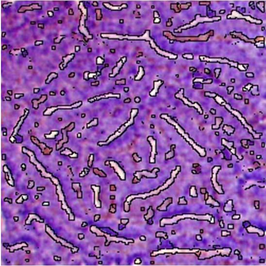
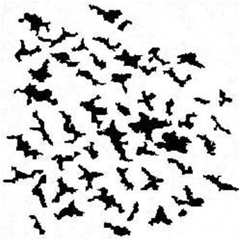
3 Takemura Y et al, Page 3 INTRODUCTION Magnifying endoscopy permits detailed visualization of the surface of the gastrointestinal tract and thus allows examination of the pit pattern (shape of the openings of 55 colorectal crypts) of colorectal tumors (Figure 1). 1 Pit pattern classification has been shown to aid in the differentiation of non-neoplastic and neoplastic colorectal lesions 2 and thus may be able to guide therapeutic decisions. Magnifying endoscopy with pit pattern recognition can be done during routine colonoscopy with indigo carmine dye spraying or crystal violet staining with little added expense or time. We have been interested in developing a software program 60 that can analyze pit patterns quantitatively and thus be used with magnifying endoscopy to diagnose colorectal tumors. We created a custom software program for this very purpose and herein describe its development and an experimental study in which we tested its clinical utility and limitations. 65 METHODS Image analysis software We developed a custom software program (HuPAS ver. 1.3) that can outline various regular pits identified on digitized endoscopic images. HuPAS is designed to mark the color 70 edges of a pit outline based on differences in color tone between the pit outline and the background; it then automatically extracts the identified pits. The software identifies regional segmentation using a watershed algorithm and combines integrated regions with excessive segmentation Endoscopic procedure The HuPAS image analysis software was tested on magnifying endoscopy images obtained from patients who had undergone diagnostic endoscopic study at Hiroshima University
4 Takemura Y et al, Page 4 Hospital. An Olympus CF-H260AZI magnifying colonoscope (Olympus Co., Tokyo, Japan) was used, which provides magnification up to 70 (optical magnification) on a 19-inch monitor. 80 After performing white light endoscopy, we examined the lesion at maximum magnification with crystal violet staining. Thereafter, images were digitized and stored on an Olympus EICP-D HDTV recorder (1,440 1,080 pixels). Informed consent was obtained from patients and/or family members for endoscopic examination. 85 Image processing Pit region extraction From each magnified endoscopic image recorded at maximum optical magnification (Figure 2a), a pixel region was cut out as a region of interest (ROI) (Figure 2b). The cut-out image was processed automatically by using the custom HuPAS ver. 1.3 software to 90 outline various pits identified on the digitized image (Figure 2c). The computer operator (S.Y.) removed non-pit regions and/or joined excessively segmented pit regions manually if necessary using Adobe Photoshop (Adobe Systems Inc. San Jose, CA) (Figure 3). Quantification of pit features 95 Using ImageJ software (National Institutes of Health, Bethesda, MD), we quantified the following six shape descriptors for each pit on the extracted images: area, perimeter, major and minor axes of the best fit ellipse, circularity (represented as 4π(area/perimeter 2 )), wherein 1.0 = a precise circle that becomes an elongated polygon as it approaches 0.0), and Feret s diameter (longest distance between any two points within the selected frame) (Figure 4). These six shape 100 descriptors were chosen for quantification because statistical analysis (Kruskal-Wallis test) revealed that differences in the values of these descriptors between types of regular pit patterns (Kudo and Tsuruta classes si, sii, siiil, siiis, siv) were significant. Quantitative analysis of regular pit patterns
5 Takemura Y et al, Page Feature extraction and quantification of the regular pit Colorectal magnifying endoscopy was performed in 72 cases at Hiroshima University Hospital between June 2007 and September Images of regular pits were obtained from the magnified endoscopic images (type I: 20 cases; type II: 10 cases; type IIIL: 10 cases; type IIIs: 2 cases; and type IV: 30 cases) as a set of training images. After extracting the pit region of the 110 ROI using HuPAS, an endoscopist (Y.T.) selected separate images of regular pits based on the morphology of the opening of the colorectal crypt and classified each pit, according to the Kudo and Tsuruta classification system (Figure 3b), as type si (round), type sii (asteroid), type siiil (larger than type si, ranging from tubular to round), type siiis (smaller than type si, ranging from tubular to round), or type siv (dendritic or gyrus-like). Based on the image processing 115 described above, quantitative features were defined for each pit type. Computer-aided identification of regular pit patterns A set of validation images was gathered from among images obtained in other cases examined at Hiroshima University Hospital. We excluded lesions that were not 120 suitable for evaluation (exclusion criteria: out-of-focus images, images that showed insufficient staining, images that were blurred). A total of 134 regular pit pattern images (type I: 32 cases; type II: 43 cases; type IIIL: 29 cases; and type IV: 30 cases) were sequentially obtained and comprised the validation set. After the separate pit types were determined based on the imaging processing described above, a discriminant analysis using JMP statistical software (SAS 125 Institute Inc. Cary, NC) was conducted by referring to the quantitative characteristics of the set of training images for automated recognition of pit patterns on the endoscopic images. Discriminant analysis is used for estimating the population to which sample data belong when the population in which the sample data reside is unknown. Thus, we first obtained quantitative characteristics for each pit type as reference data and then conducted discriminant analysis for 130 each pit based on these data. Together with the subsequent step of weighing the proportion of each pit within the respective images, we defined these steps as the two steps required to
6 Takemura Y et al, Page 6 definitively identify the patterns of the pits identified on endoscopic images. In addition, the pit patterns on the validation images were classified according to the Kudo and Tsuruta system by the same endoscopist (Y.T.), who was blinded to the computer-aided results. 135 Statistical analysis Values are reported as mean ± SD. Differences in the six quantitative features (shape descriptors) between the various pit patterns were analyzed by Kruskal-Wallis test, with significance accepted at P < RESULTS Quantification of the regular pit patterns Values for each of the six shape descriptors are shown per pit pattern in Table 1. For 145 each of the six features, differences in values between the five regular pit patterns were significant. Automated identification of regular pit patterns Performance of the automated computer-aided system for pit pattern classification of 150 colorectal lesions is shown relative to endoscopy findings in Table 2. Overall accuracy of the automated computer-aided system for identification of regular pit patterns was 132/134 (98.5%). DISCUSSION 155 To our knowledge, there are no reports of computerized quantitative analysis of pit patterns of the colorectal mucosal surface. Computerized quantification of the pit pattern of a
7 Takemura Y et al, Page 7 colorectal lesion should allow for objective diagnosis, avoiding subjectivity and eliminating the need for extensive training in evaluating pit patterns. 160 We developed a software program, known as HuPAS, that can be used to outline and characterize various pits in the colonic mucosa on endoscopically obtained images. We found that values for six shape descriptors differed significantly between regular separate pit patterns (types si-siv), so these shape descriptors became the basis for our quantitative analysis. We also analyzed the accuracy of the automated computerized identification of the pit 165 patterns in reference to endoscopic diagnosis. The overall diagnostic accuracy of the computer-aided diagnosis based on automated calculation of the pit area was 98.5%. Thus, the custom software and computer-aided diagnosis algorithm together approached the diagnostic ability of the trained endoscopist. Our automated system is limited in that some non-pit regions are extracted with the pit regions and 170 some pit regions are excessively segmented. This is because some non-pit regions differ in color tone from the background and some pit regions are of low contrast. Thus, it was necessary to remove these non-pit regions and/or join excessively segmented pit regions using Adobe Photoshop in 11% of cases. This procedure does not make the process subjective, but it does add a manual step. In addition, endoscopic images that were out-of-focus, that showed insufficient staining, or that were blurred could not be 175 evaluated. It might be possible to overcome these limitations by adding another algorithm. Unfortunately, our computer analysis takes several minutes, so the results are not available during colonoscopic examination. Improvements are needed that will allow real-time computerized evaluation of the pit patterns of colorectal lesions. If rapid, accurate differentiation between neoplastic and non-neoplastic polyps can be made by magnification endoscopy with computer-aided diagnosis, this technique could 180 reduce the number of polypectomies required and also reduce complications. We succeeded in developing a computerized system for automated recognition of regular pit patterns on magnified endoscopy images. With its limitations, our system is perhaps best characterized as semi-automated but a step toward development of a fully automated system to assist in the diagnosis of colorectal lesions. We anticipate development of a fully
8 Takemura Y et al, Page automated system that will recognize both regular and irregular pit patterns and will meet the rigors of blinded prospective evaluation comparing the results of computerized analysis against pathologic classification as the gold standard.
9 Takemura Y et al, Page 9 REFERENCES Tanaka S, Kaltenbach T, Chayama K, et al. High-magnification colonoscopy (with videos). Gastrointest Endosc 2006;64: Fu KI, Sano Y, Fujii T, et al. Chromoendoscopy using indigo carmine dye spraying with magnifying observation is the most reliable method for differential diagnosis between non-neoplastic and 195 neoplastic colorectal lesions: a prospective study. Endoscopy 2004;36: Hirota M, Tamaki T, Kaneda K, et al. Feature extraction from images of endoscopic large intestine. Proceedings of FCV2008; The 14th Korea-Japan Joint Workshop on Frontiers of Computer Vision 2008;01:
10 Takemura Y et al, Page 10 Figure Legends Figure 1. Classification of pit patterns of colorectal lesions. 205 Figure 2. Pits are outlined on the magnified endoscopic images with the use of our custom HuPAS ver.1.3 software. a: Observation and recording of the stained (crystal violet) image at maximum optical magnification (x70). b: A region of interest (ROI) measuring pixels is cut out for analysis. c: Example of a pit region automatically extracted by HuPAS. 210 Figure 3. Extracted images of pit outlines within the region of interest. a: Original image of a pit region automatically extracted by HuPAS. b: The image generated by HuPAS required some Adobe Photoshop editing. The separate pit images (si, sii, siiis, siiil, siv) (arrows) were classified according to the Kudo and Tsuruta criteria. 215 Figure 4. Diagram of the six shape descriptors used for quantitative analysis of pit patterns. (1) area, (2) perimeter, (3) major axis of the best fit ellipse, (4) minor axis of the best fit ellipse, (5) circularity: 4 (area/perimeter 2 ), wherein1.0 = precise circle that becomes an elongated polygon as it approaches 0.0, and (6) Feret s diameter, which is the longest distance between any two points within the selected frame. 220


 neoplastic Type IIIL larger")


11 Type I Round pit (normal pit) non-neoplastic Type II Asteroid pit non-neoplastic Type IIIS Tubular or round pit that is smaller than the normal pit (Type I) neoplastic Type IIIL Tubular or round pit that is larger than the normal pit (Type I) neoplastic Type IV Dendritic or gyrus-like pit neoplastic Type VI Irregular arrangement and sizes of IIIL, IIIS, IV type pit pattern neoplastic Type VN Loss or decrease of pits with an amorphous structure neoplastic Figure 1. Classification of pit patterns of colorectal lesions. Figure 1, Takemura et al


12 Figure 2, Takemura et al Figure 2 a b c


13 Figure 3, Takemura et al Figure 3 stype I stype II stype IIIL stype IV a b


14 Figure 4 Figure 4, Takemura et al Quantitative characteristics for six examined items Minor Fit Ellipse Feret s Diameter Major Fit Ellipse Area Perimeter Major Fit Ellipse Minor Fit Ellipse Circularity: 4π(area/perimeter 2 ) (1.0=precise circle; becomes an elongated polygon as it approaches 0.0) Feret s Diameter: longest distance between any two points within the selected frame.
15 Table 1. Quantitative analysis of regular pit patterns Pit pattern Number of pits Area Perimeter Major fit ellipse Minor fit ellipse Circularity Feret s diameter Type si ± ± ± ± ± ±3.1 Type sii ± ± ± ± ± ±13.6 Type siiil ± ± ± ± ± ±26.5 Type siiis ± ± ± ± ± ±1.14 Type siv ± ± ± ± ± ±42.5 P< P< P< P< P< P< Data are mean±sd. P values were obtained by Kruskal-Wallis test, which was used to analyze between-pattern differences in the values of each of the six features.
16 Table 2. Performance of the semi-automated CAD algorithm for pit pattern classification of colorectal lesions Classification using the CAD software Endoscopic diagnosis Type I Type II Type IIIL Type IV Total Type I 32 (100) /32 (100) Type II 0 43 (100) /43 (100) Type IIIL (96.6) 1 (3.4) 28/29 (96.6) Type IV (3.3) 29 (96.7) 29/30 (96.7) Data are number (percentage) of lesions. Overall accuracy: 132/134 (98.5%) CAD : computer-aided diagnosis
Magnified Examination of Small Colorectal Polyps Using a Prototype Electronic Endoscope: Preliminary Experience
 Diagnostic and Therapeutic Endoscopy, Vol. 6, pp. 77-82 Reprints available directly from the publisher Photocopying permitted by license only (C) 2000 OPA (Overseas Publishers Association) N.V. Published
Real-Time in vivo Observation of Cells and Nuclei Opens New Possibilities for Diagnostic Endoscopy
 Beyond Imagination Introducing Endocyto, Olympus has broken a new ground in endoscopy. Ultra-high magnification with up to 520x magnification ratio enables observation on microscopic level and helps to
Novel Endoscopic Imaging Techniques
 ENDOSCOPY 2006 152nd Falk Symposium, Berlin May 4-5, 2006 Novel Endoscopic Imaging Techniques Paul Fockens, MD PhD Academic Medical Center, University of Amsterdam Novel Endoscopic Imaging Techniques Current
Cellular Bioengineering Boot Camp. Image Analysis
 Cellular Bioengineering Boot Camp Image Analysis Overview of the Lab Exercises Microscopy and Cellular Imaging The purpose of this laboratory exercise is to develop an understanding of the measurements
Image Extraction using Image Mining Technique
 IOSR Journal of Engineering (IOSRJEN) e-issn: 2250-3021, p-issn: 2278-8719 Vol. 3, Issue 9 (September. 2013), V2 PP 36-42 Image Extraction using Image Mining Technique Prof. Samir Kumar Bandyopadhyay,
EXpERIENCE THE power Of LIGHT
 EXpERIENCE THE power Of LIGHT GASTROENTEROLOGY MAKING YOUR DAILY WORK EaSIER ENDOSCOpES processors preparation & HYGIENICS ULTRa- SONOGRapHY ENDOSCOpY SYSTEM fujifilm is a pioneer in diagnostic imaging
MINING COLONOSCOPY VIDEOS TO MEASURE QUALITY OF COLONOSCOPIC PROCEDURES
 MINING COLONOSCOPY VIDEOS TO MEASURE QUALITY OF COLONOSCOPIC PROCEDURES Danyu Liu a, Yu Cao a, Wallapak Tavanapong a, Johnny Wong a, JungHwan Oh b, and Piet C. de Groen c a Department of Computer Science,
The Trend of Medical Image Work Station
 The Trend of Medical Image Work Station Abstract Image Work Station has rapidly improved its efficiency and its quality along the development of biomedical engineering. The quality improvement of image
Advancing the Art of Endoscopy
 Advancing the Art of Endoscopy Advancing the Art of Endoscopy with an array of opto-digital innovations. OLYMPUS technology continues to advance the art of endoscopy. As the world leader in endoscopy,
Differentiation of Malignant and Benign Masses on Mammograms Using Radial Local Ternary Pattern
 Differentiation of Malignant and Benign Masses on Mammograms Using Radial Local Ternary Pattern Chisako Muramatsu 1, Min Zhang 1, Takeshi Hara 1, Tokiko Endo 2,3, and Hiroshi Fujita 1 1 Department of Intelligent
Centre for Computational and Numerical Studies, Institute of Advanced Study in Science and Technology 2. Dept. of Statistics, Gauhati University
 Cervix Cancer Diagnosis from Pap Smear Images Using Structure Based Segmentation and Shape Analysis 1 Lipi B. Mahanta, 2 Dilip Ch. Nath, 1 Chandan Kr. Nath 1 Centre for Computational and Numerical Studies,
Multilayer scanning enhances sensitivity of artificial intelligence-aided Mycobacterium tuberculosis detection
 Multilayer scanning enhances sensitivity of artificial intelligence-aided Mycobacterium tuberculosis detection Yan Xiong Peking University First Hospital, China. yanxiong1109@163.com Ao Hou ao_sure@foxmail.com
InScape: Making Virtual Pathology a Reality
 InScape: Making Virtual Pathology a Reality Sally S. Agersborg, M.D., Ph.D. Quest Diagnostics, Nichols Institute San Juan Capistrano, CA Company Overview Quest Diagnostics, Nichols Institute the world
Fuji Intelligent Chromo Endoscopy
 Fuji Intelligent Chromo Endoscopy The next generation of endoscopic diagnosis has arrived with Fujinon's new EPX-4400 video processor. F.I.C.E. (FUJI Intelligent Chromo Endoscopy, ) installed in the EPX-4400,
Omnidirectional Vision Attachment for Medical Endoscopes
 Omnidirectional Vision Attachment for Medical Endoscopes Ryusuke Sagawa 1, Takurou Sakai 1, Tomio Echigo 2, Keiko Yagi 3, Masatsugu Shiba 4, Kazuhide Higuchi 5, Tetsuo Arakawa 4, and Yasushi Yagi 1 1 The
Towards Optical Biopsies with an Integrated Fibered Confocal Fluorescence Microscope
 Towards Optical Biopsies with an Integrated Fibered Confocal Fluorescence Microscope Georges Le Goualher, Aymeric Perchant, Magalie Genet, Charlotte Cavé, Bertrand Viellerobe, Fredéric Berier, Benjamin
Advancing the Art of Endoscopy
 Advancing the Art of Endoscopy Advancing the Art of Endoscopy with an array of opto-digital innovations. OLYMPUS technology continues to advance the art of endoscopy. As the world leader in endoscopy,
CHAPTER-4 FRUIT QUALITY GRADATION USING SHAPE, SIZE AND DEFECT ATTRIBUTES
 CHAPTER-4 FRUIT QUALITY GRADATION USING SHAPE, SIZE AND DEFECT ATTRIBUTES In addition to colour based estimation of apple quality, various models have been suggested to estimate external attribute based
OPTIVISTA TM EPK-i7010
 OPTIVIST TM EPK-i7010 unique combination of optical and digital enhancement in one endoscopy processor Courtesy of Dr. Federico uffoli, Cremona Hospital, Italy PENTX MEDICL OPTIVIST TM EPK-i7010 Enlighten
Malignancy Detection of Candidate for Basal Cell Carcinoma Using Image Processing and Artificial Neural Network
 DLSU Engineering e-journal Vol. 1 No. 1, March 2007, pp.70-79 Malignancy Detection of Candidate for Basal Cell Carcinoma Using Image Processing and Artificial Neural Network Armida R. Bayot Louise Ann
GenePix Application Note
 GenePix Application Note Biological Relevance of GenePix Results Shawn Handran, Ph.D. and Jack Y. Zhai, Ph.D. Axon Instruments, Inc. 3280 Whipple Road, Union City, CA 94587 Last Updated: Aug 22, 2003.
Morphologi. Advanced image analysis for high sensitivity particle characterization. Particle size. Particle shape
 Particle size Particle shape Morphologi detailed specification sheets from www.malvern.co.uk Introducing a new concept in image analysis The Morphologi high sensitivity particle analyzer is more than just
Segmenting Reddish lesions in Capsule Endoscopy Images Using a GastroIntestinal Color Space
 2014 22nd International Conference on Pattern Recognition Segmenting Reddish lesions in Capsule Endoscopy Images Using a GastroIntestinal Color Space Hai Vu, Tomio Echigo, Yuma Imura, Yukiko Yanagawa,
Development of a Robotic Colonoscopic Manipulation System, Using Haptic Feedback Algorithm
 Original Article Yonsei Med J 2017 Jan;58(1):139-143 pissn: 0513-5796 eissn: 1976-2437 Development of a Robotic Colonoscopic Manipulation System, Using Haptic Feedback Algorithm Jaehong Woo 1 *, Jae Hyuk
ATLAS. Atlas of Spectral Endoscopic Images
 Atlas of Spectral Endoscopic Images ATLAS Atlas of Spectral Endoscopic Images Academic Research Report for Health Care Professionals Supervised by Yoichi Miyake, Research Center for Frontier Medical Engineering,
endoscope for observing vocal fold
 NAOSITE: Nagasaki University's Ac Title Author(s) Citation High-speed digital imaging system w endoscope for observing vocal fold Kaneko, Kenichi; Watanabe, Takeshi; Takahashi, Haruo Acta medica Nagasakiensia,
An Image Processing Approach for Screening of Malaria
 An Image Processing Approach for Screening of Malaria Nagaraj R. Shet 1 and Dr.Niranjana Sampathila 2 1 M.Tech Student, Department of Biomedical Engineering, Manipal Institute of Technology, Manipal University,
Computational approach for diagnosis of malaria through classification of malaria parasite from microscopic image of blood smear.
 Biomedical Research 2018; 29 (18): 3464-3468 ISSN 0970-938X www.biomedres.info Computational approach for diagnosis of malaria through classification of malaria parasite from microscopic image of blood
ImageJ: Introduction to Image Analysis 3 May 2012 Jacqui Ross
 Biomedical Imaging Research Unit School of Medical Sciences Faculty of Medical and Health Sciences The University of Auckland Private Bag 92019 Auckland 1142, NZ Ph: 373 7599 ext. 87438 http://www.fmhs.auckland.ac.nz/sms/biru/.
Observer Performance of Reduced X-Ray Images on Liquid Crystal Displays
 Original Paper Forma, 29, S45 S51, 2014 Observer Performance of Reduced X-Ray Images on Liquid Crystal Displays Akiko Ihori 1, Chihiro Kataoka 2, Daigo Yokoyama 2, Naotoshi Fujita 3, Naruomi Yasuda 4,
USING SIGNATURE IDENTIFICATION FOR RAPID AND EFFECTIVE X-RAY INSPECTION OF BALL GRID ARRAYS
 USING SIGNATURE IDENTIFICATION FOR RAPID AND EFFECTIVE X-RAY INSPECTION OF BALL GRID ARRAYS Gil Zweig Glenbrook Technologies, Inc. Randolph, New Jersey USA gzweig@glenbrooktech.com ABSTRACT Although X-ray
Arcturus XT Laser Capture Microdissection System AutoScanXT Software Module. User Manual
 Arcturus XT Laser Capture Microdissection System AutoScanXT Software Module User Manual For Research Use Only. Not intended for any animal or human therapeutic or diagnostic use. Information in this document
A Method of Using Digital Image Processing for Edge Detection of Red Blood Cells
 Sensors & Transducers 013 by IFSA http://www.sensorsportal.com A Method of Using Digital Image Processing for Edge Detection of Red Blood Cells 1 Jinping LI, Hongshan MU, Wei XU 1 Software School, East
ENDOSCOPIC ULTRASOUND SYSTEMS
 ENDOSCOPIC ULTRASOUND SYSTEMS DISCOVER HIGH-PRECISION DIAGNOSES AND PROCEDURES NEW ENDOSCOPIC ULTRASOUND Ultrasonography revolutionized the clinical approach to patients with digestive and respiratory
Color Based Stool Region Detection in Colonoscopy Videos for Quality Measurements
 Color Based Stool Region Detection in Colonoscopy Videos for Quality Measurements Jayantha Muthukudage 1, JungHwan Oh 1, Wallapak Tavanapong 2, Johnny Wong 2, and Piet C. de Groen 3 1 Department of Computer
Fovea and Optic Disc Detection in Retinal Images with Visible Lesions
 Fovea and Optic Disc Detection in Retinal Images with Visible Lesions José Pinão 1, Carlos Manta Oliveira 2 1 University of Coimbra, Palácio dos Grilos, Rua da Ilha, 3000-214 Coimbra, Portugal 2 Critical
3-D Computer-Automated Threshold Amsler Grid Test
 Introduction The 3-D Computer-Automated Threshold Amsler Grid Test is a five-minute vision test using a laptop computer with a touchsensitive screen that can help diagnose the onset of eye diseases and
AGRICULTURE, LIVESTOCK and FISHERIES
 Research in ISSN : P-2409-0603, E-2409-9325 AGRICULTURE, LIVESTOCK and FISHERIES An Open Access Peer Reviewed Journal Open Access Research Article Res. Agric. Livest. Fish. Vol. 2, No. 2, August 2015:
Clinical Natural Language Processing: Unlocking Patient Records for Research
 Clinical Natural Language Processing: Unlocking Patient Records for Research Mark Dredze Computer Science Malone Center for Engineering Healthcare Center for Language and Speech Processing Natural Language
Received on: Accepted on:
 ISSN: 0975-766X CODEN: IJPTFI Available Online through Research Article www.ijptonline.com AUTOMATIC FLUOROGRAPHY SEGMENTATION METHOD BASED ON HISTOGRAM OF BRIGHTNESS SUBMISSION IN SLIDING WINDOW Rimma
Image Analysis for Fluorescence
 Image Analysis for Fluorescence Terminology Table Image Analysis Macro Colocalization Intensity Dye AFI The extraction of meaningful information from digital images by means of digital image processing
Analysis and Identification of Rice Granules Using Image Processing and Neural Network
 International Journal of Electronics and Communication Engineering. ISSN 0974-2166 Volume 10, Number 1 (2017), pp. 25-33 International Research Publication House http://www.irphouse.com Analysis and Identification
Imaging with hyperspectral sensors: the right design for your application
 Imaging with hyperspectral sensors: the right design for your application Frederik Schönebeck Framos GmbH f.schoenebeck@framos.com June 29, 2017 Abstract In many vision applications the relevant information
Single balloon Enteroscope system SIF-Q180 / OBCU
 Single Balloon Enteroscope System Single balloon Enteroscope system SIF-Q180 / OBCU There s only one answer to your requirements: the Single Balloon System from Olympus Despite the rapid technological
(51) Int Cl.: A61B 1/04 ( )
 Int Cl.: A61B 1/04 ( )") (19) (12) EUROPEAN PATENT APPLICATION published in accordance with Art. 158 (3) EPC (11) EP 1 849 402 A1 (43) Date of publication: 31.10.2007 Bulletin 2007/44 (21) Application number: 06713523.6 (22) Date
(19) (12) EUROPEAN PATENT APPLICATION published in accordance with Art. 158 (3) EPC (11) EP 1 849 402 A1 (43) Date of publication: 31.10.2007 Bulletin 2007/44 (21) Application number: 06713523.6 (22) Date
Automated Detection of Early Lung Cancer and Tuberculosis Based on X- Ray Image Analysis
 Proceedings of the 6th WSEAS International Conference on Signal, Speech and Image Processing, Lisbon, Portugal, September 22-24, 2006 110 Automated Detection of Early Lung Cancer and Tuberculosis Based
IMAGING TECHNIQUES FOR MEASURING PARTICLE SIZE SSA AND GSV
 IMAGING TECHNIQUES FOR MEASURING PARTICLE SIZE SSA AND GSV APPLICATION NOTE SSA-001 (A4) Particle Sizing through Imaging TSI provides several optical techniques for measuring particle size. Two of the
Table of Contents 1. Image processing Measurements System Tools...10
 Introduction Table of Contents 1 An Overview of ScopeImage Advanced...2 Features:...2 Function introduction...3 1. Image processing...3 1.1 Image Import and Export...3 1.1.1 Open image file...3 1.1.2 Import
Iris Segmentation & Recognition in Unconstrained Environment
 www.ijecs.in International Journal Of Engineering And Computer Science ISSN:2319-7242 Volume - 3 Issue -8 August, 2014 Page No. 7514-7518 Iris Segmentation & Recognition in Unconstrained Environment ABSTRACT
ROBOT VISION. Dr.M.Madhavi, MED, MVSREC
 ROBOT VISION Dr.M.Madhavi, MED, MVSREC Robotic vision may be defined as the process of acquiring and extracting information from images of 3-D world. Robotic vision is primarily targeted at manipulation
COMPONENT II CANDIDATE STUDY GUIDE PEDIATRIC DENTISTRY
 COMPONENT II CANDIDATE STUDY GUIDE PEDIATRIC DENTISTRY Introduction The intent of this guide is to provide the candidate with an understanding of the format used for the Component II of the National Dental
A Novel Approach for Automated Color Segmentation of Tuberculosis Bacteria through Region Growing
 A Novel Approach for Automated Color Segmentation of Tuberculosis Bacteria through Region Growing M. Hemalatha S.V College of Engineering. A.V. Kiranmai S.V Engineering College for Women. D.Sreehari S.V
Digital Retinal Images: Background and Damaged Areas Segmentation
 Digital Retinal Images: Background and Damaged Areas Segmentation Eman A. Gani, Loay E. George, Faisel G. Mohammed, Kamal H. Sager Abstract Digital retinal images are more appropriate for automatic screening
THEORY AND APPROACHES TO AUTOMATED IMAGE ANALYSIS IN DIGITAL PATHOLOGY
 THEORY AND APPROACHES TO AUTOMATED IMAGE ANALYSIS IN DIGITAL PATHOLOGY Kyle Takayama, MS Charles River Laboratories EVERY STEP OF THE WAY EVERY STEP OF THE WAY MORPHOMETRY Measurements or counts performed
Intelligent Traffic Sign Detector: Adaptive Learning Based on Online Gathering of Training Samples
 2011 IEEE Intelligent Vehicles Symposium (IV) Baden-Baden, Germany, June 5-9, 2011 Intelligent Traffic Sign Detector: Adaptive Learning Based on Online Gathering of Training Samples Daisuke Deguchi, Mitsunori
Content Based Image Retrieval Using Color Histogram
 Content Based Image Retrieval Using Color Histogram Nitin Jain Assistant Professor, Lokmanya Tilak College of Engineering, Navi Mumbai, India. Dr. S. S. Salankar Professor, G.H. Raisoni College of Engineering,
Automatic Locating the Centromere on Human Chromosome Pictures
 Automatic Locating the Centromere on Human Chromosome Pictures M. Moradi Electrical and Computer Engineering Department, Faculty of Engineering, University of Tehran, Tehran, Iran moradi@iranbme.net S.
Biometrics Final Project Report
 Andres Uribe au2158 Introduction Biometrics Final Project Report Coin Counter The main objective for the project was to build a program that could count the coins money value in a picture. The work was
NSERC Summer Project 1 Helping Improve Digital Camera Sensors With Prof. Glenn Chapman (ENSC)
") NSERC Summer 2016 Digital Camera Sensors & Micro-optic Fabrication ASB 8831, phone 778-782-319 or 778-782-3814, Fax 778-782-4951, email glennc@cs.sfu.ca http://www.ensc.sfu.ca/people/faculty/chapman/ Interested
NSERC Summer 2016 Digital Camera Sensors & Micro-optic Fabrication ASB 8831, phone 778-782-319 or 778-782-3814, Fax 778-782-4951, email glennc@cs.sfu.ca http://www.ensc.sfu.ca/people/faculty/chapman/ Interested
MEASUREMENT CAMERA USER GUIDE
 How to use your Aven camera s imaging and measurement tools Part 1 of this guide identifies software icons for on-screen functions, camera settings and measurement tools. Part 2 provides step-by-step operating
ADVANCED MEDICAL SYSTEMS PTE LTD Singapore Malaysia India Australia
 Innovative design is combined with cutting-edge technology to yield a definitive diagnosis and never before seen ergonomics GIOTTO CLASS is the result of 25 years of experience in the research and development
Locating Blood Vessels in Retinal Images by Piece-wise Threshold Probing of a Matched Filter Response
 Locating Blood Vessels in Retinal Images by Piece-wise Threshold Probing of a Matched Filter Response Adam Hoover, Ph.D. +, Valentina Kouznetsova, Ph.D. +, Michael Goldbaum, M.D. + Electrical and Computer
Microwave Measurement and Quantitative Evaluation of Wall Thinning in Metal Pipes
 th World Conference on Nondestructive Testing, 25-28 Oct 2008, Shanghai, China Microwave Measurement and Quantitative Evaluation of Wall Thinning in Metal Pipes Yang JU, Linsheng LIU, Masaharu ISHIKAWA
MEASUREMENT OF ROUGHNESS USING IMAGE PROCESSING. J. Ondra Department of Mechanical Technology Military Academy Brno, Brno, Czech Republic
 MEASUREMENT OF ROUGHNESS USING IMAGE PROCESSING J. Ondra Department of Mechanical Technology Military Academy Brno, 612 00 Brno, Czech Republic Abstract: A surface roughness measurement technique, based
Image Database and Preprocessing
 Chapter 3 Image Database and Preprocessing 3.1 Introduction The digital colour retinal images required for the development of automatic system for maculopathy detection are provided by the Department of
Dynamic Distortion Correction for Endoscopy Systems with Exchangeable Optics
 Lehrstuhl für Bildverarbeitung Institute of Imaging & Computer Vision Dynamic Distortion Correction for Endoscopy Systems with Exchangeable Optics Thomas Stehle and Michael Hennes and Sebastian Gross and
On-site Safety Management Using Image Processing and Fuzzy Inference
 1013 On-site Safety Management Using Image Processing and Fuzzy Inference Hongjo Kim 1, Bakri Elhamim 2, Hoyoung Jeong 3, Changyoon Kim 4, and Hyoungkwan Kim 5 1 Graduate Student, School of Civil and Environmental
Problems and Solutions in Medical Color Imaging
 Problems and Solutions in Medical Color Imaging Masahiro NISHIBORI Clinical Laboratory, Tokyo Medical and Dental University Medical Hospital 1-5-45, Yushima, Bunkyo-ku, Tokyo, 113-8519, Japan E-mail: mn.mlab@tmd.ac.jp
At Fujinon we are committed to delivering innovative endoscopic solutions exactly as envisioned by our customers. Our latest achievement, the Fujinon
 At Fujinon we are committed to delivering innovative endoscopic solutions exactly as envisioned by our customers. Our latest achievement, the Fujinon 4400, is a crowning example of the most advanced optical
Building Damage Mapping of the 2006 Central Java, Indonesia Earthquake Using High-Resolution Satellite Images
 4th International Workshop on Remote Sensing for Post-Disaster Response, 25-26 Sep. 2006, Cambridge, UK Building Damage Mapping of the 2006 Central Java, Indonesia Earthquake Using High-Resolution Satellite
Novel 3D Computerized Threshold Amsler Grid Test CA, USA
 Novel 3D Computerized Threshold Amsler Grid Test Wolfgang Fink 1,2 and Alfredo A. Sadun 2 1 California Institute of Technology, Pasadena, CA, USA 2 Doheny Eye Institute, Keck School of Medicine, University
A new method for segmentation of retinal blood vessels using morphological image processing technique
 A new method for segmentation of retinal blood vessels using morphological image processing technique Roya Aramesh Faculty of Computer and Information Technology Engineering,Qazvin Branch,Islamic Azad
Urban Feature Classification Technique from RGB Data using Sequential Methods
 Urban Feature Classification Technique from RGB Data using Sequential Methods Hassan Elhifnawy Civil Engineering Department Military Technical College Cairo, Egypt Abstract- This research produces a fully
MATLAB DIGITAL IMAGE/SIGNAL PROCESSING TITLES
 MATLAB DIGITAL IMAGE/SIGNAL PROCESSING TITLES -2018 S.NO PROJECT CODE 1 ITIMP01 2 ITIMP02 3 ITIMP03 4 ITIMP04 5 ITIMP05 6 ITIMP06 7 ITIMP07 8 ITIMP08 9 ITIMP09 `10 ITIMP10 11 ITIMP11 12 ITIMP12 13 ITIMP13
1. Queries are issued to the image archive for information about computed tomographic (CT)
") Appendix E1 Exposure Extraction Method examinations. 1. Queries are issued to the image archive for information about computed tomographic (CT) 2. Potential dose report screen captures (hereafter, dose
Appendix E1 Exposure Extraction Method examinations. 1. Queries are issued to the image archive for information about computed tomographic (CT) 2. Potential dose report screen captures (hereafter, dose
Completely Rewrite Industry s Understanding of Transmitting. High Quality Laser Processing Light over Long Distances
 25 April, 2018 Nippon Telegraph and Telephone Corporation Mitsubishi Heavy Industries, Ltd. Completely Rewrite Industry s Understanding of Transmitting High Quality Laser Processing Light over Long Distances
Speed Traffic-Sign Recognition Algorithm for Real-Time Driving Assistant System
 R3-11 SASIMI 2013 Proceedings Speed Traffic-Sign Recognition Algorithm for Real-Time Driving Assistant System Masaharu Yamamoto 1), Anh-Tuan Hoang 2), Mutsumi Omori 2), Tetsushi Koide 1) 2). 1) Graduate
An Improved Method of Computing Scale-Orientation Signatures
 An Improved Method of Computing Scale-Orientation Signatures Chris Rose * and Chris Taylor Division of Imaging Science and Biomedical Engineering, University of Manchester, M13 9PT, UK Abstract: Scale-Orientation
MULTI-PARAMETER ANALYSIS IN EDDY CURRENT INSPECTION OF
 MULTI-PARAMETER ANALYSIS IN EDDY CURRENT INSPECTION OF AIRCRAFT ENGINE COMPONENTS A. Fahr and C.E. Chapman Structures and Materials Laboratory Institute for Aerospace Research National Research Council
2 nd generation TOMOSYNTHESIS
 2 nd generation TOMOSYNTHESIS 2 nd generation DBT true innovation in breast imaging synthesis graphy Combo mode Stereotactic Biopsy Works in progress: Advanced Technology, simplicity and ergonomics Raffaello
Keywords: - Gaussian Mixture model, Maximum likelihood estimator, Multiresolution analysis
 Volume 4, Issue 2, February 2014 ISSN: 2277 128X International Journal of Advanced Research in Computer Science and Software Engineering Research Paper Available online at: www.ijarcsse.com Expectation
2017 ADVANCING DEEPER INSIGHTS IN ENDOSCOPY
 2017 ADVANCING DEEPER INSIGHTS IN ENDOSCOPY CONTENT About Fujifilm 4 5 Technologies 6 13 ELUXEO 700 series 14 19 600 Series 20 23 580 Series 24 27 Double Balloon Endoscopy System 28 33 530 Series 34 39
CHAPTER 4 LOCATING THE CENTER OF THE OPTIC DISC AND MACULA
 90 CHAPTER 4 LOCATING THE CENTER OF THE OPTIC DISC AND MACULA The objective in this chapter is to locate the centre and boundary of OD and macula in retinal images. In Diabetic Retinopathy, location of
A Solution for Identification of Bird s Nests on Transmission Lines with UAV Patrol. Qinghua Wang
 International Conference on Artificial Intelligence and Engineering Applications (AIEA 2016) A Solution for Identification of Bird s Nests on Transmission Lines with UAV Patrol Qinghua Wang Fuzhou Power
SECTION I - CHAPTER 2 DIGITAL IMAGING PROCESSING CONCEPTS
 RADT 3463 - COMPUTERIZED IMAGING Section I: Chapter 2 RADT 3463 Computerized Imaging 1 SECTION I - CHAPTER 2 DIGITAL IMAGING PROCESSING CONCEPTS RADT 3463 COMPUTERIZED IMAGING Section I: Chapter 2 RADT
TR7500 SIII 3D SERIES. AutomAted optical InsPeCtIon
 TR7500 SIII 3D SERIES AutomAted optical InsPeCtIon TR7500 SIII 3D F E A T U R E S TR7500 SIII 3D AOI with Total Inspection Coverage Defects The TR7500 SIII 3D AOI combines the best of 2D and 3D technologies
Impact of Endoscopic Image Degradations on LBP based Features using One-Class SVM for Classification of Celiac Disease
 Impact of Endoscopic Image Degradations on based Features using One-Class SVM for Classification of Celiac Disease Sebastian Hegenbart, Andreas Uhl Department of Computer Sciences Salzburg University,
About This Survey. General Concepts and Definitions
 THECB Survey of Research Expenditures Universities and Health-Related Institutions Instructions and Definitions for Survey About This Survey The Texas Higher Education Coordinating Board collects data
Student Attendance Monitoring System Via Face Detection and Recognition System
 IJSTE - International Journal of Science Technology & Engineering Volume 2 Issue 11 May 2016 ISSN (online): 2349-784X Student Attendance Monitoring System Via Face Detection and Recognition System Pinal
An Essential Health and Biomedical R&D Treaty
 An Essential Health and Biomedical R&D Treaty Submission by Health Action International Global, Initiative for Health & Equity in Society, Knowledge Ecology International, Médecins Sans Frontières, Third
BASIC PATTERN RECOGNITION AND DIGITAL IMAGE PROCESSING USING
 BASIC PATTERN RECOGNITION AND DIGITAL IMAGE PROCESSING USING SAS/AF FRAME Abhishek Lall Department of Mathematics and Statistics, Sam Houston State University, Huntsville, Texas Abstract The principal
Digital Pathology at Johns Hopkins Practical Research and Clinical Considerations
 Digital Pathology at Johns Hopkins Practical Research and Clinical Considerations July 10, 2017 Alexander Baras, MD, PhD Assistant Professor of Pathology, Urology, and Oncology Associate Director of Pathology
Prediction Method of Beef Marbling Standard Number Using Parameters Obtained from Image Analysis for Beef Ribeye
 Prediction Method of Beef Marbling Standard Number Using Parameters Obtained from Image Analysis for Beef Ribeye Keigo KUCHIDA, Shogo TSURUTA1, a, L. D. Van Vleck2, Mitsuyoshi SUZUKI and Shunzo MIYOSHI
Instruments Commonly Used For Examination of the Eye
 Instruments Commonly Used For Examination of the Eye There are many instruments that the eye doctor might use to evaluate the eye and the vision system. This report presents some of the more commonly used
Comparison of Segmentation Framework on Digital Microscope Images for Acute Lymphoblastic Leukemia Diagnosis using RGB and HSV Color Spaces
 ` VOLUME 2 ISSUE 2 Comparison of Segmentation Framework on Digital Microscope Images for Acute Lymphoblastic Leukemia Diagnosis using RGB and HSV Color Spaces 1 Kamal A. ElDahshan, 2 Mohammed I. Youssef,
Scanned Image Segmentation and Detection Using MSER Algorithm
 Scanned Image Segmentation and Detection Using MSER Algorithm P.Sajithira 1, P.Nobelaskitta 1, Saranya.E 1, Madhu Mitha.M 1, Raja S 2 PG Students, Dept. of ECE, Sri Shakthi Institute of, Coimbatore, India
Extraction and Recognition of Text From Digital English Comic Image Using Median Filter
 Extraction and Recognition of Text From Digital English Comic Image Using Median Filter S.Ranjini 1 Research Scholar,Department of Information technology Bharathiar University Coimbatore,India ranjinisengottaiyan@gmail.com
Traffic Sign Recognition Senior Project Final Report
 Traffic Sign Recognition Senior Project Final Report Jacob Carlson and Sean St. Onge Advisor: Dr. Thomas L. Stewart Bradley University May 12th, 2008 Abstract - Image processing has a wide range of real-world
UNIT VI. Current approaches to programming are classified as into two major categories:
 Unit VI 1 UNIT VI ROBOT PROGRAMMING A robot program may be defined as a path in space to be followed by the manipulator, combined with the peripheral actions that support the work cycle. Peripheral actions
A Study On Preprocessing A Mammogram Image Using Adaptive Median Filter
 A Study On Preprocessing A Mammogram Image Using Adaptive Median Filter Dr.K.Meenakshi Sundaram 1, D.Sasikala 2, P.Aarthi Rani 3 Associate Professor, Department of Computer Science, Erode Arts and Science