MAMMOGRAPHY - HIGH LEVEL TROUBLESHOOTING
|
|
|
- Brianne McCormick
- 6 years ago
- Views:
Transcription
1 MAMMOGRAPHY - HIGH LEVEL TROUBLESHOOTING Maynard High New York Medical College SS2001-M.High 1
2 Objectives: Review MQSA and ACR annual QC tests as opportunities for troubleshooting before a significant image quality problem develops. Review MQSA equipment evaluations, and the role they play in preventing the need for troubleshooting at a later date. Present a logical decision tree approach to troubleshooting specific image quality problems. SS2001-M.High 2
3 The Medical Physicist CAN & SHOULD be an important resource for improving image quality of: Phantom images & Clinical images SS2001-M.High 3
4 Medical Physicist s Services: Compliance or Quality? Growing tendency toward compliance easier and quicker to meet minimum standards than to improve on them the MQSA inspector accepts it Both ACR and MQSA encourage the physicist to go beyond compliance checks, and to make recommendations for quality improvement. SS2001-M.High 4
5 Medical Physicist s Services: Compliance or Quality? During annual testing, make suggestions that may lead to improved image quality. this may prevent more intensive troubleshooting in the future Example: You see the maximum compression force is set to 30#. Although compliant, you should advise increasing the max to 45#. SS2001-M.High 5
6 Physicist Level Performance Evaluations & Troubleshooting: ANNUAL quality control compliance testing MQSA required testing ACR recommended testing Equipment evaluations New, repaired, relocated equipment MQSA required SS2001-M.High 6
7 Physicist Level Performance Evaluations & Troubleshooting: Troubleshooting (specific problems) Improving contrast, detail, noise, artifacts, etc Goes beyond performance testing of x-ray units screen-film appropriateness film processing evaluation technique factors proper use of AEC viewing conditions SS2001-M.High 7
8 Medical Physicist s Services in Mammography: ANNUAL quality control compliance testing Equipment evaluations Troubleshooting SS2001-M.High 8
9 MQSA Required Annual Physicist QC Survey: The 12 equipment tests in Table 1 testing methods not generally specified see Guidance ( Evaluation of technologist QC testing A written summary report format of report not specified to include recommendations physicist feels may improve the quality of mammography. SS2001-M.High 9
10 ACR Recommended Annual Physicist QC Tests: The 11 equipment tests in Table 2 testing methods detailed in 1999 ACR Mammography Quality Control Manual tests, action limits, scope of testing are very similar to MQSA, but there are several differences which Medical Physicist must consider, and use profesional judgement to develop a comprehensive performance evaluation. SS2001-M.High 10
11 ACR Recommended Annual Physicist QC Tests: Evaluation of technologist QC testing A written report including a summary model report formats are in the ACR manual to include recommendations physicist feels may improve the quality of mammography. SS2001-M.High 11

12 Resources: SS2001-M.High 12
13 Medical Physicist s Professional Judgment: Tailoring the Tests Guidance documents can never set the scope of testing and limits for all mammography units because: many different existing designs new designs with new features State Department of Health requirements Professional judgment & knowledge of unit must be used to tailor the tests to the unit SS2001-M.High 13
14 Multiple Selectable ma Stations: All tests affected by ma should be performed at each clinically used ma station kvp radiation output rate system resolution small focus is always a different ma station SS2001-M.High 14
15 Multiple Filters (Mo( Mo, Rh,, Al): All tests affected by filtration should be performed for each clinically used filter HVL average glandular dose phantom image quality AEC performance artifact evaluation SS2001-M.High 15
16 Multiple Targets (Mo( Mo, Rh,, W): All tests affected by anode selection should be performed for each clinically used target HVL average glandular dose phantom image quality AEC performance artifact evaluation SS2001-M.High 16
17 Multiple Auto-kV, Auto-filter Algorithms: Selectable maximum exposure time (0.8, 1.6s) Selectable dose (STD, CNT, DOSE) HVL average glandular dose phantom image quality AEC performance Test pulses may affect dosemeters SS2001-M.High 17
18 Multiple AEC Sensors: Some units use separate sensors for small and large Bucky s Must test thickness/kvp tracking for each SS2001-M.High 18
19 Multiple AEC Sensors: Some units use a matrix of fixed detectors which can be used to find the densest portion of the breast Must test each detector independently in some way SS2001-M.High 19
20 Multiple Film-Screen Combos: Some facilities use a faster speed screen for mag and/or large Bucky uniformity of screen speed average glandular dose phantom image quality AEC performance system resolution SS2001-M.High 20
21 Medical Physicist s Professional Judgment: Tailoring the Limits Action limits can often be tightened kvp accuracy & reproducibility AEC performance can be made better than +/ OD for 2-8 cm on some units Action limits can be tailored at acceptance testing. SS2001-M.High 21
22 The Annual Medical Physicist s Survey Report: Requirements Communication of results with recommendations is just as important as performing the tests. Needed on-site for annual MQSA inspections Needed to be submitted with applications for accreditation. SS2001-M.High 22
23 The Medical Physicist s Report: Useful for - Information resource for technologist Image quality and dose of Rh filter, AEC mode Information resource for service engineer. Details of calibration deficiencies Information resource for troubleshooting Performance baseline to which future measurements can be compared SS2001-M.High 23
24 The Medical Physicist s Report: Useful only if - There is sufficient information in report. Adequate description of test conditions kvp, ma, filter, target SID, magnification, receptor size phantom type and thickness AEC settings, compression force screen, film, processing conditions Test results for complete range of machine settings and modes of operation clinically used SS2001-M.High 24
25 The Medical Physicist s Report: Useful only if - Contains detailed recommendations for corrective action. And contains: recommendations that will improve image quality, including [those] concerning image receptors, technique factors, processing, viewing conditions and technologist QC. (ACR QC Manual) SS2001-M.High 25
26 Medical Physicist s Services in Mammography: ANNUAL quality control compliance testing Equipment evaluations Troubleshooting SS2001-M.High 26
27 Equipment Evaluations: Requirements Before use on patients, a qualified medical physicist must perform an MQSA equipment evaluation on : newly installed mammography equipment (x-ray unit or processor) disassembled and reassembled equipment equipment which has had a major component changed or repaired. SS2001-M.High 27
28 Equipment Evaluations: New Equipment Acceptance Test ACR Mammography QC Manual: It is assumed that mammography equipment will have been subjected to more extensive acceptance testing or a thorough performance evaluation prior to initiation of [annual] QC testing. SS2001-M.High 28
29 Equipment Evaluations: New Equipment Acceptance Test MQSA Compliance Guidance The equipment evaluation is more extensive than the survey. It may be regarded as an acceptance test for equipment and an annual survey alone is not sufficient to meet this requirement. SS2001-M.High 29
30 Equipment Evaluations: Disassembled, Reassembled Relocated, disassembled and reassembled equipment is to be evaluated as newly installed. A complete acceptance test needed SS2001-M.High 30
31 Equipment Evaluations: Major Repairs New x-ray tube AEC component replacement Collimator replacement Beam filter replacement Processor reassembly Generator replacement or re-calibration are considered major repairs. SS2001-M.High 31
32 Equipment Evaluations: Scope for Major Repairs Tests would include all those affected by the component repaired or replaced. SS2001-M.High 32
33 Equipment Evaluations: Example - New AEC sensor AEC testing Dose determination Phantom tests SS2001-M.High 33
34 Equipment Evaluations: Rebuilt/Replaced Processor Sensitometric testing Phantom tests Artifact evaluation Dose determination AEC testing Verification of proper processing solutions SS2001-M.High 34
35 Equipment Repairs: Physicist Involvement Medical physicist evaluates in person Medical physicist provides oversight Medical physicist involvement optional Should be discussion between facility and medical physicist SS2001-M.High 35
36 The Mammography Quality Standards Act Final Regulations Document #4 May 23, SS2001-M.High 36
37 From CDRH Guidance Document #4 Table: Medical Physicist Involvement in Equipment Adjustments, Changes, or Repairs For any adjustment, change, or repair not listed in the table below, or if the facility is unsure as to the full extent of the adjustment, change, or repair, the facility should consult their medical physicist to determine the proper extent of his or her involvement in evaluating the item. Item Major Repair Medical Physicist Involvement Automatic Exposure Control AEC Replacement Y MP conducts evaluation in person Thickness compensation internal* MP conducts evaluation Y adjustment in person AEC sensor replacement Y MP conducts evaluation in person AEC circuit board replacement Y MP conducts evaluation in person Density control - internal* adjustment N MP oversight Bucky (New to Facility) Replacement AEC also replaced Y MP conducts evaluation in person AEC not replaced N MP oversight SS2001-M.High 37
38 Medical Physicist s Services in Mammography: ANNUAL quality control compliance testing Equipment evaluations Troubleshooting SS2001-M.High 38
39 Manufacturer s Troubleshooting Resources: Film processor service company Film manufacturer representative Mammography unit service engineer Mammography unit applications person Should be advised where appropriate SS2001-M.High 39
40 Manufacturer s Troubleshooting Resources may not resolve issue - Poorly trained service person Narrow focus and expertise of individual Management decisions resulting in poorly compatible system components film/screen/chemistry/processor Physicist can analyze entire imaging system to determine where improvement can be made. SS2001-M.High 40
41 Troubleshooting by the Medical Physicist Involvement of medical physicist in solving specific problems concerning image quality Will evaluate entire imaging chain Film processing Film-Screen combination X-ray unit Technologist technique SS2001-M.High 41
42 Triggering Events for Physicist s Troubleshooting: #1 Failure of Technologist s QC test processor sensitometry phantom image quality Physicist must be knowledgeable in performing technologist s tests and in all variables which affect test results. SS2001-M.High 42
43 Triggering Events for Physicist s Troubleshooting: #2 Patient s current mammograms perceived to be not as good as previous images. Physicist must separate reality from impression establish which image quality parameter degraded establish possible cause of degraded parameter make recommendations for improvement SS2001-M.High 43
44 Triggering Events for Physicist s Troubleshooting: #3 Facility fails its application for accreditation because of image quality This risk can be reduced if physicist reviews all images before submission. Of course physicist must be familiar with the image quality criteria used by the ACR for both phantom and clinical images SS2001-M.High 44
45 ACR Image Quality Criteria for Clinical Images: ACR Mammography QC Manual Accreditation Application Package Accreditation Results Report returned to facility Literature (Bassett et al. 2000) SS2001-M.High 45
46 Clinical Image Failures : 1997* Positioning 20% Exposure 15% Compression 14% Sharpness 13% Contrast 13% Artifacts 11% Labeling 8% Noise 5% SS2001-M.High *Bassett, et al;radiology 2000;215:
47 SS2001-M.High *Bassett, et al; Radiology 2000; 215: Inadequate pectoralis on MLO Sagging breast on MLO Poor visualization post. tissue on MLO Skin folds Poor visualization post. tissue on CC Post. Nipple line on CC <1cm of MLO Positioning Problems : 1997* 20 % 22% 14% 14% 12% 12% 10% 47


48 Posterior nipple line on CC not within 1 cm of that on MLO 6.5 cm 8.0 cm SS2001-M.High 48
49 Mammography Troubleshooting Categories: Film processing Film-Screen combination X-ray unit Technologist technique SS2001-M.High 49
50 Troubleshooting the Film Processing: General principles Film must be processed as recommended by film manufacturer (or equivalently) physicist can compare processing at another site using recommended processing. Sensitometry does not guarantee proper processing, only consistency. SS2001-M.High 50
51 Troubleshooting the Film Processing: General principles Look for changes film type, emulsion number chemistry type, supplier, mixer staffing, hours of operation, films/day PM schedule, PM personnel Film processing must be evaluated before troubleshooting the x-ray unit SS2001-M.High 51

52 Troubleshooting Resources: SS2001-M.High 52
53 Troubleshooting Resources: SS2001-M.High 53

54 Troubleshooting Resources: SS2001-M.High 54
55 Mammography Troubleshooting Categories: Film processing Film-Screen combination X-ray unit Technologist technique SS2001-M.High 55
56 Troubleshooting the Film-Screen Combination: High system speed Analysis of 31,000 phantom images submitted to ACR showed: High failure for masses & fibers (30-40%) associated with doses lower than 0.75 mgy (Haus, Yaffe, Feig, et al. 2000) Low doses should trigger a careful evaluation of image noise. SS2001-M.High 56

57 Troubleshooting the Film-Screen Combination: Low System Speed May result in blur due to long exposure times May result in low contrast if kvp is raised to achieve proper exposure times Dose and risk may be needlessly high. SS2001-M.High 57
58 Average Glandular Dose: An important troubleshooting tool It should be within an appropriate range SS2001-M.High 58
59 Mammography Troubleshooting Categories: Film processing Film-Screen combination X-ray unit Technologist technique SS2001-M.High 59
60 Troubleshooting the Mammographic X-Ray Unit: Physicist may feel this is an easy category Make a few measurements Compare with last report Voila! The offending parameter pops out. Things are seldom so simple SS2001-M.High 60
61 Example: Troubleshooting AEC because of Low OD on Images Seldom fruitful to have service change AEC calibration without determining reason for low OD Need a troubleshooting algorithm SS2001-M.High 61
62 Troubleshooting Low OD: A possible algorithm First, get the facts: Under what conditions were images light? All images, parts of images, dense, fatty, thick, thin, high kvp, Rh filter, large Bucky only, etc Was there any correlation with x-ray unit, cassette #, technologist, radiologist, time of day, day of week, PM schedule, etc SS2001-M.High 62
63 Areas of film with OD < 1.0 are UNDER-EXPOSED EXPOSED OD = 1.6 OD = 0.4 SS2001-M.High 63
64 Troubleshooting Low OD: A possible algorithm Next, check film and film processing: Nothing can be learned about AEC if processing is not in control Any film, chemistry, processing changes? Review sensitometry records Is processing in control today so AEC testing will be meaningful? SS2001-M.High 64
65 Troubleshooting Low OD: A possible algorithm Next, check phantom image QC records: Has mas been stable indicating stability of AEC? Review phantom OD plot for stability If there are changes, are they correlated with changes in film sensitometry? If needed, go back to original images. SS2001-M.High 65
66 Troubleshooting Low OD: A possible algorithm Next, review technologist technique: Check Technique chart Query all technologists about AEC modes used, filters chosen, sensor positioning Compression adequate to spread tissue? Physicist needs working knowledge of the various modes available on machine. SS2001-M.High 66
67 Troubleshooting Low OD: A possible algorithm Review inputs used by AEC: Thickness (position of compression paddle) Compression force kvp ma, exposure time Attenuation measurements (test pulse) Film type selected Dose or time mode chosen SS2001-M.High 67
68 Troubleshooting Low OD: A possible algorithm Now expose some phantoms: Simulate patient exposure by using normal compression force with normally used AEC mode. Process films as technologists do emulsion down lengthwise on right side of feed tray SS2001-M.High 68

69 Film must be fed properly MinR 2000 Emulsion side down MinR 2000 Emulsion side up OD=1.51 OD=1.41 SS2001-M.High 69
70 Troubleshooting Low OD: A possible algorithm Analyze the data: What is the proper OD? Phantom > 1.4, Patient >1.0 in densest portion But, Depends on type of film viewbox luminance radiologist SS2001-M.High 70
71 Troubleshooting Low OD: A possible algorithm Finally review all information and make recommendations: There may be more than one cause. Areas of possible improvement unrelated to the light films may be uncovered. Make recommendations concerning these SS2001-M.High 71
72 LESSON LEARNED #1 Troubleshooting in mammography generally involves several problem sources. They all need to be considered. SS2001-M.High 72
73 LESSON LEARNED #2 The medical physicist must be knowledgeable about mammography equipment design and how it is used clinically. SS2001-M.High 73
74 LESSON LEARNED #3 The medical physicist must develop a logical and efficient troubleshooting algorithm. SS2001-M.High 74
75 Troubleshooting Decision Trees: Identify the particular image quality problem. Make a list of the possible causes or contributing factors. Create a decision tree algorithm to test causes. SS2001-M.High 75
76 Sensitometry Density and/or Density Difference Change: Film emulsion # (use reserved film) Developer temp Immersion time Replenish rates Contaminated dev. Improperly mixed developer. Change in developer type/brand Oxidized developer Expired/improperly stored film SS2001-M.High 76
77 Sensitometry Base + Fog Increase Contaminated developer Light leak Expired/improperly stored film Improper safelight SS2001-M.High 77
78 Phantom OD or Contrast Change Film emulsion # (use clinical film) kvp calibration or setting See all items for sensitometry AEC calibration or setting Target/filter setting Cassette/screen changed Position of phantom & AEC sensor SS2001-M.High 78
79 Phantom mas Change AEC calibration or setting Cassette/screen changed kvp calibration or setting Position of phantom & AEC sensor Target/filter setting SS2001-M.High 79
80 Phantom Score Change See all items for sensitometry See all items for Phantom OD or Contrast change See items for low contrast, blur and noise Excessive artifacts SS2001-M.High 80

81 Low Contrast on Patient Images Most common cause generalized underexposure, or underpenetration of dense portions of breast OD = 0.4 AEC sensor not under densest portion of breast. SS2001-M.High 81
82 Exposure Problems : 1997* 15% 46% 40% Generalized under-exposure Inadequate penetration of dense areas 9% Generalized over- exposure SS2001-M.High *Basset,et al;radiology 2000;215:
83 Contrast Problems : 1997* 89% 13% 10% Inadequate Contrast Excessive Contrast SS2001-M.High *Bassett,et al;radiology 2000;215:
84 Low Contrast on Patient Images See all items for sensitometry See all items for Phantom OD or Contrast change Film processing does not meet manufacturer s recommendations High fog level SS2001-M.High 84
85 Low Contrast Case Study #1 Chemistry changed to non film manufacturer by department manager, but not caught by sensitometry or phantom tests because mistakes were made in plotting. Non-optimal optimal film processing. SS2001-M.High 85
86 Low Contrast on Patient Images kvp calibration or setting (may be affected by AEC mode) AEC calibration or setting leading to low OD (may affect kvp or target/filter) Target/filter setting (may be affected by AEC mode) Inadequate compression SS2001-M.High 86

87 Compression Force is often operator adjustable SS2001-M.High 87

88 Deflection Should be < 1 cm 6.5 cm 5.5 cm SS2001-M.High 88
89 Low Contrast Case Study #2 Compression force miscalibrated,, indicated 20 dn, measured <25 # Resulted in 9 cm compressed thickness (compared with 5 cm previous year) and AOP changed filter to Rh. SS2001-M.High 89
90 Excessive Blur on Patient Images Marginal focal spot performance Inadequate compression (at least 25#, but as much as possible) Long exposure times (kvp may be too low) time should be between 1-2 sec. Poor film-screen contact SS2001-M.High 90
91 Compression Problems : 1997* 59% 14% 23% 9% Poor separation of parenchymal tissues Patient motion Non uniform exposure levels SS2001-M.High *Bassett, et al; Radiology 2000; 215:
92 Excessive Noise on Patient Images Film-screen combination too fast Correlates with low Average Glandular Dose Film processing does not meet manufacturer s recommendations (this is a special concern in case of non-dedicated processor) SS2001-M.High 92
93 Noise Problems : 1997* 74% 5% 26% Visually Striking Mottle Pattern Limited Visualization of Detail because of Noise SS2001-M.High *Bassett, et al; Radiology 2000; 215:
94 Excessive Artifacts on Patient Images X-ray unit filter Screen artifacts Film handling Darkroom dust Improperly installed or vented processor Dirty/worn or misaligned rollers Nightly cleaning of cross-over rollers Not following manufacturer s recommendations for processing SS2001-M.High 94
95 Artifact Problems : 1997* 11% 37% 29% 12% 9% 12% Dirt or Lint Scratches or Pickoff Grid Roller Marks Other SS2001-M.High *Bassett, et al; Radiology 2000; 215:
96 Mammography Troubleshooting Categories: Film processing Film-Screen combination X-ray unit Technologist technique SS2001-M.High 96
97 Technologist skill affects image quality: Responsible for positioning & compression Controls OD by AEC sensor positioning Controls contrast by kvp and target/filter selection Controls motion blur (exposure time) by kvp selection SS2001-M.High 97
98 Compression is one of most important image quality factors Separates structures within breast Reduces thickness of breast more uniform OD less motion shorter exposure time reduced geometric blur SS2001-M.High 98
99 Physicist can & should evaluate compression Is compression force properly set and calibrated? Physicist should perform test personally and compare with technologist s records Does compression force hold for length of time it takes to complete patient exposure? SS2001-M.High 99
100 Physicist can & should evaluate compression Is compression mode used properly by technologist? Some units have programmable, multi-step compression modes, that if not understood, can result in incomplete compression. SS2001-M.High 100
101 Physicist can & should evaluate compression Is the technologist using adequate force? Many units print compression force on the film or on a sticker The physicist should review patient films to see that at least 25# of force is being applied. SS2001-M.High 101
102 kvp controls both contrast and exposure time RULE of THUMB Maximize contrast by selecting the lowest kvp consistent with an exposure time between 1 and 2 sec to reduce motion blurring. SS2001-M.High 102
103 kvp controls both contrast and exposure time Is the technologist using a kvp which properly balances contrast and time? Review auto-kvp AEC modes used with technologists and explain their influence on kvp Review exposure times on films Consult technique charts SS2001-M.High 103
104 Technique charts are valuable Is technique chart conspicuously posted? If not, images may not be consistent among all technologists. Is technique chart current? Chart may not have been changed to reflect changes in screen/film/chemistry SS2001-M.High 104
105 Technique charts are valuable Do all technologists follow chart? Need to query all technologists. Are recommended settings appropriate? Do AEC phantom tests result in an acceptable exposure time and kvp? SS2001-M.High 105
106 Mammography Image Quality Troubleshooting Requires good acceptance and periodic QC testing data Requires analysis of entire imaging chain Film processing Film-Screen combination X-ray unit Technologist technique SS2001-M.High 106
107 Mammography Image Quality Troubleshooting The medical physicist needs to expand his/her knowledge and expertise beyond x- ray unit testing to be a more valuable resource for improving image quality. SS2001-M.High 107

108 My recommended solution to film processing problems: * * May not be FDA approved SS2001-M.High 108
THE ART OF THE IMAGE: IDENTIFICATION AND REMEDIATION OF IMAGE ARTIFACTS IN MAMMOGRAPHY
 THE ART OF THE IMAGE: IDENTIFICATION AND REMEDIATION OF IMAGE ARTIFACTS IN MAMMOGRAPHY William Geiser, MS DABR Senior Medical Physicist MD Anderson Cancer Center Houston, Texas wgeiser@mdanderson.org INTRODUCTION
Quality Control for Stereotactic Breast Biopsy. Robert J. Pizzutiello, Jr., F.A.C.M.P. Upstate Medical Physics, Inc
 Quality Control for Stereotactic Breast Biopsy Robert J. Pizzutiello, Jr., F.A.C.M.P. Upstate Medical Physics, Inc. 716-924-0350 Methods of Imaging Guided Breast Biopsy Ultrasound guided, hand-held needle
Acceptance Testing of a Digital Breast Tomosynthesis Unit
 Acceptance Testing of a Digital Breast Tomosynthesis Unit 2012 AAPM Spring Clinical Meeting Jessica Clements, M.S., DABR Objectives Review of technology and clinical advantages Acceptance Testing Procedures
Practical Aspects of Medical Physics Surveys of Mammography Equipment and Facilities
 Practical Aspects of Medical Physics Surveys of Mammography Equipment and Facilities Melissa Martin, M.S., FAAPM, FACR, FACMP AAPM Annual Meeting - Philadelphia July 19, 2010 MO-B-204C-1 Educational Objectives
Mammography is a radiographic procedure specially designed for detecting breast pathology Approximately 1 woman in 8 will develop breast cancer over
 Mammography is a radiographic procedure specially designed for detecting breast pathology Approximately 1 woman in 8 will develop breast cancer over a lifetime Breast cancer screening programs rely on
STEREOTACTIC BREAST BIOPSY EQUIPMENT SURVEYS
 STEREOTACTIC BREAST BIOPSY EQUIPMENT SURVEYS JAMES A. TOMLINSON, M.S. Diagnostic Radiological Physicist American Board of Radiology Certified Medical Physics Consultants, Inc. Bio 28 yrs experience 100%
Surveying and QC of Stereotactic Breast Biopsy Units for ACR Accreditation
 Surveying and QC of Stereotactic Breast Biopsy Units for ACR Accreditation AAPM Annual Clinical Meeting Indianapolis, IN August 5, 2013 Learning Objectives Become familiar with the recommendations and
Quality Control of Full Field Digital Mammography Units
 Quality Control of Full Field Digital Mammography Units Melissa C. Martin, M.S., FACMP, FACR, FAAPM Melissa@TherapyPhysics.com 310-612-8127 ACMP Annual Meeting Virginia Beach, VA May 2, 2009 History of
Breast Tomosynthesis. Bob Liu, Ph.D. Department of Radiology Massachusetts General Hospital And Harvard Medical School
 Breast Tomosynthesis Bob Liu, Ph.D. Department of Radiology Massachusetts General Hospital And Harvard Medical School Outline Physics aspects of breast tomosynthesis Quality control of breast tomosynthesis
8/2/2017. Radiologist Responsibilities. Radiologist Responsibilities. Medical Physicist Mammography Equipment Evaluation and Annual Survey
 Implementation of the 2016 ACR Digital Mammography QC Manual Medical Physicist Mammography Equipment Evaluation and Annual Survey Eric A Berns, PhD, FACR Radiologist Responsibilities Radiologist Responsibilities
Introduction. Digital Mammography QA: Comparing the Manufacturers Recommendations. What is QC and why is it important? Review & compare QC tests
 Slide 1 Digital Mammography QA: Comparing the Manufacturers Recommendations Eric A. Berns, Ph.D. Slide 2 Introduction What is QC and why is it important? Review & compare QC tests Key take home points
Aspire HD. Program Manual. 2nd Edition - October 2012
 Quality Control 1 Aspire HD Quality Control Program Manual 2nd Edition - October 2012 Overview Installation of FDR Mammography QC Program Weekly Test 2 3 4 Quarterly Test 5 Semi-annual Test 6 Annual Test
7/20/2014. Outline. Outline. Disclosures. Learning Objectives. SBB: Practical Aspects of ACR Accreditation, QC and ACR On Site Surveys
 Outline SBB: Practical Aspects of ACR Accreditation, QC and ACR On Site Surveys Robert J. Pizzutiello, MS, FACR, FAAPM, FAC Residency Program Director, Upstate Medical Physics, PC Senior Vice President,
Nuclear Associates
 Nuclear Associates 07-647 R/F QC Phantom Operators Manual March 2005 Manual No. 07-647-1 Rev. 2 2004, 2005 Fluke Corporation, All rights reserved. All product names are trademarks of their respective companies
Investigation of the line-pair pattern method for evaluating mammographic focal spot performance
 Investigation of the line-pair pattern method for evaluating mammographic focal spot performance Mitchell M. Goodsitt, a) Heang-Ping Chan, and Bob Liu Department of Radiology, University of Michigan, Ann
Ansur TNT Users Manual. Plug-In
 Ansur TNT 12000 Plug-In Users Manual August 2009, Rev. 2, 12/09 2009 Fluke Corporation. All rights reserved. Specifications are subject to change without notice. All product names are trademarks of their
GE Healthcare. Performa. High-performance breast imaging
 GE Healthcare Performa High-performance breast imaging Moving mammography forward. And patients faster. GE Healthcare s unparalleled leadership across mammography begins with a deep understanding of breast
MILADY. Product Data. Page 1 of 8
 Page 1 of 8 The MILADY Mammographic Unit offers the best quality-to-price ratio to our customers worldwide. The unit advanced technology together with the application of industrial production standards,
NJDEP Medical Physicist s Radiographic QC Survey Registration Number:
 Facility Name NJDEP ID # NJDEP Medical Physicist s Radiographic QC Survey PLEASE PRINT Facility Information Unit Information Manufacturer Model Console Model # Console serial # Tube serial # Location (room)
Facility, Unit and Test Equipment Data
 Facility, Unit and Test Equipment Data Medical hysicist's Tests - SAMLE FORMS Facility Information Facility Name Happy Valley Mammography Address Suite 1 Address 1 Oak Street City, State, Zip Anywhere
LECTURE 1 The Radiographic Image
 LECTURE 1 The Radiographic Image Prepared by:- KAMARUL AMIN ABDULLAH @ ABU BAKAR UiTM Faculty of Health Sciences Medical Imaging Department 11/23/2011 KAMARUL AMIN (C) 1 Lesson Objectives At the end of
COMPUTED RADIOGRAPHY CHAPTER 4 EFFECTIVE USE OF CR
 This presentation is a professional collaboration of development time prepared by: Rex Christensen Terri Jurkiewicz and Diane Kawamura New Technology https://www.youtube.com/watch?v=ptkzznazb 7U COMPUTED
I. PERFORMANCE OF X-RAY PRODUCTION COMPONENTS FLUOROSCOPIC ACCEPTANCE TESTING: TEST PROCEDURES & PERFORMANCE CRITERIA
 FLUOROSCOPIC ACCEPTANCE TESTING: TEST PROCEDURES & PERFORMANCE CRITERIA EDWARD L. NICKOLOFF DEPARTMENT OF RADIOLOGY COLUMBIA UNIVERSITY NEW YORK, NY ACCEPTANCE TESTING GOALS PRIOR TO 1st CLINICAL USAGE
Mammography: Physics of Imaging
 Mammography: Physics of Imaging Robert G. Gould, Sc.D. Professor and Vice Chair Department of Radiology and Biomedical Imaging University of California San Francisco, California Mammographic Imaging: Uniqueness
4/19/2016. Quality Control Activities for the RadiologicTechnologist. Objectives. 3D Tomosynthesis QC differences
 Quality Control Activities for the RadiologicTechnologist Quality Control Tests 2D QC Tomosynthesis QC DICOM Printer Quality Control Weekly Detector Flat Field Calibration Weekl Artifact Evaluation Weekly
Digital Breast Tomosynthesis
 Digital Breast Tomosynthesis OLIVE PEART MS, RT(R) (M) HTTP://WWW.OPEART.COM 2D Mammography Not 100% effective Limited by tissue superimposition Overlapping tissue can mask tumors False negative Overlapping
RAD 150 RADIOLOGIC EXPOSURE TECHNIQUE II
 RAD 150 RADIOLOGIC EXPOSURE TECHNIQUE II APPROVED 12/O2/2011 EFFECTIVE SPRING 2013-14 Prefix & Number RAD 150 Course Title: Radiologic Exposure Technique II & Lab Purpose of this submission: New Change/Updated
Overview of Safety Code 35
 Common Quality Control Procedures for All s Quality Control Procedures Film All s Daily Quality Control Tests Equipment Warm-up (D1) According to manufacturers instructions Can include auto calibration(d1)
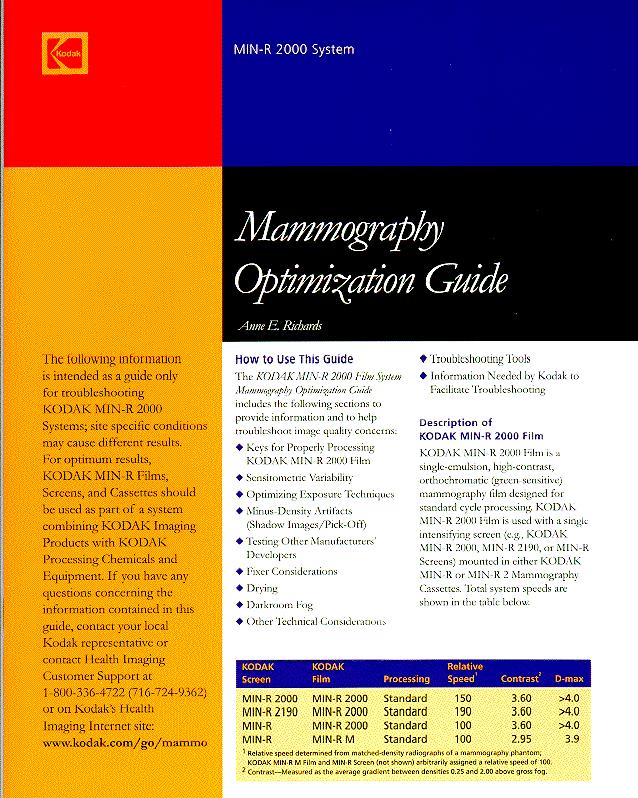
KODAK MIN-R S Film / 4906
 TECHNICAL INFORMATION DATA SHEET Copyright, Eastman Kodak Company, 2002 KODAK MIN-R S Film / 4906 1) Description KODAK MIN-R S Film / 4906 is a medium speed, dual coated, ortho-sensitive medical x-ray
Digital Imaging started in the 1972 with Digital subtraction angiography Clinical digital imaging was employed from the 1980 ~ 37 years ago Amount of
 Digital Imaging started in the 1972 with Digital subtraction angiography Clinical digital imaging was employed from the 1980 ~ 37 years ago Amount of radiation to the population due to Medical Imaging
Collimation Assessment Using GAFCHROMIC XR-M2
 Collimation Assessment Using GAFCHROMIC XR-M2 I. Introduction A method of collimation assessment for GE Senographe full-field digital mammography (FFDM) systems is described that uses a self-developing
Compliance Guidance for RADIOGRAPHIC QUALITY CONTROL (5 th Edition)
") Compliance Guidance for RADIOGRAPHIC QUALITY CONTROL (5 th Edition) New Jersey Department of Environmental Protection Bureau of X-ray Compliance PO Box 420 Mail Code 25-01 Trenton, NJ 08625-0420 FAX 609-984-5811
Compliance Guidance for RADIOGRAPHIC QUALITY CONTROL (5 th Edition) New Jersey Department of Environmental Protection Bureau of X-ray Compliance PO Box 420 Mail Code 25-01 Trenton, NJ 08625-0420 FAX 609-984-5811
FFDM in the Field: Physicist's Role in the QC of Mammography Laser Printers May Carl R. Keener, Ph.D., DABMP, DABR
 FFDM in the Field: Physicist's Role in the QC of Mammography Laser Printers May 2010 Carl R. Keener, Ph.D., DABMP, DABR keener@marpinc.com MARP Medical & Radiation Physics, Inc. Physicist's Role in the
Exposure System Selection
 Principles of Imaging Science II (RAD120) Exposure Systems Exposure System Selection Radiographic exposure is a very complex process Best technique systems manipulate one variable while holding others
KODAK DIRECTVIEW CR Mammography Feature User s Guide
 KODAK DIRECTVIEW CR Mammography Feature User s Guide 17 September 2010 9G3741 Version 1.0 Carestream Health, Inc. 150 Verona Street Rochester, NY 14608 CARESTREAM, DIRECTVIEW, and DRYVIEW are trademarks
Safelight Fog does what to contrast and density on film?
 Terri Jurkiewicz Safelight Fog does what to contrast and density on film? ANSWER INCREASES DENSITY DECREASES CONTRAST Explain how you determine if the focal spot size is within appropriate limits.
Patient-Assisted Compression Impact on Image Quality and Workflow
 Patient-Assisted Compression Impact on Image Quality and Workflow Senographe Pristina In 2017, GE Healthcare s Senographe Pristina ( Pristina ) was approved by the FDA using the standard technologist-controlled
Image Quality. HTC Grid High Transmission Cellular Grid provides higher contrast images
 B R E A S T I M A G I N G S O L U T I O N S Setting the benchmark for mammography M-IV Series Innovations in breast imaging The Lorad M-IV Series exemplifies Hologic's commitment to developing advanced
Exposure Indices and Target Values in Radiography: What Are They and How Can You Use Them?
 Exposure Indices and Target Values in Radiography: What Are They and How Can You Use Them? Definition and Validation of Exposure Indices Ingrid Reiser, PhD DABR Department of Radiology University of Chicago
New spectral benefi ts, proven low dose
 New spectral benefi ts, proven low dose Philips MicroDose mammography SI, technical data sheet Philips MicroDose SI with single-shot spectral imaging is a fullfi eld digital mammography solution that delivers
RADIOGRAPHIC EXPOSURE
 RADIOGRAPHIC EXPOSURE Receptor Exposure Receptor Exposure the that interacts with the receptor. Computed Radiography ( ) requires a. Direct Digital Radiography (DR) requires a. Exposure Indicators Exposure
Beam-Restricting Devices
 Beam-Restricting Devices Three factors contribute to an increase in scatter radiation: Increased kvp Increased Field Size Increased Patient or Body Part Size. X-ray Interactions a some interact with the
FFDM -FCRm QC Requirements- What You REALLY Need to Know
 FFDM -FCRm QC Requirements- What You REALLY Need to Know Melissa C. Martin, M.S., FACR, FAAPM, FACMP AAPM Annual Meeting July 28, 2008 FDA Approval for Mammography Fuji FCRm System approved for Mammography
GE Healthcare. Senographe 2000D Full-field digital mammography system
 GE Healthcare Senographe 2000D Full-field digital mammography system Digital has arrived. The Senographe 2000D Full-Field Digital Mammography (FFDM) system gives you a unique competitive advantage. That
Image Quality. HTC Grid High Transmission Cellular Grid provides higher contrast images
 B R E A S T I M A G I N G S O L U T I O N S Setting the benchmark for mammography M-IV Series Innovations in breast imaging The Lorad M-IV Series exemplifies Hologic s commitment to developing advanced
Multiple Choice Identify the letter of the choice that best completes the statement or answers the question.
 RA110 test 3 Multiple Choice Identify the letter of the choice that best completes the statement or answers the question. 1. An object 35 cm in width is radiographed at 100 cm SID and at a 50 cm SOD. What
REQUIREMENTS FOR LICENCE HOLDERS WITH RESPECT TO QUALITY CONTROL TESTS FOR DIAGNOSTIC X-RAY IMAGING SYSTEMS
 REQUIREMENTS FOR LICENCE HOLDERS WITH RESPECT TO QUALITY CONTROL TESTS FOR DIAGNOSTIC X-RAY IMAGING SYSTEMS DEPARTMENT OF HEALTH DIRECTORATE: RADIATION CONTROL Implementation date: 31 March 2009 Contents
X-ray Imaging. PHYS Lecture. Carlos Vinhais. Departamento de Física Instituto Superior de Engenharia do Porto
 X-ray Imaging PHYS Lecture Carlos Vinhais Departamento de Física Instituto Superior de Engenharia do Porto cav@isep.ipp.pt Overview Projection Radiography Anode Angle Focal Spot Magnification Blurring
Breast Imaging Basics: Module 10 Digital Mammography
 Module 10 Transcript For educational and institutional use. This test bank is licensed for noncommercial, educational inhouse or online educational course use only in educational and corporate institutions.
Digital Mammography Quality Control for the Mammographic Technologist
 Ontario Breast Screening Program Digital Mammography Quality Control for the Mammographic Technologist Authors: G.E. Mawdsley, A.K. Bloomquist, M.J. Yaffe October 2011 Revision 3.1 Mammographic Physics
Imaging Technique Optimization of Tungsten Anode FFDM System
 Imaging Technique Optimization of Tungsten Anode FFDM System Biao Chen a*, Andrew P. Smith b, Zhenxue Jing a, Elena Ingal a a Hologic, Inc. 600 Technology Drive, DE 1970 b Hologic, Inc. 35 Crosby Drive,
Multiple Choice Identify the letter of the choice that best completes the statement or answers the question.
 RA202 image production class two Multiple Choice Identify the letter of the choice that best completes the statement or answers the question. 1. What removes excess chemistry from the film prior to it
DIGITAL RADIOGRAPHY ARTIFACTS
 IMAGING LAB MPHY 487 DIGITAL RADIOGRAPHY ARTIFACTS Mohammad Esmael Alsulimane B.Sc, M.Sc Medical Physics Lecturer - Physics Department All Rights Reserved: Some information and figures in this presentation
History of digital imaging
 CR/QA RADCHEX History of digital imaging Early, crude digital detectors were developed in the 1970 s Image quality was problematic Processing time of digital images was untenable Viewing, transfer and
Breast Imaging Basics: Module 3 Producing Quality Images
 Module 3 Transcript For educational and institutional use. This test bank is licensed for noncommercial, educational inhouse or online educational course use only in educational and corporate institutions.
Nuclear Associates
 Nuclear Associates 07-644 Grid Alignment Test Tool Users Manual March 2005 Manual No. 07-644-1 Rev. 2 2004, 2005 Fluke Corporation, All rights reserved. Printed in U.S.A. All product names are trademarks
Y11-DR Digital Radiography (DR) Image Quality
 Image Quality") Y11-DR Digital Radiography (DR) Image Quality Image quality is stressed for all systems in Safety Code 35. In the relevant sections Health Canada s advice is the manufacturer s recommended test procedures
Y11-DR Digital Radiography (DR) Image Quality Image quality is stressed for all systems in Safety Code 35. In the relevant sections Health Canada s advice is the manufacturer s recommended test procedures
Published text: Institute of Cancer Research Repository Please direct all s to:
 This is an author produced version of an article that appears in: MEDICAL PHYSICS The internet address for this paper is: https://publications.icr.ac.uk/1316/ Copyright information: http://www.aip.org/pubservs/web_posting_guidelines.html
diagnostic examination
 RADIOLOGICAL PHYSICS 2011 Raphex diagnostic examination Adel A. Mustafa, Ph.D., Editor PUBLISHED FOR: RAMPS (Radiological and Medical Physics Society of New York) preface The RAPHEX Diagnostic exam 2011
DIAGNOSTIC ACCREDITATION PROGRAM. Radiology and CT Quality Control Procedures Workbook
 DIAGNOSTIC ACCREDITATION PROGRAM Radiology and CT Quality Control Procedures Workbook Quality Control Procedures Radiography/CR/DR Safety Code 35 Summary For more detail about each quality control (QC)
X-RAY IMAGING EE 472 F2017. Prof. Yasser Mostafa Kadah
 X-RAY IMAGING EE 472 F2017 Prof. Yasser Mostafa Kadah www.k-space.org Recommended Textbook Stewart C. Bushong, Radiologic Science for Technologists: Physics, Biology, and Protection, 10 th ed., Mosby,
PRACTICE GUIDELINE FOR DETERMINANTS OF IMAGE QUALITY IN DIGITAL MAMMOGRAPHY
 The American College of Radiology, with more than 30,000 members, is the principal organization of radiologists, radiation oncologists, and clinical medical physicists in the United States. The College
Introduction of Computed Radiography in Two Mammography Services: Image Quality and Dose Analysis
 Introduction of Computed Radiography in Two Mammography Services: Image Quality and Dose Analysis Rosangela Requi Jakubiak* a, Humberto Remigio Gamba a, Maria Manuela Ramos a, Gislene Gabrielle Faversani
1. Carlton, Richard R., and Arlene M. Adler. Principles of Radiographic Imaging: An Art and a Science, 5th edition (2013).
.") CODE: RADT 151 INSTITUTE: Health Science TITLE: Radiographic Exposure DEPARTMENT: Radiologic Technology COURSE DESCRIPTION: This course covers the principles of radiographic exposure selection and manipulation
CODE: RADT 151 INSTITUTE: Health Science TITLE: Radiographic Exposure DEPARTMENT: Radiologic Technology COURSE DESCRIPTION: This course covers the principles of radiographic exposure selection and manipulation
TECHNICAL DATA. GIOTTO IMAGE SDL/W is pre-arranged for Full Field Digital Biopsy examination with the patient in prone position.
 Ver. 01/06/07 TECHNICAL DATA GIOTTO IMAGE SDL/W LOW DOSE, FULL FIELD DIGITAL MAMMOGRAPHY UNIT USING AMORPHOUS SELENIUM (a-se) TECHNOLOGY DETECTOR (pre-arranged for stereotactic biopsy with the same digital
Teaching Digital Radiography and Fluoroscopic Radiation Protection
 Teaching Digital Radiography and Fluoroscopic Radiation Protection WCEC 20 th Student Educator Radiographer Conference Dennis Bowman, RT(R), CRT (R)(F) Community Hospital of the Monterey Peninsula (CHOMP)
TESTING FLAT-PANEL IMAGING SYSTEMS: What the Medical Physicist Needs to Know. JAMES A. TOMLINSON, M.S., D.A.B.R. Diagnostic Radiological Physicist
 TESTING FLAT-PANEL IMAGING SYSTEMS: What the Medical Physicist Needs to Know JAMES A. TOMLINSON, M.S., D.A.B.R. Diagnostic Radiological Physicist Topics Image Uniformity and Artifacts Image Quality - Detail
Nuclear Associates
 Nuclear Associates 07-649 CDRH Fluoroscopic Phantom Users Manual March 2005 Manual No. 07-649-1 Rev. 2 2004, 2005 Fluke Corporation, All rights reserved. Printed in U.S.A. All product names are trademarks
Quality control for digital mammography: Part II recommendations from the ACRIN DMIST trial
 Quality control for digital mammography: Part II recommendations from the ACRIN DMIST trial Martin J. Yaffe, Aili K. Bloomquist, Gordon E. Mawdsley, Etta D. Pisano, R. Edward Hendrick, Laurie L. Fajardo,
Predicted image quality of a CMOS APS X-ray detector across a range of mammographic beam qualities
 Journal of Physics: Conference Series PAPER OPEN ACCESS Predicted image quality of a CMOS APS X-ray detector across a range of mammographic beam qualities Recent citations - Resolution Properties of a
Test Equipment for Radiology and CT Quality Control Contents
 Test Equipment for Radiology and CT Quality Control Contents Quality Control Testing...2 Photometers for Digital Clinical Display QC...3 Primary Workstations...3 Secondary Workstations...3 Testing of workstations...3
Introduction. Chapter 16 Diagnostic Radiology. Primary radiological image. Primary radiological image
 Introduction Chapter 16 Diagnostic Radiology Radiation Dosimetry I Text: H.E Johns and J.R. Cunningham, The physics of radiology, 4 th ed. http://www.utoledo.edu/med/depts/radther In diagnostic radiology
Enhanced Functionality of High-Speed Image Processing Engine SUREengine PRO. Sharpness (spatial resolution) Graininess (noise intensity)
 Graininess (noise intensity)") Vascular Enhanced Functionality of High-Speed Image Processing Engine SUREengine PRO Medical Systems Division, Shimadzu Corporation Yoshiaki Miura 1. Introduction In recent years, digital cardiovascular
Vascular Enhanced Functionality of High-Speed Image Processing Engine SUREengine PRO Medical Systems Division, Shimadzu Corporation Yoshiaki Miura 1. Introduction In recent years, digital cardiovascular
Learning Objectives: What s my motivation? (unknown screen actor) Workshop Overview
 Workshop Overview") Practical Medical Physics Adapting Traditional Clinical Medical Physics to Digital Radiography Charles E. Willis, Ph.D., DABR Associate Professor Department of Imaging Physics The University of Texas M.D.
Practical Medical Physics Adapting Traditional Clinical Medical Physics to Digital Radiography Charles E. Willis, Ph.D., DABR Associate Professor Department of Imaging Physics The University of Texas M.D.
CHAPTER 6 QC Test For Fluoroscopic Equipment. Prepared by:- Kamarul Amin bin Abu Bakar School of Medical Imaging KLMUC
 CHAPTER 6 QC Test For Fluoroscopic Equipment Prepared by:- Kamarul Amin bin Abdullah @ Abu Bakar School of Medical Imaging KLMUC Lesson Outcomes Describe the objectives of each QC test done. Identify QC
Photomultiplier Tube
 Nuclear Medicine Uses a device known as a Gamma Camera. Also known as a Scintillation or Anger Camera. Detects the release of gamma rays from Radionuclide. The radionuclide can be injected, inhaled or
NEXT Protocol for the Survey of Pediatric Chest Radiography
 NEXT 1998 Protocol for the Survey of Pediatric Chest Radiography Prepared by David C. Spelic Division of Mammography Quality and Radiation Programs Center for Devices and Radiological Health Office of
DISC QC/QA Program for Digital Imaging Systems using the DR Radchex Plus Meter
 DISC QC/QA Program for Digital Imaging Systems using the DR Radchex Plus Meter Revision Date: January 5th, 2017 www.disc-imaging.com Table of Contents Section A: Preliminary Setup Requirements... 4 Tools
of sufficient quality and quantity
 of sufficient quality and quantity The patient s body attenuates the beam as it passes though the body More energy is deposited in organs located near the entry of the beam than near the exit of the beam
GE Healthcare. Essential for life. Senographe Essential Full-Field Digital Mammography system
 GE Healthcare Essential for life Senographe Essential Full-Field Digital Mammography system Excellence in FFDM is a process. An ongoing quest, fueled by our continuing breakthroughs in breast cancer detection
Digital radiography: Practical advantages of Digital Radiography. Practical Advantages in image quality
 Digital radiography: Digital radiography is set to become the most common form of processing radiographic images in the next 10 years. This is due to a number of practical and image quality issues. Practical
Maximizing clinical outcomes
 Maximizing clinical outcomes Digital Tomosynthesis Dual Energy Subtraction Automated Long Length Imaging Improved image quality at a low dose Xray Xray Patented ISS capture technology promotes high sensitivity
Image Display and Perception
 Image Display and Perception J. Anthony Seibert, Ph.D. Department of Radiology UC Davis Medical Center Sacramento, California, USA Image acquisition, display, & interpretation X-rays kvp mas Tube filtration
Third Edition DECEMBER Collaborating institutes: QARAD, BE LUCK, BE ARCADES, FR LRCB, NL EMIFMA, BE NCCPM, UK. Contributors:
 Third Edition DECEMBER 1999 Collaborating institutes: QARAD, BE LUCK, BE ARCADES, FR LRCB, NL EMIFMA, BE NCCPM, UK Contributors: M. Fitzgerald, London, UK P. Heid, Marseille, FR R. van Loon, Brussels,
X-RAYS - NO UNAUTHORISED ENTRY
 Licencing of premises Premises Refer Guidelines A radiation warning sign and warning notice, X-RAYS - NO UNAUTHORISED ENTRY must be displayed at all entrances leading to the rooms where x-ray units are
Ludlum Medical Physics
 Ludlum Medical Physics Medical Imaging Radiology QA Test Tools NEW LUDLUM PRODUCT LINE Medical Physics Products Medical Physics Products What are they? Products used to measure radiation output and to
Digital radiography (DR) post processing techniques for pediatric radiology
 post processing techniques for pediatric radiology") Digital radiography (DR) post processing techniques for pediatric radiology St Jude Children s Research Hospital Samuel Brady, MS PhD DABR samuel.brady@stjude.org Purpose Review common issues and solutions
Digital radiography (DR) post processing techniques for pediatric radiology St Jude Children s Research Hospital Samuel Brady, MS PhD DABR samuel.brady@stjude.org Purpose Review common issues and solutions
Features and Weaknesses of Phantoms for CR/DR System Testing
 Physics testing of image detectors Parameters to test Features and Weaknesses of Phantoms for CR/DR System Testing Spatial resolution Contrast resolution Uniformity/geometric distortion Dose response/signal
Dosepix Detector as kvp-meter in Radiology and Mammography: First steps
 Dosepix Detector as kvp-meter in Radiology and Mammography: First steps F.Bisello, I.Ritter, F.Tennert, A.Zang MediPix Collaboration Meeting, 19th February 2014, CERN Protect, Enhance, and Save Lives -
Overview. Professor Roentgen was a Physicist!!! The Physics of Radiation Oncology X-ray Imaging
 The Physics of Radiation Oncology X-ray Imaging Charles E. Willis, Ph.D. DABR Associate Professor Department of Imaging Physics The University of Texas M.D. Anderson Cancer Center Houston, Texas Overview
RaySafe X2. Effortless measurements of X-ray
 RaySafe X2 Effortless measurements of X-ray At your fingertips We ve grown accustomed to intuitive interactions with our devices. After all, it s not the device that s most important, but what you can
TOPICS: CT Protocol Optimization over the Range of Patient Age & Size and for Different CT Scanner Types: Recommendations & Misconceptions
 CT Protocol Optimization over the Range of Patient Age & Size and for Different CT Scanner Types: Recommendations & Misconceptions TOPICS: Computed Tomography Quick Overview CT Dosimetry Effects of CT
NATIONWIDE EVALUATION OF X-RAY TRENDS (NEXT) TABULATION AND GRAPHICAL SUMMARY OF THE 1999 DENTAL RADIOGRAPHY SURVEY
 TABULATION AND GRAPHICAL SUMMARY OF THE 1999 DENTAL RADIOGRAPHY SURVEY") CRCPD Publication E-3-6-a Available Online at No Charge $15. for a Computer-Generated Copy NATIONWIDE EVALUATION OF X-RAY TRENDS (NEXT) TABULATION AND GRAPHICAL SUMMARY OF THE 1999 DENTAL RADIOGRAPHY SURVEY
CRCPD Publication E-3-6-a Available Online at No Charge $15. for a Computer-Generated Copy NATIONWIDE EVALUATION OF X-RAY TRENDS (NEXT) TABULATION AND GRAPHICAL SUMMARY OF THE 1999 DENTAL RADIOGRAPHY SURVEY
Digital Mammography Quality Control for the Mammographic Physicist
 Ontario Breast Screening Program Digital Mammography Quality Control for the Mammographic Physicist Authors: G.E. Mawdsley, A.K. Bloomquist, M.J. Yaffe March 2014 Revision 3.2 Mammographic Physics Consulting
Mammography Solution. AMULET Innovality. The new leader in the AMULET series. Tomosynthesis, 3D mammography and biopsy are all available.
 Mammography Solution AMULET Innovality The new leader in the AMULET series. Tomosynthesis, 3D mammography and biopsy are all available. FUJIFILM supports the Pink Ribbon Campaign for early detection of
1.1.Clinical Artefacts
 1.1.Clinical Artefacts While the incidence of artefact on digital mammographic images 1 is typically less than with film based mammography, artefacts can be produced on digital systems. This section provides
X-ray Tube and Generator Basic principles and construction
 X-ray Tube and Generator Basic principles and construction Dr Slavik Tabakov - Production of X-rays OBJECTIVES - X-ray tube construction - Anode - types, efficiency - X-ray tube working characteristics
Assessment of Beam Alignment, Collimation and Half Value Layer of Some Selected X-Ray Machines in Plateau State, Nigeria
 International Journal of Innovative Scientific & Engineering Technologies Research 5(4):-5, Oct.-Dec., 07 SEAHI PUBLICATIONS, 07 www.seahipaj.org ISSN: 60-896X Assessment of Beam Alignment, Collimation
Optimization of Digital Mammography Resolution Using Magnification Technique in Computed Radiography 1
 Optimization of Digital Mammography Resolution Using Magnification Technique in Computed Radiography 1 Gham Hur, M.D., Yoon Joon Hwang, M.D., Soon Joo Cha, M.D., Su Young Kim, M.D., Yong Hoon Kim, M.D.
SPRINGFIELD TECHNICAL COMMUNITY COLLEGE ACADEMIC AFFAIRS
 SPRINGFIELD TECHNICAL COMMUNITY COLLEGE ACADEMIC AFFAIRS Course Number: RADG 112 Department: Radiography Course Title: Image Production & Eval. Semester: Spring Year: 1997 Objectives/ Unit One: Introduction