Introduction to BioImage Analysis using Fiji
|
|
|
- Rose Harrington
- 5 years ago
- Views:
Transcription


1 Introduction to BioImage Analysis using Fiji CellNetworks Math-Clinic core facility Qi Gao Carlo A. Beretta


2 Math-Clinic core facility Data analysis services on bioinformatics & bioimage analysis: 1-to-1 consultancies research collaboration courses and workshops internship, MSc/BSc thesis Room 001, BioQuant (INF 267) +49 (0)
3 Agenda Introduction to BioImage Analysis using Fiji 9:00-10:30 Getting to know digital images using Fiji Qi Gao 10:30-11:00 Coffee break 11:00-12:30 Basic bioimage analysis methods Qi Gao 12:30-13:30 Lunch break 13:30-15:00 Automating image analysis (ImageJ Macro) I Carlo Beretta 15:00-15:30 Coffee break 15:30-17:00 Automating image analysis (ImageJ Macro) II Carlo Beretta
4 Getting to know digital images using Fiji
5 with slides and figures from Peter Bankhead Kota Miura Chong Zhang Daniel White


6 1.1 Digital images which are digital images?


7 Images are composed of pixels each pixel corresponds to a number brighter region - more photons - larger pixel value an image is usually display based on grey scale [figure by PB] 0 255
8 A pixel is NOT a little square!!! A pixel is a point sample. It exists only at a point. It generally lies on a grid pattern. X X X X X X X X X = [DW]
9 Look-Up Table (LUT) pixels representing color is determined by the LUT [figure by PB] 0 255
10 Look-Up Table (LUT) pixels representing color is determined by the LUT changing the LUT won t affect pixel values [figure by PB] 0 255
11 The numbers contain all information of an image an image can be displayed arbitrarily what we really care in image analysis are the numbers (pixel values) [figure by PB] 0 255


12 Do not trust your eyes! What I think I see What is actually there Green and yellow circles? A and B: which is brighter?

13 Fiji is just imagej The main window [DW]
14 Fiji is just imagej Overview of the menus Help, Links Selection/ROI handling File input/output Image filters Statistics Visualization parameters Windows Plugins, Macros and Utilities [DW]
15 Fiji is just imagej The status bar (message & progress) Shows information about long-running processes. Clicking in the status bar shows information about memory consumption. [DW]

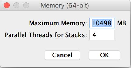
16 Set up memory
 in the plugins folder of Fiji (ImageJ) without unzip it restart Fiji")
17 Install plugins download the ImageJ plugin files (xxx.jar) put the files (xxx.jar) in the plugins folder of Fiji (ImageJ) without unzip it restart Fiji (ImageJ)


18 Check updates Check Update Status: [Help > Update ] After confirming to be up-to-date, Click Manage Update Sites : to add optional plugins [KM]

19 Open an image, check the pixel values 1. [File -> Open -> Cell_Colony.tif] width x height (0,0) x y

20 Tip: press L to use the Command Finder memorise the menu? not necessary!
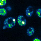
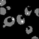
![Check and change the LUT 1. [File -> Open -> Cell_Colony.tif] 2.](/docs-images/88/116199931/images/21-0.jpg "[Image -> Color -> Show LUT] 3.")
![Change the LUT by [Image -> Lookup Tables -> Spectrum] 4.](/docs-images/88/116199931/images/21-1.jpg "Check the LUT again by [Image -> Color -> Show LUT] 5.")
21 Check and change the LUT 1. [File -> Open -> Cell_Colony.tif] 2. [Image -> Color -> Show LUT] 3. Change the LUT by [Image -> Lookup Tables -> Spectrum] 4. Check the LUT again by [Image -> Color -> Show LUT] 5. [Image -> Color -> Display LUTs] Do the pixel values also change? [KM]
22 Image depth measured intensity by detector [digitization] corresponding level in image digital intensity resolution: 10 9 real analogue intensities digital intensity resolution: [DW]
23 Bit-depth determines the dynamic range of image pixel values 1bit: 2 1 = 2 steps (segmentation) 2bit: 2 2 = 4 steps 4bit: 2 4 = 16 steps 8bit: 2 8 = 256 steps 16bit: 2 16 = 65,536 steps 32bit: 2 32 = 4,294,967,296 steps (~ limit of human eye) (intensity-based measurements) Images can contain far more different pixel values than our eyes can distinguish!

 2 bit (4 values) 1 bit (2")
24 Image bit-depth A higher bit-depth allows pixels to have more different values 8 bit (256 values) 4 bit (16 values) 2 bit (4 values) 1 bit (2 values) [PB]

25 Reducing bit-depth will lose information data scaling: pixel values are rescaled and rounded to the nearest valid integer 16-bit image 2 16 = values 8-bit image 2 8 = 256 values Values changed by rounding [PB]
26 Choosing bit-depth during image acquisition Exciting, high-risk method Use the minimum bit-depth that gives the accuracy you need Safer method Use the maximum bit-depth you can (but that doesn t make the computer crash) [PB]

![tif] then line of selection tools 2.](/docs-images/88/116199931/images/27-1.jpg "[Analyze -> Plot Profile..] 3.")
![[Edit -> Option -> Conversion] (ON!) 4.](/docs-images/88/116199931/images/27-2.jpg "[Image -> Type -> 8-bit] 5.")
![[Analyze -> Plot Profile..] without scaling 1.](/docs-images/88/116199931/images/27-3.jpg "tif] [Edit -> Selection -> Restore..] 2.")
27 Convert bit-depth 16bit 8bit with scaling 1. [File -> Open -> m51.tif] then line of selection tools 2. [Analyze -> Plot Profile..] 3. [Edit -> Option -> Conversion] (ON!) 4. [Image -> Type -> 8-bit] 5. [Analyze -> Plot Profile..] without scaling 1. [File -> Open -> m51.tif] [Edit -> Selection -> Restore..] 2. [Edit -> Option -> Conversion] (OFF!) 3. [Image -> Type -> 8-bit] 4. [Analyze -> Plot Profile..] [KM]


28 Image dimension image can be multi-dimensional x, y, z coordinate color channel time point 2D: x-y 4D: x-y-z-ch 3D: x-y-ch [figure by PB]

29 ImageJ makes it (relatively) straightforward to work with images that have up to 5 dimensions Colour channels Time point z-slice [PB]

30 Stack basics Open listeriacells.stk. [Start Animation] [Stop Animation] [Animation Options] [KM]


31 Orthogonal view Open mitosis_anaphase_3d.tif [Image > Stacks > Orthogonal Views] Interactive Reslice. Drag the crossing lines. [KM]

![tif [Plugins > 3D Viewer]](/docs-images/88/116199931/images/32-2.jpg "rotate and zoom (wheel)!")
32 3D viewer Open mitosis_anaphase_3d.tif [Plugins > 3D Viewer] rotate and zoom (wheel)! pan: shift-drag [KM]

33 Color image type composite RGB data from the microscope converted after acquisition # channels any 3 bit-depth any for each channel 8-bit for each channels adaptability special scientific softwares appearance varies most softwares appearance consistent
34 When converting a composite image to RGB, information is usually lost Convert to RGB Composite RGB 16-bit channels 8-bit channels [PB]

35 When converting a composite image to RGB, information is usually lost Convert to RGB Composite RGB 16-bit channels 8-bit channels [PB]
36 RGB image for analysis unless you are really really really sure you have not lost vital information for display journal figures, websites, presentations [PB]
37 RGB image 1. Open FluorescentCells.tif 2. [Image -> Type -> RGB Color] what is different than the original? 3. [Image -> Color -> Split Channels] [CZ]

38 Composite image Merge 3 frames [Image -> Color -> Merge Channels ]. [CZ]


39 Composite image Composite: you could process individual channels. -- Do [Image -> Color -> Channel Tool ] and try unchecking some channels! [KM]
40 Image format image image file contains 2 parts header: the metadata (data about data) image data: numbers (pixel values) ics_version 1.0 filename 3a-z-stack (cropped) layout parameters 6 layout order bits x y z channels t layout sizes parameter units relative um um um undefined s parameter scale sensor model Hamamatsu C , 462, 438, 447, 442, 451, 480, 467, 467, 440, 447, 461, 482, 493, 432, 490, 445, 459, 473, 455, 443, 443, 430, 457, 423, 442, 469, 437, 422, 438, 461, 455, 447, 446, 458, 446, 441, 477, 470, 452, 449, 461, 446, 472, 452, 461, 454, 471, 462, 464, 456, 434, 440, 446, 463, 438, 449, 483, 473, 470, 442, 438, 472, 464, 450, 454, 453, 445, 469, 441, 434, 459, 435, 465, 454, 433, 459, 427, 445, 457, 434, 424, 467, 444, 467, 458, 445, 455, 454, 436, 489, 427, 433, 466, 474, 461, 458, 449, 458, 467, 456, 464, 487, 496, 463, 453, 460, 465, 456, 464, 448, 458, 455, 476, 494, 444, 491, 420, 478, 451, 468, 465, 467, 456, 450, 460, 450, 496, 430, 486, 481, 468, 453, 477, 458, 470, 436, 476, 446, 471, 455, 440, 454, 462, 466, 463, 459, 446, 441, [figure by PB]


41 Image format in some formats, image data is compressed lossy compression may make the image no longer suitable for quantitative analysis original filtered original jpeg compressed filtered compressed [PB]


![]](/docs-images/88/116199931/images/42-5.jpg)
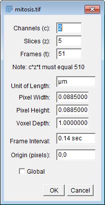
42 Metadata [open > mitosis.tif] [image -> show info ] [image -> properties ] [figure by PB]
43 Image format always keep your original files and metadata avoid using lossy compression (eg, jpeg format) save your images using tiff format

![tif] 2.](/docs-images/88/116199931/images/44-1.jpg "[Analysis > Tools >")
44 Draw scale bar 1. [Open > hela-cell.tif] 2. [Analysis > Tools > Scale Bar] 3. Click OK! [KM]
45 Sometimes there is no scale information


46 Adding real world scale 1. [open -> micrometer.jpg] 2. Draw line between large bars. (50µm). 3. [Analysis > Set Scale ] known distance: 50. Unit of length: µm 4. Click OK [KM]
47 1.2 Image quality good quality of images always benefit analysis images need not only proper storage high bit-depth multi-channel lossless file format but also proper acquisition high resolution low noise and blur properly distributed pixel values fast acquisition
48 Pixel size how big a structure in my image? = how big is a pixel? a pixel is a sample of intensity of a point in space pixel size is pixel spacing distance not the imaginary pixel edge length y x Yes! A pixel is NOT a little square!!! No! [DW]

49 Resolution / pixel size # of pixels in unit length Pixel size = 64.2 µm / 600 = µm 64.2 µm 600 px [figure by PB]
50 Resolution / pixel size # of pixels in unit length resolution affects spatial information Pixel size = 64.2 µm / 75 = µm 64.2 µm 75 px [figure by PB]


51 Higher resolution, more details 4 x 4 pixels 16 x 16 pixels 512 x 512 pixels 64 x 64 pixels 256 x 256 pixels [PB]


52 128 x 128 pixels 256 x 256 pixels 512 x 512 pixels 1024 x 1024 pixels But increasing resolution doesn t add more details indefinitely 8 x 8 pixels 16 x 16 pixels 32 x 32 pixels 64 x 64 pixels


53 Why? An image we can record is the result of replacing each point with a corresponding PSF Point PSF [PB]

54 Why? An image we can record is the result of replacing each point with a corresponding PSF Point PSF [PB]
 detecting more light helps to overcome both noise 1 10 100 1000 exposure time")
55 Noise adds randomness to the pixel values 2 main sources of noise in fluorescence microscopy photon noise - from the random emission of photons read noise - from sources in the detector (microscope) detecting more light helps to overcome both noise exposure time (ms) [PB]

56 Extra light can be obtained with costs longer exposure time loses temporal information beware of over-exposure increase the pixel size loses spatial information [PB]


![histogram] 3.](/docs-images/88/116199931/images/57-6.jpg "Compare the pixel")
57 Understanding histograms Find the corresponding histograms! 1. Open images 2D_Gel.tif and gel_inv.tif 2. Do [Analyze -> histogram] 3. Compare the pixel value in the image and the histogram.


58 under-/over-exposure occur when storing values too low/high for the bit-depth don t know what happens in the darkest/brightest regions
59 Which image is better? a wider and evener distributed histogram means more details stored and good contrast

60 1.3 ROI (region of interest) & measurements selection tools Oval Polygon Freehand Rectangular Rounded rectangle Elliptical Line Segmented Freehand Brush Arrow
![ROI Open any image and then Cropping. [image -> Crop]. Masking. Select a region by rectangular ROI. [Edit -> Clear outside]. [Edit -> Fill]. ( same as [Edit -> Selection -> Create Mask]) Invert ROI.](/docs-images/88/116199931/images/61-0.jpg "[Edit -> Selection -> Make Inverse]. Redirecting ROI. Open any two images. In one of the image, select a region by rectangular ROI.")
61 ROI Open any image and then Cropping. [image -> Crop]. Masking. Select a region by rectangular ROI. [Edit -> Clear outside]. [Edit -> Fill]. ( same as [Edit -> Selection -> Create Mask]) Invert ROI. [Edit -> Selection -> Make Inverse]. Redirecting ROI. Open any two images. In one of the image, select a region by rectangular ROI. Then activate the other image [Edit -> Selection -> Restore Selection]. ROI manager. [Analysis -> Tools -> Roi Manager]. Click Add button to store ROI information. Stored ROI can be saved as a file, and could be loaded again when you restart the ImageJ. [KM]
![Intensity measurements 1. [Analyze -> Set measurements] 2. Open cells_actin.tif 3. Use Polygon ROI and select a cell.](/docs-images/88/116199931/images/62-1.jpg "4. [Analyze -> Measure] 5. Measure also the background. Measure Background as well Intensity = Cell - Background [KM]")
62 Intensity measurements 1. [Analyze -> Set measurements] 2. Open cells_actin.tif 3. Use Polygon ROI and select a cell. 4. [Analyze -> Measure] 5. Measure also the background. Measure Background as well Intensity = Cell - Background [KM]
![Intensity measurements 1. [Analyze > Tools > ROI manager] 2. Open [cells_actin.](/docs-images/88/116199931/images/63-2.jpg "tif] zoom up! ( + key) 3. Use Polygon ROI and select a cell. 4.")

63 Intensity measurements 1. [Analyze > Tools > ROI manager] 2. Open [cells_actin.tif] zoom up! ( + key) 3. Use Polygon ROI and select a cell. 4. In ROI manager, click Add. 5. Use Rectangular ROI and select background. 3. In ROI manager, click Add. Measure Background as well 7. Click Measure! Intensity = Cell - Background [KM]


64 Creating tricky ROIs Marking a whole cell in hela-cell.tif, excluding the nucleus Draw 2 ROIs & add to the ROI Manager (press t) a polygon around the cell an ellipse around the nucleus under More >>, remove the nucleus ROI from the cell ROI, using XOR [Edit -> Selection -> Create Mask] [CZ]
 roi1 roi2 AND OR XOR")
65 Combination of ROIs (binary images) roi1 roi2 AND OR XOR exclusive or
Introduction to BioImage Analysis
 Introduction to BioImage Analysis Qi Gao CellNetworks Math-Clinic core facility 22-23.02.2018 MATH- CLINIC Math-Clinic core facility Data analysis services on bioimage analysis & bioinformatics: 1-to-1
EPFL BIOP Image Processing Practicals R. Guiet, O. Burri
 EPFL BIOP Image Processing Practicals 23-25.03.2015 R. Guiet, O. Burri Overview DAY 1 Intensity/Histogram Look up table (LUT) Contrast Image Depth RGB images Image Math File Formats Resizing Images Regions
IMAGE PROCESSING PRACTICALS
 EPFL PTBIOP IMAGE PROCESSING PRACTICALS 14.03.2011-16.03.2011 ACKNOWLEDGEMENTS This presentation and the exercises are based on the script CMCI Image processing & Analysis Course Series I which was kindly
Introduction to Image Analysis with
 Introduction to Image Analysis with PLEASE ENSURE FIJI IS INSTALLED CORRECTLY! WHAT DO WE HOPE TO ACHIEVE? Specifically, the workshop will cover the following topics: 1. Opening images with Bioformats
Introduction to ImageJ 8 Sept 2009
 Biomedical Imaging Research Unit School of Medical Sciences Faculty of Medical and Health Sciences The University of Auckland Private Bag 92019 Auckland, NZ Ph: 373 7599 ext. 87438 http://www.auckland.ac.nz/biru/
A guide to SalsaJ. This guide gives step-by-step instructions on how to use SalsaJ to carry out basic data analysis on astronomical data files.
 A guide to SalsaJ SalsaJ is free, student-friendly software developed originally for the European Hands- On Universe (EU-HOU) project. It is designed to be easy to install and use. It allows students to
What is a digital image?
 Chapter 4 What is a digital image? 4.1 How is the image represented by the computer? Pixels Images can have 2 or 3 spacial dimensions, a time dimensions and a number of colour-channels. An image is a rectilinear
Adobe Photoshop CC 2018 Tutorial
 Adobe Photoshop CC 2018 Tutorial GETTING STARTED Adobe Photoshop CC 2018 is a popular image editing software that provides a work environment consistent with Adobe Illustrator, Adobe InDesign, Adobe Photoshop,
Topic 04 What is a digital image?
 Topic 04 What is a digital image? Exercise 4.1 How is the image represented by the computer - Pixels Images can have 2 or 3 spacial dimensions, a time dimensions and a number of colour-channels. An image
CTE BASIC DIGITAL PHOTOGRAPHY STUDY GUIDE
 CTE BASIC DIGITAL PHOTOGRAPHY STUDY GUIDE VOCABULARY Histogram a graph of all tones in an image Image/adjust (hue/saturation, brightness/contrast) hue: color name (like green), saturation: how opaque (rich
Point Spread Function Estimation Tool, Alpha Version. A Plugin for ImageJ
 Tutorial Point Spread Function Estimation Tool, Alpha Version A Plugin for ImageJ Benedikt Baumgartner Jo Helmuth jo.helmuth@inf.ethz.ch MOSAIC Lab, ETH Zurich www.mosaic.ethz.ch This tutorial explains
Photoshop 1. click Create.
 Photoshop 1 Step 1: Create a new file Open Adobe Photoshop. Create a new file: File->New On the right side, create a new file of size 600x600 pixels at a resolution of 300 pixels per inch. Name the file
CATEGORY SKILL SET REF. TASK ITEM
 ECDL / ICDL Image Editing This module sets out essential concepts and skills relating to the ability to understand the main concepts underlying digital images and to use an image editing application to
Zeiss Axiovert 135 Fluorescence Microscope Quick Guide / Operations Manual (v. 1.0 February 09)
") University of Chicago Integrated Light Microscopy Core Dr. Vytas Bindokas, Director http://digital.bsd.uchicago.edu By: Christine Labno, Assistant Director Room: AB-129 Phone: 4-9040 Zeiss Axiovert 135
University of Chicago Integrated Light Microscopy Core Dr. Vytas Bindokas, Director http://digital.bsd.uchicago.edu By: Christine Labno, Assistant Director Room: AB-129 Phone: 4-9040 Zeiss Axiovert 135
μscope Microscopy Software
 μscope Microscopy Software Pixelink μscope Essentials (ES) Software is an easy-to-use robust image capture tool optimized for productivity. Pixelink μscope Standard (SE) Software had added features, making
Basics of Quantitative Imaging and Image Processing Using ImageJ / Fiji. Dan White Nov 2008
 MPI-CBG LMF / IPF Basics of Quantitative Imaging and Image Processing Using ImageJ / Fiji Dan White Nov 2008 Before you start writing... Presentations soon available at: http://tu-dresden.de/med/ifn Light
ISCapture User Guide. advanced CCD imaging. Opticstar
 advanced CCD imaging Opticstar I We always check the accuracy of the information in our promotional material. However, due to the continuous process of product development and improvement it is possible
Supplemental Method Information Zeiss LSM710
 Supplemental Method Information Zeiss LSM710 1 Under the Light Path window set up the confocal for imaging a green dye (Alexa488-EGFP). For example, set up the light path as shown here using the 488 nm
BZ-X800 Analyzer BZ-H4A/BZ-H4M/BZ-H4R/BZ-H4C/BZ-H4CM/BZ-H4K
 96M14854 All-in-One Fluorescence Microscope BZ-X800 Analyzer BZ-H4A/BZ-H4M/BZ-H4R/BZ-H4C/BZ-H4CM/BZ-H4K 1 Getting Started 2 Analyzer Names and Functions 3 Loading and Saving Images 4 Processing Images
DIGITAL-MICROSCOPY CAMERA SOLUTIONS USB 3.0
 DIGITAL-MICROSCOPY CAMERA SOLUTIONS USB 3.0 PixeLINK for Microscopy Applications PixeLINK will work with you to choose and integrate the optimal USB 3.0 camera for your microscopy project. Ideal for use
(Quantitative Imaging for) Colocalisation Analysis
 Colocalisation Analysis") (Quantitative Imaging for) Colocalisation Analysis or Why Colour Merge / Overlay Images are EVIL! Special course for DIGS-BB PhD program What is an Image anyway..? An image is a representation of reality
(Quantitative Imaging for) Colocalisation Analysis or Why Colour Merge / Overlay Images are EVIL! Special course for DIGS-BB PhD program What is an Image anyway..? An image is a representation of reality
Fuzzy Image Editor. User Manual
 Fuzzy Image Editor User Manual I. Installation To install the program, run Fuzzy Image Editor.msi and follow the prompts. Then go to your Program Files/Team6/FuzzyImageEditor folder, right click and run
Digital Imaging - Photoshop
 Digital Imaging - Photoshop A digital image is a computer representation of a photograph. It is composed of a grid of tiny squares called pixels (picture elements). Each pixel has a position on the grid
Chroma Mask. Manual. Chroma Mask. Manual
 Chroma Mask Chroma Mask Tooltips If you let your mouse hover above a specific feature in our software, a tooltip about this feature will appear. Load Image Here an image is loaded which has been shot in
Mask Integrator. Manual. Mask Integrator. Manual
 Mask Integrator Mask Integrator Tooltips If you let your mouse hover above a specific feature in our software, a tooltip about this feature will appear. Load Image Load the image with the standard lighting
Adobe Photoshop CS5 Tutorial
 Adobe Photoshop CS5 Tutorial GETTING STARTED Adobe Photoshop CS5 is a popular image editing software that provides a work environment consistent with Adobe Illustrator, Adobe InDesign, Adobe Photoshop
Table of Contents 1. Image processing Measurements System Tools...10
 Introduction Table of Contents 1 An Overview of ScopeImage Advanced...2 Features:...2 Function introduction...3 1. Image processing...3 1.1 Image Import and Export...3 1.1.1 Open image file...3 1.1.2 Import
START-UP PROCEDURE 1 THE MICROSCOPE STAND 3 OBJECTIVES 5 STARTING WITH LAS (SOFTWARE) AND SETTING UP THE MICROSCOPE STAND 7
 AND SETTING UP THE MICROSCOPE STAND 7") Leica DMI AF6000LX Table of contents START-UP PROCEDURE 1 THE MICROSCOPE STAND 3 OBJECTIVES 5 STARTING WITH LAS (SOFTWARE) AND SETTING UP THE MICROSCOPE STAND 7 ACQUIRE MODULE 6 SETTING THE LIGHTPATH 6
Leica DMI AF6000LX Table of contents START-UP PROCEDURE 1 THE MICROSCOPE STAND 3 OBJECTIVES 5 STARTING WITH LAS (SOFTWARE) AND SETTING UP THE MICROSCOPE STAND 7 ACQUIRE MODULE 6 SETTING THE LIGHTPATH 6
Applying mathematics to digital image processing using a spreadsheet
 Jeff Waldock Applying mathematics to digital image processing using a spreadsheet Jeff Waldock Department of Engineering and Mathematics Sheffield Hallam University j.waldock@shu.ac.uk Introduction When
ImagesPlus Basic Interface Operation
 ImagesPlus Basic Interface Operation The basic interface operation menu options are located on the File, View, Open Images, Open Operators, and Help main menus. File Menu New The New command creates a
ADOBE PHOTOSHOP CS TUTORIAL
 ADOBE PHOTOSHOP CS TUTORIAL A D O B E P H O T O S H O P C S Adobe Photoshop CS is a popular image editing software that provides a work environment consistent with Adobe Illustrator, Adobe InDesign, Adobe
Using the Nikon TE2000 Inverted Microscope
 Wellcome Trust Centre for Human Genetics Molecular Cytogenetics and Microscopy Core Using the Nikon TE2000 Inverted Microscope Fluorescence image acquisition using Scanalytic s IPLab software and the B&W
Dr. Bob on Colocalization or MSL Experiments In Learning Colocalization Using Image J
 Dr. Bob on Colocalization or MSL Experiments In Learning Colocalization Using Image J Confocal microscopy is used to test whether two fluorescently labeled molecules are associated with one another. If
Quick Guide for Zeiss 710 Laser Scanning Confocal MGH Cancer Center
 Quick Guide for Zeiss 710 Laser Scanning Confocal MGH Cancer Center For any questions or concerns, please contact: Linda Nieman lnieman@mgh.harvard.edu Office: (617) 643-9684 Cell: (512) 565-8076 Chenyue
GXCapture 8.1 Instruction Manual
 GT Vision image acquisition, managing and processing software GXCapture 8.1 Instruction Manual Contents of the Instruction Manual GXC is the shortened name used for GXCapture Square brackets are used to
ImageJ: Introduction to Image Analysis 3 May 2012 Jacqui Ross
 Biomedical Imaging Research Unit School of Medical Sciences Faculty of Medical and Health Sciences The University of Auckland Private Bag 92019 Auckland 1142, NZ Ph: 373 7599 ext. 87438 http://www.fmhs.auckland.ac.nz/sms/biru/.
Revosoft Operators Manual
 1 Revosoft Operators Manual Contents LAUNCHING REVOSOFT:... 2 Entering a New Patient:... 2 IMAGE VIEWER:... 6 Mouse Functions:... 6 Panning the image:... 6 Magnification:... 6 Annotations:... 7 Text and
HOW TO CREATE A SUPER SHINY PENCIL ICON
 HOW TO CREATE A SUPER SHINY PENCIL ICON Tutorial from http://psd.tutsplus.com/ Compiled by INTRODUCTION The Pencil is one of the visual metaphors most used to express creativity. In this tutorial,
Image Pro Ultra. Tel:
 Image Pro Ultra www.ysctech.com info@ysctech.com Tel: 510.226.0889 Instructions for installing YSC VIC-USB and IPU For software and manual download, please go to below links. http://ysctech.com/support/ysc_imageproultra_20111010.zip
LSM 780 Confocal Microscope Standard Operation Protocol
 LSM 780 Confocal Microscope Standard Operation Protocol Basic Operation Turning on the system 1. Sign on log sheet according to Actual start time 2. Check Compressed Air supply for the air table 3. Switch
Title: Leica SP5 Confocal User Manual
 Title: Leica SP5 Confocal User Manual Date of first issue: 23/10/2015 Date of review: Version: Admin For assistance or to report an issue Office: CG07 or 05 Email: Igmm-imaginghelpdesk@igmm.ed.ac.uk Website:
FIJI/Image J for Quantification Hands on session
 FIJI/Image J for Quantification Hands on session Dr Paul McMillan Biological Optical Microscopy Platform Hands on demonstrations FIJI set up Line Profile Thresholding Area of stain Cell confluence Nuclei
Manual. Cell Border Tracker. Jochen Seebach Institut für Anatomie und Vaskuläre Biologie, WWU Münster
 Manual Cell Border Tracker Jochen Seebach Institut für Anatomie und Vaskuläre Biologie, WWU Münster 1 Cell Border Tracker 1. System Requirements The software requires Windows XP operating system or higher
Module 11 Digital image processing
 Introduction Geo-Information Science Practical Manual Module 11 Digital image processing 11. INTRODUCTION 11-1 START THE PROGRAM ERDAS IMAGINE 11-2 PART 1: DISPLAYING AN IMAGE DATA FILE 11-3 Display of
Quick Operation Guide
 Quick Operation Guide Power ON Mounting specimens Set the specimen on the sample holder, and install the sample holder to the holder frame. Attach the holder frame to the XY stage. Type of holder Main
Optika ISview. Image acquisition and processing software. Instruction Manual
 Optika ISview Image acquisition and processing software Instruction Manual Key to the Instruction Manual IS is shortened name used for OptikaISview Square brackets are used to indicate items such as menu
Motic Live Imaging Module. Windows OS User Manual
 Motic Live Imaging Module Windows OS User Manual Motic Live Imaging Module Windows OS User Manual CONTENTS (Linked) Introduction 05 Menus, bars and tools 06 Title bar 06 Menu bar 06 Status bar 07 FPS 07
GIMP (GNU Image Manipulation Program) MANUAL
 MANUAL") Selection Tools Icon Tool Name Function Select Rectangle Select Ellipse Select Hand-drawn area (lasso tool) Select Contiguous Region (magic wand) Selects a rectangular area, drawn from upper left (or lower
Selection Tools Icon Tool Name Function Select Rectangle Select Ellipse Select Hand-drawn area (lasso tool) Select Contiguous Region (magic wand) Selects a rectangular area, drawn from upper left (or lower
TRAINING MANUAL. Olympus FV1000
 TRAINING MANUAL Olympus FV1000 September 2014 TABLE OF CONTENTS A. Start-Up Procedure... 1 B. Visual Observation under the Microscope... 1 C. Image Acquisition... 4 A brief Overview of the Settings...
Stitching distortion-free mosaic images for QWA using PTGui. Georg von Arx
 Stitching distortion-free mosaic images for QWA using PTGui Georg von Arx Index A. Introduction and overview... 2 B. Taking microscopic images... 2 C. Installing PTGui... 3 D. Initial Setup... 3 E. Preparing
Digital Photography 1
 Digital Photography 1 Photoshop Lesson 3 Resizing and transforming images Name Date Create a new image 1. Choose File > New. 2. In the New dialog box, type a name for the image. 3. Choose document size
Editing Using Photoshop CS5
 The Photoshop CS4 Editing Workspace - shown is the document (image) window, ToolBox, Info, Navigator, History, Adjustments and Layers Palettes, Windows Menus and Options Bar (on top). USING THE LAYERS
Index of Command Functions
 Index of Command Functions version 2.3 Command description [keyboard shortcut]:description including special instructions. Keyboard short for a Windows PC: the Control key AND the shortcut key. For a MacIntosh:
Photoshop Notes and Application Study Packet
 Basic Parts of Photoshop Interface Photoshop Notes and Application Study Packet PANELS Photoshop Study Packet Copyright Law The World Intellectual Property Organization (WIPO) Copyright treaty restrict
Nikon. King s College London. Imaging Centre. N-SIM guide NIKON IMAGING KING S COLLEGE LONDON
 N-SIM guide NIKON IMAGING CENTRE @ KING S COLLEGE LONDON Starting-up / Shut-down The NSIM hardware is calibrated after system warm-up occurs. It is recommended that you turn-on the system for at least
Using Photoshop Elements
 Using Photoshop Elements Created By: Rick Williams August 2004 Table of Contents Photoshop Element Tools...Page 1 Tool Descriptions... Page 3 Starting Photoshop Elements... Page 7 Resizing an Image...
Guidance on Using Scanning Software: Part 5. Epson Scan
 Guidance on Using Scanning Software: Part 5. Epson Scan Version of 4/29/2012 Epson Scan comes with Epson scanners and has simple manual adjustments, but requires vigilance to control the default settings
In Photoshop you can change the size of an image by going to:
 Change an images size In Photoshop you can change the size of an image by going to: Image Image Size and change the dimensions of the pictures in pixels. Once you adjust the top number the bottom number
Images and Graphics. 4. Images and Graphics - Copyright Denis Hamelin - Ryerson University
 Images and Graphics Images and Graphics Graphics and images are non-textual information that can be displayed and printed. Graphics (vector graphics) are an assemblage of lines, curves or circles with
Recitation 2 Introduction to Photoshop
 Recitation 2 Introduction to Photoshop What is Adobe Photoshop? Adobe Photoshop is a tool for creating digital graphics either by starting with a scanned photograph or artwork or by creating the graphics
I. File Format Tips: For image (raster) files you make (microscope images, scans, photos, screen captures, etc).
 files you make (microscope images, scans, photos, screen captures, etc).") Image Handling Notes Figure Making Workshop Jan/Feb 2018 Quick Guide to Using Images (TIFF, JPEG, PNG, BMP) in/as figures 1) Open the image in Photoshop or GIMP. 2) Adjust Levels and Crop as needed * 3)
Image Handling Notes Figure Making Workshop Jan/Feb 2018 Quick Guide to Using Images (TIFF, JPEG, PNG, BMP) in/as figures 1) Open the image in Photoshop or GIMP. 2) Adjust Levels and Crop as needed * 3)
GETTING STARTED. 0 P a g e B a s i c s o f A d o b e P h o t o s h o p A g a P r i v a t e I n s t i t u t e f o r c o m p u t e r s c i e n c e
 GETTING STARTED 0 P a g e B a s i c s o f A d o b e P h o t o s h o p Adobe Photoshop: is a popular image editing software that provides a work environment consistent with Adobe Illustrator, Adobe InDesign,
Tutorial: Correcting images
 Welcome to Corel PHOTO-PAINT, a powerful tool for editing photos and creating bitmaps. In this tutorial, you'll learn how to perform basic image corrections to a scanned photo. This is what the image looks
Color, Resolution, & Other Image Essentials
 www.gilbertconsulting.com blog.gilbertconsulting.com kgilbert@gilbertconsulting.com Twitter: @gilbertconsult lynda.com/keithgilbert Every Photoshop image consists of three specific attributes: image resolution,
By Washan Najat Nawi
 By Washan Najat Nawi how to get started how to use the interface how to modify images with basic editing skills Adobe Photoshop: is a popular image-editing software. Two general usage of Photoshop Creating
Image Processing Tutorial Basic Concepts
 Image Processing Tutorial Basic Concepts CCDWare Publishing http://www.ccdware.com 2005 CCDWare Publishing Table of Contents Introduction... 3 Starting CCDStack... 4 Creating Calibration Frames... 5 Create
LSM 710 Confocal Microscope Standard Operation Protocol
 LSM 710 Confocal Microscope Standard Operation Protocol Basic Operation Turning on the system 1. Switch on Main power switch 2. Switch on System / PC power button 3. Switch on Components power button 4.
CHAPTER1: QUICK START...3 CAMERA INSTALLATION... 3 SOFTWARE AND DRIVER INSTALLATION... 3 START TCAPTURE...4 TCAPTURE PARAMETER SETTINGS... 5 CHAPTER2:
 Image acquisition, managing and processing software TCapture Instruction Manual Key to the Instruction Manual TC is shortened name used for TCapture. Help Refer to [Help] >> [About TCapture] menu for software
Using Adobe Photoshop
 Using Adobe Photoshop 4 Colour is important in most art forms. For example, a painter needs to know how to select and mix colours to produce the right tones in a picture. A Photographer needs to understand
Leica TCS SP8 Quick Start Guide
 Leica TCS SP8 Quick Start Guide Leica TCS SP8 System Overview Start-Up Procedure 1. Turn on the CTR Control Box, EL6000 fluorescent light source for the microscope stand. 2. Turn on the Scanner Power
Grid Assembly. User guide. A plugin developed for microscopy non-overlapping images stitching, for the public-domain image analysis package ImageJ
 BIOIMAGING AND OPTIC PLATFORM Grid Assembly A plugin developed for microscopy non-overlapping images stitching, for the public-domain image analysis package ImageJ User guide March 2008 Introduction In
ImageJ, A Useful Tool for Image Processing and Analysis Joel B. Sheffield
 ImageJ, A Useful Tool for Image Processing and Analysis Joel B. Sheffield Temple University Dedicated to the memory of Dan H. Moore (1909-2008) Presented at the 2008 meeting of the Microscopy and Microanalytical
Photoshop Elements Hints by Steve Miller
 2015 Elements 13 A brief tutorial for basic photo file processing To begin, click on the Elements 13 icon, click on Photo Editor in the first box that appears. We will not be discussing the Organizer portion
Portrait Pro User Manual
 Portrait Pro User Manual Version 17.0 Anthropics Technology Ltd www.portraitpro.com Contents 3 Table of Contents Part I Getting Started 6 1 Quick Start... Guide 7 2 Top Tips... For Best Results 8 3 Portrait...
PHOTOSHOP. pixel based image editing software (pixel=picture element) several small dots or pixels make up an image.
 several small dots or pixels make up an image.") Photoshop PHOTOSHOP pixel based image editing software (pixel=picture element) several small dots or pixels make up an image. RESOLUTION measurement of the total number of pixels displayed determines the
Photoshop PHOTOSHOP pixel based image editing software (pixel=picture element) several small dots or pixels make up an image. RESOLUTION measurement of the total number of pixels displayed determines the
One method for removing and replacing the sky in an image
 One method for removing and replacing the sky in an image Reality versus Artistic License I am pretty sure that when Monet painted these they did not reflect reality. What I Look For I look for an image
Leica TCS SP8 Quick Start Guide
 Leica TCS SP8 Quick Start Guide Leica TCS SP8 System Overview Start-Up Procedure 1. Turn on the CTR Control Box, Fluorescent Light for the microscope stand. 2. Turn on the Scanner Power (1) on the front
MC3 Motion Control System Shutter Stream Quickstart
 MC3 Motion Control System Shutter Stream Quickstart Revised 7/6/2016 Carousel USA 6370 N. Irwindale Rd. Irwindale, CA 91702 www.carousel-usa.com Proprietary Information Carousel USA has proprietary rights
Swept-Field User Guide
 Swept-Field User Guide Note: for more details see the Prairie user manual at http://www.prairietechnologies.com/resources/software/prairieview.html Please report any problems to Julie Last (jalast@wisc.edu)
Remote Sensing Instruction Laboratory
 Laboratory Session 217513 Geographic Information System and Remote Sensing - 1 - Remote Sensing Instruction Laboratory Assist.Prof.Dr. Weerakaset Suanpaga Department of Civil Engineering, Faculty of Engineering
Photoshop CC: Essentials
 Photoshop CC: Essentials Summary Workspace Overview... 2 Exercise Files... 2 Selection Tools... 3 Select All, Deselect, And Reselect... 3 Adding, Subtracting, and Intersecting... 3 Working with Layers...
BV NNET User manual. V0.2 (Draft) Rémi Lecerf, Marie Weiss
 Rémi Lecerf, Marie Weiss") BV NNET User manual V0.2 (Draft) Rémi Lecerf, Marie Weiss 1. Introduction... 2 2. Installation... 2 3. Prerequisites... 2 3.1. Image file format... 2 3.2. Retrieving atmospheric data... 3 3.2.1. Using
BV NNET User manual V0.2 (Draft) Rémi Lecerf, Marie Weiss 1. Introduction... 2 2. Installation... 2 3. Prerequisites... 2 3.1. Image file format... 2 3.2. Retrieving atmospheric data... 3 3.2.1. Using
MRI Grid. The MRI Grid is a tool in MRI Cell Image Analyzer, that can be used to associate measurements with labeled positions on a board.
 Abstract The is a tool in MRI Cell Image Analyzer, that can be used to associate measurements with labeled positions on a board. Illustration 2: A grid on a binary image. Illustration 1: The interface
Supplemental Figure 1: Histogram of 63x Objective Lens z axis Calculated Resolutions. Results from the MetroloJ z axis fits for 5 beads from each
 Supplemental Figure 1: Histogram of 63x Objective Lens z axis Calculated Resolutions. Results from the MetroloJ z axis fits for 5 beads from each lens with a 1 Airy unit pinhole setting. Many water lenses
Zeiss LSM 510 Confocor III Training Notes. Center for Cell Analysis & Modeling
 Zeiss LSM 510 Confocor III Training Notes Center for Cell Analysis & Modeling Confocor 3 Start Up Go to System Module Turn on Main Switch, System/ PC, and Components Switches Do you need the arc lamp?
Image Capture TOTALLAB
 1 Introduction In order for image analysis to be performed on a gel or Western blot, it must first be converted into digital data. Good image capture is critical to guarantee optimal performance of automated
PhotoFiltre DEPARTMENT OF EDUCATION
 DEPARTMENT OF EDUCATION PhotoFiltre Updated on 20 February 2010 This resource is part of the resource collection available through the ecentre for teachers. www.ecentre.education.tas.gov.au PhotoFiltre
Definiens. Tissue Studio 4.2. Tutorial 1: Composer and Nuclear Markers
 Definiens Tissue Studio 4.2 Tutorial 1: Composer and Nuclear Markers Tutorial 1: Composer and Nuclear Markers Imprint and Version Copyright 2015 Definiens AG. All rights reserved. This document may be
Workshop: Image processing and analysis with ImageJ
 Workshop: Montpellier RIO Imaging Volker Baecker 02.03.2012 1/88 Table of Contents 1. Introduction...4 2. Installing and setting up ImageJ...4 2.1 Installing ImageJ...4 2.2 Memory Settings...4 2.3 Upgrading
Capture V2.0 Software Instruction Manual
 Capture V2.0 Software Instruction Manual Contents General Introduction 3 1. Starting Interface 4 2. Windows 5-2.1 Status Bar 5-2.2 Control Bar 5-2.3 Preview Window 6-2.4 Data Bar 6-2.5 Image Bar 7 3. Capture
Zeiss LSM 780 Protocol
 Zeiss LSM 780 Protocol 1) System Startup F Please note the sign-up policy. You must inform the facility at least 24 hours beforehand if you can t come; otherwise, you will receive a charge for unused time.
Welcome to Photoshop CS
 Chapter 1 Welcome to Photoshop CS COPYRIGHTED MATERIAL Photoshop CS is the latest version of Photoshop, Adobe s powerful image-editing program. It s part of Adobe s Creative Suite, a package of design
in the list below are available in the Pro version of Scan2CAD
 Scan2CAD features Features marked only. in the list below are available in the Pro version of Scan2CAD Scan Scan from inside Scan2CAD using TWAIN (Acquire). Use any TWAIN-compliant scanner of any size.
Panoramas and the Info Palette By: Martin Kesselman 5/25/09
 Panoramas and the Info Palette By: Martin Kesselman 5/25/09 Any time you have a color you would like to copy exactly, use the info palette. When cropping to achieve a particular size, it is useful to use
Solution for Image & Video Processing
 Solution for Image & Video Processing December-2015 Index Q.1) a). 2-3 b). 4 (N.A.) c). 4 (N.A.) d). 4 (N.A.) e). 4-5 Q.2) a). 5 to 7 b). 7 (N.A.) Q.3) a). 8-9 b). 9 to 12 Q.4) a). 12-13 b). 13 to 16 Q.5)
Photoshop Elements Week 1 - Photoshop Elements Work Environment
 Menu Bar Just like any computer program, you have several dropdown menus to work with. Explore them all! But, most importantly remember to SAVE! Photoshop Elements Toolbox (with keyboard shortcut) Photoshop
PHOTO 11: INTRODUCTION TO DIGITAL IMAGING
 PHOTO 11: INTRODUCTION TO DIGITAL IMAGING Instructor: Sue Leith Exam Review On your camera, what are the following and what are they used for? WB matches the color temperature of light ISO - The sensitivity
Contents Foreword 1 Feedback 2 Legal information 3 Getting started 4 Installing the correct Capture One version 4 Changing the version type 5 Getting
 Contents Foreword 1 Feedback 2 Legal information 3 Getting started 4 Installing the correct Capture One version 4 Changing the version type 5 Getting to know Capture One Pro 6 The Grand Overview 6 The
Color Correction with Curves
 Lecture 02 wk 07 with Curves Channels o Photoshop uses Channels to provide access to the different color components of an image. o In most cases, an image is composed of one or more 8-bit channels. o Channels
Adobe Photoshop CS2 Workshop
 COMMUNITY TECHNICAL SUPPORT Adobe Photoshop CS2 Workshop Photoshop CS2 Help For more technical assistance, open Photoshop CS2 and press the F1 key, or go to Help > Photoshop Help. Selection Tools - The