Novel 3D Computerized Threshold Amsler Grid Test CA, USA
|
|
|
- Valerie Riley
- 6 years ago
- Views:
Transcription
1 Novel 3D Computerized Threshold Amsler Grid Test Wolfgang Fink 1,2 and Alfredo A. Sadun 2 1 California Institute of Technology, Pasadena, CA, USA 2 Doheny Eye Institute, Keck School of Medicine, University of Southern California, Los Angeles, CA, USA First author and corresponding author: Wolfgang Fink, Ph.D. Visiting Associate in Physics at Caltech Research Assistant Professor of Ophthalmology, Keck School of Medicine at USC California Institute of Technology Mail Code Pasadena, CA Phone: (818) wfink@krl.caltech.edu Alfredo A. Sadun, M.D., Ph.D. Professor of Neuro-ophthalmology and Neurosurgery at the Doheny Eye Institute Keck School of Medicine at the University of Southern California, Los Angeles, CA, USA Phone: (323) asadun@usc.edu 1
2 Abstract Purpose: To introduce a novel 3D computerized threshold Amsler grid test, developed by Fink and Sadun, that allows for a novel characterization of the structure of visual field defects in three dimensions (see Methods: With one eye covered, patients are placed in front of a computer monitor displaying an Amsler grid. While focusing on a varying central fixation marker, the patients trace the areas, on a touch sensitive screen overlaying the displayed grid, that are missing from their field of vision, with their finger. The procedure is repeated with varying degrees of grid contrast. The results are recorded and later displayed as topographical contour rings, in a 3D depiction of the central hill-of-vision. Results: Several clinical pilot studies have been conducted at the Doheny Eye Institute and over 200 patients have been examined so far. Conditions such as optic neuritis, anterior ischemic optic neuropathy, age-related macular degeneration, glaucoma, and ocular hypertension have been examined with the 3D visual field test. The tested visual field is limited to the central 20 to 25 degrees (depending on the computer monitor size). The physiological blind spot cannot be detected with this test due to the fill-in phenomenon. Conclusions: The 3D computerized threshold Amsler grid test is an innovative and fast (4-5 min. per eye) visual field test. It provides several novel features, including: a) additional information through immediate 3D rather than 2D depiction of scotomas, such as location, extent, slope, depth, and shape; b) simple test-setup; c) good patient compliance. In light of results from pilot studies, the 3D visual field test appears to have the potential for the early detection and monitoring of various diseases, in particular glaucoma and macular degeneration. 2
3 1. Introduction Visual field testing (perimetry) has always been an important part of an ophthalmological evaluation. In 1947 Amsler [1, 2] introduced a suprathreshold grid for evaluating the central ten degrees of the visual field. The high contrast of the standard Amsler test may fail to detect subtle field defects, such as relative scotomas. In 1986, threshold Amsler grid testing was introduced by Wall and Sadun [3]. This test utilizes near threshold, rather than suprathreshold, visual stimuli. Contrast between the target and the background was controlled with cross-polarizing filters. With this modification it was possible to detect subtle scotomas and relative visual field defects. Taking this a step further, Fink and Sadun computerized threshold Amsler grid testing in 2000 [4, 5]. The examination results were used to generate a three-dimensional (3D) map of the central visual field that illustrates the location, extent, slope, depth, and shape of visual field defects. Fig. 1. The 3D computerized threshold Amsler grid test, using a computer-monitor with touch sensitive screen. The 3D computerized threshold Amsler grid test (see Fig. 1) is the outcome of a close collaboration over the past three years between the California Institute of Technology and the Doheny Eye Institute, Keck School of Medicine at the University of Southern California (for further details see 2. Examination method The 3D computerized threshold Amsler grid test uses an IBM compatible PC with a 17" monitor and touch sensitive screen (see Fig. 1). Each patient is positioned 30cm in front of the computer monitor. An eye-patch is used to cover the eye that is not being examined. Refractive correction is used with the patient s contact lenses or spectacles when necessary. Fig. 2. Screenshot of Amsler grid at a mid grayscale level with changing central fixation marker and filled in area of scotoma. An Amsler grid, at a preselected contrast level, is displayed on the monitor (see Fig. 2). The patient is first asked to focus on a changing stimulus at the center of the grid. In order to suppress the central Troxler effect and keep the patient's attention, the stimulus is regularly changed (changing set of characters such as letters and numbers, see Fig. 2). The patient is asked to mark the areas, on the Amsler grid, that are missing from his or her field of vision by tracing this region with their finger on the touch screen. Areas that are missing are defined as areas where the grid lines are missing, bent, wavy or distorted, or areas where the grid contrast differs 3
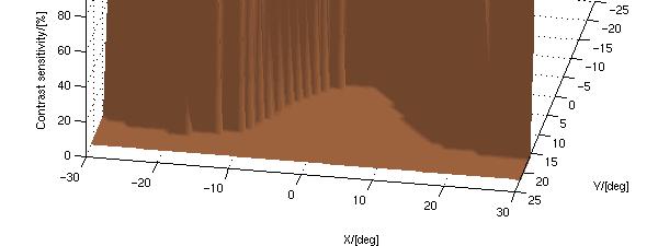
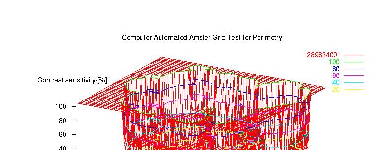
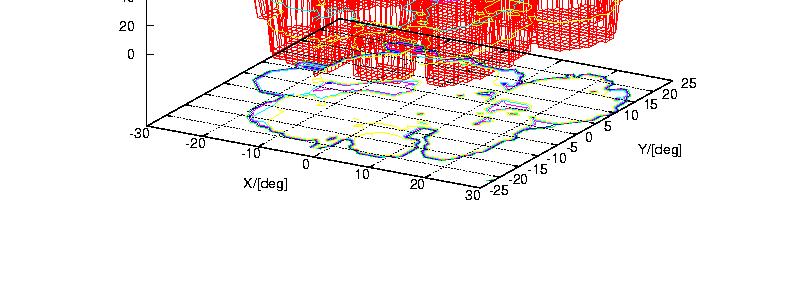
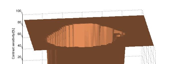
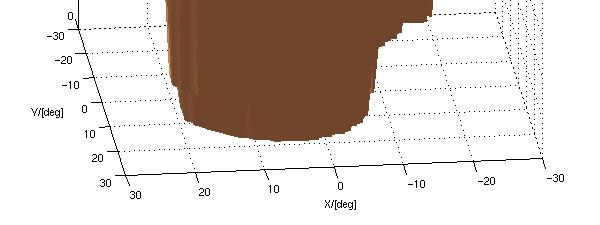
4 from the rest of the grid. The test is repeated with varying degrees of contrast. The results are recorded for later conversion into 3D format at the end of the test (see Fig. 3). The third dimension being screen contrast. 3. Results Each eye, depending on the patient compliance, required a total of approximately 4 to 5 minutes to be tested. Fig. 3. 3D display of visual field with a diagnosis of: a) optic neuropathy; b) anterior ischemic optic neuropathy; c) age-related macular degeneration; d) glaucoma; e) unimpaired central hill-of-vision; recorded by the 3D computerized threshold Amsler grid test. The x/y-axis denotes the horizontal/vertical coordinates of the visual field in degrees with (0, 0) being the center of fixation. The z-axis denotes the screen contrast expressed as a percent. Examples from several ophthalmic disorders are depicted in Figure 3. In figure 3a the 3D plot exhibits a more scalloped-shape visual field defect in optic neuropathy, with islands of relative sensitivity. This is in contrast to a clean-cut division between vision and no vision in a case of anterior ischemic optic neuropathy (Fig. 3b) (see also [5]). This feature may have potential for differential diagnosis. Figures 3c and 3d depict typical cases of age-related macular degeneration (see also [6]) and advanced glaucoma (see also [7]). The 3D representation of visual field defects caused by these two diseases is both intuitive (macular degeneration being a central hole and advanced glaucoma being a confined mesa ) and informative: The 3D representation can be further characterized by a slope along the circumference of the defects. Figure 3e shows a normal central hill-of-vision at 100% contrast, for the central 20 degrees. The 3D computerized threshold Amsler grid test has been in use since 2000 at the Doheny Eye Institute, Keck School of Medicine at the University of Southern California. Over 200 patients have been examined or screened for glaucoma, age-related macular degeneration, optic neuritis, anterior ischemic optic neuropathy, and ocular hypertension (see also [8]). In these pilot studies [5-8] the 3D test has proven to be reliable, fast, and accurate. Subtle scotomas, hard to detect with standard automated perimetry (mainly because of limited spatial resolution, e.g., light stimulus every 6 degrees both horizontally and vertically), were repeatedly identified by the 3D test [8], suggesting a potential role as a screening instrument. The physiological blind spot was not detected with this test because of cortical fill-in phenomenon for a steady grid, which is a known limitation of all Amsler grid-based tests. 4
5 Further (larger scale) clinical studies are needed to corroborate and extend the initial findings gathered with the 3D computerized threshold Amsler grid test. 4. Conclusions The 3D computerized threshold Amsler grid test is a novel approach to visual field testing that provides several additional features over conventional perimetry. The test delivers 3D rather than 2D depictions of scotomas (the z-axis reflecting contrast sensitivity) and provides insight into visual field defects, including shape and slope information about the scotoma boundaries (see Fig. 3). This test provides characterization of the 3D structure of scotomas, thus laying the foundation for monitoring the development of scotoma profiles. The 3D computerized threshold Amsler grid test only requires a touch sensitive screen, the test software, and an off-the-shelf computer system. The new test is simple to use and tests are performed quickly (4-5 minutes per eye), making frequent testing feasible [9]. 5. References 1. Amsler M. L'examen qualitatif de la fonction maculaire. Ophthalmologica 1947;144: Amsler M. Earliest symptoms of diseases of the macula. Br J Ophthalmol. 1955;37: Wall M, Sadun AA. Threshold Amsler grid testing. Cross-polarizing lenses enhance yield. Arch Ophthalmol 1986;104(4): For further information on the 3-D Computer-automated Threshold Amsler Grid Test : and 5. Fink W, Hsieh AK, Sadun AA. Computer-automated 3-D visual field testing in distinguishing paracentral scotomas of Optic Neuritis vs. AION. [ARVO Abstract]. Invest Ophthalmol Vis Sci. 2000;41(4):S311. Abstract nr Nazemi PP, Fink W, Lim JI, Sadun AA. Paracentral scotomas of age-related macular degeneration detected by means of a novel computer-automated 3-D visual field test. [ARVO Abstract] Invest Ophthalmol Vis Sci Abstract nr Fahimi A, Sadun AA, Fink W. Computer Automated 3D Visual Field Testing of Scotomas in Glaucoma. [ARVO Abstract] Invest Ophthalmol Vis Sci Abstract nr Nazemi PP, Fink W, Sadun AA, Minckler D, Francis B. Early detection of glaucoma by means of a novel computer-automated 3-D visual field test. Poster and Abstract, American Academy of Ophthalmology Meeting in New Orleans 2001, Scientific Poster 33, p Fink W, Sadun AA (2001) Prospects for Autonomous Visual Field Testing on Space Missions, lecture and poster, NanoSpace 2001, Exploring Interdisciplinary Frontiers, The International Conference on Integrated Nano/Microtechnology for Space and Biomedical Applications, March 13-16, 2001, Houston, Texas. 5

6 Figure 1 6

7 Figure 2 7
 b) c)")
8 Figure 3 a) b) c) d) e) 8
Three-dimensional computer-automated threshold Amsler grid test
 Journal of Biomedical Optics 9(1), 149 153 (January/February 2004) Three-dimensional computer-automated threshold Amsler grid test Wolfgang Fink California Institute of Technology Pasadena, California
3-D Computer-Automated Threshold Amsler Grid Test
 Introduction The 3-D Computer-Automated Threshold Amsler Grid Test is a five-minute vision test using a laptop computer with a touchsensitive screen that can help diagnose the onset of eye diseases and
Examination of the ten degrees of visual field surrounding fixation
 Examination of the ten degrees of visual field surrounding fixation Michael Wall, M.D. Tulane University School of Medicine NANOS, 1987 Albrecht von Graete :ntroduced visual field testing into clinical
Invited Paper ABSTRACT
 Invited Paper Ceeable Visual Field Analyzer (CVFA TM ) for the Portable, Comprehensive, and Tele-medical Assessment of Visual Performance over Time in Warfighters, Pilots, Veterans, and Civilians Chris
THRESHOLD AMSLER GRID TESTING AND RESERVING POWER OF THE POTIC NERVE by MOUSTAFA KAMAL NASSAR. M.D. MENOFIA UNIVERSITY.
 THRESHOLD AMSLER GRID TESTING AND RESERVING POWER OF THE POTIC NERVE by MOUSTAFA KAMAL NASSAR. M.D. MENOFIA UNIVERSITY. Since Amsler grid testing was introduced by Dr Marc Amsler on 1947and up till now,
PERIMETRY A STANDARD TEST IN OPHTHALMOLOGY
 7 CHAPTER 2 WHAT IS PERIMETRY? INTRODUCTION PERIMETRY A STANDARD TEST IN OPHTHALMOLOGY Perimetry is a standard method used in ophthalmol- It provides a measure of the patient s visual function - performed
We focus on progress OCULUS CENTERFIELD 2
 We focus on progress OCULUS CENTERFIELD 2 Oculus Centerfield 2 Projection perimeter for visual field tests up to 70 Our know-how to your benefit Take advantage of the more than 50 years experience of Oculus
OCULUS Twinfield 2. Perimeter. We focus on progress
 OCULUS Twinfield 2 Perimeter We focus on progress Ophthalmologist Definitely my all-round favourite! For glaucoma care, macular examinations and neurological cases as well as for formulation of expert
EXAMINATION OF THE CENTRAL VISUAL FIELD AT
 Brit. J. Ophthal. (1968) 52, 408 EXAMINATION OF THE CENTRAL VISUAL FIELD AT A READING DISTANCE*t BY V. N. HIGHMAN Moorfields Eye Hospital, City Road, London THIS investigation was started in an attempt
Automated Perimeter PTS 1000
 PTS 1000 Automated Perimeter PTS 1000 is a modern diagnostic instrument for precise and fast testing of field of vision. It offers static and kinetic stimuli with all Goldmann stimuli sizes and all stimuli
Frey AP-300 Humphrey Zeiss HFA II 740 perimeters
 Frey AP-300 Humphrey Zeiss HFA II 740 perimeters COMPARISON of the results of visual field testing according to the 30-2 test pattern using Frey AP-300 and Humphrey Zeiss HFA II 740 perimeters in patients
A New Method for Estimating Effects of Visual Field Loss in a Panoramic Driving Environment
 University of Iowa Iowa Research Online Driving Assessment Conference 2017 Driving Assessment Conference Jun 27th, 12:00 AM A New Method for Estimating Effects of Visual Field Loss in a Panoramic Driving
DEFECTS OF VISION THROUGH APHAKIC SPECTACLE LENSES*t
 Brit. J. Ophthal. (1967) 51, 306 DEFECTS OF VISION THROUGH APHAKIC SPECTACLE LENSES*t BY ROBERT C. WELSH Miami, Florida BY the use of a series of scale diagrams an attempt is made to explain the following:
OCULUS Easyfield. Perimeter. We focus on progress
 OCULUS Easyfield Perimeter We focus on progress OCULUS Easyfield The right choice for all your needs The OCULUS Easyfield is a full-fledged compact perimeter capable of performing standard automated perimetry
A reduction of visual fields during changes in the background image such as while driving a car and looking in the rearview mirror
 Original Contribution Kitasato Med J 2012; 42: 138-142 A reduction of visual fields during changes in the background image such as while driving a car and looking in the rearview mirror Tomoya Handa Department
Goldmann vs. Humphrey
 Get Ready: Visual Field Correction and Calibration Pitfalls Correction For the Humphrey & Goldmann Visual Fields Goldmann vs. Humphrey When performing visual fields, you must take the patients correction
Introduction. scotoma. Effects of preferred retinal locus placement on text navigation and development of adventageous trained retinal locus
 Effects of preferred retinal locus placement on text navigation and development of adventageous trained retinal locus Gale R. Watson, et al. Journal of Rehabilitration Research & Development 2006 Introduction
Multifocal Electroretinograms in Normal Subjects
 Multifocal Electroretinograms in Normal Subjects Akiko Nagatomo, Nobuhisa Nao-i, Futoshi Maruiwa, Mikki Arai and Atsushi Sawada Department of Ophthalmology, Miyazaki Medical College, Miyazaki, Japan Abstract:
Retina. Convergence. Early visual processing: retina & LGN. Visual Photoreptors: rods and cones. Visual Photoreptors: rods and cones.
 Announcements 1 st exam (next Thursday): Multiple choice (about 22), short answer and short essay don t list everything you know for the essay questions Book vs. lectures know bold terms for things that
Limitations of the Oriented Difference of Gaussian Filter in Special Cases of Brightness Perception Illusions
 Short Report Limitations of the Oriented Difference of Gaussian Filter in Special Cases of Brightness Perception Illusions Perception 2016, Vol. 45(3) 328 336! The Author(s) 2015 Reprints and permissions:
Nursing Assessment of Visual Acuity and Colour Discrimination
 Nursing Assessment of Visual Acuity and Colour Discrimination Visual impairment is the most significant adverse reaction of ethambutol and may occur in individuals receiving ethambutol for longer than
The First True Color Confocal Scanner on the Market
 The First True Color Confocal Scanner on the Market White color and infrared confocal images: the advantages of white color and confocality together for better fundus images. The infrared to see what our
Varilux Comfort. Technology. 2. Development concept for a new lens generation
 Dipl.-Phys. Werner Köppen, Charenton/France 2. Development concept for a new lens generation In depth analysis and research does however show that there is still noticeable potential for developing progresive
The TRC-NW8F Plus: As a multi-function retinal camera, the TRC- NW8F Plus captures color, red free, fluorescein
 The TRC-NW8F Plus: By Dr. Beth Carlock, OD Medical Writer Color Retinal Imaging, Fundus Auto-Fluorescence with exclusive Spaide* Filters and Optional Fluorescein Angiography in One Single Instrument W
Raise your expectations. Deliver theirs.
 66 EXTENDED RANGE OF VISION MONOFOCAL-LIKE DISTANCE Raise your expectations. Deliver theirs. Now you can give your patients the best of both worlds with the first and only hybrid designed monofocal-multifocal
MEASUREMENT OF ECCENTRIC FIXATION BY THE
 Brit. J. Ophthal. (1959) 43, 461. MEASUREMENT OF ECCENTRIC FIXATION BY THE BJERRUM SCREEN* BY G. BROCKBANK AND R. DOWNEY General Infirmary, Leeds Introduction by G. W. Black andj. Foster.-The forward movement
Image Modeling of the Human Eye
 Image Modeling of the Human Eye Rajendra Acharya U Eddie Y. K. Ng Jasjit S. Suri Editors ARTECH H O U S E BOSTON LONDON artechhouse.com Contents Preface xiiii CHAPTER1 The Human Eye 1.1 1.2 1. 1.4 1.5
ORIGINAL ARTICLE. Vision Evaluation of Eccentric Refractive Correction. LINDA LUNDSTRÖM, PhD, JÖRGEN GUSTAFSSON, OD, PhD, and PETER UNSBO, PhD
 1040-5488/07/8411-1046/0 VOL. 84, NO. 11, PP. 1046 1052 OPTOMETRY AND VISION SCIENCE Copyright 2007 American Academy of Optometry ORIGINAL ARTICLE Vision Evaluation of Eccentric Refractive Correction LINDA
ABSTRACT. Keywords: Glaucoma, videopupillography, intraocular pressure, pupil, pupil size, pupillometer 1. INTRODUCTION
 An improved apparatus of infrared videopupillography for monitoring pupil size T.-W. Huang* a, M.-L. Ko a, b, Y. Ouyang c, Y.-Y. Chen d, B.-S. Sone d, M. Ou-Yang a, J.-C. Chiou a a Department of Electrical
The First True-Color Wide-Field Confocal Scanner
 The First True-Color Wide-Field Confocal Scanner 2 Company Profile CenterVue designs and manufactures highly automated medical devices for the diagnosis and management of ocular pathologies, including
CCVIP Early Intervention Pearls
 CCVIP Early Intervention Pearls Table of Contents Page 1: Page 2: Page 3: Page 4: Page 5: Page 6: Page 8: Page 9: Page 10: Page 11: Page 12: Page 13: Page 14: Past & Present Table of Contents Functional
Contact Lenses Didn t Work! Now What? Evaluation and Treatment of Aniseikonia
 Contact Lenses Didn t Work! Now What? Evaluation and Treatment of Aniseikonia Andrew J Toole, OD, PhD, FAAO The Ohio State University College of Optometry Disclosure Statement: Nothing to disclose Aniseikonia
Impressive Wide Field Image Quality with Small Pupil Size
 Impressive Wide Field Image Quality with Small Pupil Size White color and infrared confocal images: the advantages of white color and confocality together for better fundus images. The infrared to see
Modified Bagolini striated glass test: clinical applications of starlight test in binocular visual field screening
 1288 Department of Ophthalmology, Nagoya University, School of Medicine, Nagoya, Japan T Hirai M Arai Y Ito MSato Correspondence to: Toshie Hirai, Department of Ophthalmology, Nagoya University, School
PERIPHERAL VISON PATTERN DETECTION DYNAMIC TEST
 PERIPHERAL VISON PATTERN DETECTION DYNAMIC TEST João P Rodrigues, João D Semedo, Fernando M Melicio Institute Systems and Robotics,Technical University, Av Rovisco Pais 1 TN6.21, Lisbon, Portugal jrodrigues@laseeb.org,
United States Patent (19) Sadun
 Sadun") United States Patent (19) Sadun 54 METHOD AND SYSTEM FOR DETECTING, CHARACTERIZING AND MONITORING OPTIC NERVE DISEASES 75) Inventor: 73 Assignee: Alfredo A. Sadun, San Marino, Calif. University of Southern
United States Patent (19) Sadun 54 METHOD AND SYSTEM FOR DETECTING, CHARACTERIZING AND MONITORING OPTIC NERVE DISEASES 75) Inventor: 73 Assignee: Alfredo A. Sadun, San Marino, Calif. University of Southern
CS 565 Computer Vision. Nazar Khan PUCIT Lecture 4: Colour
 CS 565 Computer Vision Nazar Khan PUCIT Lecture 4: Colour Topics to be covered Motivation for Studying Colour Physical Background Biological Background Technical Colour Spaces Motivation Colour science
Instruments Commonly Used For Examination of the Eye
 Instruments Commonly Used For Examination of the Eye There are many instruments that the eye doctor might use to evaluate the eye and the vision system. This report presents some of the more commonly used
Conventional photographs do not show how, at any moment of visual fixation, neural
 SPEIAL ARTILE Simulating Vision With and Without Macular Disease David J. Marmor, MFA; Michael F. Marmor, MD onventional photographs do not show how, at any moment of visual fixation, neural vision is
Reading With a Macular Scofoma
 Reading With a Macular Scofoma //. Retinal Locus For Scanning Text George T. Timberlake, Eli Peli,* Edward A. EssocKf and Reed A. Augliere To elucidate how patients with macular scotomas use residual functional
LIQUID CRYSTAL LENSES FOR CORRECTION OF P ~S~YOP
 LIQUID CRYSTAL LENSES FOR CORRECTION OF P ~S~YOP GUOQIANG LI and N. PEYGHAMBARIAN College of Optical Sciences, University of Arizona, Tucson, A2 85721, USA Email: gli@ootics.arizt~ii~.e~i~ Correction of
Geometric Optics. This is a double-convex glass lens mounted in a wooden frame. We will use this as the eyepiece for our microscope.
 I. Before you come to lab Read through this handout in its entirety. II. Learning Objectives As a result of performing this lab, you will be able to: 1. Use the thin lens equation to determine the focal
Research Article Testing of Visual Field with Virtual Reality Goggles in Manual and Visual Grasp Modes
 Hindawi Publishing Corporation BioMed Research International Volume, Article ID, pages http://dx.doi.org/.// Research Article Testing of Visual Field with Virtual Reality Goggles in Manual and Visual Grasp
Going beyond the surface of your retina
 Going beyond the surface of your retina OCT-HS100 Optical Coherence Tomography Canon s expertise in optics and innovative technology have resulted in a fantastic 3 μm optical axial resolution for amazing
Biology 70 Slides for Lecture 1 Fall 2007
 Biology 70 Part II Sensory Systems www.biology.ucsc.edu 1 2 intensity vs spatial position (image formation) color 3 4 motion depth (monocular) 5 6 1 depth (binocular) 1. In the lectures on perception we
Fitting Peripheral Prisms for Patients with Hemianopia. The workshop is sponsored by Chadwick Optical, Inc.
 Fitting Peripheral Prisms for Patients with Hemianopia Eli Peli, MSc, OD, FAAO Professor of Ophthalmology, Harvard Medical School Moakley Scholar in Aging Eye Research Please silence all mobile devices
OCULAR MEDIA* PHOTOGRAPHIC RECORDING OF OPACITIES OF THE. development by the control of diabetes, the supply of a deficient hormone
 Brit. J. Ophthal. (1955) 39, 85. PHOTOGRAPHIC RECORDING OF OPACITIES OF THE OCULAR MEDIA* BY E. F. FINCHAM Institute of Ophthalmology, University of London THE value of photography for recording pathological
Human Visual System. Prof. George Wolberg Dept. of Computer Science City College of New York
 Human Visual System Prof. George Wolberg Dept. of Computer Science City College of New York Objectives In this lecture we discuss: - Structure of human eye - Mechanics of human visual system (HVS) - Brightness
The MoviText method: Efficient pre-optical reading training in persons with central visual field loss
 Technology and Disability 6 (2004) 211 221 211 IOS Press The MoviText method: Efficient pre-optical reading training in persons with central visual field loss Jögen Gustafsson and Krister Inde Division
Automated Spectrophotometric Spatial Profiling of Coated Optical Wafers
 Automated Spectrophotometric Spatial Profiling of Coated Optical Wafers Application note Materials testing and research Authors Travis Burt Fabian Zieschang Agilent Technologies, Inc. Parts of this work
Vision Science I Exam 2 31 October 2016
 Vision Science I Exam 2 31 October 2016 1) Mr. Jack O Lantern, pictured here, had an unfortunate accident that has caused brain damage, resulting in unequal pupil sizes. Specifically, the right eye is
Peripheral Prism Glasses for Hemianopia Giorgi et al. APPENDIX 1
 1 Peripheral Prism Glasses for Hemianopia Giorgi et al. APPENDIX 1 Monocular and binocular sector prisms are commonly used for hemianopia.3, 10, 14 The impact of these prisms on the visual field is not
Gravitational Lensing Experiment
 EKA Advanced Physics Laboratory Gravitational Lensing Experiment Getting Started Guide In this experiment you will be studying gravitational lensing by simulating the phenomenon with optical lenses. The
Keeler Catalogue. All Keeler products are designed to offer the ultimate in professional performance.
 Keeler Catalogue All Keeler products are designed to offer the ultimate in professional performance. They are the result of the constant pursuit of perfection, combining the aesthetic and ergonomic. 34
Visual evoked cortical potential can be used to differentiate between uncorrected refractive error and macular disorders
 Documenta Ophthalmologica 102: 41 62, 2001. 2001 Kluwer Academic Publishers. Printed in the Netherlands. Visual evoked cortical potential can be used to differentiate between uncorrected refractive error
Going beyond the surface of your retina OCT-HS100 OPTICAL COHERENCE TOMOGRAPHY
 Going beyond the surface of your retina OCT-HS100 OPTICAL COHERENCE TOMOGRAPHY Automatic functions make examinations short and simple. Perform the examination with only two simple mouse clicks! 1. START
Going beyond the surface of your retina OCT-HS100 OPTICAL COHERENCE TOMOGRAPHY
 Going beyond the surface of your retina OCT-HS100 OPTICAL COHERENCE TOMOGRAPHY Full Auto OCT High specifications in a very compact design Automatic functions make examinations short and simple. Perform
This is the author s version of a work that was submitted/accepted for publication in the following source:
 This is the author s version of a work that was submitted/accepted for publication in the following source: Atchison, David A. & Mathur, Ankit (2014) Effects of pupil center shift on ocular aberrations.
CLINICAL SCIENCES. The Visual Performance and Metamorphopsia of Patients With Macular Holes
 CLINICAL SCIENCES The Visual Performance and Metamorphopsia of Patients With Macular Holes Yoshihiro Saito, MD; Yoshiko Hirata, MD; Atsushi Hayashi, MD; Takashi Fujikado, MD; Masahito Ohji, MD; Yasuo Tano,
Low Vision Assessment Components Job Aid 1
 Low Vision Assessment Components Job Aid 1 Eye Dominance Often called eye dominance, eyedness, or seeing through the eye, is the tendency to prefer visual input a particular eye. It is similar to the laterality
V isual restitution training1 2
 3 EXTENDED REPORT Does visual restitution training change absolute homonymous visual field defects? A fundus controlled study J Reinhard, A Schreiber, U Schiefer, E Kasten, B A Sabel, S Kenkel, R Vonthein,
HEREDITARY RETINAL DEGENERATIVE DISEASES,
 Visual Performance Using a Retinal Prosthesis in Three Subjects With Retinitis Pigmentosa DOUGLAS YANAI, JAMES D. WEILAND, MANJUNATHA MAHADEVAPPA, ROBERT J. GREENBERG, IONE FINE, AND MARK S. HUMAYUN PURPOSE:
4th International Congress of Wavefront Sensing and Aberration-free Refractive Correction ADAPTIVE OPTICS FOR VISION: THE EYE S ADAPTATION TO ITS
 4th International Congress of Wavefront Sensing and Aberration-free Refractive Correction (Supplement to the Journal of Refractive Surgery; June 2003) ADAPTIVE OPTICS FOR VISION: THE EYE S ADAPTATION TO
VISUAL PROSTHESIS FOR MACULAR DEGENERATION AND RETINISTIS PIGMENTOSA
 VISUAL PROSTHESIS FOR MACULAR DEGENERATION AND RETINISTIS PIGMENTOSA 1 SHWETA GUPTA, 2 SHASHI KUMAR SINGH, 3 V K DWIVEDI Electronics and Communication Department 1 Dr. K.N. Modi University affiliated to
Special Publication: Ophthalmochirurgie Supplement 2/2009 (Original printed issue available in the German language)
") Special Publication: Ophthalmochirurgie Supplement 2/2009 (Original printed issue available in the German language) LENTIS Mplus - The one -and and-only Non--rotationally Symmetric Multifocal Lens Multi-center
Special Publication: Ophthalmochirurgie Supplement 2/2009 (Original printed issue available in the German language) LENTIS Mplus - The one -and and-only Non--rotationally Symmetric Multifocal Lens Multi-center
What s New in Ocular Biomechanics?
 What s New in Ocular Biomechanics? The International Congress of Wavefront Sensing & Optimized Refractive Corrections Wavefront Course January 28, 2006 Torrence A. Makley Research Professor Department
Normal Wavefront Error as a Function of Age and Pupil Size
 RAA Normal Wavefront Error as a Function of Age and Pupil Size Raymond A. Applegate, OD, PhD Borish Chair of Optometry Director of the Visual Optics Institute College of Optometry University of Houston
Eyes. Inspection Visual Acuity Visual Fields Pupillary Response Fundoscopic Exam
 Eyes Inspection Visual Acuity Visual Fields Pupillary Response Fundoscopic Exam Eye Examination Inspection 11.Inspects external ocular (eye) structures (lids, conjunctiva, iris, cornea, pupils) 12.Gently
CHAPTER 4 LOCATING THE CENTER OF THE OPTIC DISC AND MACULA
 90 CHAPTER 4 LOCATING THE CENTER OF THE OPTIC DISC AND MACULA The objective in this chapter is to locate the centre and boundary of OD and macula in retinal images. In Diabetic Retinopathy, location of
The New World of. Virtual Reality
 The New World of Virtual Reality Virtual reality technology is showing promise in augmenting traditional assessment and interventional strategies. A look at some lines of investigation. By Linda Roach,
Be aware that there is no universal notation for the various quantities.
 Fourier Optics v2.4 Ray tracing is limited in its ability to describe optics because it ignores the wave properties of light. Diffraction is needed to explain image spatial resolution and contrast and
Tracking retinal motion with a scanning laser ophthalmoscope
 JRRD Volume 42, Number 3, Pages 373 380 May/June 2005 Journal of Rehabilitation Research & Development Tracking retinal motion with a scanning laser ophthalmoscope Zhiheng Xu, MS; 1 Ronald Schuchard, PhD;
Image processing of computerised visual field data
 ritish Journal of Ophthalmology 995; 79: 27-22 Image processing of computerised visual field data 27 Department of Visual Science, Institute of Ophthalmology, London F W Fitzke School of Mathematics, ctuarial
Developments of a peripheral vision system using immersive virtual environment
 Developments of a peripheral vision system using immersive virtual environment Y Tabata 1, M Suga 2, K Minato 3, O Oshiro 4, K Chihara 5 and S Nagata 6 1-5 Graduate School of Information Science, Nara
OPTIC DISC LOCATION IN DIGITAL FUNDUS IMAGES
 OPTIC DISC LOCATION IN DIGITAL FUNDUS IMAGES Miss. Tejaswini S. Mane 1,Prof. D. G. Chougule 2 1 Department of Electronics, Shivaji University Kolhapur, TKIET,Wrananagar (India) 2 Department of Electronics,
The human electroretinogram and occipital potential in response to focal illumination of the retina
 The human electroretinogram and occipital potential in response to focal illumination of the retina Jerry H. Jacobson, Kazuo Kawasaki,* and Tatsuo Hirose* Electrical responses to light stimuli of 1 angular
10/25/2017. Financial Disclosures. Do your patients complain of? Are you frustrated by remake after remake? What is wavefront error (WFE)?
?") Wavefront-Guided Optics in Clinic: Financial Disclosures The New Frontier November 4, 2017 Matthew J. Kauffman, OD, FAAO, FSLS STAPLE Program Soft Toric and Presbyopic Lens Education Gas Permeable Lens
Wavefront-Guided Optics in Clinic: Financial Disclosures The New Frontier November 4, 2017 Matthew J. Kauffman, OD, FAAO, FSLS STAPLE Program Soft Toric and Presbyopic Lens Education Gas Permeable Lens
IMAGE FORMATION. Light source properties. Sensor characteristics Surface. Surface reflectance properties. Optics
 IMAGE FORMATION Light source properties Sensor characteristics Surface Exposure shape Optics Surface reflectance properties ANALOG IMAGES An image can be understood as a 2D light intensity function f(x,y)
Driving simulators in hemianopia rehabilitation research
 Schepens Eye Research Institute Massachusetts Eye and Ear Harvard Medical School Affiliate Driving simulators in hemianopia rehabilitation research Alex Bowers, PhD No disclosures Hemianopia Loss of half
Monochromatic Aberrations and Emmetropization
 Monochromatic Aberrations and Emmetropization Howard C. Howland* Department of Neurobiology and Behavior Cornell University, Ithaca N.Y. Jennifer Kelly Toshifumi Mihashi Topcon Corporation Tokyo *paid
Introduction to Psychology Prof. Braj Bhushan Department of Humanities and Social Sciences Indian Institute of Technology, Kanpur
 Introduction to Psychology Prof. Braj Bhushan Department of Humanities and Social Sciences Indian Institute of Technology, Kanpur Lecture - 10 Perception Role of Culture in Perception Till now we have
Kinder products for your most precious patients
 Kinder products for your most precious patients Keeler kinder products Keeler is a global leader in the supply and design of ophthalmic diagnostic products. Our products are used throughout the world to
LOW VISION ENHANCEMENT SYSTEM
 ROBERT W. MASSOF, DOUGLAS L. RICKMAN, and PETER A. LALLE LOW VISION ENHANCEMENT SYSTEM The Johns Hopkins University, the National Aeronautics and Space Administration, and the Veterans Administration are
Spatial variations in field data
 Chapter 2 Spatial variations in field data This chapter illustrates strong spatial variability in a multi-component surface seismic data set. One of the simplest methods for analyzing variability is looking
Optical Performance of Nikon F-Mount Lenses. Landon Carter May 11, Measurement and Instrumentation
 Optical Performance of Nikon F-Mount Lenses Landon Carter May 11, 2016 2.671 Measurement and Instrumentation Abstract In photographic systems, lenses are one of the most important pieces of the system
Surveying & Measurement. Detail Survey Topographic Surveying
 Surveying & Measurement Detail Survey Topographic Surveying Introduction Mapping surveys are made to determine the relief of the earth s surface and locate critical points on it. to determine the locations
Patients in your area are ready to set appointments with you. Keep reading on to learn why they re eager to use our system.
 Hello Doctor! If you re reading this, the person who gave it to you is one of over 11,000 people who visited our website looking for a provider of Vivid Vision - Vision Therapy in Virtual Reality. They
Blood Vessel Tree Reconstruction in Retinal OCT Data
 Blood Vessel Tree Reconstruction in Retinal OCT Data Gazárek J, Kolář R, Jan J, Odstrčilík J, Taševský P Department of Biomedical Engineering, FEEC, Brno University of Technology xgazar03@stud.feec.vutbr.cz
IMPLANTABLE MINIATURIZED TELESCOPE (IMT) FOR LOW-VISION
 FOR LOW-VISION") IMPLANTABLE MINIATURIZED TELESCOPE (IMT) FOR LOW-VISION Eli Peli 1, 2, Isaac Lipshitz 2, and Gideon Dotan 2 1 The Schepens Eye Research Institute, Harvard Medical School, Boston MA, USA 2 VisionCare, Inc.,
IMPLANTABLE MINIATURIZED TELESCOPE (IMT) FOR LOW-VISION Eli Peli 1, 2, Isaac Lipshitz 2, and Gideon Dotan 2 1 The Schepens Eye Research Institute, Harvard Medical School, Boston MA, USA 2 VisionCare, Inc.,
MEDICAL & LIFE SCIENCES
 MEDICAL & LIFE SCIENCES Basler cameras - the power of sight for medical and life science technology Broad industrial camera portfolio for digital imaging -year warranty, long-term availability Trust in
CLARUS 500 from ZEISS HD ultra-widefield fundus imaging
 CLARUS 500 from ZEISS HD ultra-widefield fundus imaging Imaging ultra-wide without compromise. ZEISS CLARUS 500 // INNOVATION MADE BY ZEISS Compromising image quality may leave some pathology unseen. Signs
Comparison of two algorithms in the automatic segmentation of blood vessels in fundus images
 Comparison of two algorithms in the automatic segmentation of blood vessels in fundus images ABSTRACT Robert LeAnder, Myneni Sushma Chowdary, Swapnashri Mokkapati, and Scott E Umbaugh Effective timing
CR-2 AF DIGITAL NON-MYDRIATIC RETINAL CAMERA. Superior Image Resolution and Auto Functionality
 DIGITAL NON-MYDRIATIC RETINAL CAMERA Superior Image Resolution and Auto Functionality 1 superior RESOLUTION for earlier, more accurate detection GOOD ENOUGH IS NOT GOOD ENOUGH If you were having your vision
CLARUS 500 from ZEISS HD ultra-widefield fundus imaging
 CLARUS 500 from ZEISS HD ultra-widefield fundus imaging Imaging ultra-wide without compromise. ZEISS CLARUS 500 // INNOVATION MADE BY ZEISS Compromising image quality may leave some pathology unseen. Signs
Locating Blood Vessels in Retinal Images by Piece-wise Threshold Probing of a Matched Filter Response
 Locating Blood Vessels in Retinal Images by Piece-wise Threshold Probing of a Matched Filter Response Adam Hoover, Ph.D. +, Valentina Kouznetsova, Ph.D. +, Michael Goldbaum, M.D. + Electrical and Computer
Spatial coding: scaling, magnification & sampling
 Spatial coding: scaling, magnification & sampling Snellen Chart Snellen fraction: 20/20, 20/40, etc. 100 40 20 10 Visual Axis Visual angle and MAR A B C Dots just resolvable F 20 f 40 Visual angle Minimal
you can See more with fundus autofluorescence. CR-2 PLUS NON-MYDRIATIC DIGITAL RETINAL CAMERA
 you can See more with fundus autofluorescence. CR-2 PLUS NON-MYDRIATIC DIGITAL RETINAL CAMERA High-performance non-mydriatic retinal camera with integrated FAF photography. Canon EOS Camera Technology
Linear Collider Collaboration Tech Notes
 LCC-0123 Rev. 3 August 2003 Rev. June 2004 Linear Collider Collaboration Tech Notes Design Guideline Summary Based on the GEOVISION Report of Stanford Linear Accelerator Tunnel Vibration Measurements Parsons
Wide-Band Enhancement of TV Images for the Visually Impaired
 Wide-Band Enhancement of TV Images for the Visually Impaired E. Peli, R.B. Goldstein, R.L. Woods, J.H. Kim, Y.Yitzhaky Schepens Eye Research Institute, Harvard Medical School, Boston, MA Association for
USE YOUR PC TO QUICKLY MAP REMAINING VISION AFTER FOVEAL VISION LOSS
 Use your PC to quickly map remaining vision 307 USE YOUR PC TO QUICKLY MAP REMAINING VISION AFTER FOVEAL VISION LOSS MANFRED MACKEBEN, AUGUST COLENBRANDER and ALEKSANDR GOFEN The Smith-Kettlewell Eye Research