Biomedical Imaging and Image Analysis
|
|
|
- Kristina Richard
- 6 years ago
- Views:
Transcription

1 Biomedical Imaging and Image Analysis Lecture in Medical Informatics Course Ewert Bengtsson Professor of computerized image analysis Division of Visual Information and Interaction Department of Information Technology


2 The theme Images are of central importance in medical diagnosis There has been a dramatic development in medical imaging during the last few decades In this lecture we will briefly describe different ways of Lecture 1: Creating Lecture 2: Analyzing medical images
3 Medical imaging vs. microscopy There are two main types of medical images: Microscopic and macroscopic. The macroscopic images are typically taken in situ from the patient while microscopic images are based on small samples taken out of the patient. The term Medical imaging is typically limited to images of the patient and his/her organs on a macroscopic scale.


4 Medical imaging motivations Medical imaging is used to visualize and understand the patient Anatomy Physiology (function) Discovering injuries or pathology Different imaging methods are better at one or the other aspect


5 Medical imaging modalities

6 Imaging motivations - microscopy Visual evaluation of tissue samples in a microscope is usually the final basis for diagnosis of all cancers and many other diseases Used to be all optical Is rapidly becoming digital Also a rapid development of new microscopy imaging techniques


7 Microscopic imaging modalities Brightfield light microscopy The classical microscopy Fluorescent microscopy Used for research Several versions: Full field Confocal Two-photon STED Electron Microscopy The highest resolution but complex and tedious


8 Microscopic images Brightfield microscopy Fluorescent microscopy Tissue microarray Electron microscopy

9 Medical imaging Using different parts of the electromagnetic spectrum PET hard gamma rays, 511keV X-ray images, CT Visible light Heat images, thermography Radio waves from nuclear spinn, MRT The electric activity of the body, EEG Sound waves, ultrasound


10 Classical X-ray projection, gives a 2D shadow image


11 X-rays: Röntgen the inventor

12 X-ray technology trends Since about 100 years X- ray imaging through analogue electronic technology and photography Since about 30 years with digital technology Digital technology is rapidly taking over in this field as in most other
13 Fluoroscopy vs radiography Fluoroscopy transillumination, Creates a live image of the patient Can support real time diagnosis Shows dynamics Can control certain invasive diagnostic procedures Gives a relative high dose also to the medical doctor Radiography X-ray photography Creates a frozen permanent image Can be interpreted without rush Gives medical and legal documentation
14 Fluoroscopy Fluoroscope, originally zinkcadmiumsulphide screen, 7% efficiency Electro-optical image amplifiers with fluorescent screen (> x amplification) Image amplifier with TV-camera (tube or CCD) Digitally registering the image from the TVcamera Digital fluoroscopy Digital subtraction angiography


15 Blood vessels - Angiography

16 Digital fluoroscopy
17 Radiography Original direct film exposure, gives the sharpest images but low efficiency, only used in special cases such as dental imaging Amplification screens converts X-rays to light, gain x Can use secondary aperture, a grid to decrease scattered light and increase contrast The film can be replaced by image plates, gives a greater dynamic range and possibilities of directly digitizing and improving the image through image processing
18 Muscles and bones

19 Conventional vs digital, high-frequency amplified X-ray image
20 Digital radiography, advantages Greater contrast range gives fewer retakes because of poor exposure Digital image handling gives fewer lost films and simplified archiving More enviromentally friendly through less use of film and chemicals Easier to consult other experts over the network


21 Computed Tomography (CT) Creates images of slices through the body

22 How the tomograph functions
23 CT-functional principles In a large number of projection rays though the body the X-ray absorption is measured, this yields many density profiles. These can be reprojected into the slice through Radons formula or through filtered back projection CT gives good contrast resolution and very good geometric accuracy

24 Computed tomography CT gives anatomical information

25
26 CT image properties CT measures X-ray density in absolute units according to the Hounsfields scale for air 0 for water for bone Through different contrast windows in the display different tissues can be displayed optimally

27 A modern CT can have 64 parallel channels Typical specifications; 64 x 0.625mm acquisition 0.34mm x 0.34mm x 0.34mm isotropic resolution 0.4 second rotation time Up to 24 Lp/cm ultra-high spatial resolution High resolution 768 and 1024 reconstruction matrices Reconstruction up to 40 images per second


28 CT examples
 Based on magnetic pulse")


29 Magnetic Resonance Tomography (MRT) Based on magnetic pulse sequences in a strong magnetic field Different pulse sequences gives different contrast The orientation of the slices can be chosen freely through manipulation of the magnetic fields

30 Magnetic Resonance Imaging MRI gives anatomical information

31 Magnetic Resonance Imaging MRI the main parts of the machine

32 How MRT works Nuclei with odd number of protons/neutrons has spin The spin vector can be aligned to a (very) strong magnetic field Can be disturbed by a radio signal in resonance with the spin frequency, the so called Larmorfrequency When the atoms returns to rest position they become radio transmitters which can be detected by sensitive receivers Through conrol of field gradients and pulse sequences one can determine which atoms are activated and listened to respectively and thus images can be created in 2D and 3D
33 Some fundamental MR-concepts MR-images can be weighted to show two time constants giving different contrast: T1 is the time constant that determines how fast the spin M Z returns to equilibrium, it is called spin lattice relaxation time M z = M o ( 1 - e -t/t1 ) T2 is the time constant that determines the return to equilibrium for the transversal magnetisation M XY, it is called spin-spin relaxation time M XY =M XYo ( e -t/t2 )
34 MRT image properties Very good contrast resolution for soft tissue Very flexible, different pulse sequences gives different contrast Usually not possible to determine the signal levels in absolute terms Poor geometric precision No known harmful effects Still under strong development


35 MR Neuro


36 Muscles and bones (joints)

37 Microscopic resolution for mm in-plane resolution of wrist Observe clear delineation of fine structures such as the vessel walls Technical details: T1 FLASH TR 591 ms, TE 7.5 ms, TA 6:09 min, SL 3 mm, slices 19, matrix 1024, FoV 80 mm. orthopedics


38 Whole body MR imaging


39 Neurological Multiple sclerosis


40 Angiography

41 The heart
42 MRT technologies The image properties are influenced by many factors: Radio antenna coils can be adapted to anatomy and pathology Closer coil gives better image Different pulse sequences gives different contrast, resolution, signal noise and registration times Triggering by heart beat, blood motion and breading can increase the resolution Contrast media can enhance certain structures With functional MR, fmri activity in the brain can be registered and imaged
43 MRT technologies The image properties are influenced by many factors: Radio antenna coils can be adapted to anatomy and pathology Closer coil gives better image Different pulse sequences gives different contrast, resolution, signal noise and registration times Triggering by heart beat, blood motion and breading can increase the resolution Contrast media can enhance certain structures With functional MR, fmri activity in the brain can be registered and imaged

44 Functional imaging SHOWS WHERE THE BRAIN IS PARTICULARLY ACTIVE

45 Functional imaging - how it works


46 MR diffusion tensor imaging Showing the connections of fibers in the brain

47 Brain fiber visualization by Anders Brun CMIV/CBA
48 For further studies about MRT A good description of the MRI technology at: A good popular description at: ss-sv.html A leading research group in MR and other medical image visualisation, CMIV:
49 Positron Emission Tomograpy (PET) PET shows the concentration and distribution of positron emitting tracer substances in the patient. These images are functional, not anatomical, i.e. they show physiological parameters


50 PET functional principle 50
51 PET functional principles A positron emitting compound is injected into the body (must be produced in an accelerator) The positrons will, within a couple of mm, collide with an electron and create two co-linear 511keV gamma rays These are detected by two detectors located in opposite locations in rings around the person and based on this one can figure out where the event took place Re-projection based on the tomographic principle

52 Positron Emission Tomography PET gives functional information 52

53 Positron Emission Tomografi : accelerator for creating the radioactive tracer substances 53
54 The properties of PET images Gives functional images with rather good resolution at least 1 cm Glucose can be labelled with C11 and this makes it possible to see where in the brain fuel is needed i.e. where the brain is working Very specific substances can be labelled so PET has many applications in pharmaceutical research The need for an accelerator and a chemical lab which can handle high speed synthesis of radioactive compounds makes the technology very expensive
55 PET in Uppsala The PET-research in Uppsala is in the international front-line In 2001 the university PET-centre was sold to Amersham-Biosciences and Imanet AB was created Amersham-Biosciences was bought by GE Medical a few years later Now the PET-centre medical activity is back at Uppsala university hospital Will get the first PET-MR system in Sweden next year

56 Typical result from PCA image enhancement of PET images HV NK1-receptor tracer GLD Pasha Razifar PhD thesis work at IMANET AB and CBA

57 Single Photon Emission Computed Tomography (SPECT) SPECT is similar to PET and shows the concentration and distribution of a radioactive tracer in the patient. The images are functional, not anatomical.


58 Scintigraphy - SPECT camera 58
59 SPECT functional principles A radioactive tracer is injected into the body With a matrix of detectors arranged above the body the location of the radioactive disintegrations is approximately determined The detector can be moved into different positions, which makes tomographic reconstruction possible Alternatively a collimator with slanted holes can be used - ectomography

60 Single Photon Emission Tomography SPECT gives functional information 60
61 The SPECT image properties SPECT gives a functional image with relatively low resolution, some cm The images are intrinsically 3D The radioactive compounds can be obtained from long lived mother isotopes which is much cheaper than accelerators Dynamic processes can be studied through long registrations
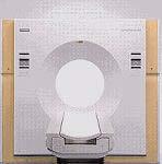
62 Ultrasound, US Based on the sonar, acoustic echo principle. Sound with high frequency, typically a few MHz is sent into the body and the echoes are studied. Can with a small, compact equipment give dynamic images in 2D or 3D. The images has problems with coherent noise, specle, and with non-linearities in the sound propagation.


63 Ultrasound equipment 63

64 Ultrasound, best at showing soft tissue

65 Heart 65

66 Ultrasound images of a heart Sharp images of structures in a moving heart
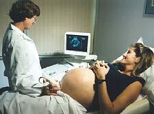

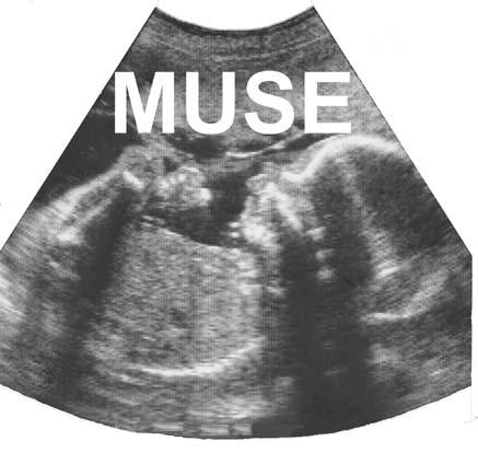
67 Ultrasound for fetal examinations

68 3D rendering of dynamic Ultrasound

69 Ultrasound can show flow through Doppler technology
70 Digital image analysis Most modern medical imaging devices are digital This creates opportunities for also using digital image processing techniques to help interpret the images Actually digital techniques are necessary even for being able to handle some types of images
71 Advantages of digital technology Can create images with greater contrast range with less radiation Can handle the images more efficiently through PACS Picture Archiving and Communication Systems Can create completely new types of images Slice images, computer tomography Three dimensional volume images Images of new physiological aspects e.g. oxygen consumption or flow Can visualize the images in new ways, 3D Can extract quantitative information from the images
72 Man vs computer Man is superior when it comes to recognising and interpreting patterns The computer is superior when it comes to Store Transport Present Count and measure The computer can make the images better for human visual analysis
73 PACS the computer as an administrative tool Large amounts of images are registered dayly at a modern hospital. Administration and storage of these requires great resources A Picture Archiving and Communication System, PACS, can make this more rational Requires high capacity storage units and networks. Typically several TB needs to be handled and stored. Sectra-Imtec in Linköping is a leading company in this field
74 Digital image enhancement When the images are available in digital format the computer can be used to help presenting them optimally In order to enhance the images they are filtered point-wise through neighbourhood filters or in the spectral domain
75 Point-wise greyscale transforms

76 Example of simple greyscale transforms: Contrast inverted mammograms

77 Contrastenhancement with nonlinear greyscaletransform

78 Image subtraction image with contrast image without


79 Spatial filtering

80 Mean filtering Linear quadratic mean filter with increasing size 3,5,9,15,35
81 Noise reducing filtering Original image 3x3 mean filter 3x3 medianfilter
82 Laplace filter 3x3

83 Edge sharpening filter
 Laplace filtered c) Sum a and")
 c*e g) a+f h) Greyscale transf.")
84 Image filtering example a) Whole body image b) Laplace filtered c) Sum a and b d) Sobel filtered a e) 5x5 mean of a f) c*e g) a+f h) Greyscale transf. of g
")
85 Image enhancement with the Context Vision method (adaptive neighboorhood filtering)


86 Context Vision filtering of MR
87 Medical image analysis: CAD - Computer Aided Diagnosis To filter an image so that it becomes significantly better for visual analysis is difficult, the visual system is very adaptive and can handle rather poor images To automatically find abnormalities in images is even harder, requires advanced image analysis The techology is about to mature in this area
88 Typical Mammography image
89 Typical Mammography image


90 Typical Mammography image Automated detection of suspicious cancerous lesions
91 Quantitative Microscopy


92 Exp_Wt7777, num ber of blobs per cell green red 150


93 GUI by Amin Allalou BlobFinder Special features z-stack input and enhancement slice by slice zooming slide-bar for object thresholding in real time (at configuration setup)


94 3D MRI An MR camera gives a 3D image. Classical X- ray image handling works with 2D film. 3D images gives a whole stack of 2D images to be interpreted jointly

95 Volume rendering An imaginative light ray is sent through each pixel in the image plane. The colour and intensity is determined through the interaction between the ray and the volume elements in the volume in combination with different light sources.
96 Volume rendering methods Single modalities Greylevel gradient shading Maximum intensity projection (MIP) Integrated projection Multiple modalities Combined rendering Implicit segmentation Surface projection of cortical activity 96


97 Greylevel gradient shading A greylevel threshold is set and rays are sent into the volume until a volume element with a value greater than the threshold is encountered The intensity gradients at these positions are combined with the light sources to render the the image Cutting planes can be used to remove parts of the volume to make other parts more visible

98 3D volume rendering used for CT Much easier than for MR because of fixed Hounsfield units
99 Maximum intensity projection (MIP) Along each ray the maximal density/intensity value is determined This is particularly useful for small intense structures such as the vessels in angiography Can become complex if several vessels are crossing and overlapping each other
 the different vessel")

100 With special image analysis (based on greyscale connectivity) the different vessel can be separated MIP projections of a contrast enhanced MRA volume. Original MIP Arteries Veins

101 Rotating MIP gives good 3D effect
102 Image Fusion Different modalities give complimentary information, anatomy and physiology respectively. There are therefore needs to fuse data from different modalities Image fusion includes spatial registration combined visualisation combined analysis 102
 Choose new set of parameters Yes Converged?")
103 Reference Study Choose starting par Transform Study Evaluate similarity (cost function) Choose new set of parameters Yes Converged? No

104 PET-MRI 104

105 SPECT-MRI


106 Multimodal registration can also be combined with 3D visualization - =


107 Surface projection of cortical activity


108

109 Stereo rendering


110


111
112 3D visualisation requires segmentation Small differences in the properties of different tissue types makes advanced segmentation methods necessary High demands of correct reproduction of small details in the anatomy Need for rapid interaction between man and the system Greate needs for research
113 Summary Humans are good at recognising patterns Computers are good at counting and measuring The 3D reality is hard to represent accurately in 2D images Computers can significantly improve and facilitate medical diagnostics So far mainly by producing new types of images In the future 3D visualisation and CAD will probably also have great importance
114 THE END That's all, thanks for your attention!
Medical Images Analysis and Processing
 Medical Images Analysis and Processing - 25642 Emad Course Introduction Course Information: Type: Graduated Credits: 3 Prerequisites: Digital Image Processing Course Introduction Reference(s): Insight
Explain what is meant by a photon and state one of its main properties [2]
![Explain what is meant by a photon and state one of its main properties [2]](/thumbs/80/82516318.jpg "Explain what is meant by a photon and state one of its main properties [2]") 1 (a) A patient has an X-ray scan taken in hospital. The high-energy X-ray photons interact with the atoms inside the body of the patient. Explain what is meant by a photon and state one of its main properties....
1 (a) A patient has an X-ray scan taken in hospital. The high-energy X-ray photons interact with the atoms inside the body of the patient. Explain what is meant by a photon and state one of its main properties....
Medical Imaging. X-rays, CT/CAT scans, Ultrasound, Magnetic Resonance Imaging
 Medical Imaging X-rays, CT/CAT scans, Ultrasound, Magnetic Resonance Imaging From: Physics for the IB Diploma Coursebook 6th Edition by Tsokos, Hoeben and Headlee And Higher Level Physics 2 nd Edition
Introduction. Chapter 16 Diagnostic Radiology. Primary radiological image. Primary radiological image
 Introduction Chapter 16 Diagnostic Radiology Radiation Dosimetry I Text: H.E Johns and J.R. Cunningham, The physics of radiology, 4 th ed. http://www.utoledo.edu/med/depts/radther In diagnostic radiology
SYLLABUS. 1. Identification of Subject:
 SYLLABUS Date/ Revision : 30 January 2017/1 Faculty : Life Sciences Approval : Dean, Faculty of Life Sciences SUBJECT : Biophysics 1. Identification of Subject: Name of Subject : Biophysics Code of Subject
SECTION I - CHAPTER 1 DIGITAL RADIOGRAPHY: AN OVERVIEW OF THE TEXT. Exam Content Specifications 8/22/2012 RADT 3463 COMPUTERIZED IMAGING
 RADT 3463 - COMPUTERIZED IMAGING Section I: Chapter 1 RADT 3463 Computerized Imaging 1 SECTION I - CHAPTER 1 DIGITAL RADIOGRAPHY: AN OVERVIEW OF THE TEXT RADT 3463 COMPUTERIZED IMAGING Section I: Chapter
Digital Image Processing
 What is an image? Digital Image Processing Picture, Photograph Visual data Usually two- or three-dimensional What is a digital image? An image which is discretized, i.e., defined on a discrete grid (ex.
PD233: Design of Biomedical Devices and Systems
 PD233: Design of Biomedical Devices and Systems (Lecture-8 Medical Imaging Systems) (Imaging Systems Basics, X-ray and CT) Dr. Manish Arora CPDM, IISc Course Website: http://cpdm.iisc.ac.in/utsaah/courses/
SECTION I - CHAPTER 2 DIGITAL IMAGING PROCESSING CONCEPTS
 RADT 3463 - COMPUTERIZED IMAGING Section I: Chapter 2 RADT 3463 Computerized Imaging 1 SECTION I - CHAPTER 2 DIGITAL IMAGING PROCESSING CONCEPTS RADT 3463 COMPUTERIZED IMAGING Section I: Chapter 2 RADT
Radiology Physics Lectures: Digital Radiography. Digital Radiography. D. J. Hall, Ph.D. x20893
 Digital Radiography D. J. Hall, Ph.D. x20893 djhall@ucsd.edu Background Common Digital Modalities Digital Chest Radiograph - 4096 x 4096 x 12 bit CT - 512 x 512 x 12 bit SPECT - 128 x 128 x 8 bit MRI -
Introduction, Review of Signals & Systems, Image Quality Metrics
 Introduction, Review of Signals & Systems, Image Quality Metrics Yao Wang Polytechnic University, Brooklyn, NY 11201 Based on Prince and Links, Medical Imaging Signals and Systems and Lecture Notes by
Radionuclide Imaging MII Single Photon Emission Computed Tomography (SPECT)
") Radionuclide Imaging MII 3073 Single Photon Emission Computed Tomography (SPECT) Single Photon Emission Computed Tomography (SPECT) The successful application of computer algorithms to x-ray imaging in
Radionuclide Imaging MII 3073 Single Photon Emission Computed Tomography (SPECT) Single Photon Emission Computed Tomography (SPECT) The successful application of computer algorithms to x-ray imaging in
Photomultiplier Tube
 Nuclear Medicine Uses a device known as a Gamma Camera. Also known as a Scintillation or Anger Camera. Detects the release of gamma rays from Radionuclide. The radionuclide can be injected, inhaled or
Introduction. Stefano Ferrari. Università degli Studi di Milano Methods for Image Processing. academic year
 Introduction Stefano Ferrari Università degli Studi di Milano stefano.ferrari@unimi.it Methods for Image Processing academic year 2015 2016 Image processing Computer science concerns the representation,
Introduction. MIA1 5/14/03 4:37 PM Page 1
 MIA1 5/14/03 4:37 PM Page 1 1 Introduction The last two decades have witnessed significant advances in medical imaging and computerized medical image processing. These advances have led to new two-, three-
X-rays in medical diagnostics
 X-rays in medical diagnostics S.Dolanski Babić 2017/18. History W.C.Röntgen (1845-1923) discovered a new type of radiation Nature, Jan. 23. 1896.; Science, Feb.14. 1896. X- rays: Induced the ionization
COMPUTED TOMOGRAPHY 1
 COMPUTED TOMOGRAPHY 1 Why CT? Conventional X ray picture of a chest 2 Introduction Why CT? In a normal X-ray picture, most soft tissue doesn't show up clearly. To focus in on organs, or to examine the
DURING the past 15 years the use of digitized
 DIGITAL IMAGING BASICS Properties of Digital Images in Radiology DURING the past 15 years the use of digitized images in radiology has proliferated. It is reasonable to expect that within a few years virtually
Digital Imaging CT & MR
 Digital Imaging CT & MR January 22, 2008 Digital Radiography, CT and MRI generate images in a digital format What is a Digital Image? A digital image is made up of picture elements, pixels row by column
Cardiac MR. Dr John Ridgway. Leeds Teaching Hospitals NHS Trust, UK
 Cardiac MR Dr John Ridgway Leeds Teaching Hospitals NHS Trust, UK Cardiac MR Physics for clinicians: Part I Journal of Cardiovascular Magnetic Resonance 2010, 12:71 http://jcmr-online.com/content/12/1/71
PRODUCT 4.06 IMAGE MANAGEMENT
 IT EDUCTRA TELEMATICS APPLICATIONS PROGRAMME Sector: Healthcare PRODUCT 4.06 IMAGE MANAGEMENT Arie HASMAN This Product Section outlines the different methods of generating and analysing images, where each
V SALAI SELVAM, AP & HOD, ECE, Sriram Engg. College, Perumalpattu 1 MEDICAL ELECTRONICS UNIT IV
 V SALAI SELVAM, AP & HOD, ECE, Sriram Engg. College, Perumalpattu 1 MEDICAL ELECTRONICS UNIT IV Ionizing and non-ionizing radiations: The radiation that ionizes the gases through which it travels is known
Chapter 3 Medical Image Processing
 Chapter 3 Medical Image Processing Medical image processing is application area of digital image processing in which the signal is medical image. The technique or process works as creating visual representations
1 Introduction. 2 The basic principles of NMR
 1 Introduction Since 1977 when the first clinical MRI scanner was patented nuclear magnetic resonance imaging is increasingly being used for medical diagnosis and in scientific research and application
Introduction, Review of Signals & Systems, Image Quality Metrics
 EL-GY 5823 / BE-GY 6203 / G16.4426 Medical Imaging Introduction, Review of Signals & Systems, Image Quality Metrics Jonathan Mamou & Yao Wang Tandon School of Engineering New York University, Brooklyn,
PET/CT Instrumentation Basics
 / Instrumentation Basics 1. Motivations for / imaging 2. What is a / Scanner 3. Typical Protocols 4. Attenuation Correction 5. Problems and Challenges with / 6. Examples Motivations for / Imaging Desire
Multi-Access Biplane Lab
 Multi-Access Biplane Lab Advanced technolo gies deliver optimized biplane imaging Designed in concert with leading physicians, the Infinix VF-i/BP provides advanced, versatile patient access to meet the
Radionuclide Imaging MII 3073 RADIONUCLIDE IMAGING SYSTEM
 Radionuclide Imaging MII 3073 RADIONUCLIDE IMAGING SYSTEM Preamplifiers and amplifiers The current from PMT must be further amplified before it can be processed and counted (the number of electrons yielded
INNOVATION BY DESIGN. Toshiba A History of Leadership REMOTE CONTROL R/F SYSTEM
 INNOVATION BY DESIGN For over 130 years, Toshiba has led the world in developing technology to improve the quality of life. This Made for Life TM commitment is reflected in our family of leading-edge imaging
Changing the Shape of Nuclear Medicine
 TRUTH IN IMAGING Changing the Shape of Nuclear Medicine Multi-Purpose SPECT Scanner Nothing Gets Closer Introducing 360 Body Contour Scanning With 360 degree detector coverage, and unique proximity sensors
Multimodal Co-registration Using the Quantum GX, G8 PET/CT and IVIS Spectrum Imaging Systems
 TECHNICAL NOTE Preclinical In Vivo Imaging Authors: Jen-Chieh Tseng, Ph.D. Jeffrey D. Peterson, Ph.D. PerkinElmer, Inc. Hopkinton, MA Multimodal Co-registration Using the Quantum GX, G8 PET/CT and IVIS
Introduction to image processing
 Part I Introduction to image processing 1 Introduction Overview Imaging systems construct an (output) image in response to (input) signals from diverse types of objects. They can be classified in a number
Maximum Performance, Minimum Space
 TECHNOLOGY HISTORY For over 130 years, Toshiba has been a world leader in developing technology to improve the quality of life. Our 50,000 global patents demonstrate a long, rich history of leading innovation.
X-ray Imaging. PHYS Lecture. Carlos Vinhais. Departamento de Física Instituto Superior de Engenharia do Porto
 X-ray Imaging PHYS Lecture Carlos Vinhais Departamento de Física Instituto Superior de Engenharia do Porto cav@isep.ipp.pt Overview Projection Radiography Anode Angle Focal Spot Magnification Blurring
2. Sources of medical images and their general characteristics
 2. Sources of medical images and their general characteristics 2.1. X-ray images In 1895, the German physicist Wilhelm Roentgen (Fig. 2.1.a) noted that a cathode tube exposes paper coated with a barium
PET Detectors. William W. Moses Lawrence Berkeley National Laboratory March 26, 2002
 PET Detectors William W. Moses Lawrence Berkeley National Laboratory March 26, 2002 Step 1: Inject Patient with Radioactive Drug Drug is labeled with positron (β + ) emitting radionuclide. Drug localizes
DICOM Conformance. DICOM Detailed Specification for Diagnostic Labs and Radiology Center Connectivity
 DICOM Detailed Specification for Diagnostic Labs and Radiology Center Connectivity Authored by Global Engineering Team, Health Gorilla April 10, 2014 Table of Contents About Health Gorilla s Online Healthcare
JEFFERSON COLLEGE COURSE SYLLABUS BET220 DIAGNOSTIC IMAGING. 3 Credit Hours. Prepared by: Scott Sebaugh Date: 2/20/2012
 JEFFERSON COLLEGE COURSE SYLLABUS BET220 DIAGNOSTIC IMAGING 3 Credit Hours Prepared by: Scott Sebaugh Date: 2/20/2012 Mary Beth Ottinger, Division Chair Elizabeth Check, Dean, Career & Technical Education
10/3/2012. Study Harder
 This presentation is a professional collaboration of development time prepared by: Rex Christensen Terri Jurkiewicz and Diane Kawamura Study Harder CR detection is inefficient, inferior to film screen
X-RAYS - NO UNAUTHORISED ENTRY
 Licencing of premises Premises Refer Guidelines A radiation warning sign and warning notice, X-RAYS - NO UNAUTHORISED ENTRY must be displayed at all entrances leading to the rooms where x-ray units are
Infrared Screening. with TotalVision anatomy software
 Infrared Screening with TotalVision anatomy software Unlimited possibilities with our high-quality infrared screening systems Energetic Health Systems leads the fi eld in infrared screening and is the
ELE 882: Introduction to Digital Image Processing (DIP)
") ELE882 Introduction to Digital Image Processing Course Instructor: Prof. Ling Guan Department of Electrical & Computer Engineering Room 315, ENG Building Tel: (416)979-5000 ext 6072 Email: lguan@ee.ryerson.ca
ELE882 Introduction to Digital Image Processing Course Instructor: Prof. Ling Guan Department of Electrical & Computer Engineering Room 315, ENG Building Tel: (416)979-5000 ext 6072 Email: lguan@ee.ryerson.ca
Medical Imaging and its Associated Analysis
 Medical Imaging and its Associated Analysis Saurabh Singh 1, Anurag Singh 2,Pranay Surana3, Priyen Dang 4, Anand Ranka 5, Saurabh Burange 6 1 Department of Electronics and Communication Engineering 2,3,4,5,6
12/21/2016. Siemens Medical Systems Research Agreement Philips Healthcare Research Agreement AAN and ASN Committees
 Joseph V. Fritz, PhD Nandor Pintor, MD Dent Neurologic Institute ASN 2017 Friday, January 20, 2017 Siemens Medical Systems Research Agreement Philips Healthcare Research Agreement AAN and ASN Committees
A Study On Preprocessing A Mammogram Image Using Adaptive Median Filter
 A Study On Preprocessing A Mammogram Image Using Adaptive Median Filter Dr.K.Meenakshi Sundaram 1, D.Sasikala 2, P.Aarthi Rani 3 Associate Professor, Department of Computer Science, Erode Arts and Science
Magnetic Resonance Imaging Principles, Methods, and Techniques
 Magnetic Resonance Imaging Principles, Methods, and Techniques Perry Sprawls Jr., Emory University Publisher: Medical Physics Publishing Corporation Publication Place: Madison, Wisconsin Publication Date:
Biomedical Imaging Informatics
 Biomedical Imaging Informatics Daniel L. Rubin, Hayit Greenspan, and James F. Brinkley 9 After reading this chapter, you should know the answers to these questions: What makes images a challenging type
10/26/2015. Study Harder
 This presentation is a professional collaboration of development time prepared by: Rex Christensen Terri Jurkiewicz and Diane Kawamura Study Harder CR detection is inefficient, inferior to film screen
Veterinary Science Preparatory Training for the Veterinary Assistant. Floron C. Faries, Jr., DVM, MS
 Veterinary Science Preparatory Training for the Veterinary Assistant Floron C. Faries, Jr., DVM, MS Radiology Floron C. Faries, Jr., DVM, MS Objectives Determine the appropriate machine settings for making
2 nd generation TOMOSYNTHESIS
 2 nd generation TOMOSYNTHESIS 2 nd generation DBT true innovation in breast imaging synthesis graphy Combo mode Stereotactic Biopsy Works in progress: Advanced Technology, simplicity and ergonomics Raffaello
Digitization and fundamental techniques
 Digitization and fundamental techniques Chapter 2.2-2.6 Robin Strand Centre for Image analysis Swedish University of Agricultural Sciences Uppsala University Outline Imaging Digitization Sampling Labeling
10/15/2012 SECTION III - CHAPTER 6 DIGITAL FLUOROSCOPY RADT 3463 COMPUTERIZED IMAGING
 RADT 3463 - COMPUTERIZED IMAGING Section III: Chapter 6 RADT 3463 Computerized Imaging 1 SECTION III - CHAPTER 6 DIGITAL FLUOROSCOPY RADT 3463 COMPUTERIZED IMAGING Section III: Chapter 6 RADT 3463 Computerized
Digital human technology and its application in the field of sports. medicine and Prospect
 Digital human technology and its application in the field of sports medicine and Prospect WU Hong-jiang 1, a, ZHAO Hai-yan 2,b 1 Department of Physical Education, Northwest University, Xi an 710069, China
Magnetic Resonance Imaging
 Magnetic Resonance Imaging Principles, Methods, and Techniques Perry Sprawls, Ph.D., FACR, FAAPM, FIOMP Distinguished Emeritus Professor Department of Radiology Emory University Atlanta, Georgia Medical
Light Microscopy. Upon completion of this lecture, the student should be able to:
 Light Light microscopy is based on the interaction of light and tissue components and can be used to study tissue features. Upon completion of this lecture, the student should be able to: 1- Explain the
Positron Emission Tomography
 Positron Emission Tomography UBC Physics & Astronomy / PHYS 409 1 Introduction Positron emission tomography (PET) is a non-invasive way to produce the functional 1 image of a patient. It works by injecting
25 CP Generalize Concepts in Abstract Multi-dimensional Image Model Component Semantics Page 1
 25 CP-1390 - Generalize Concepts in Abstract Multi-dimensional Image Model Component Semantics Page 1 1 STATUS Letter Ballot 2 Date of Last Update 2014/09/08 3 Person Assigned David Clunie 4 mailto:dclunie@dclunie.com
CR Basics and FAQ. Overview. Historical Perspective
 Page: 1 of 6 CR Basics and FAQ Overview Computed Radiography is a term used to describe a system that electronically records a radiographic image. Computed Radiographic systems use unique image receptors
Chapter 21. Alternating Current Circuits and Electromagnetic Waves
 Chapter 21 Alternating Current Circuits and Electromagnetic Waves AC Circuit An AC circuit consists of a combination of circuit elements and an AC generator or source The output of an AC generator is sinusoidal
Do you have any other questions? Please call us at (Toll Free) or , or
 or , or") INSTRUCTIONS Read the appropriate course/ textbook. This is an open book test. A score of 75% or higher is needed to receive CE credit. You will have a maximum of three attempts to pass this course. Please
INSTRUCTIONS Read the appropriate course/ textbook. This is an open book test. A score of 75% or higher is needed to receive CE credit. You will have a maximum of three attempts to pass this course. Please
Enhanced Functionality of High-Speed Image Processing Engine SUREengine PRO. Sharpness (spatial resolution) Graininess (noise intensity)
 Graininess (noise intensity)") Vascular Enhanced Functionality of High-Speed Image Processing Engine SUREengine PRO Medical Systems Division, Shimadzu Corporation Yoshiaki Miura 1. Introduction In recent years, digital cardiovascular
Vascular Enhanced Functionality of High-Speed Image Processing Engine SUREengine PRO Medical Systems Division, Shimadzu Corporation Yoshiaki Miura 1. Introduction In recent years, digital cardiovascular
Mammography: Physics of Imaging
 Mammography: Physics of Imaging Robert G. Gould, Sc.D. Professor and Vice Chair Department of Radiology and Biomedical Imaging University of California San Francisco, California Mammographic Imaging: Uniqueness
Seminar 8. Radiology S8 1
 Seminar 8 Radiology Medical imaging. X-ray image formation. Energizing and controlling the X-ray tube. Image detectors. The acquisition of analog and digital images. Digital image processing. Selected
Digital Image Fundamentals
 Digital Image Fundamentals Computer Science Department The University of Western Ontario Presenter: Mahmoud El-Sakka CS2124/CS2125: Introduction to Medical Computing Fall 2012 October 31, 2012 1 Objective
BASICS OF FLUOROSCOPY
 Medical Physics Residents Training Program BASICS OF FLUOROSCOPY Dr. Khalid Alyousef, PhD Department of Medical Imaging King Abdulaziz Medical City- Riyadh Edison examining the hand of Clarence Dally with
Visibility of Detail
 Visibility of Detail Radiographic Quality Quality radiographic images represents the, and information is for diagnosis. The of the anatomic structures and the accuracy of their ( ) determine the overall
Digital Image Processing
 Digital Image Processing Digital Imaging Fundamentals Christophoros Nikou cnikou@cs.uoi.gr Images taken from: R. Gonzalez and R. Woods. Digital Image Processing, Prentice Hall, 2008. Digital Image Processing
X-ray phase-contrast imaging
 ...early-stage tumors and associated vascularization can be visualized via this imaging scheme Introduction As the selection of high-sensitivity scientific detectors, custom phosphor screens, and advanced
Optical coherence tomography
 Optical coherence tomography Peter E. Andersen Optics and Plasma Research Department Risø National Laboratory E-mail peter.andersen@risoe.dk Outline Part I: Introduction to optical coherence tomography
Computerized transverse axial scanning (tomography): Part I. Description of system
: Part I. Description of system") 1973, British Journal of Radiology, 46, 1016-1022 Computerized transverse axial scanning (tomography): Part I. Description of system Central Research Laboratories of EMI Limited, Hayes, Middlesex (Received
1973, British Journal of Radiology, 46, 1016-1022 Computerized transverse axial scanning (tomography): Part I. Description of system Central Research Laboratories of EMI Limited, Hayes, Middlesex (Received
Practical work no. 3: Confocal Live Cell Microscopy
 Practical work no. 3: Confocal Live Cell Microscopy Course Instructor: Mikko Liljeström (MIU) 1 Background Confocal microscopy: The main idea behind confocality is that it suppresses the signal outside
Digital Image Fundamentals. Digital Image Processing. Human Visual System. Contents. Structure Of The Human Eye (cont.) Structure Of The Human Eye
 Structure Of The Human Eye") Digital Image Processing 2 Digital Image Fundamentals Digital Imaging Fundamentals Christophoros Nikou cnikou@cs.uoi.gr Those who wish to succeed must ask the right preliminary questions Aristotle Images
Digital Image Processing 2 Digital Image Fundamentals Digital Imaging Fundamentals Christophoros Nikou cnikou@cs.uoi.gr Those who wish to succeed must ask the right preliminary questions Aristotle Images
Image Display and Perception
 Image Display and Perception J. Anthony Seibert, Ph.D. Department of Radiology UC Davis Medical Center Sacramento, California, USA Image acquisition, display, & interpretation X-rays kvp mas Tube filtration
Digital Image Fundamentals. Digital Image Processing. Human Visual System. Contents. Structure Of The Human Eye (cont.) Structure Of The Human Eye
 Structure Of The Human Eye") Digital Image Processing 2 Digital Image Fundamentals Digital Imaging Fundamentals Christophoros Nikou cnikou@cs.uoi.gr Images taken from: R. Gonzalez and R. Woods. Digital Image Processing, Prentice Hall,
Digital Image Processing 2 Digital Image Fundamentals Digital Imaging Fundamentals Christophoros Nikou cnikou@cs.uoi.gr Images taken from: R. Gonzalez and R. Woods. Digital Image Processing, Prentice Hall,
LECTURE 20 ELECTROMAGNETIC WAVES. Instructor: Kazumi Tolich
 LECTURE 20 ELECTROMAGNETIC WAVES Instructor: Kazumi Tolich Lecture 20 2 25.6 The photon model of electromagnetic waves 25.7 The electromagnetic spectrum Radio waves and microwaves Infrared, visible light,
Digital Image Processing
 Digital Image Processing Digital Imaging Fundamentals Christophoros Nikou cnikou@cs.uoi.gr Images taken from: R. Gonzalez and R. Woods. Digital Image Processing, Prentice Hall, 2008. Digital Image Processing
15/12/2017. What is digital image processing? What is digital image processing? History of digital images. History of digital images
 What is digital image processing? Image: a two-dimensional function f(x,y), where x and y are spatial coordinates and the amplitude f at any pair of coordinates (x,y) is called the intensity or gray level.
GE Healthcare. Senographe 2000D Full-field digital mammography system
 GE Healthcare Senographe 2000D Full-field digital mammography system Digital has arrived. The Senographe 2000D Full-Field Digital Mammography (FFDM) system gives you a unique competitive advantage. That
ADVANCED MEDICAL SYSTEMS PTE LTD Singapore Malaysia India Australia
 Innovative design is combined with cutting-edge technology to yield a definitive diagnosis and never before seen ergonomics GIOTTO CLASS is the result of 25 years of experience in the research and development
Medical Imaging. Digital R/F remote controlled table. radiology ahead
 Medical Imaging Digital R/F remote controlled table radiology ahead the evolution of the digital remote controlled table Apollo DRF is Villa s reference product in the panorama of digital remote controlled
In-Vivo IMAGING SYSTEMS. A complete line of high resolution optical & X-ray systems for pre-clinical imaging
 In-Vivo IMAGING SYSTEMS A complete line of high resolution optical & X-ray systems for pre-clinical imaging In-Vivo Imaging Systems Carestream is a strong, successful, multi-billion dollar, international
CHAPTER 8 GENERIC PERFORMANCE MEASURES
 GENERIC PERFORMANCE MEASURES M.E. DAUBE-WITHERSPOON Department of Radiology, University of Pennsylvania, Philadelphia, Pennsylvania, United States of America 8.1. INTRINSIC AND EXTRINSIC MEASURES 8.1.1.
Mammography is a radiographic procedure specially designed for detecting breast pathology Approximately 1 woman in 8 will develop breast cancer over
 Mammography is a radiographic procedure specially designed for detecting breast pathology Approximately 1 woman in 8 will develop breast cancer over a lifetime Breast cancer screening programs rely on
The physics of ultrasound. Dr Graeme Taylor Guy s & St Thomas NHS Trust
 The physics of ultrasound Dr Graeme Taylor Guy s & St Thomas NHS Trust Physics & Instrumentation Modern ultrasound equipment is continually evolving This talk will cover the basics What will be covered?
Digital Imaging started in the 1972 with Digital subtraction angiography Clinical digital imaging was employed from the 1980 ~ 37 years ago Amount of
 Digital Imaging started in the 1972 with Digital subtraction angiography Clinical digital imaging was employed from the 1980 ~ 37 years ago Amount of radiation to the population due to Medical Imaging
National 4. Waves and Radiation. Summary Notes. Name:
 National 4 Waves and Radiation Summary Notes Name: Mr Downie 2014 1 Sound Waves To produce a sound the particles in an object must vibrate. This means that sound can travel through solids, liquids and
MAGNETIC RESONANCE IMAGING
 CSEE 4620 Homework 3 Fall 2018 MAGNETIC RESONANCE IMAGING 1. THE PRIMARY MAGNET Magnetic resonance imaging requires a very strong static magnetic field to align the nuclei. Modern MRI scanners require
In-Vivo Imaging: IVIS Lumina XR. William R. Anderson IVIS Product Specialist
 In-Vivo Imaging: IVIS Lumina XR William R. Anderson IVIS Product Specialist 1 What will be covered? Introduction Principles of optical In Vivo Imaging Key IVIS Hardware components Overview of Living Image
X-ray Tube and Generator Basic principles and construction
 X-ray Tube and Generator Basic principles and construction Dr Slavik Tabakov - Production of X-rays and Patient Dose OBJECTIVES - X-ray tube construction - Anode - types, efficiency - Classical X-ray generator
Reconstruction Filtering in Industrial gamma-ray CT Application
 Reconstruction Filtering in Industrial gamma-ray CT Application Lakshminarayana Yenumula *, Rajesh V Acharya, Umesh Kumar, and Ashutosh Dash Industrial Tomography and Instrumentation Section, Isotope Production
A Module for Visualisation and Analysis of Digital Images in DICOM File Format
 A Module for Visualisation and Analysis of Digital Images in DICOM File Format Rumen Rusev Abstract: This paper deals with design and realisation of software module for visualisation and analysis of digital
X-RAY IMAGING EE 472 F2017. Prof. Yasser Mostafa Kadah
 X-RAY IMAGING EE 472 F2017 Prof. Yasser Mostafa Kadah www.k-space.org Recommended Textbook Stewart C. Bushong, Radiologic Science for Technologists: Physics, Biology, and Protection, 10 th ed., Mosby,
Performance and care. all in one
 Performance and care all in one INNOVATION IS WHAT DRIVES US THINKING ABOUT THE FUTURE Preventive diagnostics remains an essential weapon in defeating breast cancer. Metaltronica s forward-thinking design
2017 West Coast Educators Conference Orlando. Projection Geometry. 1. Review hierarchy of image qualities (amplified version):
:") Spatial Resolution in the Digital Age: NOTES Quinn B. Carroll, MEd, RT 2017 West Coast Educators Conference Orlando Projection Geometry 1. Review hierarchy of image qualities (amplified version): a. Maximum
Spatial Resolution in the Digital Age: NOTES Quinn B. Carroll, MEd, RT 2017 West Coast Educators Conference Orlando Projection Geometry 1. Review hierarchy of image qualities (amplified version): a. Maximum
Longitudinal and transverse waves Waves transfer energy from one place to another. There are two types of wave.
 Wave Characteristics Longitudinal and transverse waves Waves transfer energy from one place to another. There are two types of wave. Transverse wave. Examples of a transverse wave are water waves and light.
Pulse Sequence Design and Image Procedures
 Pulse Sequence Design and Image Procedures 1 Gregory L. Wheeler, BSRT(R)(MR) MRI Consultant 2 A pulse sequence is a timing diagram designed with a series of RF pulses, gradients switching, and signal readout
Current technology in digital image production (CR/DR and other modalities) Jaroonroj Wongnil 25 Mar 2016
 Jaroonroj Wongnil 25 Mar 2016") Current technology in digital image production (CR/DR and other modalities) Jaroonroj Wongnil 25 Mar 2016 Current technology in digital image production (CR/DR and other modalities) 2/ Overview Digital
Current technology in digital image production (CR/DR and other modalities) Jaroonroj Wongnil 25 Mar 2016 Current technology in digital image production (CR/DR and other modalities) 2/ Overview Digital
MRI Summer Course Lab 2: Gradient Echo T1 & T2* Curves
 MRI Summer Course Lab 2: Gradient Echo T1 & T2* Curves Experiment 1 Goal: Examine the effect caused by changing flip angle on image contrast in a simple gradient echo sequence and derive T1-curves. Image
X-ray detectors in healthcare and their applications
 X-ray detectors in healthcare and their applications Pixel 2012, Inawashiro September 4th, 2012 Martin Spahn, PhD Clinical applications of X-ray imaging Current X-ray detector technology (case study radiography
ELECTROMAGNETIC WAVES AND LIGHT. Physics 5 th Six Weeks
 ELECTROMAGNETIC WAVES AND LIGHT Physics 5 th Six Weeks What are Electromagnetic Waves Electromagnetic Waves Sound and water waves are examples of waves resulting from energy being transferred from particle