More fancy SPIM, Even fancier SPIM
|
|
|
- Janis Rogers
- 5 years ago
- Views:
Transcription
1 More fancy SPIM, Even fancier SPIM
2 Last class Light sheet microscopy Fancy SPIM (ispim, dspim, etc ) This class Multi camera SPIM SIM SPIM Bessels


3 d x,y = λ em 2 NA d z = 2 NA λ ex + n(1 cosθ λ em 1

4 IsoView SPIM 4x the cameras, 16x the alignement IsoView use lasers and cameras across all 4 axes Isotropic resolution in all dimensions Extremely rapid sectioning of thick specimens 2Hz imaging of drosophila larva at 1 mm resolution Multicolor imaging at.25 Hz 400 x larger specimen sizes


5 IsoView Improvements

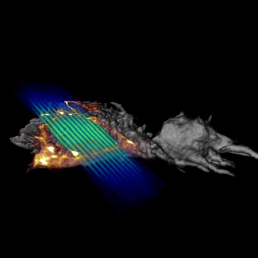
6 IsoView Fruit fly larvae

7 Fruit fly gastrualtion

8 Zebrafish calcium imaging


9 SPIM STORM Use SPIM to confine in 3 rd dimension Use improved signal to localize single molecules


10 SPIM STORM 2 Use an AFM tip as mirror Image single copies of RNA polymerase

11 Fruitfly embryo


12 SPIM STED Pattern STED beam around axial dimensions of SPIM beam Illuminate thinner axial slice Improved axial resolution Only a few publications, probably not really useful
13 SPIM SIM, with a twist

14 Bessel beams Use pencil rather than sheet Bessel beams are perfectly non-diffracting and self reconstructing Can maintain a pencil of illumination across very long distances Like one section of the sheet Formed by imaging annulus at the back aperture of the objective Larger side lobes than Gaussian beam d = λ ex 2NA ex Infinitely thin annulus will create perfect Bessel beam

15 Two photon Bessel imaging Bessel beam excitation enforces isotropic resolution, but side lobes remain Since side lobes can cause a large problem, we can get rid of them by requiring 2 photon excitation Lower intensity side lobes don t have enough energy to excite fluorescence Typical limitations of 2 photon very expensive and hard to image 2 colors

16 Bessel microscope setup Not easy to align No commercial vendor of Bessel scopes, although Zeiss has licensed the IP Even placing the sample in the chamber becomes a challenge

17 SIM SPIM (with Bessel beams) The Bessel beam enables periodicity Rather than scanning through sample rapidly, illuminate discrete spots in time You can illuminate an effective grating Reconstruct as per normal SIM experiment Unlike normal SIM, typically need 7-9 illuminations per sample plane Can still work faster than normal SIM imaging
18 SIM SPIM Also possible to use high intensities to achieve SSIM Just like normal SSIM, have to take more individual frames per SIM frame Also very easy to do 2 photon SIM SPIM Tradeoffs in time, photobleaching, and resolutions
19


20 Two color HeLa cell with actin and connexin


21

22 Lattice sheet the (current) ultimate imaging A combination of Bessel beams and a SIM like illumination By playing with the intensity in the Fourier space, it is possible to make lattices of light in the image plane Sweep lattices around in space to complete entire 3D image Requires opposing excitation and detection objectives just like SPIM


23 Lattice sheet is hard to make Optics are laid out vertically Requires spatial light modulator and annulus mask to pattern light at objective Sample chamber must be waterproof, and objectives must be realigned often

24 Lattice advantages High spatial resolution and high temporal resolution Each Bessel lattice is close to diffraction limited beam Multiple Bessel beams MUCH less phototoxic than confocal or even SPIM, even with same power delivered to sample Similar tradeoff between thinness of sheet and effective field of view Can do SIM exactly the same way as the single Bessel sheet HeLa with labeled actin
25


26 HeLa cell comparing Bessel and Lattice


27 Multicolor lattice sheets

28

29

30

31

32 Lattice STORM Due to thinness of Bessel beams, it s possible to track and localize single molecules that are much more densely packed as compared to widefield High speed imaging allows tracking with high temporal and spatial resolution

33
34 On to Matlab
Point Spread Function. Confocal Laser Scanning Microscopy. Confocal Aperture. Optical aberrations. Alternative Scanning Microscopy
 Bi177 Lecture 5 Adding the Third Dimension Wide-field Imaging Point Spread Function Deconvolution Confocal Laser Scanning Microscopy Confocal Aperture Optical aberrations Alternative Scanning Microscopy
High-resolution, low light-dose lightsheet microscope LATTICE LIGHTSHEET
 LATTICE LIGHTSHEET High-resolution, low light-dose lightsheet microscope First developed by Nobel Laureate Dr. Eric Betzig, the 3i Lattice LightSheet microscope is capable of imaging biological systems
MOM#3: LIGHT SHEET MICROSCOPY (LSM) Stanley Cohen, MD
 Stanley Cohen, MD") MOM#3: LIGHT SHEET MICROSCOPY (LSM) Stanley Cohen, MD Introduction. Although the technical details of light sheet imaging and its various permutations appear at first glance to be complex and require some
MOM#3: LIGHT SHEET MICROSCOPY (LSM) Stanley Cohen, MD Introduction. Although the technical details of light sheet imaging and its various permutations appear at first glance to be complex and require some
Multicolor 4D Fluorescence Microscopy using Ultrathin Bessel Light sheets
 SUPPLEMENTARY MATERIAL Multicolor 4D Fluorescence Microscopy using Ultrathin Bessel Light sheets Teng Zhao, Sze Cheung Lau, Ying Wang, Yumian Su, Hao Wang, Aifang Cheng, Karl Herrup, Nancy Y. Ip, Shengwang
Introduction to light microscopy
 Center for Microscopy and Image Anaylsis Introduction to light Imaging with light / Overview of techniques Urs Ziegler ziegler@zmb.uzh.ch Light interacting with matter Absorbtion Refraction Diffraction
Microscopy Live Animal Imaging
 Microscopy Live Animal Imaging A collaborative environment that provides the knowledge, instruments, and expertise needed to visualize life at scales ranging from single molecules to entire animals. Project
Introduction to light microscopy
 Center for Microscopy and Image Anaylsis Introduction to light microscopy Basic concepts of imaging with light Urs Ziegler ziegler@zmb.uzh.ch Light interacting with matter Absorbtion Refraction Diffraction
Bio 407. Applied microscopy. Introduction into light microscopy. José María Mateos. Center for Microscopy and Image Analysis
 Center for Microscopy and Image Analysis Bio 407 Applied Introduction into light José María Mateos Fundamentals of light Compound microscope Microscope composed of an objective and an additional lens (eyepiece,
Nikon. King s College London. Imaging Centre. N-SIM guide NIKON IMAGING KING S COLLEGE LONDON
 N-SIM guide NIKON IMAGING CENTRE @ KING S COLLEGE LONDON Starting-up / Shut-down The NSIM hardware is calibrated after system warm-up occurs. It is recommended that you turn-on the system for at least
Light Microscopy. Upon completion of this lecture, the student should be able to:
 Light Light microscopy is based on the interaction of light and tissue components and can be used to study tissue features. Upon completion of this lecture, the student should be able to: 1- Explain the
Administrative details:
 Administrative details: Anything from your side? www.photonics.ethz.ch 1 What are we actually doing here? Optical imaging: Focusing by a lens Angular spectrum Paraxial approximation Gaussian beams Method
Reflecting optical system to increase signal intensity. in confocal microscopy
 Reflecting optical system to increase signal intensity in confocal microscopy DongKyun Kang *, JungWoo Seo, DaeGab Gweon Nano Opto Mechatronics Laboratory, Dept. of Mechanical Engineering, Korea Advanced
EUV microscopy - a user s perspective Dimitri Scholz EUV,
 EUV microscopy - a user s perspective Dimitri Scholz EUV, 09.11.2011 Imaging technologies: available at UCD now and in the next future Begin ab ovo - Simple approaches direct to the goal - Standard methods
Why and How? Daniel Gitler Dept. of Physiology Ben-Gurion University of the Negev. Microscopy course, Michmoret Dec 2005
 Why and How? Daniel Gitler Dept. of Physiology Ben-Gurion University of the Negev Why use confocal microscopy? Principles of the laser scanning confocal microscope. Image resolution. Manipulating the
Deep and fast live imaging with two-photon scanned light-sheet microscopy
 Nature Methods Deep and fast live imaging with two-photon scanned light-sheet microscopy Thai V Truong, Willy Supatto, Davis S Koos, John M Choi & Scott E Fraser Supplementary Figure 1 Supplementary Figure
Shaping light in microscopy:
 Shaping light in microscopy: Adaptive optical methods and nonconventional beam shapes for enhanced imaging Martí Duocastella planet detector detector sample sample Aberrated wavefront Beamsplitter Adaptive
Basics of confocal imaging (part I)
") Basics of confocal imaging (part I) Swiss Institute of Technology (EPFL) Faculty of Life Sciences Head of BIOIMAGING AND OPTICS BIOP arne.seitz@epfl.ch Lateral resolution BioImaging &Optics Platform Light
Basics of confocal imaging (part I) Swiss Institute of Technology (EPFL) Faculty of Life Sciences Head of BIOIMAGING AND OPTICS BIOP arne.seitz@epfl.ch Lateral resolution BioImaging &Optics Platform Light
Confocal Microscopy. Kristin Jensen
 Confocal Microscopy Kristin Jensen 17.11.05 References Cell Biological Applications of Confocal Microscopy, Brian Matsumoto, chapter 1 Studying protein dynamics in living cells,, Jennifer Lippincott-Schwartz
5/4/2015 INTRODUCTION TO LIGHT MICROSCOPY. Urs Ziegler MICROSCOPY WITH LIGHT. Image formation in a nutshell. Overview of techniques
 INTRODUCTION TO LIGHT MICROSCOPY Urs Ziegler ziegler@zmb.uzh.ch MICROSCOPY WITH LIGHT INTRODUCTION TO LIGHT MICROSCOPY Image formation in a nutshell Overview of techniques Widefield microscopy Resolution
Examination, TEN1, in courses SK2500/SK2501, Physics of Biomedical Microscopy,
 KTH Applied Physics Examination, TEN1, in courses SK2500/SK2501, Physics of Biomedical Microscopy, 2009-06-05, 8-13, FB51 Allowed aids: Compendium Imaging Physics (handed out) Compendium Light Microscopy
BASICS OF CONFOCAL IMAGING (PART I)
") BASICS OF CONFOCAL IMAGING (PART I) INTERNAL COURSE 2012 LIGHT MICROSCOPY Lateral resolution Transmission Fluorescence d min 1.22 NA obj NA cond 0 0 rairy 0.61 NAobj Ernst Abbe Lord Rayleigh Depth of field
BASICS OF CONFOCAL IMAGING (PART I) INTERNAL COURSE 2012 LIGHT MICROSCOPY Lateral resolution Transmission Fluorescence d min 1.22 NA obj NA cond 0 0 rairy 0.61 NAobj Ernst Abbe Lord Rayleigh Depth of field
Nature Methods: doi: /nmeth Supplementary Figure 1. sospim principle and representation of the sospim beam-steering unit.
 Supplementary Figure 1 sospim principle and representation of the sospim beam-steering unit. Schematic representation of the sospim principle showing a sample holder comprising 45 micromirrored cavities
a) How big will that physical image of the cells be your camera sensor?
 How big will that physical image of the cells be your camera sensor?") 1. Consider a regular wide-field microscope set up with a 60x, NA = 1.4 objective and a monochromatic digital camera with 8 um pixels, properly positioned in the primary image plane. This microscope is
1. Consider a regular wide-field microscope set up with a 60x, NA = 1.4 objective and a monochromatic digital camera with 8 um pixels, properly positioned in the primary image plane. This microscope is
Lecture 16. OMX - Structured Illumination Microscopy Ian Dobbie x Microscopy Course Lecture 16 1
 Lecture 16 OMX - Structured Illumination Microscopy Ian Dobbie x13323 Microscopy Course 2014 - Lecture 16 1 Super-resolution fluorescence microscopy Specificity Sensitivity Non-invasive (in situ & in vivo)
Adaptive optimisation of illumination beam profiles in fluorescence microscopy
 Adaptive optimisation of illumination beam profiles in fluorescence microscopy T. J. Mitchell a, C. D. Saunter a, W. O Nions a, J. M. Girkin a, G. D. Love a a Centre for Advanced nstrumentation & Biophysical
3D light microscopy techniques
 3D light microscopy techniques The image of a point is a 3D feature In-focus image Out-of-focus image The image of a point is not a point Point Spread Function (PSF) 1D imaging 1 1 2! NA = 0.5! NA 2D imaging
Practical Flatness Tech Note
 Practical Flatness Tech Note Understanding Laser Dichroic Performance BrightLine laser dichroic beamsplitters set a new standard for super-resolution microscopy with λ/10 flatness per inch, P-V. We ll
INTRODUCTION TO MICROSCOPY. Urs Ziegler THE PROBLEM
 INTRODUCTION TO MICROSCOPY Urs Ziegler ziegler@zmb.uzh.ch THE PROBLEM 1 ORGANISMS ARE LARGE LIGHT AND ELECTRONS: ELECTROMAGNETIC WAVES v = Wavelength ( ) Speed (v) Frequency ( ) Amplitude (A) Propagation
Imaging Retreat - UMASS Customized real-time confocal and 2-photon imaging
 Imaging Retreat - UMASS 2012 Customized real-time confocal and 2-photon imaging Mike Sanderson Department of Microbiology and Physiological Systems University of Massachusetts Medical School Thanks for
Rapid three-dimensional isotropic imaging of living cells using Bessel beam plane illumination
 Nature Methods Rapid three-dimensional isotropic imaging of living cells using beam plane illumination Thomas A Planchon, Liang Gao, Daniel E Milkie, Michael W Davidson, James A Galbraith, Catherine G
Enhancement of the lateral resolution and the image quality in a line-scanning tomographic optical microscope
 Summary of the PhD thesis Enhancement of the lateral resolution and the image quality in a line-scanning tomographic optical microscope Author: Dudás, László Supervisors: Prof. Dr. Szabó, Gábor and Dr.
Shreyash Tandon M.S. III Year
 Shreyash Tandon M.S. III Year 20091015 Confocal microscopy is a powerful tool for generating high-resolution images and 3-D reconstructions of a specimen by using point illumination and a spatial pinhole
Introduction to light microscopy
 Center for Microscopy and Image Anaylsis Introduction to light Basic concepts of imaging with light Urs Ziegler ziegler@zmb.uzh.ch Microscopy with light 1 Light interacting with matter Absorbtion Refraction
Development of a High-speed Super-resolution Confocal Scanner
 Development of a High-speed Super-resolution Confocal Scanner Takuya Azuma *1 Takayuki Kei *1 Super-resolution microscopy techniques that overcome the spatial resolution limit of conventional light microscopy
Advanced Optical Microscopy lecture. 03. December 2012 Kai Wicker
 Advanced Optical Microscopy lecture 03. December 2012 Kai Wicker Today: Optical transfer functions (OTF) and point spread functions (PSF) in incoherent imaging. 1. Quick revision: the incoherent wide-field
Boulevard du Temple Daguerrotype (Paris,1838) a busy street? Nyquist sampling for movement
 a busy street? Nyquist sampling for movement") Boulevard du Temple Daguerrotype (Paris,1838) a busy street? Nyquist sampling for movement CONFOCAL MICROSCOPY BioVis Uppsala, 2017 Jeremy Adler Matyas Molnar Dirk Pacholsky Widefield & Confocal Microscopy
Boulevard du Temple Daguerrotype (Paris,1838) a busy street? Nyquist sampling for movement CONFOCAL MICROSCOPY BioVis Uppsala, 2017 Jeremy Adler Matyas Molnar Dirk Pacholsky Widefield & Confocal Microscopy
High resolution extended depth of field microscopy using wavefront coding
 High resolution extended depth of field microscopy using wavefront coding Matthew R. Arnison *, Peter Török #, Colin J. R. Sheppard *, W. T. Cathey +, Edward R. Dowski, Jr. +, Carol J. Cogswell *+ * Physical
Durham Research Online
 Durham Research Online Deposited in DRO: March Version of attached le: Accepted Version Peer-review status of attached le: Peer-reviewed Citation for published item: Girkin, John M. and Carvalho, Mariana
Supplementary Information. Stochastic Optical Reconstruction Microscopy Imaging of Microtubule Arrays in Intact Arabidopsis thaliana Seedling Roots
 Supplementary Information Stochastic Optical Reconstruction Microscopy Imaging of Microtubule Arrays in Intact Arabidopsis thaliana Seedling Roots Bin Dong 1,, Xiaochen Yang 2,, Shaobin Zhu 1, Diane C.
Confocal and 2-photon Imaging. October 15, 2010
 Confocal and 2-photon Imaging October 15, 2010 Review Optical Elements Adapted from Sluder & Nordberg 2007 Review Optical Elements Collector Lens Adapted from Sluder & Nordberg 2007 Review Optical Elements
Travel to New Dimensions- LSM 880. The Resolution of a Microscope is limited. The Resolution of a Microscope is limited. Image. Image. Object.
 Travel to New Dimensions- LSM 880 LSM 880: The Power of Sensitivity Our Latest Member of the LSM 880 with GaAsP Detectors Sensitivity, and Ease of Use Innovative High-End Laser Scanning Microscopes from
Resolution. Diffraction from apertures limits resolution. Rayleigh criterion θ Rayleigh = 1.22 λ/d 1 peak at 2 nd minimum. θ f D
 Microscopy Outline 1. Resolution and Simple Optical Microscope 2. Contrast enhancement: Dark field, Fluorescence (Chelsea & Peter), Phase Contrast, DIC 3. Newer Methods: Scanning Tunneling microscopy (STM),
3D light microscopy techniques
 3D light microscopy techniques The image of a point is a 3D feature In-focus image Out-of-focus image The image of a point is not a point Point Spread Function (PSF) 1D imaging 2D imaging 3D imaging Resolution
Bi/BE 227 Winter Assignment #3. Adding the third dimension: 3D Confocal Imaging
 Bi/BE 227 Winter 2016 Assignment #3 Adding the third dimension: 3D Confocal Imaging Schedule: Jan 20: Assignment Jan 20-Feb 8: Work on assignment Feb 10: Student PowerPoint presentations. Goals for this
ADVANCED METHODS FOR CONFOCAL MICROSCOPY II. Jean-Yves Chatton Sept. 2006
 ADVANCED METHODS FOR CONFOCAL MICROSCOPY II Jean-Yves Chatton Sept. 2006 Workshop outline Confocal microscopy of living cells and tissues X-Z scanning Time series Bleach: FRAP, photoactivation Emission
CFIM MICROSCOPY COURSE PROGRAMME PRINCIPLES OF MICROSCOPY CONFOCAL AND FLUORESCENCE MICROSCOPY
 CFIM MICROSCOPY COURSE PROGRAMME PRINCIPLES OF MICROSCOPY 11.01.16-15.01.2016 CONFOCAL AND FLUORESCENCE MICROSCOPY 25.01.16-29.01.2016 PhD Course - University of Copenhagen Department of Biomedical Sciences
Introduction to light microscopy
 Center for Microscopy and Image Anaylsis Introduction to light microscopy (an overview) Microscopy with light Components of a light microscope 1. Light source 2. Objective 3. Sample or specimen holder
Characteristics of point-focus Simultaneous Spatial and temporal Focusing (SSTF) as a two-photon excited fluorescence microscopy
 as a two-photon excited fluorescence microscopy") Characteristics of point-focus Simultaneous Spatial and temporal Focusing (SSTF) as a two-photon excited fluorescence microscopy Qiyuan Song (M2) and Aoi Nakamura (B4) Abstracts: We theoretically and experimentally
Characteristics of point-focus Simultaneous Spatial and temporal Focusing (SSTF) as a two-photon excited fluorescence microscopy Qiyuan Song (M2) and Aoi Nakamura (B4) Abstracts: We theoretically and experimentally
Aberrations and adaptive optics for biomedical microscopes
 Aberrations and adaptive optics for biomedical microscopes Martin Booth Department of Engineering Science And Centre for Neural Circuits and Behaviour University of Oxford Outline Rays, wave fronts and
Spectral Imaging with the Opterra Multipoint Scanning Confocal
 Spectral Imaging with the Opterra Multipoint Scanning Confocal Outline Opterra design overview Scan Modes Light Path Spectral Imaging with Opterra Drosophila larva heart. Opterra Design Overview Supravideo
Multidimensional Imaging with the Opterra Multipoint Scanning Confocal System
 Multidimensional Imaging with the Opterra Multipoint Scanning Confocal System Outline Opterra design overview Light Path Scan Modes Performance Application data Drosophila larva heart. Courtesy of Nikon
Confocal Microscopy and Related Techniques
 Confocal Microscopy and Related Techniques Chau-Hwang Lee Associate Research Fellow Research Center for Applied Sciences, Academia Sinica 128 Sec. 2, Academia Rd., Nankang, Taipei 11529, Taiwan E-mail:
Opterra. Multipoint Scanning Confocal Microscope. Innovation with Integrity. Cell-Friendly, High-Speed, High-Resolution Imaging
 Opterra Multipoint Scanning Confocal Microscope Cell-Friendly, High-Speed, High-Resolution Imaging Innovation with Integrity Fluorescence Microscopy Opterra Multipoint Scanning Confocal Microscope Superior
attocfm I for Surface Quality Inspection NANOSCOPY APPLICATION NOTE M01 RELATED PRODUCTS G
 APPLICATION NOTE M01 attocfm I for Surface Quality Inspection Confocal microscopes work by scanning a tiny light spot on a sample and by measuring the scattered light in the illuminated volume. First,
Invitation for a walk through microscopy. Sebastian Schuchmann Jörg Rösner
 Invitation for a walk through microscopy Sebastian Schuchmann Jörg Rösner joerg.roesner@charite.de Techniques in microscopy Conventional (light) microscopy bright & dark field, phase & interference contrast
Katarina Logg, Kristofer Bodvard, Mikael Käll. Dept. of Applied Physics. 12 September Optical Microscopy. Supervisor s signature:...
 Katarina Logg, Kristofer Bodvard, Mikael Käll Dept. of Applied Physics 12 September 2007 O1 Optical Microscopy Name:.. Date:... Supervisor s signature:... Introduction Over the past decades, the number
contents TABLE OF The SECOM platform Applications - sections Applications - whole cells Features Integrated workflow Automated overlay
 S E C O M TABLE OF contents The SECOM platform 4 Applications - sections 5 Applications - whole cells 8 Features 9 Integrated workflow 12 Automated overlay ODEMIS - integrated software Specifications 13
Light Sheet Microscopy
 Light Sheet Microscopy Introduction During the last decade, microscopy has been constantly trying to exploit new boundaries. By aiming for smaller details, cameras and other detectors needed to become
Nature Methods: doi: /nmeth Supplementary Figure 1
 . Supplementary Figure 1 Schematics and characterization of our AO two-photon fluorescence microscope. (a) Essential components of our AO two-photon fluorescence microscope: Ti:Sapphire laser; optional
Multicolor two-photon light-sheet microscopy Pierre Mahou, Julien Vermot, Emmanuel Beaurepaire & Willy Supatto
 Mahou et al Supplementary Information Page 1 / 16 Multicolor two-photon light-sheet microscopy Pierre Mahou, Julien Vermot, Emmanuel Beaurepaire & Willy Supatto SUPPLEMENTAR INFORMATION SUPPLEMENTAR ITEM
FLUORESCENCE MICROSCOPY. Matyas Molnar and Dirk Pacholsky
 FLUORESCENCE MICROSCOPY Matyas Molnar and Dirk Pacholsky 1 The human eye perceives app. 400-700 nm; best at around 500 nm (green) Has a general resolution down to150-300 μm (human hair: 40-250 μm) We need
Nikon Instruments Europe
 Nikon Instruments Europe Recommendations for N-SIM sample preparation and image reconstruction Dear customer, We hope you find the following guidelines useful in order to get the best performance out of
Microscope anatomy, image formation and resolution
 Microscope anatomy, image formation and resolution Ian Dobbie Buy this book for your lab: D.B. Murphy, "Fundamentals of light microscopy and electronic imaging", ISBN 0-471-25391-X Visit these websites:
Multiphoton Microscopy
 Multiphoton Microscopy A. Neumann, Y. Kuznetsova Introduction Multi-Photon Fluorescence Microscopy is a relatively novel imaging technique in cell biology. It relies on the quasi-simultaneous absorption
Confocal, hyperspectral, spinning disk
 Confocal, hyperspectral, spinning disk Administrative HW 6 due on Fri Midterm on Wed Covers everything since previous midterm 8.5 x 11 sheet allowed, 1 side Guest lecture by Joe Dragavon on Mon 10/30 Last
Application Note. The New 2D Superresolution Mode for ZEISS Airyscan 120 nm Lateral Resolution without Acquiring a Z-stack
 The New 2D Superresolution Mode for ZEISS Airyscan 120 nm Lateral Resolution without Acquiring a Z-stack The New 2D Superresolution Mode for ZEISS Airyscan 120 nm Lateral Resolution without Acquiring a
TRAINING MANUAL. Multiphoton Microscopy LSM 510 META-NLO
 TRAINING MANUAL Multiphoton Microscopy LSM 510 META-NLO September 2010 Multiphoton Microscopy Training Manual Multiphoton microscopy is only available on the LSM 510 META-NLO system. This system is equipped
Rapid Non linear Image Scanning Microscopy, Supplementary Notes
 Rapid Non linear Image Scanning Microscopy, Supplementary Notes Calculation of theoretical PSFs We calculated the electrical field distribution using the wave optical theory developed by Wolf 1, and Richards
Fundamentals of Light Microscopy II: Fluorescence, Deconvolution, Confocal, Multiphoton, Spectral microscopy. Integrated Microscopy Course
 Fundamentals of Light Microscopy II: Fluorescence, Deconvolution, Confocal, Multiphoton, Spectral microscopy Integrated Microscopy Course Review Lecture 1: Microscopy Basics Light train Kohler illumination*
3. are adherent cells (ie. cells in suspension are too far away from the coverslip)
") Before you begin, make sure your sample... 1. is seeded on #1.5 coverglass (thickness = 0.17) 2. is an aqueous solution (ie. fixed samples mounted on a slide will not work - not enough difference in refractive
Before you begin, make sure your sample... 1. is seeded on #1.5 coverglass (thickness = 0.17) 2. is an aqueous solution (ie. fixed samples mounted on a slide will not work - not enough difference in refractive
Fast, high-contrast imaging of animal development with scanned light sheet based structured-illumination microscopy
 nature methods Fast, high-contrast imaging of animal development with scanned light sheet based structured-illumination microscopy Philipp J Keller, Annette D Schmidt, Anthony Santella, Khaled Khairy,
1.The Problem LIGHT-LEVEL LEVEL IMAGING. light-level level Cameras. 3. Solutions. 2. Low-light LOW-LIGHT
 LOW-LIGHT LIGHT-LEVEL LEVEL IMAGING 1.The Problem 2. Low-light light-level level Cameras 3. Solutions How Much Light? I. Illumination system: 75 W Xenon Arc (~1mW/nm in visible) 490/10 nm exciter filter
Multifluorescence The Crosstalk Problem and Its Solution
 Multifluorescence The Crosstalk Problem and Its Solution If a specimen is labeled with more than one fluorochrome, each image channel should only show the emission signal of one of them. If, in a specimen
Opterra II Multipoint Scanning Confocal Microscope. Innovation with Integrity
 Opterra II Multipoint Scanning Confocal Microscope Enabling 4D Live-Cell Fluorescence Imaging through Speed, Sensitivity, Viability and Simplicity Innovation with Integrity Fluorescence Microscopy The
Microscopic Structures
 Microscopic Structures Image Analysis Metal, 3D Image (Red-Green) The microscopic methods range from dark field / bright field microscopy through polarisation- and inverse microscopy to techniques like
OPERATING INSTRUCTIONS
 Zeiss LSM 510 M eta Confocal M icroscope OPERATING INSTRUCTIONS Starting the System: 1. Turn the black knob on the laser box one-quarter turn from Off to On. You will hear the laser cooling mechanisms
Technology Note ZEISS LSM 880 with Airyscan
 Technology Note ZEISS LSM 880 with Airyscan Introducing the Fast Acquisition Mode ZEISS LSM 880 with Airyscan Introducing the Fast Acquisition Mode Author: Dr. Annette Bergter Carl Zeiss Microscopy GmbH,
Optical Microscopy and Imaging ( Part 2 )
") 1 Optical Microscopy and Imaging ( Part 2 ) Chapter 7.1 : Semiconductor Science by Tudor E. Jenkins Saroj Kumar Patra, Department of Electronics and Telecommunication, Norwegian University of Science and
1 Optical Microscopy and Imaging ( Part 2 ) Chapter 7.1 : Semiconductor Science by Tudor E. Jenkins Saroj Kumar Patra, Department of Electronics and Telecommunication, Norwegian University of Science and
Heisenberg) relation applied to space and transverse wavevector
 relation applied to space and transverse wavevector") 2. Optical Microscopy 2.1 Principles A microscope is in principle nothing else than a simple lens system for magnifying small objects. The first lens, called the objective, has a short focal length (a
2. Optical Microscopy 2.1 Principles A microscope is in principle nothing else than a simple lens system for magnifying small objects. The first lens, called the objective, has a short focal length (a
Training Guide for Leica SP8 Confocal/Multiphoton Microscope
 Training Guide for Leica SP8 Confocal/Multiphoton Microscope LAS AF v3.3 Optical Imaging & Vital Microscopy Core Baylor College of Medicine (2017) Power ON Routine 1 2 Turn ON power switch for epifluorescence
Training Guide for Carl Zeiss LSM 5 LIVE Confocal Microscope
 Training Guide for Carl Zeiss LSM 5 LIVE Confocal Microscope AIM 4.2 Optical Imaging & Vital Microscopy Core Baylor College of Medicine (2017) Power ON Routine 1 2 Verify that main power switches on the
Nature Methods: doi: /nmeth Supplementary Figure 1. Schematic of 2P-ISIM AO optical setup.
 Supplementary Figure 1 Schematic of 2P-ISIM AO optical setup. Excitation from a femtosecond laser is passed through intensity control and shuttering optics (1/2 λ wave plate, polarizing beam splitting
Optical Sectioning Deep Inside Live Embryos by Selective Plane Illumination Microscopy
 Supporting Online Material Optical Sectioning Deep Inside Live Embryos by Selective Plane Illumination Microscopy 1 Material and methods 1.1 Setup Jan Huisken, Jim Swoger, Filippo Del Bene, Joachim Wittbrodt,
D2.1 Operating 2D STED Microscope
 D2.1 Operating 2D STED Microscope Nature: Report Dissemination Level: Public Lead Beneficiary: UNIVDUN Author(s): Piotr Zdankowski Work Package: WP2 Task: ESR5 Version: 0.02 Last modified: 24/04/2017 Status:
長庚大學共軛焦顯微鏡課程 長庚大學共軛焦顯微鏡課程. Spot light 長庚大學
 長庚大學共軛焦顯微鏡課程 Spot light 長庚大學共軛焦顯微鏡課程 20071030 長庚大學 Basic principle of Laser Scanning Confocal Microscopy The application of LSM 510 META detector Multiphoton microscopy basic principle and introduction
Beam Shaping and Simultaneous Exposure by Diffractive Optical Element in Laser Plastic Welding
 Beam Shaping and Simultaneous Exposure by Diffractive Optical Element in Laser Plastic Welding AKL`12 9th May 2012 Dr. Daniel Vogler Page 1 Motivation: Quality and flexibility diffractive spot shaping
Fast, Two-Dimensional Optical Beamscanning by Wavelength Switching T. K. Chan, E. Myslivets, J. E. Ford
 Photonics Systems Integration Lab University of California San Diego Jacobs School of Engineering Fast, Two-Dimensional Optical Beamscanning by Wavelength Switching T. K. Chan, E. Myslivets, J. E. Ford
A Compact Perpendicular Microscopy and Imaging System for the Detection of Fluorescent Solution Flow
 Progress In Electromagnetics Research Letters, Vol. 67, 75 79, 2017 A Compact Perpendicular Microscopy and Imaging System for the Detection of Fluorescent Solution Flow Fuhong Cai 1, *,MengZhao 1,andDanWang
3D live fluorescence imaging of cellular dynamics using Bessel beam plane illumination microscopy
 3D live fluorescence imaging of cellular dynamics using Bessel beam plane illumination microscopy Liang Gao 1,2, Lin Shao 1, Bi-Chang Chen 1 & Eric Betzig 1 1 Janelia Farm Research Campus, Howard Hughes
Principles of Light-Sheet Microscopy. Alon Greenbaum, PhD Gradinaru Lab Antti Lignell, PhD
 Principles of Light-Sheet Microscopy Alon Greenbaum, PhD Gradinaru Lab Antti Lignell, PhD Overview Different imaging modalities pros and cons Introduction to light-sheet microscopy Light-sheet issues Light-sheet
Supplementary information, Figure S1A-S1H The thickness and the uniformity of the light sheet at different DOFs. By
 Supplementary information, Figure S1A-S1H The thickness and the uniformity of the light sheet at different DOFs. By imaging FITC-containing solution, the thickness of the light sheet generated by the P3A-DSLM
6/3/15. The Anatomy of a Digital Image. Representative Intensities. Specimen: (molecular distribution)
") 2015 LMIC Imaging Workshop Sidney L. Shaw Technical Director An introduction of concepts for Super-Resolution Light Microscopy The Anatomy of a Digital Image Representative Intensities Specimen: (molecular
2015 LMIC Imaging Workshop Sidney L. Shaw Technical Director An introduction of concepts for Super-Resolution Light Microscopy The Anatomy of a Digital Image Representative Intensities Specimen: (molecular
STORM/ PALM ANSWER KEY
 STORM/ PALM ANSWER KEY Phys598BP Spring 2016 University of Illinois at Urbana-Champaign Questions for Lab Report 1. How do you define a resolution in STORM imaging? If you are given a STORM setup, how
Maria Smedh, Centre for Cellular Imaging. Maria Smedh, Centre for Cellular Imaging
 Nonlinear microscopy I: Two-photon fluorescence microscopy Multiphoton Microscopy What is multiphoton imaging? Applications Different imaging modes Advantages/disadvantages Scattering of light in thick
MASSACHUSETTS INSTITUTE OF TECHNOLOGY Department of Electrical Engineering and Computer Science
 Student Name Date MASSACHUSETTS INSTITUTE OF TECHNOLOGY Department of Electrical Engineering and Computer Science 6.161 Modern Optics Project Laboratory Laboratory Exercise No. 3 Fall 2005 Diffraction
Training Guide for Carl Zeiss LSM 510 META Confocal Microscope
 Training Guide for Carl Zeiss LSM 510 META Confocal Microscope AIM 4.2 Optical Imaging & Vital Microscopy Core Baylor College of Medicine (2017) Power ON Routine 1 2 Turn ON Components and System/PC switches
Dynamic beam shaping with programmable diffractive optics
 Dynamic beam shaping with programmable diffractive optics Bosanta R. Boruah Dept. of Physics, GU Page 1 Outline of the talk Introduction Holography Programmable diffractive optics Laser scanning confocal
Rates of excitation, emission, ISC
 Bi177 Lecture 4 Fluorescence Microscopy Phenomenon of Fluorescence Energy Diagram Rates of excitation, emission, ISC Practical Issues Lighting, Filters More on diffraction Point Spread Functions Thus Far,
Confocal Microscopy. (Increasing contrast and resolu6on using op6cal sec6oning) Lecture 7. November 2017
 Lecture 7. November 2017") Confocal Microscopy (Increasing contrast and resolu6on using op6cal sec6oning) Lecture 7 November 2017 3 Flavours of Microscope Confocal Laser Scanning Problem: Out of Focus Light Spinning disc 2-Photon
Confocal Microscopy (Increasing contrast and resolu6on using op6cal sec6oning) Lecture 7 November 2017 3 Flavours of Microscope Confocal Laser Scanning Problem: Out of Focus Light Spinning disc 2-Photon
Zero Focal Shift in High Numerical Aperture Focusing of a Gaussian Laser Beam through Multiple Dielectric Interfaces. Ali Mahmoudi
 1 Zero Focal Shift in High Numerical Aperture Focusing of a Gaussian Laser Beam through Multiple Dielectric Interfaces Ali Mahmoudi a.mahmoudi@qom.ac.ir & amahmodi@yahoo.com Laboratory of Optical Microscopy,