ADVANCED METHODS FOR CONFOCAL MICROSCOPY II. Jean-Yves Chatton Sept. 2006
|
|
|
- Francis Morrison
- 5 years ago
- Views:
Transcription

1 ADVANCED METHODS FOR CONFOCAL MICROSCOPY II Jean-Yves Chatton Sept. 2006
2 Workshop outline Confocal microscopy of living cells and tissues X-Z scanning Time series Bleach: FRAP, photoactivation Emission spectra and spectral deconvolution Ratio imaging
3 Workshop outline Confocal microscopy of living cells and tissues X-Z scanning Time series Bleach: FRAP, photoactivation Emission spectra and spectral deconvolution Ratio imaging
 and objective are used to focus a diffraction-limited spot The field lens projects emitted light from this point to the infinite as a collimated beam A pinhole (or diaphragm)")
4 Out-of of-focus focus signal rejection by scanning confocal microscopy (epifluorescence) modied from J. Pawley A field lens (L) and objective are used to focus a diffraction-limited spot The field lens projects emitted light from this point to the infinite as a collimated beam A pinhole (or diaphragm) is placed in the intermediate plane in front of the detector and rejects all but the signal originating from the focal point
5 Comparison of sample illumination in conventional and confocal fluorescence imaging Conventional microscope (Widefield illumination) Confocal microscope (Illumination by a diffraction-limited spot)
6 Weaknesses of confocal microscopy 1. The luminous flux of excitation needed to detect enough signal is very high, which has the following consequences: - Photobleaching of dyes - Photodynamic dammage to living cells 2. Speed limitation caused by point-by-point image formation (non-parallel scanning). E.g.: 512x512 pixel image, video rate = 40 msec/image (25 fps) --> each pixel is visited during only 152 nsec 3. No direct visual observation 4. Laser illuminatin limits the range of available wavelengths 5. Complexity and high cost of the instrument
7 Strengths of the confocal microscope 1. Out-of-focus light is not recorded. Moving the focus does not cause blur in the image but gradually cuts the parts of the object that are not in the focal plane: optical sections of the sample. 2. Because of the small dimension of the spot illuminating the focal plane, scattered and diffused light is minimized. 3. A set of tri-dimensional data can be recorded. 4. Scanning an object in the x/y direction (i.e. in the plane) and in the z direction (along the optical axis) enables visualizing the object from all sides. 5. Using image processing, optical sections can be superimposed and produce an extended focus, which can be obtained in conventional microscopy only by reducing the aperture and thus diminishing resolution.
8 Other possibilities offered by the confocal microscope (Very) Fast measurements by line scan (rather than frame scanning). Measurement of cell height or volume change by X-Z scanning (piezo focus). As laser light is normally polarized, excitation light that has not been absorbed by the sample can be collected by the condenser and be used to build a transmitted Nomarski image. By rapid change of laser light intensity using an acousto-optical modulator, it is possible to perform experiments of: - fluorescence recovery after photobleaching (FRAP) experiments to measure motility or diffusion of fluroescent compounds - photoactivation of caged compounds (flash photolysis) - photoactivation of photoactivatable proteins (e.g. Kaede)
9 Workshop outline Confocal microscopy of living cells and tissues X-Z scanning Time series Bleach: FRAP, photoactivation Emission spectra and spectral deconvolution Ratio imaging


10 X-Z Z scanning Z Y X XY Single optical sections

11 X-Z Z scanning After 3D reconstruction and rendering
12 X-Z Z scanning Z Y X XY Line scan along the X axis and rapid Z movement
13 X-Z Z scanning X Z Line scan along the X axis and rapid Z movement

14 X-Z Z scanning X XZ Z Optimal using piezo Z-drive (speed and resolution)
15 X-Z Z scanning Applications Experimental direct and rapid measurement of specimen thickness XZ over time : dynamic cell volume measurement (e.g. swelling, regulatory volume decrease, etc.) Technical / teaching direct assessment of point spread function (PSF) assessment of pinhole effect
16 Workshop outline Confocal microscopy of living cells and tissues X-Z scanning Time series Bleach: FRAP, photoactivation Emission spectra and spectral deconvolution Ratio imaging
17 Time series Idea: recording of fluorescence signal over time Applicable for recording: entire images, sub-regions, lines, points in 2D, in 3D (z-stacks), spectra, etc. which becomes "4D", "5D", "6D", etc. Acquisition of time series can be combined and synchronized with: internal actions (e.g. bleach) external events (opening of valves, stimulus, etc.)
18 "TIME SERIES" module of Zeiss LSM 510 Meta software


19 Lateral mouvement Frame averaging Line averaging
20 "TIME SERIES" module of Zeiss LSM 510 Meta software
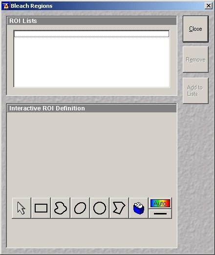
21 to record only intensity in a region (!) 4

22 To visualize the results: Gallery

23 To visualize the results: Graphs
24 To visualize the results: Movies


25 To visualize the results: Movies MAP GFP (B. Riederer)
 with")
26 Stacks (3D) with time


27
28 "TIME SERIES" module of Zeiss LSM 510 Meta software
29 LINE SCAN : to go very fast
")
30 LINE SCAN : to go very fast X Each line is scanned very rapidly (in this example <1 msec) Time
31 Workshop outline Confocal microscopy of living cells and tissues X-Z scanning Time series Bleach: FRAP, photoactivation Emission spectra and spectral deconvolution Ratio imaging
32 Fluorescence recovery after Definition photobleaching (FRAP) Rapid and localized photodestruction of fluorescence used as an experimental tool...
33 Photobleaching Weakening of fluorescent signal Poorly understood, but critical in fluorescence microscopy Transition to a triplet state, reaction with oxygen Quantum yield * of fluorescein: ~ 0.9 Quantum yield of bleaching of fluorescein: ~ The molecule will emit 30'000-40'000 photons in its photochemical life. This is independent on the type of illumination (continuous or pulsed). At low intensity of illumination, bleaching is not eliminated by only its kinetics is slowed down. * Quantum yield: Q= Number of photons emitted / Number of photons absorbed
34 FRAP - Fluorescence Recovery After Photobleaching Applications: - rates of diffusion and convection - motility of proteins (e.g. in membrane, reticulum, etc.) - protein interactions - protein synthesis

35 AOTF (acousto-optical tunable filter) Working wavelengths: nm Speed of selection: < 1ms Angle of acceptance: ± 5 (i.e. laser) Efficiency of transmission/diffraction: 80% Maximum input power: 500W/cm 2 Selection of both wavelength and intensity Source: Chatton&Spring, Microsc. Soc. Am. Bull. 23:324 (1993)
36 Mesurement of diffusion by FRAP Non-reversible Reversible Diffusion rate FITC Taken from Periasamy (2001) "Methods in cellular imaging", p. 115


37 "TIME SERIES" module of Zeiss LSM 510 Meta software

38

39

40

41 Example: Gap jonctions in mouse astrocytes Loading with BCECF AM False color intensity LUT

42 Astrocytes functional gap jonctions Percentage of recovery: 55% Time to 90% : 17 seconds

43 Inhibition of gap junction using octanol Loading with BCECF AM False color intensity LUT

44 Inhibition of gap junction using octanol Pecentage of recovery: 7% Time to 90%: 9 seconds

45 UV Photoactivation using the Bleach module UV Photoactivation of caged ATP. Astrocytes loaded with calcium probe Fluo-4

46 UV Photoactivation using the Bleach module Event markers
47 Workshop outline Confocal microscopy of living cells and tissues X-Z scanning Time series Bleach: FRAP, photoactivation Emission spectra and spectral deconvolution Ratio imaging
48 Making use of the spectral information Transitions from different vibrational levels define absorption spectrum of a molecule A similar set of transitions from S1 to S0 define its emission spectrum.


49 Microscope with spectral detector: Diffraction grating Configuration Zeiss PMT Array 32 canaux Zeiss LSM 510 Meta Detector : array of 32 photomultipliers in parallel
50 Spectral detector: Zeiss configuration Diffraction grating PMT array (32 channels) Simultaneous recording of 8 channels (images) Emitted Fluorescence Wavelength (nm) Emitted light (Polychromatic)

51 Possibiities offered by the spectral detector Define band-pass "filters" Spectral scanning

52 Emission spectra

53 Gallery by wavelength 492nm 566nm 513nm 566nm 534nm 545nm 556nm 566nm 577nm 588nm 599nm 609nm 620nm 631nm

54 Spectra in subregions of images

 Application example: Demonstration")

55 Spectra in subregions of images A B C D Fluorescence emission E F Wavelength (nm) Bernadinelli, Azarias, Chatton GLIA 54: (2006) Application example: Demonstration of colocalization of two mitochondrial dyes, CoroNa Red and Mitotracker Green. Spectra taken in one single mitochondrion
56 Possibiities offered by the spectral detector Define band-pass "filters" Spectral scanning

57 Create Meta-channels

58
59 Linear spectral deconvolution Fluorescein Lucifer Yellow Fluorescence (Emission) Longueur d'onde (nm) Resulting spectrum Leica: Spectral unmixing Zeiss: Emission fingerprinting, Linear unmixing

 0 500 510 520 530")

60 Spectral deconvolution EGFP SYTOX Green YFP Fluorescence Emission Fluorescence Emission Fluorescence Emission Wavelength (nm) Wavelength (nm) Wavelength (nm) 250 Fluorescence Emission Excitation 488nm Zeiss LSM 510 Meta Wavelength (nm)

61 Spectral deconvolution EGFP SYTOX YFP Combined Excitation 488nm Zeiss LSM 510 Meta
62 See you next week
63 Workshop outline Confocal microscopy of living cells and tissues X-Z scanning Time series Bleach: FRAP, photoactivation Emission spectra and spectral deconvolution Ratio imaging
64 Ratio imaging Excitation ratio : fixed emission, excitation with two wavelengths Examples : indicators like Fura-2 (calcium), BCECF (ph), SBFI (sodium) Emission ratio : fixed excitation, emission with two wavelengths Examples : indicator like Indo-1 (calcium), SNARF-1 (ph)
65 Ratio imaging Emission ratio : fixed excitation, emission with two wavelengths Examples : - organic probes like Indo-1 (Calcium) - FRET probes (eg : cameleons)
 -5-(and-6)-carboxyfluorescein pka = 6.98")
66 BCECF : Sensitivity to ambient ph Isosbestic point (440 nm) BCECF = 2',7'-bis-(2-carboxyethyl) -5-(and-6)-carboxyfluorescein pka = 6.98
67 Example of ratio dye: Fura nm 380 nm Emission High Ca 2+ 0-Ca 2+ Isosbestic point
68 Comparison : single wavelength and ratio fluorescence changes F 340 F 340 ou F 380 F 380 Ratio F 340 / F 380 Ratio Rem : simulated plots Temps

69 Ratio Module of Zeiss LSM software
Why and How? Daniel Gitler Dept. of Physiology Ben-Gurion University of the Negev. Microscopy course, Michmoret Dec 2005
 Why and How? Daniel Gitler Dept. of Physiology Ben-Gurion University of the Negev Why use confocal microscopy? Principles of the laser scanning confocal microscope. Image resolution. Manipulating the
LSM 510 META in Chang Gung University
 Content LSM 510 META in Chang ung University LSM 510 META 路 理 The features and applications of LSM 510 META 01-09 Introduction of the hardware 10-12 Fluorescence observation in conventional microscope
Multifluorescence The Crosstalk Problem and Its Solution
 Multifluorescence The Crosstalk Problem and Its Solution If a specimen is labeled with more than one fluorochrome, each image channel should only show the emission signal of one of them. If, in a specimen
Ratio Imaging. Dividing one image by another to detect changing conditions. Images collected at different times, wavelengths, polarities, etc
 Ratio Imaging Dividing one image by another to detect changing conditions Images collected at different times, wavelengths, polarities, etc Most common use of ratio imaging is to provide a quick spectral
Shreyash Tandon M.S. III Year
 Shreyash Tandon M.S. III Year 20091015 Confocal microscopy is a powerful tool for generating high-resolution images and 3-D reconstructions of a specimen by using point illumination and a spatial pinhole
長庚大學共軛焦顯微鏡課程 長庚大學共軛焦顯微鏡課程. Spot light 長庚大學
 長庚大學共軛焦顯微鏡課程 Spot light 長庚大學共軛焦顯微鏡課程 20071030 長庚大學 Basic principle of Laser Scanning Confocal Microscopy The application of LSM 510 META detector Multiphoton microscopy basic principle and introduction
Boulevard du Temple Daguerrotype (Paris,1838) a busy street? Nyquist sampling for movement
 a busy street? Nyquist sampling for movement") Boulevard du Temple Daguerrotype (Paris,1838) a busy street? Nyquist sampling for movement CONFOCAL MICROSCOPY BioVis Uppsala, 2017 Jeremy Adler Matyas Molnar Dirk Pacholsky Widefield & Confocal Microscopy
Boulevard du Temple Daguerrotype (Paris,1838) a busy street? Nyquist sampling for movement CONFOCAL MICROSCOPY BioVis Uppsala, 2017 Jeremy Adler Matyas Molnar Dirk Pacholsky Widefield & Confocal Microscopy
Dynamic Confocal Imaging of Living Brain. Advantages and risks of multiphoton microscopy in physiology
 Dynamic Confocal Imaging of Living Brain Advantages and risks of multiphoton microscopy in physiology Confocal laser scanning microscopy In conventional optical microscopy focused and out-offocus light
Bio 407. Applied microscopy. Introduction into light microscopy. José María Mateos. Center for Microscopy and Image Analysis
 Center for Microscopy and Image Analysis Bio 407 Applied Introduction into light José María Mateos Fundamentals of light Compound microscope Microscope composed of an objective and an additional lens (eyepiece,
Point Spread Function. Confocal Laser Scanning Microscopy. Confocal Aperture. Optical aberrations. Alternative Scanning Microscopy
 Bi177 Lecture 5 Adding the Third Dimension Wide-field Imaging Point Spread Function Deconvolution Confocal Laser Scanning Microscopy Confocal Aperture Optical aberrations Alternative Scanning Microscopy
Fundamentals of Light Microscopy II: Fluorescence, Deconvolution, Confocal, Multiphoton, Spectral microscopy. Integrated Microscopy Course
 Fundamentals of Light Microscopy II: Fluorescence, Deconvolution, Confocal, Multiphoton, Spectral microscopy Integrated Microscopy Course Review Lecture 1: Microscopy Basics Light train Kohler illumination*
Rates of excitation, emission, ISC
 Bi177 Lecture 4 Fluorescence Microscopy Phenomenon of Fluorescence Energy Diagram Rates of excitation, emission, ISC Practical Issues Lighting, Filters More on diffraction Point Spread Functions Thus Far,
3D light microscopy techniques
 3D light microscopy techniques The image of a point is a 3D feature In-focus image Out-of-focus image The image of a point is not a point Point Spread Function (PSF) 1D imaging 1 1 2! NA = 0.5! NA 2D imaging
BASICS OF CONFOCAL IMAGING (PART I)
") BASICS OF CONFOCAL IMAGING (PART I) INTERNAL COURSE 2012 LIGHT MICROSCOPY Lateral resolution Transmission Fluorescence d min 1.22 NA obj NA cond 0 0 rairy 0.61 NAobj Ernst Abbe Lord Rayleigh Depth of field
BASICS OF CONFOCAL IMAGING (PART I) INTERNAL COURSE 2012 LIGHT MICROSCOPY Lateral resolution Transmission Fluorescence d min 1.22 NA obj NA cond 0 0 rairy 0.61 NAobj Ernst Abbe Lord Rayleigh Depth of field
Basics of confocal imaging (part I)
") Basics of confocal imaging (part I) Swiss Institute of Technology (EPFL) Faculty of Life Sciences Head of BIOIMAGING AND OPTICS BIOP arne.seitz@epfl.ch Lateral resolution BioImaging &Optics Platform Light
Basics of confocal imaging (part I) Swiss Institute of Technology (EPFL) Faculty of Life Sciences Head of BIOIMAGING AND OPTICS BIOP arne.seitz@epfl.ch Lateral resolution BioImaging &Optics Platform Light
Confocal Laser Scanning Microscopy
 Name of the Core Facility: Confocal Laser Scanning Microscopy CORE Forschungszentrum Immunologie Mainz Welcome to the CSLM Core Facility: The CLSM Core Facility enables working groups to incorporate high
Zeiss 780 Training Notes
 Zeiss 780 Training Notes Turn on Main Switch, System PC and Components Switches 780 Start up sequence Do you need the argon laser (458, 488, 514 nm lines)? Yes Turn on the laser s main power switch and
Final Exam, 150 points PMB 185: Techniques in Light Microscopy
 Final Exam, 150 points Name PMB 185: Techniques in Light Microscopy Point value is in parentheses at the end of each question. Note: GFP = green fluorescent protein ; CFP = cyan fluorescent protein ; YFP
Confocal Microscopy. Kristin Jensen
 Confocal Microscopy Kristin Jensen 17.11.05 References Cell Biological Applications of Confocal Microscopy, Brian Matsumoto, chapter 1 Studying protein dynamics in living cells,, Jennifer Lippincott-Schwartz
Confocal Microscopy. (Increasing contrast and resolu6on using op6cal sec6oning) Lecture 7. November 2017
 Lecture 7. November 2017") Confocal Microscopy (Increasing contrast and resolu6on using op6cal sec6oning) Lecture 7 November 2017 3 Flavours of Microscope Confocal Laser Scanning Problem: Out of Focus Light Spinning disc 2-Photon
Confocal Microscopy (Increasing contrast and resolu6on using op6cal sec6oning) Lecture 7 November 2017 3 Flavours of Microscope Confocal Laser Scanning Problem: Out of Focus Light Spinning disc 2-Photon
Imaging Retreat - UMASS Customized real-time confocal and 2-photon imaging
 Imaging Retreat - UMASS 2012 Customized real-time confocal and 2-photon imaging Mike Sanderson Department of Microbiology and Physiological Systems University of Massachusetts Medical School Thanks for
Maria Smedh, Centre for Cellular Imaging. Maria Smedh, Centre for Cellular Imaging
 Nonlinear microscopy I: Two-photon fluorescence microscopy Multiphoton Microscopy What is multiphoton imaging? Applications Different imaging modes Advantages/disadvantages Scattering of light in thick
ANSWER KEY Lab 2 (IGB): Bright Field and Fluorescence Optical Microscopy and Sectioning
: Bright Field and Fluorescence Optical Microscopy and Sectioning") Phys598BP Spring 2016 University of Illinois at Urbana-Champaign ANSWER KEY Lab 2 (IGB): Bright Field and Fluorescence Optical Microscopy and Sectioning Location: IGB Core Microscopy Facility Microscope:
Phys598BP Spring 2016 University of Illinois at Urbana-Champaign ANSWER KEY Lab 2 (IGB): Bright Field and Fluorescence Optical Microscopy and Sectioning Location: IGB Core Microscopy Facility Microscope:
Components of confocal and two-photon microscopes
 Components of confocal and two-photon microscopes Internal training 07/04/2016 A. GRICHINE Platform Optical microscopy Cell imaging, IAB, ISdV Plan Confocal laser scanning microscope o o o Principle Main
TRAINING MANUAL. Multiphoton Microscopy LSM 510 META-NLO
 TRAINING MANUAL Multiphoton Microscopy LSM 510 META-NLO September 2010 Multiphoton Microscopy Training Manual Multiphoton microscopy is only available on the LSM 510 META-NLO system. This system is equipped
Quick Start Guide. Leica SP5 X
 Quick Start Guide Leica SP5 X Please note: Some of the information in this guide was taken from Leica Microsystems Leica TCS SP5 LAS AF Guide for New Users. This work is licensed under the Creative Commons
Opterra II Multipoint Scanning Confocal Microscope. Innovation with Integrity
 Opterra II Multipoint Scanning Confocal Microscope Enabling 4D Live-Cell Fluorescence Imaging through Speed, Sensitivity, Viability and Simplicity Innovation with Integrity Fluorescence Microscopy The
Practical work no. 3: Confocal Live Cell Microscopy
 Practical work no. 3: Confocal Live Cell Microscopy Course Instructor: Mikko Liljeström (MIU) 1 Background Confocal microscopy: The main idea behind confocality is that it suppresses the signal outside
3D light microscopy techniques
 3D light microscopy techniques The image of a point is a 3D feature In-focus image Out-of-focus image The image of a point is not a point Point Spread Function (PSF) 1D imaging 2D imaging 3D imaging Resolution
Travel to New Dimensions- LSM 880. The Resolution of a Microscope is limited. The Resolution of a Microscope is limited. Image. Image. Object.
 Travel to New Dimensions- LSM 880 LSM 880: The Power of Sensitivity Our Latest Member of the LSM 880 with GaAsP Detectors Sensitivity, and Ease of Use Innovative High-End Laser Scanning Microscopes from
Invitation for a walk through microscopy. Sebastian Schuchmann Jörg Rösner
 Invitation for a walk through microscopy Sebastian Schuchmann Jörg Rösner joerg.roesner@charite.de Techniques in microscopy Conventional (light) microscopy bright & dark field, phase & interference contrast
1.The Problem LIGHT-LEVEL LEVEL IMAGING. light-level level Cameras. 3. Solutions. 2. Low-light LOW-LIGHT
 LOW-LIGHT LIGHT-LEVEL LEVEL IMAGING 1.The Problem 2. Low-light light-level level Cameras 3. Solutions How Much Light? I. Illumination system: 75 W Xenon Arc (~1mW/nm in visible) 490/10 nm exciter filter
Resolution. Diffraction from apertures limits resolution. Rayleigh criterion θ Rayleigh = 1.22 λ/d 1 peak at 2 nd minimum. θ f D
 Microscopy Outline 1. Resolution and Simple Optical Microscope 2. Contrast enhancement: Dark field, Fluorescence (Chelsea & Peter), Phase Contrast, DIC 3. Newer Methods: Scanning Tunneling microscopy (STM),
Zeiss 880 Training Notes Zen 2.3
 Zeiss 880 Training Notes Zen 2.3 1 Turn on the HXP 120V Lamp 2 Turn on Main Power Switch Turn on the Systems PC Switch Turn on the Components Switch. 3 4 5 Turn on the PC and log into your account. Start
Technology Note ZEISS LSM 880 with Airyscan
 Technology Note ZEISS LSM 880 with Airyscan Introducing the Fast Acquisition Mode ZEISS LSM 880 with Airyscan Introducing the Fast Acquisition Mode Author: Dr. Annette Bergter Carl Zeiss Microscopy GmbH,
Confocal and 2-photon Imaging. October 15, 2010
 Confocal and 2-photon Imaging October 15, 2010 Review Optical Elements Adapted from Sluder & Nordberg 2007 Review Optical Elements Collector Lens Adapted from Sluder & Nordberg 2007 Review Optical Elements
Precision-tracking of individual particles By Fluorescence Photo activation Localization Microscopy(FPALM) Presented by Aung K.
 Presented by Aung K.") Precision-tracking of individual particles By Fluorescence Photo activation Localization Microscopy(FPALM) Presented by Aung K. Soe This FPALM research was done by Assistant Professor Sam Hess, physics
Precision-tracking of individual particles By Fluorescence Photo activation Localization Microscopy(FPALM) Presented by Aung K. Soe This FPALM research was done by Assistant Professor Sam Hess, physics
Bi Imaging. Multicolor Imaging: The Important Question of Co-Localization. Anna Smallcombe Bio-Rad Laboratories, Hemel Hempstead, UK
 Multicolor Imaging: The Important Question of Co-Localization Anna Smallcombe Bio-Rad Laboratories, Hemel Hempstead, UK The use of specific fluorescent probes, combined with confocal or multiphoton microscopy
Confocal Microscopy Confocal Microscopy Acousto-optic products
 Confocal Microscopy Confocal Microscopy Acousto-optic products AA OPTO-ELECTRONIC QUANTA TECH Introduction Confocal microscopy is an imaging technique used to increase micrograph contrast and/or to reconstruct
Microscopy from Carl Zeiss
 Microscopy from Carl Zeiss Contents Page Contents... 1 Introduction... 1 Starting the System... 2 Introduction to ZEN Efficient Navigation... 5 Setting up the microscope... 10 Configuring the beam path
Supplemental Figure 1: Histogram of 63x Objective Lens z axis Calculated Resolutions. Results from the MetroloJ z axis fits for 5 beads from each
 Supplemental Figure 1: Histogram of 63x Objective Lens z axis Calculated Resolutions. Results from the MetroloJ z axis fits for 5 beads from each lens with a 1 Airy unit pinhole setting. Many water lenses
Introduction to light microscopy
 Center for Microscopy and Image Anaylsis Introduction to light Basic concepts of imaging with light Urs Ziegler ziegler@zmb.uzh.ch Microscopy with light 1 Light interacting with matter Absorbtion Refraction
Non-Descanned FLIM Detection in Multiphoton Microscopes
 Non-Descanned FLIM Detection in Multiphoton Microscopes Abstract. Multiphoton microscopes use a femtosecond NIR laser to excite fluorescence in the sample. Excitation is performed via a multi-photon absorption
Training Guide for Carl Zeiss LSM 510 META Confocal Microscope
 Training Guide for Carl Zeiss LSM 510 META Confocal Microscope AIM 4.2 Optical Imaging & Vital Microscopy Core Baylor College of Medicine (2017) Power ON Routine 1 2 Turn ON Components and System/PC switches
Operation Guide for the Leica SP2 Confocal Microscope Bio-Imaging Facility Hunter College October 2009
 Operation Guide for the Leica SP2 Confocal Microscope Bio-Imaging Facility Hunter College October 2009 Introduction of Fluoresence Confocal Microscopy The first confocal microscope was invented by Princeton
Training Guide for Leica SP8 Confocal/Multiphoton Microscope
 Training Guide for Leica SP8 Confocal/Multiphoton Microscope LAS AF v3.3 Optical Imaging & Vital Microscopy Core Baylor College of Medicine (2017) Power ON Routine 1 2 Turn ON power switch for epifluorescence
Introduction to light microscopy
 Center for Microscopy and Image Anaylsis Introduction to light microscopy Basic concepts of imaging with light Urs Ziegler ziegler@zmb.uzh.ch Light interacting with matter Absorbtion Refraction Diffraction
Guide to Confocal 5. Starting session
 Guide to Confocal 5 Remember that when booking and before starting session you can check for any problems at https://www.bris.ac.uk/biochemistry/uobonly/cif/index.html Starting session Switch on microscope
Supplemental Method Information Zeiss LSM710
 Supplemental Method Information Zeiss LSM710 1 Under the Light Path window set up the confocal for imaging a green dye (Alexa488-EGFP). For example, set up the light path as shown here using the 488 nm
Last updated: May 2014 Y.DeGraaf
 FLINDERS MICROSCOPY BIOMEDICAL SERVICES AVAILABLE MICROSCOPES AND SPECIFICATIONS & INFORMATION REGARDING TRAINING FOR NEW USERS Last updated: May 2014 Y.DeGraaf If you have new staff or students (Honours/Masters
ZEISS LSM510META confocal manual
 ZEISS LSM510META confocal manual Switching on the system 1) Switch on the Remote Control button located on the table to the right of the microscope. This is the main switch for the whole system including
INTRODUCTION TO MICROSCOPY. Urs Ziegler THE PROBLEM
 INTRODUCTION TO MICROSCOPY Urs Ziegler ziegler@zmb.uzh.ch THE PROBLEM 1 ORGANISMS ARE LARGE LIGHT AND ELECTRONS: ELECTROMAGNETIC WAVES v = Wavelength ( ) Speed (v) Frequency ( ) Amplitude (A) Propagation
LSM 510 NLO and LSM 510 META NLO Multiphoton Laser Scanning Microscopes Deep Insights Carefully Gained
 Microscopy from Carl Zeiss LSM 510 NLO and LSM 510 META NLO Multiphoton Laser Scanning Microscopes Deep Insights Carefully Gained LSM 510 NLO and LSM 510 META NLO Deep Insights Carefully Gained In multiphoton
Akinori Mitani and Geoff Weiner BGGN 266 Spring 2013 Non-linear optics final report. Introduction and Background
 Akinori Mitani and Geoff Weiner BGGN 266 Spring 2013 Non-linear optics final report Introduction and Background Two-photon microscopy is a type of fluorescence microscopy using two-photon excitation. It
FLUORESCENCE MICROSCOPY. Matyas Molnar and Dirk Pacholsky
 FLUORESCENCE MICROSCOPY Matyas Molnar and Dirk Pacholsky 1 The human eye perceives app. 400-700 nm; best at around 500 nm (green) Has a general resolution down to150-300 μm (human hair: 40-250 μm) We need
EUV microscopy - a user s perspective Dimitri Scholz EUV,
 EUV microscopy - a user s perspective Dimitri Scholz EUV, 09.11.2011 Imaging technologies: available at UCD now and in the next future Begin ab ovo - Simple approaches direct to the goal - Standard methods
Imaging Beyond the Basics: Optimizing Settings on the Leica SP8 Confocal
 Imaging Beyond the Basics: Optimizing Settings on the Leica SP8 Confocal Todays Goal: Introduce some additional functionalities of the Leica SP8 confocal HyD vs. PMT detectors Dye Assistant Scanning By
BIOIMAGING AND OPTICS PLATFORM EPFL SV PTBIOP LASER SCANNING CONFOCAL MICROSCOPY PRACTICAL CONSIDERATIONS
 LASER SCANNING CONFOCAL MICROSCOPY PRACTICAL CONSIDERATIONS IMPORTANT PARAMETERS Pixel dwell time Zoom and pixel number PIXEL DWELL TIME How much time signal is collected at every pixel Very small values,
Introduction to light microscopy
 Center for Microscopy and Image Anaylsis Introduction to light Imaging with light / Overview of techniques Urs Ziegler ziegler@zmb.uzh.ch Light interacting with matter Absorbtion Refraction Diffraction
Training Guide for Carl Zeiss LSM 5 LIVE Confocal Microscope
 Training Guide for Carl Zeiss LSM 5 LIVE Confocal Microscope AIM 4.2 Optical Imaging & Vital Microscopy Core Baylor College of Medicine (2017) Power ON Routine 1 2 Verify that main power switches on the
5/4/2015 INTRODUCTION TO LIGHT MICROSCOPY. Urs Ziegler MICROSCOPY WITH LIGHT. Image formation in a nutshell. Overview of techniques
 INTRODUCTION TO LIGHT MICROSCOPY Urs Ziegler ziegler@zmb.uzh.ch MICROSCOPY WITH LIGHT INTRODUCTION TO LIGHT MICROSCOPY Image formation in a nutshell Overview of techniques Widefield microscopy Resolution
Confocal Application Letter No. 13. Sequential Scan for Leica TCS NT/SP systems
 Confocal Application Letter No. 13 Sequential Scan for Leica TCS NT/SP systems Leica Microsystems Heidelberg GmbH Im Neuenheimer Feld 518 D-69120 Heidelberg Telephone +49 6221 4148 0 Fax +49 6221 414833
Advanced Live Cell Imaging
 FRET Analysis in Laser Scanning Microscopy What is FRET? FRET (fluorescence resonance energy transfer) is the non-radiative transfer of photon energy from an excited fluorophore (the donor) to another
Multicolor 4D Fluorescence Microscopy using Ultrathin Bessel Light sheets
 SUPPLEMENTARY MATERIAL Multicolor 4D Fluorescence Microscopy using Ultrathin Bessel Light sheets Teng Zhao, Sze Cheung Lau, Ying Wang, Yumian Su, Hao Wang, Aifang Cheng, Karl Herrup, Nancy Y. Ip, Shengwang
LSM 710 Confocal Microscope Standard Operation Protocol
 LSM 710 Confocal Microscope Standard Operation Protocol Basic Operation Turning on the system 1. Switch on Main power switch 2. Switch on System / PC power button 3. Switch on Components power button 4.
Fluorescent Indicators. Martin Thomas, Cairn Research Ltd
 Fluorescent Indicators Martin Thomas, Cairn Research Ltd ptical Measurements Are Sensitive! Electric current 1A = 6.25 x 10 18 electrons/sec Squid axon voltage clamp 1mA 10 16 charges/sec Microelectrode
The DCS-120 Confocal Scanning FLIM System
 he DCS-120 Confocal Scanning FLIM System he bh DCS-120 confocal scanning FLIM system converts a conventional microscope into a high-performance fluorescence lifetime imaging system. he system is based
1 Co Localization and Working flow with the lsm700
 1 Co Localization and Working flow with the lsm700 Samples -1 slide = mousse intestine, Dapi / Ki 67 with Cy3/ BrDU with alexa 488. -1 slide = mousse intestine, Dapi / Ki 67 with Cy3/ no BrDU (but with
Systems Biology. Optical Train, Köhler Illumination
 McGill University Life Sciences Complex Imaging Facility Systems Biology Microscopy Workshop Tuesday December 7 th, 2010 Simple Lenses, Transmitted Light Optical Train, Köhler Illumination What Does a
Chemical Imaging. Whiskbroom Imaging. Staring Imaging. Pushbroom Imaging. Whiskbroom. Staring. Pushbroom
 Chemical Imaging Whiskbroom Chemical Imaging (CI) combines different technologies like optical microscopy, digital imaging and molecular spectroscopy in combination with multivariate data analysis methods.
Development of a High-speed Super-resolution Confocal Scanner
 Development of a High-speed Super-resolution Confocal Scanner Takuya Azuma *1 Takayuki Kei *1 Super-resolution microscopy techniques that overcome the spatial resolution limit of conventional light microscopy
Sensitive imaging of spectrally overlapping fluorochromes using the LSM 510 META
 Invited Paper Sensitive imaging of spectrally overlapping fluorochromes using the LSM 510 META Mary E. Dickinson a*, Christopher W. Waters a, Gregory Bearman b, Ralf Wolleschensky c, Sebastian Tille d
Leica SP8 TCS Users Manual
 Leica SP8 TCS Users Manual Follow the procedure for start up and log on as posted in the lab. Please log on with your account only and do not share your password with anyone. We track and confirm usage
Leica TCS SP8 Quick Start Guide
 Leica TCS SP8 Quick Start Guide Leica TCS SP8 System Overview Start-Up Procedure 1. Turn on the CTR Control Box, EL6000 fluorescent light source for the microscope stand. 2. Turn on the Scanner Power
Things to check before start-up.
 Byeong Cha Page 1 11/24/2009 Manual for Leica SP2 Confocal Microscope Enter you name, the date, the time, and the account number in the user log book. Things to check before start-up. Make sure that your
MULTIPHOTON MICROSCOPY. Matyas Molnar Dirk Pacholsky
 MULTIPHOTON MICROSCOPY Matyas Molnar Dirk Pacholsky Information Information given here about 2 Photon microscopy were mainly taken from these sources: Background information on 2-Photon microscopy: http://micro.magnet.fsu.edu/primer/techniques/fluorescence/multiphoton/
Inside the LSM 880 NLO + Airyscan
 Inside the LSM 880 NLO + Airyscan Overview of the Newest High-End Point Scanning Solution from Carl Zeiss Microscopy Matt Curtis 3D Imaging Specialist John Dirnberger Account Manager Washington University
Training Guide for Carl Zeiss LSM 880 with AiryScan FAST
 Training Guide for Carl Zeiss LSM 880 with AiryScan FAST ZEN 2.3 Optical Imaging & Vital Microscopy Core Baylor College of Medicine (2018) Power ON Routine 1 2 Turn ON Main Switch from the remote control
Modes of light microscopy Choosing the appropriate system
 Modes of light microscopy Choosing the appropriate system Wide-field microscopy Confocal microscopy Multi-photon microscopy Wide-field, inverted fluorescence microscope Nikon MicroscopyU Endosome migration
Opterra. Multipoint Scanning Confocal Microscope. Innovation with Integrity. Cell-Friendly, High-Speed, High-Resolution Imaging
 Opterra Multipoint Scanning Confocal Microscope Cell-Friendly, High-Speed, High-Resolution Imaging Innovation with Integrity Fluorescence Microscopy Opterra Multipoint Scanning Confocal Microscope Superior
Application Note. The New 2D Superresolution Mode for ZEISS Airyscan 120 nm Lateral Resolution without Acquiring a Z-stack
 The New 2D Superresolution Mode for ZEISS Airyscan 120 nm Lateral Resolution without Acquiring a Z-stack The New 2D Superresolution Mode for ZEISS Airyscan 120 nm Lateral Resolution without Acquiring a
Bi/BE 227 Winter Assignment #3. Adding the third dimension: 3D Confocal Imaging
 Bi/BE 227 Winter 2016 Assignment #3 Adding the third dimension: 3D Confocal Imaging Schedule: Jan 20: Assignment Jan 20-Feb 8: Work on assignment Feb 10: Student PowerPoint presentations. Goals for this
Leica TCS SP8 Quick Start Guide
 Leica TCS SP8 Quick Start Guide Leica TCS SP8 System Overview Start-Up Procedure 1. Turn on the CTR Control Box, Fluorescent Light for the microscope stand. 2. Turn on the Scanner Power (1) on the front
Rapid Adaptive Optical Recovery of Optimal Resolution over Large Volumes
 SUPPLEMENTARY MATERIAL Rapid Adaptive Optical Recovery of Optimal Resolution over Large Volumes Kai Wang, Dan Milkie, Ankur Saxena, Peter Engerer, Thomas Misgeld, Marianne E. Bronner, Jeff Mumm, and Eric
CFIM MICROSCOPY COURSE PROGRAMME PRINCIPLES OF MICROSCOPY CONFOCAL AND FLUORESCENCE MICROSCOPY
 CFIM MICROSCOPY COURSE PROGRAMME PRINCIPLES OF MICROSCOPY 11.01.16-15.01.2016 CONFOCAL AND FLUORESCENCE MICROSCOPY 25.01.16-29.01.2016 PhD Course - University of Copenhagen Department of Biomedical Sciences
Light microscopy BMB 173, Lecture 14, Feb. 21, 2018
 Light microscopy The Structural Biology Continuum Next two lectures: Light microscopy Many slides taken from Scott Fraser, Murphy s Fundamentals of light microscopy, Alberts Molecular Biology of the Cell,
OPERATING INSTRUCTIONS
 Zeiss LSM 510 M eta Confocal M icroscope OPERATING INSTRUCTIONS Starting the System: 1. Turn the black knob on the laser box one-quarter turn from Off to On. You will hear the laser cooling mechanisms
Microscopy Live Animal Imaging
 Microscopy Live Animal Imaging A collaborative environment that provides the knowledge, instruments, and expertise needed to visualize life at scales ranging from single molecules to entire animals. Project
Arc Lamps and Monochromators for Fluorescence Microscopy
 Topic Introduction Arc Lamps and Monochromators for Fluorescence Microscopy Rainer Uhl Fluorescence microscopy requires high photon-flux densities in the specimen plane. These intensities are only achieved
Quick Guide. LSM 5 MP, LSM 510 and LSM 510 META. Laser Scanning Microscopes. We make it visible. M i c r o s c o p y f r o m C a r l Z e i s s
 LSM 5 MP, LSM 510 and LSM 510 META M i c r o s c o p y f r o m C a r l Z e i s s Quick Guide Laser Scanning Microscopes LSM Software ZEN 2007 August 2007 We make it visible. Contents Page Contents... 1
Light Microscopy. Upon completion of this lecture, the student should be able to:
 Light Light microscopy is based on the interaction of light and tissue components and can be used to study tissue features. Upon completion of this lecture, the student should be able to: 1- Explain the
Microscopic Structures
 Microscopic Structures Image Analysis Metal, 3D Image (Red-Green) The microscopic methods range from dark field / bright field microscopy through polarisation- and inverse microscopy to techniques like
More fancy SPIM, Even fancier SPIM
 More fancy SPIM, Even fancier SPIM Last class Light sheet microscopy Fancy SPIM (ispim, dspim, etc ) This class Multi camera SPIM SIM SPIM Bessels d x,y = λ em 2 NA d z = 2 NA λ ex + n(1 cosθ λ em 1 IsoView
Reflecting optical system to increase signal intensity. in confocal microscopy
 Reflecting optical system to increase signal intensity in confocal microscopy DongKyun Kang *, JungWoo Seo, DaeGab Gweon Nano Opto Mechatronics Laboratory, Dept. of Mechanical Engineering, Korea Advanced
LSM 5 EXCITER Laser Scanning Microscope
 Microscopy from Carl Zeiss LSM 5 EXCITER Laser Scanning Microscope Tracking of Cellular Processes We make it visible. The LSM 5 EXCITER from Carl Zeiss is a confocal laser scanning microscope for fundamental
3. are adherent cells (ie. cells in suspension are too far away from the coverslip)
") Before you begin, make sure your sample... 1. is seeded on #1.5 coverglass (thickness = 0.17) 2. is an aqueous solution (ie. fixed samples mounted on a slide will not work - not enough difference in refractive
Before you begin, make sure your sample... 1. is seeded on #1.5 coverglass (thickness = 0.17) 2. is an aqueous solution (ie. fixed samples mounted on a slide will not work - not enough difference in refractive
User Guide to the IBIF Leica TCS SP8 MP Confocal Microscope
 User Guide to the IBIF Leica TCS SP8 MP Confocal Microscope This version: 7.24.14. Introduction The IBIF confocal microscope is made available on a fee-for-use-hour basis to all users who have been trained.
Light Microscopy for Biomedical Research
 Light Microscopy for Biomedical Research Tuesday 4:30 PM Quantification & Digital Images Michael Hooker Microscopy Facility Michael Chua microscopy@unc.edu 843-3268 6007 Thurston Bowles http://microscopy.unc.edu/lmbr
LSM 510 META Laser Scanning Microscope Fluorescence Signals Reliably Separated
 Microscopy from Carl Zeiss LSM 510 META Laser Scanning Microscope Fluorescence Signals Reliably Separated Highlights of Laser Scanning Microscopy 1982 The first Laser Scanning Microscope from Carl Zeiss.
High resolution extended depth of field microscopy using wavefront coding
 High resolution extended depth of field microscopy using wavefront coding Matthew R. Arnison *, Peter Török #, Colin J. R. Sheppard *, W. T. Cathey +, Edward R. Dowski, Jr. +, Carol J. Cogswell *+ * Physical