Raman Imaging: Unlocking Solid Dosage Form Evaluation. Robert Heintz, Ph.D. Senior Applications Specialist
|
|
|
- Ophelia Banks
- 5 years ago
- Views:
Transcription
1 Raman Imaging: Unlocking Solid Dosage Form Evaluation Robert Heintz, Ph.D. Senior Applications Specialist
2 Agenda Brief overview of Raman spectroscopy & Raman imaging Introducing the Thermo Scientific DXR xi Raman imaging microscope Raman imaging for pharmaceutical products Examples Pharmaceutical Tablet homogeneity and content uniformity Low Dose Tablet distribution of polymorphs Hot Melt Extruder Products component characterization 2
 spectroscopy Uses light")


3 What is Raman Spectroscopy? Complementary technique to infrared (IR) spectroscopy Uses light to probe covalent chemical bonds by looking at vibrations Provides detailed molecular information: sensitive to even slight changes in bond angle or strength Useful for identifying unknown solids and liquids, including both inorganic and organic materials Can also detect sensitive changes in structure, morphology, and even temperature! 800 R H Raman shift (cm-1) H R Anatase TiO 2 Unit Cell Rutile TiO 2 Unit Cell
4 Raman Spectroscopy The Raman Effect LASER Rayleigh scattering (filtered out) Raman scattering (Stokes shift) ~~~~~~~~~~~~~~~ V = virtual state Excitation frequency Rayleigh scattering Raman scattering V = 0 V = 1 Blocking Filter Raman shift (cm-1) 4

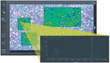
5 What Can Raman Imaging Do? Extends the advantages of Raman analysis across the sample Rapid collection of vast amounts of spectroscopic data Provides visual images depicting differences in molecular structure and chemical environment Raman images provide views of the samples that are not always apparent in the visual images 5

6 Application Areas for Raman Imaging Pharmaceuticals Other Application Areas Polymers and Packaging Semiconductors and Thin Films Carbon Nanomaterials Geology / Mineralogy Life Sciences 6
7 Introducing the Thermo Scientific DXRxi Raman Imaging Microscope A total imaging system: hardware and software integration combines powerful performance with image-centric analysis and ease of use A completely new approach to Raman imaging! 7


8 8 Intelligent Workflow with Excellent Flexibility

9 No Raman Expertise Required to get the Best Results Visual controls and instantaneous, continuous visual feedback NO lengthy trial and error NO guesswork You can see when parameters are optimal Focus quickly on the problem, not the technique 9


10 Raman Image Preview Just like using an visual image to inspect the sample now it is possible to use a Raman image preview of the sample. Don t waste time guessing at your region of interest Don t wait until an image is collected to learn if parameters were ideal Rapidly see and identify constituents and domains without the wait Preview in seconds! 10


11 Get There Faster By Getting Just What You Need Optimize image collection, not individual spectra Quickly and visually balance image collection time with necessary detail level Remove unanticipated results somewhere in an image Stop any time if results are good enough rather than wait for multiple scans of each point in entire image to finish, one at a time 20 micron image pixels 1900 spectra, 2 scans 1 minute 5 micron image pixels 30,000 spectra, 2 scans 4 minutes 1 micron image pixels 191,000 spectra, 2 scans 25 minutes 11
; 500 Hz data collection; 10 co-adds DXRxi, rastering entire images to desired quality level, like other")
12 Image-centric vs. spectral-centric at same spectral speed 100 x 100 microns 1 micron spacing (10,000 spectra); 500 Hz data collection; 10 co-adds DXRxi, rastering entire images to desired quality level, like other microscopes 20 seconds 1 minute 200 seconds Single scan of entire image with MCR, 10,000 spectra 3 scans of entire image with MCR 10 scans of entire image with MCR Some Time Later. Other Raman Imaging Systems, building images one spectrum at a time 1000 co-added spectra, no useful image information 3000 co-added spectra, no useful image information 10,000 spectra collected MCR Processed Map 12
 applied")

13 Built-In Expertise: Profiles and MCR (Multivariate Curve Resolution) Standard profiles (correlation, chemigram, peak area, peak height, peak ratios, peak shift) applied immediately via graphical interface Component analysis calculated in real-time One-click application of image profiles is a unique concept adds value at every step of the workflow 13

14 Data Processing Concurrent With Data Collection Raman images with component identification are created in real time Without configuring a spectroscopic method Without prior operator knowledge of what s in the sample Without waiting for an entire image data set to be collected Instant and obvious interpretation even if you don t know what you re looking for 14

15 Integration and Cross-Compatibility Access to raw data All data (chemical image, video image, spectra) can be quickly exported using a full array of formats HDF5 provides open-source solution for compatibility with third party packages Send data to OMNIC and Specta with a single click! 15


16 Microscopy Options Supports a wide variety of sample measurement options, including: Single and dual microscope slide holders Heating and cooling stages even during imaging! Rotating stage insert Industry standard wafer holders and SEM accessories Breadboard -style holder for custom configurations Holder for the Thermo Scientific K-Alpha XPS! Integrated Olympus research grade optics for peak performance and stability: High NA and long working distance objectives Optional brightfield and darkfield optics DIC and visual polarizers for more challenging samples Available with transmission illumination 16


17 Pharmaceutical Formulations Typically complex multi-component mixtures Need to identify and verify components Known components Impurities Identify changes in components during processing Distribution of components Homogeneity Particle size Content uniformity 17
 2500 µm Video Mosaic Image (10X objective, 100X total magnification) Inactive corn starch, microcrystalline cellulose, sodium lauryl sulfate, sodium starch, glycolate, crospovidone, polyethylene")
18 Tablet Imaging Example Migraine Relief Tablet 11 mm diameter, 676 mg APIs Acetaminophen 250 mg (37%) Aspirin 250 mg (37%) Caffeine 65 mg (9.6 %) 2500 µm Video Mosaic Image (10X objective, 100X total magnification) Inactive corn starch, microcrystalline cellulose, sodium lauryl sulfate, sodium starch, glycolate, crospovidone, polyethylene glycol, polyvinyl alcohol, povidone, stearic acid, talc, titanium dioxide 18
 532 nm laser, 8 minute collect time!")
19 Imaging the Whole Tablet Raman MCR Image Area Imaged - 11 x 11 mm 2 10X objective Image Pixel Size - 25 µm 226,000 spectra Exposure Time 1.8 ms (550 spectra per s) 532 nm laser, 8 minute collect time!! Aspirin Acetaminophen Caffeine Titanium Dioxide 19
 532 nm laser 36 GB file Size only computer limited (128 GB RAM) 6 hour collection time (3 hr estimated) Image Analysis % Area of Particles Component Calculated % (Surface")
20 Higher Resolution Image Whole Tablet Area Imaged - 11 x 11 mm 2 10X objective Image Pixel Size - 5 µm 5.4 million spectra Exposure Time 1.8 ms (550 spectra per s) 532 nm laser 36 GB file Size only computer limited (128 GB RAM) 6 hour collection time (3 hr estimated) Image Analysis % Area of Particles Component Calculated % (Surface Area) Reported % Aspirin Acetaminophen Caffeine Aspirin Acetaminophen Caffeine Titanium Dioxide 20


21 Tablets Components From Multivariate Curve Resolution (MCR) Aspirin Titanium Dioxide Acetaminophen Caffeine 21


22 Identify Area of Interest Longer Exposure Time 1.6 x 1.7 mm 2 50X Objective 5 micron image pixel size spectra Exposure time 5 ms (200 spectra per s) 532 nm laser 5 averaged scans, 55 minutes 500 µm Blue Caffeine, Green - Acetaminophen, Yellow Aspirin, and Red starch 22


23 Define Area Further, Higher Resolution, Longer Exposure Time 500 µm 225 x 250 µm 2 100X Objective 0.5 micron image pixel size spectra Exposure time 10 ms (100 spectra per s) 532 nm laser 5 averaged scans, 3 hr collect 100 µm Blue Aspirin, Green Acetaminophen, Yellow Caffiene, Red starch, Fuchsia microcrystalline cellulose, Orange sodium lauryl sulfate. 23
24 Summary of Tablet Imaging Possible to Image an entire 11 mm diameter tablet in 8 minutes Higher resolution images on whole tablets are possible but may not be necessary and there are other alternatives to imaging the whole tablet (select regions, multiple regions) Raman imaging can give spatial distribution of components including particle size estimates and relative percentages based on areas occupied by different components APIs tend to be strong Raman scatterers. Weaker excipients may require longer exposure times and possibly better spatial resolution to differentiate them. 24
 Video Mosaic Image (10X objective, 100X total magnification) Tibolone")
25 Low Dose Tablet Example Tibolone Tablet 3% Tibolone A synthetic steroid used in hormone replacement therapy 6 mm diameter Polymorphs (monoclinic, triclinic) Video Mosaic Image (10X objective, 100X total magnification) Tibolone 25
 532 nm laser 10 averaged scans 3 hr collect")
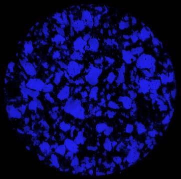
26 Raman MCR Image of Low Dose Tablet Raman MCR Image 5.7 x 5.7 mm 2 area 10X objective 25 µm image pixel size spectra Exposure Time 20 ms (50 spectra per s) 532 nm laser 10 averaged scans 3 hr collect Tibolone was not readily differentiated using MCR Starch Lactose Fluorescent Compound 26

27 A Peak Height Profile Readily Shows the Location of the Tibolone Peak Height Analysis at 2102 cm -1 Bright Spots are Tibolone 27
 532 nm laser 25 averaged scans 10 hr collect Tibolone is one")
28 Defined Area of Interest, Higher Resolution, More Scans 1.1 x 1.6 mm 2 area 10X objective 5 µm image pixel size spectra Exposure Time 20 ms (50 spectra / s) 532 nm laser 25 averaged scans 10 hr collect Tibolone is one of the components defined by the MCR analysis Tibolone Starch & Lactose Fluorescent Compound 28

")

29 Distribution of Polymorphs of Tibolone All Tibolone (peak at 2102 cm -1 ) 3273, 3254 cm cm -1 B (monoclinic) C (triclinic) 29
30 Summary of Low Dose Tablet Imaging Lower dose tablets are commonly encounter pharmaceutical products Other profile options (peak height etc.) might be better choices than MCR for displaying the spatial distribution of low concentration components. Multiple options are available in the OMNICxi software for generating different types of Raman images based on different aspects of the spectra. Whole tablet imaging is possible Evaluation of the presence and distribution of polymorphs is possible. 30
.")
31 Raman Imaging of Hot Melt Extruder (HME) Samples Hot melt extrusion is a novel way of formulating solid dosage pharmaceutical products (tablets, granules, pellets, and transdermal films) Has been used extensively for a long time in the plastics industry API and other components are combined with a pharmaceutically approved thermoplastic polymer (usually at higher temperatures). Screw threads control the mixing and transport properties at various stages. Final form depends on the die and post extruder processing. 31
32 HME General Processing Advantages Continuous process inline monitoring and control Establish stable solid solutions Increase the availability of poorly soluble ingredients Flexibility to easily produce different dosage products Availability of time release forms Taste masking Special dosage form designs (films, rods, etc ) Die change provides different shapes for special applications Reduce the consumption of solvents Compared to wet granulation process 32
33 Raman Imaging of HME Products Hot melt extrusion produces new forms using new processes Processing can effect the components Monitoring components Changes in molecular structure Spatial distribution and particle size of components Identification of unknowns (impurities and defects) 33
.")
34 Example of Raman Imaging of HME Products* HPMCAS (hydroxypropyl methyl cellulose acetate succinate or hypromellose acetate succinate) polymer carrier Ibuprofen (25-33%) and ibuprofen (25-33%) + D-mannitol (7-15%). Cross-sections mounted in epoxy for analysis Video Mosaic Image (10X objective, 100X total magnification) Darkfield Illumination To evaluate the spatial distribution of components and to look for any unforeseen changes caused by processing conditions * HME Samples Provided by Dr. Adrian Kelly, School of Engineering, Design and Technology, Bradford University, UK 34
 780 nm laser 100 averaged scans Image Analysis % Area of Particles Component Calculated")
35 Raman Image of HME Product 6.35 x 4.6 mm 2 area 10X objective 25 µm image pixel size spectra Exposure Time 10 ms (100 spectra /s) 780 nm laser 100 averaged scans Image Analysis % Area of Particles Component Calculated % (Surface Area) Reported % Raman MCR Image purple HPMCAS, green ibuprofen, yellow epoxy, red cyanoacrylate, blue inorganic impurity ibuprofen

36 Smaller Area, Higher Resolution, Raman Imaging Video Image (50X objective - brightfield illumination) Raman MCR Image blue HPMCAS, green ibuprofen, yellow ibuprofen 50X objective, 780 nm laser, 24 mw, 687 x 423 µm area, 3.0 µm image pixel size, spectra, s exposure time, 100 scans 36
37 Subtle Differences in Ibuprofen Spectra Raman Spectrum of Ibuprofen from Green Area Raman Spectrum of Ibuprofen from Yellow Area Library Spectrum of Ibuprofen 37
38 Differences in the Raman Spectra of Ibuprofen Ibuprofen is a mixture of stereoisomers Not distinguishable with this type of experiment (ROA required) The active form is S (+) ibuprofen Different polymorphs of ibuprofen have been reported Phase I (thermodynamically stable) & Phase II (metastable) Degree of crystallinity effects the Raman spectra Crystallinity versus amorphous Co-crystallization of ibuprofen with other components can alter the Raman spectrum Ibuprofen association with other carriers (polyvinyl pyrrolidone (PVP) can cause slight differences in Raman spectra The observed differences in the ibuprofen spectra in these products does not match with any of these effects However it does illustrate how sensitive Raman imaging can be 38


39 Raman Imaging From Whole Samples to Small Particles Millimeters to Microns 39


40 Hot Melt Extruder Product Ibuprofen & D-Mannitol Raman MCR Image blue - HPMCAS, green ibuprofen, orange mannitol, yellow epoxy, fuschia cyanoacrylate 10X objective, 780 nm laser, 24 mw, 6.7 x 4.7 mm area, 25 µm image pixel size, spectra, s exposure time, 100 scans 50X objective, 780 nm laser, 24 mw, 600 x 580 µm area, 5 µm image pixel size, spectra, s exposure time, 100 scans 40
41 Summary of Raman Imaging of Hot Melt Extruder Products Raman imaging allows evaluation of the spatial distribution of components in hot melt extruder products. This is not generally available with the typical inline monitoring of the products. Raman can be used to monitor any changes in molecular structure and chemical environment including molecular associations that might be induced during the HME processing. There are many options for Raman imaging from imaging whole samples down to small particles (millimeters to microns). This is important for these types of samples where there can be a significant range in particle sizes. 41


42 Let the Power of Raman Imaging Work for You Raman imaging is clearly a very useful analytical tool for evaluating pharmaceutical products Raman imaging extends the power of Raman spectroscopy across greater areas and further expands the utility of Raman spectroscopy Raman imaging gives you the ability to identify materials and to assess subtle differences in molecular structure and chemical environment The DXRxi Raman imaging microscope will help you: Get results and solve problems quickly with exceptional performance Allow more people to solve problems without the need for a central expert Walk up and use the system within minutes, anytime, without a significant learning curve 42


43 Thank you! Thank you for attending! Visit thermoscientific.com/dxrxi for more information on the Thermo Scientific DXRxi Raman imaging microscope 43
Approachable Raman Solutions The Shortest Path from Problem to Answer
 Approachable Raman Solutions The Shortest Path from Problem to Answer Michael S. Bradley The world leader in serving science Thermo Scientific Raman Spectroscopy: Discover. Solve. Assure. Raman Spectroscopy
Agilent 8700 LDIR Chemical Imaging System. Bringing Clarity and Unprecedented Speed to Chemical Imaging.
 Agilent 8700 LDIR Chemical Imaging System Bringing Clarity and Unprecedented Speed to Chemical Imaging. What if you could save time and achieve better results? The Agilent 8700 Laser Direct Infrared (LDIR)
Thermo Scientific DXRxi Raman Imaging Microscope. Accelerate your work Visualize your answers
 Thermo Scientific DXRxi Raman Imaging Microscope Accelerate your work Visualize your answers Raman imaging evolved Walk-up-and-run ease of use Visually driven image acquisition A microscopy-first approach
Chemical Imaging. Whiskbroom Imaging. Staring Imaging. Pushbroom Imaging. Whiskbroom. Staring. Pushbroom
 Chemical Imaging Whiskbroom Chemical Imaging (CI) combines different technologies like optical microscopy, digital imaging and molecular spectroscopy in combination with multivariate data analysis methods.
Fast Laser Raman Microscope RAMAN
 Fast Laser Raman Microscope RAMAN - 11 www.nanophoton.jp Fast Raman Imaging A New Generation of Raman Microscope RAMAN-11 developed by Nanophoton was created by combining confocal laser microscope technology
Fast Laser Raman Microscope RAMAN
 Fast Laser Raman Microscope RAMAN - 11 www.nanophoton.jp Fast Raman Imaging A New Generation of Raman Microscope RAMAN-11 developed by Nanophoton was created by combining confocal laser microscope technology
Fastest high definition Raman imaging. Fastest Laser Raman Microscope RAMAN
 Fastest high definition Raman imaging Fastest Laser Raman Microscope RAMAN - 11 www.nanophoton.jp Observation A New Generation in Raman Observation RAMAN-11 developed by Nanophoton was newly created by
Characteristics and Applications of Imaging Microscope FT-IR. Teo Wei Boon PerkinElmer Singapore
 Characteristics and Applications of Imaging Microscope FT-IR Teo Wei Boon PerkinElmer Singapore Infrared (IR) Spectroscopy. IR Spectroscopy measures the radiation absorbed by materials due to molecular
Spectrum 400. FT-IR and FT-NIR Spectrometer. There is only one answer.
 Spectrum 400 FT-IR and FT-NIR Spectrometer There is only one answer. The latest innovation in PerkinElmer s long history of IR technology leadership For over 60 years, PerkinElmer has been the world leader
SENTERRA II. Innovation with Integrity. The Next Level of Compact Raman Microscopy. Raman
 SENTERRA II The Next Level of Compact Raman Microscopy Innovation with Integrity Raman Research-grade spectroscopic performance Next Level Compact Raman Microscopy The SENTERRA II defines a new level of
SENTERRA II. Innovation with Integrity. The Next Level of Compact Raman Microscopy. Raman
 SENTERRA II The Next Level of Compact Raman Microscopy Innovation with Integrity Raman Next Level Compact Raman Microscopy The SENTERRA II defines a new level of spectroscopic performance and user friendliness
Spectroscopy Application: Using Raman Spectroscopy to Detect Art Forgeries Detecting art forgeries using Raman spectroscopy
 Spectroscopy Application: Using Raman Spectroscopy to Detect Art Forgeries 5 Detecting art forgeries using Raman spectroscopy In the previous experiment, you were introduced to Raman spectroscopy. This
Get the full picture of your sample. Applications
 Follow the Experts Get the full picture of your sample The new generation of confocal Raman microscopes offers a non-destructive and non-contact method of sample analysis at the sub-micron level. More
FT-IR.
 FT-IR varian, inc. 610/620-IR ft-ir MICROSCOPY AND IMAGING SoLUTIONS www.varianinc.com VARIAN, INC. Setting the Standard Again When Only the Best Will Do The world leader in molecular spectroscopy innovation
Infrared Microscope AIM-9000 C103-E103B
 Infrared Microscope C103-E103B Wide-Field Camera Automatic Recognition Automatic Qualitative High Sensitivity Finally, a wide view on micro sample analysis Surface Micro SHIMADZU AIMs to provide analysis
Introduction. Methods: Spherical Granulation. Shawn Engels, Vector Corporation
 DISCUSSION OF PROCESSES WHICH UTILIZE CONICAL ROTOR TECHNOLOGY (SPHERONIZATION OR SPHERICAL GRANULATION, POWDER LAYERING OF ACTIVES OR POLYMERS, CONVENTIONAL SOLUTION/SUSPENSION APPLICATION OF ACTIVES
Infrared Microscope. Dedicated AIMsolution Software. Hisato Fukuda. 1. Introduction. 2. Automatic Contaminant Recognition Function
 C103-E120 Vol. 28 Infrared Microscope Dedicated AIMsolution Software ------- 02 Infrared Microscope Using Imaging Analysis ------- 05 EDXIR-Analysis EDX-FTIR Contaminant Finder/Material Inspector -------
Development and Applications of a Sample Compartment FTIR Microscope
 Application Note Development and Applications of a Sample Since the early to mid-1940 s, scientists using infrared spectroscopy have been trying to obtain spectral data from ever smaller samples. Starting
Philippa Eshun. Lincoln Leadership Academy Charter School (Allentown, PA)
") Outreach Program: Plastics are all around us. Support: NSF Polymer Program NSF-1308617 (PI: Chang Ryu)/RPI Polymer Center Understanding FTIR and Polymer Plastics Philippa Eshun Lincoln Leadership Academy
Outreach Program: Plastics are all around us. Support: NSF Polymer Program NSF-1308617 (PI: Chang Ryu)/RPI Polymer Center Understanding FTIR and Polymer Plastics Philippa Eshun Lincoln Leadership Academy
Measurement of Microscopic Three-dimensional Profiles with High Accuracy and Simple Operation
 238 Hitachi Review Vol. 65 (2016), No. 7 Featured Articles Measurement of Microscopic Three-dimensional Profiles with High Accuracy and Simple Operation AFM5500M Scanning Probe Microscope Satoshi Hasumura
Agilent Cary 610/620 FTIR microscopes and imaging systems RESOLUTION FOR EVERY APPLICATION
 Agilent Cary 610/620 FTIR microscopes and imaging systems RESOLUTION FOR EVERY APPLICATION AGILENT CARY 610/620 FTIR MICROSCOPES ADVANCING FTIR MICROSCOPY AND IMAGING Agilent s 610/620 FTIR microscopes
Improving the Collection Efficiency of Raman Scattering
 PERFORMANCE Unparalleled signal-to-noise ratio with diffraction-limited spectral and imaging resolution Deep-cooled CCD with excelon sensor technology Aberration-free optical design for uniform high resolution
Raman Spectroscopy of TiO2
 Raman Spectroscopy of TiO2 Experiment #4 Characterization of Materials (96.445/545) Meg Noah Meg Noah 1 of 8 10/21/2010 Objective The purpose of this laboratory is to identify two different phases (rutile
Thermo Scientific Nicolet in10 FT-IR Microscope. Infrared microscopy. with confidence. intuitive innovative integrated
 Thermo Scientific Nicolet in10 FT-IR Microscope Infrared microscopy with confidence intuitive innovative integrated Thermo Scientific Nicolet in10 FT-IR Microscope The Infrared Microscope that Delivers
WHITE PAPER MINIATURIZED HYPERSPECTRAL CAMERA FOR THE INFRARED MOLECULAR FINGERPRINT REGION
 WHITE PAPER MINIATURIZED HYPERSPECTRAL CAMERA FOR THE INFRARED MOLECULAR FINGERPRINT REGION Denis Dufour, David Béland, Hélène Spisser, Loïc Le Noc, Francis Picard, Patrice Topart January 2018 Low-cost
Foreign Particulate Matter testing using the Morphologi G3
 Foreign Particulate Matter testing using the Morphologi G3 Introduction The Morphologi G3 with its Foreign Particle Detection capabilities allows the detection, enumeration and size classification of foreign
NOVEL APPLICATIONS OF CONFOCAL MICROSCOPY TECHNIQUES IN COATINGS RESEARCH
 ARKEMA COATING RESINS NOVEL APPLICATIONS OF CONFOCAL MICROSCOPY TECHNIQUES IN COATINGS RESEARCH DOUG MALL FOR DR. WENJUN WU 9/20/2018 Wood Coatings & Substrates Conference 2018 OUTLINE Introduction Confocal
Revisions to ASTM D7310 Standard Guide for Defect Detection and Rating of Plastic Films Using Optical Sensors
 Revisions to ASTM D7310 Standard Guide for Defect Detection and Rating of Plastic Films Using Optical Sensors ANTEC 2017 Brenda Colegrove, The Dow Chemical Company Richard Garner, Borealis Dow.com SPE
FTIR Microscopy and Imaging
 FTIR Microscopy and Imaging A world of applications and solutions Dr. Mustafa Kansiz FTIR Imaging and Microscopy Product Manager Agilent Technologies Email: mustafa.kansiz@agilent.com FTIR Spectroscopy
November 17, Mr. Jeffrey Fuller Fuller Fine Art Auctions 730 Carpenter Lane Philadelphia, PA Dear Mr. Lawrence:
 November 17, 2010 Mr. Jeffrey Fuller Fuller Fine Art Auctions 730 Carpenter Lane Philadelphia, PA 19119 Dear Mr. Lawrence: This report summarizes the analysis of two paintings by victor Manuel. You asked
Color More than meets the Eye
 TOPICS Color More than meets the Eye Anna Kreofsky Color R&D Engineer Brief introduction to RTP Company Color Division Color Fundamentals Three Sciences of Color Colorant Types & Limitations Evaluation
Material analysis by infrared mapping: A case study using a multilayer
 Material analysis by infrared mapping: A case study using a multilayer paint sample Application Note Author Dr. Jonah Kirkwood, Dr. John Wilson and Dr. Mustafa Kansiz Agilent Technologies, Inc. Introduction
Nanonics Systems are the Only SPMs that Allow for On-line Integration with Standard MicroRaman Geometries
 Nanonics Systems are the Only SPMs that Allow for On-line Integration with Standard MicroRaman Geometries 2002 Photonics Circle of Excellence Award PLC Ltd, England, a premier provider of Raman microspectral
Camera Overview. Digital Microscope Cameras for Material Science: Clear Images, Precise Analysis. Digital Cameras for Microscopy
 Digital Cameras for Microscopy Camera Overview For Materials Science Microscopes Digital Microscope Cameras for Material Science: Clear Images, Precise Analysis Passionate about Imaging: Olympus Digital
OLYMPUS Digital Cameras for Materials Science Applications: Get the Best out of Your Microscope
 Digital Cameras for Microscopy Camera Overview For Materials Science Microscopes OLYMPUS Digital Cameras for Materials Science Applications: Get the Best out of Your Microscope Passionate About Imaging
Compare and Contrast. Contrast Methods in Industrial Inspection Microscopy. Application Note. We explain how to
 Application Note Compare and Contrast Contrast Methods in Industrial Inspection Microscopy We explain how to E nhance materials inspection microscopy workflows Reveal surface and sub-surface imperfections
BaySpec SuperGamut OEM
 BaySpec SuperGamut OEM Spectrographs & Spectrometers RUGGED SOLID STATE HIGH RESOLUTION OPTIMIZED COOLING COST EFFECTIVE HIGH THROUGHPUT www.bayspec.com Specifications Model UV-NIR VIS-NIR NIR 900-1700nm
Spotlight 150 and 200 FT-IR Microscopy Systems
 S P E C I F I C A T I O N S Spotlight 150 and 200 FT-IR Microscopy Systems FT-IR Microscopy Spotlight 200 with Frontier FT-IR Spectrometer Introduction PerkinElmer Spotlight FT-IR Microscopy Systems are
Aqualog. Water Quality Measurements Made Easy PARTICLE CHARACTERIZATION ELEMENTAL ANALYSIS FLUORESCENCE
 Aqualog Water Quality Measurements Made Easy ELEMENTAL ANALYSIS FLUORESCENCE GRATINGS & OEM SPECTROMETERS OPTICAL COMPONENTS PARTICLE CHARACTERIZATION RAMAN SPECTROSCOPIC ELLIPSOMETRY SPR IMAGING Water
Aqualog. CDOM Measurements Made Easy PARTICLE CHARACTERIZATION ELEMENTAL ANALYSIS FLUORESCENCE GRATINGS & OEM SPECTROMETERS OPTICAL COMPONENTS RAMAN
 Aqualog CDOM Measurements Made Easy ELEMENTAL ANALYSIS FLUORESCENCE GRATINGS & OEM SPECTROMETERS OPTICAL COMPONENTS PARTICLE CHARACTERIZATION RAMAN SPECTROSCOPIC ELLIPSOMETRY SPR IMAGING CDOM measurements
Advancing FT-IR Imaging Synchrotron IR Imaging in Your Lab. Dr. Mustafa Kansiz FTIR Microscopy & Imaging Product Manager
 Advancing FT-IR Imaging Synchrotron IR Imaging in Your Lab Dr. Mustafa Kansiz FTIR Microscopy & Imaging Product Manager How can FTIR microscopy imaging help me? An FTIR microscope has two essential purposes:
Camera Overview. Digital Microscope Cameras for Material Science: Clear Images, Precise Analysis. Digital Cameras for Microscopy
 Digital Cameras for Microscopy Camera Overview For Materials Science Microscopes Digital Microscope Cameras for Material Science: Clear Images, Precise Analysis Passionate about Imaging: Olympus Digital
Nicolet Continuμm Infrared Microscope
 m o l e c u l a r s p e c t r o s c o p y Nicolet Continuμm Infrared Microscope Infrared Microscopy Excellence Crystal-clear Visible Microscopy Superior Purity Infrared Spectroscopy New Dimensions in Imaging
FTIR microscopy and imaging for failure analysis in electronics manufacturing
 FTIR microscopy and imaging for failure analysis in electronics manufacturing Application Note Author Steven M. Barnett, Ellen V. Miseo, and Wayne Jalenak Agilent Technologies, Inc. Introduction The electronics
A BASIC EXPERIMENTAL STUDY OF CAST FILM EXTRUSION PROCESS FOR FABRICATION OF PLASTIC MICROLENS ARRAY DEVICE
 A BASIC EXPERIMENTAL STUDY OF CAST FILM EXTRUSION PROCESS FOR FABRICATION OF PLASTIC MICROLENS ARRAY DEVICE Chih-Yuan Chang and Yi-Min Hsieh and Xuan-Hao Hsu Department of Mold and Die Engineering, National
Thermo Scientific Nicolet in10 FT-IR Microscope
 m o l e c u l a r s p e c t r o s c o p y Thermo Scientific Nicolet in10 FT-IR Microscope Infrared Microscopy with Confidence intuitive innovative integrated Part of Thermo Fisher Scientific Thermo Scientific
Add CLUE to your SEM. High-efficiency CL signal-collection. Designed for your SEM and application. Maintains original SEM functionality
 Add CLUE to your SEM Designed for your SEM and application The CLUE family offers dedicated CL systems for imaging and spectroscopic analysis suitable for most SEMs. In addition, when combined with other
Low Voltage Electron Microscope
 LVEM5 Low Voltage Electron Microscope Nanoscale from your benchtop LVEM5 Delong America DELONG INSTRUMENTS COMPACT BUT POWERFUL The LVEM5 is designed to excel across a broad range of applications in material
Bringing Answers to the Surface
 3D Bringing Answers to the Surface 1 Expanding the Boundaries of Laser Microscopy Measurements and images you can count on. Every time. LEXT OLS4100 Widely used in quality control, research, and development
NanoSpective, Inc Progress Drive Suite 137 Orlando, Florida
 TEM Techniques Summary The TEM is an analytical instrument in which a thin membrane (typically < 100nm) is placed in the path of an energetic and highly coherent beam of electrons. Typical operating voltages
FT-IR IMAGING THAT'S CLEARLY MEASURABLY AMAZING. Spotlight 400 FT-IR and 400N FT-NIR Imaging Systems
 FT-IR IMAGING THAT'S CLEARLY MEASURABLY AMAZING Spotlight 400 FT-IR and 400N FT-NIR Imaging Systems YOUR CHALLENGES COME IN ALL SHAPES AND SIZES ONE SYSTEM CAN HANDLE THEM ALL It s been called the most
Microscopic Structures
 Microscopic Structures Image Analysis Metal, 3D Image (Red-Green) The microscopic methods range from dark field / bright field microscopy through polarisation- and inverse microscopy to techniques like
PLASTICS WORKSHOP ENGINEERED. Learn About Thermoplastics Connect with Experts KING OF PRUSSIA / PENNSYLVANIA (PHILADELPHIA AREA)
") ENGINEERED PLASTICS WORKSHOP Learn About Thermoplastics Connect with Experts 2017 KING OF PRUSSIA / PENNSYLVANIA (PHILADELPHIA AREA) YOUR GLOBAL COMPOUNDER OF CUSTOM ENGINEERED THERMOPLASTICS Light and
ENGINEERED PLASTICS WORKSHOP Learn About Thermoplastics Connect with Experts 2017 KING OF PRUSSIA / PENNSYLVANIA (PHILADELPHIA AREA) YOUR GLOBAL COMPOUNDER OF CUSTOM ENGINEERED THERMOPLASTICS Light and
Proudly serving laboratories worldwide since 1979 SPECIFICATIONS
 www.ietltd.com Proudly serving laboratories worldwide since 1979 SPECIFICATIONS Scan RDI Specifications System Components Main analytical console Laser Module CRT Printer Data Manager Motorized stage (option)
Camera Overview. Olympus Digital Cameras for Materials Science Applications: For Clear and Precise Image Analysis. Digital Cameras for Microscopy
 Digital Cameras for Microscopy Camera Overview For Materials Science Microscopes Olympus Digital Cameras for Materials Science Applications: For Clear and Precise Image Analysis Passionate about Imaging
More Detail. Faster. Easier. The results will inspire you. Spotlight 400
 More Detail. Faster. Easier. The results will inspire you. Spotlight 400 FT-IR and 400N FT-NIR Imaging Systems Raising the Level of Lab Productivity to an Art From SPOTLIGHT 400 FT-IR AND 400N FT-NIR IMAGING
Leading in Desktop SEM Imaging and Analysis
 Leading in Desktop SEM Imaging and Analysis Fast. Outstanding. Reliable SEM imaging and analysis. The Phenom: World s Fastest Scanning Electron Microscope With its market-leading Phenom desktop Scanning
Dual-FL. World's Fastest Fluorometer. Measure absorbance spectra and fluorescence simultaneously FLUORESCENCE
 Dual-FL World's Fastest Fluorometer Measure absorbance spectra and fluorescence simultaneously FLUORESCENCE 100 Times Faster Data Collection The only simultaneous absorbance and fluorescence system available
Nicolet Almega XR. High Performance Micro and Macro Dispersive Raman Analysis Systems
 m o l e c u l a r s p e c t r o s c o p y Nicolet Almega XR High Performance Micro and Macro Dispersive Raman Analysis Systems Materials Research Identification/Verification Characterization Failure Analysis
INFRARED ANALYSIS OF SINGLE AND MULTILAYER FILMS IN THE PRODUCTION AREA
 INFRARED ANALYSIS OF SINGLE AND MULTILAYER FILMS IN THE PRODUCTION AREA Sandy Rintoul Wilks Enterprise, Inc. South Norwalk, CT Scott Cobranchi Sealed Air Corporation Duncan, SC Nina Tani Sealed Air Corporation
Characterisation of Photovoltaic Materials and Cells
 Standard Measurement Services and Prices No. Measurement Description Reference 1 Large area, 0.35-sun biased spectral response (SR) 2 Determination of linearity of spectral response with respect to irradiance
DMS-8220, Hot Applied Thermoplastic
 Overview Effective Date: August 2004 August 2007 (refer to 'Archived Versions' for previous versions). This Specification governs for the materials, composition, quality, sampling, and testing of thermoplastic
Dynamic Phase-Shifting Microscopy Tracks Living Cells
 from photonics.com: 04/01/2012 http://www.photonics.com/article.aspx?aid=50654 Dynamic Phase-Shifting Microscopy Tracks Living Cells Dr. Katherine Creath, Goldie Goldstein and Mike Zecchino, 4D Technology
Characterization Microscope Nikon LV150
 Characterization Microscope Nikon LV150 Figure 1: Microscope Nikon LV150 Introduction This upright optical microscope is designed for investigating up to 150 mm (6 inch) semiconductor wafers but can also
PTG NIR. Powder Analysis System
 PTG NIR Powder Analysis System The automated PTG-S4 stand-alone powder characterization system is used to measure the flow behavior of granules and powders in compliance with the EP and USP
Advanced Research Raman System Raman Spectroscopy Systems
 T600 Advanced Research Raman System Raman Spectroscopy Systems T600 Advanced Research Raman System T600 Triple stage Raman Spectrometer: The only solution for unprecedented stability and performance! Robust
SUPPLEMENTAL MATERIAL
 SUPPLEMENTAL MATERIAL 1 - Folios and areas of analysis Figure S1.1. Folio 4, areas of analysis for microxrf ( ), FORS ( ), micro-samples for Raman and FTIR ( ) and Raman in-situ ( ). Figure S1.2. Folio
Applications of Steady-state Multichannel Spectroscopy in the Visible and NIR Spectral Region
 Feature Article JY Division I nformation Optical Spectroscopy Applications of Steady-state Multichannel Spectroscopy in the Visible and NIR Spectral Region Raymond Pini, Salvatore Atzeni Abstract Multichannel
Parameter Selection and Spectral Optimization Using the RamanStation 400
 Parameter Selection and Spectral Optimization Using the RamanStation 400 RAMAN SPECTROSCOPY A P P L I C A T I O N N O T E In modern dispersive Raman spectroscopy, good quality spectra can be obtained from
Aqualog. Water Quality Measurements Made Easy FLUORESCENCE
 Aqualog Water Quality Measurements Made Easy FLUORESCENCE Water quality measurements made easy The only simultaneous absorbance and fluorescence system for water quality analysis! The new Aqualog is the
Feedback EMEA / Industry Discussion
 Feedback EMEA / Industry Discussion Eli Lilly & Co Ltd Case Study: Use of In-Line Near-Infrared Spectroscopy to Monitor Segregation of a Pharmaceutical Powder Blend in a Tablet Press Martin Diller PhD,
You won t be able to measure the incident power precisely. The readout of the power would be lower than the real incident power.
 1. a) Given the transfer function of a detector (below), label and describe these terms: i. dynamic range ii. linear dynamic range iii. sensitivity iv. responsivity b) Imagine you are using an optical
Chemical imaging of pharmaceutical granules by Raman global illumination and near-infrared mapping platforms
 analytica chimica acta 611 (2008) 73 79 available at www.sciencedirect.com journal homepage: www.elsevier.com/locate/aca Chemical imaging of pharmaceutical granules by Raman global illumination and near-infrared
Horiba LabRAM ARAMIS Raman Spectrometer Revision /28/2016 Page 1 of 11. Horiba Jobin-Yvon LabRAM Aramis - Raman Spectrometer
 Page 1 of 11 Horiba Jobin-Yvon LabRAM Aramis - Raman Spectrometer The Aramis Raman system is a software selectable multi-wavelength Raman system with mapping capabilities with a 400mm monochromator and
Diamond Analysis. Innovation with Integrity. Reliable identification and type determination by FTIR spectroscopy FTIR
 Diamond Analysis Reliable identification and type determination by FTIR spectroscopy Innovation with Integrity FTIR FTIR Diamond Analysis Since the appearance of synthetic diamonds, nearly perfect imitates
Digital Camera Technologies for Scientific Bio-Imaging. Part 2: Sampling and Signal
 Digital Camera Technologies for Scientific Bio-Imaging. Part 2: Sampling and Signal Yashvinder Sabharwal, 1 James Joubert 2 and Deepak Sharma 2 1. Solexis Advisors LLC, Austin, TX, USA 2. Photometrics
Lecture 20: Optical Tools for MEMS Imaging
 MECH 466 Microelectromechanical Systems University of Victoria Dept. of Mechanical Engineering Lecture 20: Optical Tools for MEMS Imaging 1 Overview Optical Microscopes Video Microscopes Scanning Electron
Raman images constructed from. Raman Imaging: Defining the Spatial Resolution of the Technology
 18 Raman Technology for Today s Spectroscopists June 26 Raman Imaging: Defining the Spatial Resolution of the Technology Chemical images of polystyrene beads on silicon acquired using Raman mapping and
NanoFocus Inc. Next Generation Scanning Probe Technology. Tel : Fax:
 NanoFocus Inc. Next Generation Scanning Probe Technology www.nanofocus.kr Tel : 82-2-864-3955 Fax: 82-2-864-3956 Albatross SPM is Multi functional research grade system Flexure scanner and closed-loop
Fein. High Sensitivity Microscope Camera with Advanced Software 3DCxM20-20 Megapixels
 Fein High Sensitivity Microscope Camera with Advanced Software 3DCxM20-20 Megapixels 3DCxM20 Camera Features High Sensitivity Camera This microscopy camera was designed with high sensitivity and ultra
Introduction of New Products
 Field Emission Electron Microscope JEM-3100F For evaluation of materials in the fields of nanoscience and nanomaterials science, TEM is required to provide resolution and analytical capabilities that can
Light, Color, Spectra 05/30/2006. Lecture 17 1
 What do we see? Light Our eyes can t t detect intrinsic light from objects (mostly infrared), unless they get red hot The light we see is from the sun or from artificial light When we see objects, we see
Composite Thermal Damage Measurement with Handheld FTIR. April 9, 2013 Brian D. Flinn, Ashley Tracey, and Tucker Howie University of Washington
 Composite Thermal Damage Measurement with Handheld FTIR April 9, 2013 Brian D. Flinn, Ashley Tracey, and Tucker Howie University of Washington Composite Thermal Damage Measurement with Handheld FTIR Motivation
ASM Webinar Digital Microscopy for Materials Science
 Digital Microscopy Defined The term Digital Microscopy applies to any optical platform that integrates a digital camera and software to acquire images; macroscopes, stereomicroscopes, compound microscopes
CHAPTER 2 POLARIZATION SPLITTER- ROTATOR BASED ON A DOUBLE- ETCHED DIRECTIONAL COUPLER
 CHAPTER 2 POLARIZATION SPLITTER- ROTATOR BASED ON A DOUBLE- ETCHED DIRECTIONAL COUPLER As we discussed in chapter 1, silicon photonics has received much attention in the last decade. The main reason is
Terahertz Spectroscopic/ Imaging Analysis Systems
 Terahertz Spectroscopic/ Series Non-Destructive Analysis of Pharmaceuticals, Chemicals, Communication Materials, etc. Compact, High-Speed Terahertz Spectroscopic/ High-speed measurement functionality Compact,
Observing Microorganisms through a Microscope LIGHT MICROSCOPY: This type of microscope uses visible light to observe specimens. Compound Light Micros
 PHARMACEUTICAL MICROBIOLOGY JIGAR SHAH INSTITUTE OF PHARMACY NIRMA UNIVERSITY Observing Microorganisms through a Microscope LIGHT MICROSCOPY: This type of microscope uses visible light to observe specimens.
Print microfluidic devices in minutes for as little as $1 each.
 Print microfluidic devices in minutes for as little as $1 each www.dolomite-microfluidics.com fluidic factory» overview Fluidic Factory is the world s first commercially available 3D printer for quick
Pigments Coatings renol HW 30 Pigment preparations
 Pigments Coatings renol HW 30 Pigment preparations renol Product name Colour Index Chemical Characterization Yellow GG-HW 30 Pigment Yellow 17 Disazo pigment Yellow HR-HW 30 Pigment Yellow 83 Disazo pigment
Spectroscopy of Ruby Fluorescence Physics Advanced Physics Lab - Summer 2018 Don Heiman, Northeastern University, 1/12/2018
 1 Spectroscopy of Ruby Fluorescence Physics 3600 - Advanced Physics Lab - Summer 2018 Don Heiman, Northeastern University, 1/12/2018 I. INTRODUCTION The laser was invented in May 1960 by Theodor Maiman.
3D light microscopy techniques
 3D light microscopy techniques The image of a point is a 3D feature In-focus image Out-of-focus image The image of a point is not a point Point Spread Function (PSF) 1D imaging 1 1 2! NA = 0.5! NA 2D imaging
Quick and simple installation and no maintenance needed. 3 Times More affordable Than a normal SEM. Obtaining results in less than 4 minutes
 INTRODUCTION We believe that every laboratory working in the field of nanotechnology needs an SEM, therefore we would like to introduce to you our IEM series of SEM. In short space of time, our device
Ultrafast Light-Controlled Growth of Silver Nanoparticles for Direct Plasmonic Color Printing
 Supporting Materials for Ultrafast Light-Controlled Growth of Silver Nanoparticles for Direct Plasmonic Color Printing Yangxi Zhang, Qiang Zhang, Xia Ouyang, Dang Yuan Lei, A. Ping Zhang * and Hwa-Yaw
Timescales for Change A Look at Innovation in the Pharmaceutical Industry
 Timescales for Change A Look at Innovation in the Pharmaceutical Industry 3rd FDA/PQRI Conference on Advancing Product Quality 23 Mar 2017 Robert F. Meyer, Ph.D. Global Pharmaceutical Commercialization
INTRODUCTION We believe that every laboratory working in the field of nanotechnology needs an SEM, therefore we would like to introduce to you our IEM
 INTRODUCTION We believe that every laboratory working in the field of nanotechnology needs an SEM, therefore we would like to introduce to you our IEM series of SEM. In short space of time, our device
picoemerald Tunable Two-Color ps Light Source Microscopy & Spectroscopy CARS SRS
 picoemerald Tunable Two-Color ps Light Source Microscopy & Spectroscopy CARS SRS 1 picoemerald Two Colors in One Box Microscopy and Spectroscopy with a Tunable Two-Color Source CARS and SRS microscopy
Confocal Raman Microscopy (WITec Alpha 300R)
") Confocal Raman Microscopy (WITec Alpha 300R) Please refer to Witec Alpha300R Confocal Raman Microscope User Manual for the details of the operating procedure. Sample preparation 1. Attach your sample on
Confocal Raman Microscopy (WITec Alpha 300R) Please refer to Witec Alpha300R Confocal Raman Microscope User Manual for the details of the operating procedure. Sample preparation 1. Attach your sample on
PGx11 series. Transform Limited Broadly Tunable Picosecond OPA APPLICATIONS. Available models
 PGx1 PGx3 PGx11 PT2 Transform Limited Broadly Tunable Picosecond OPA optical parametric devices employ advanced design concepts in order to produce broadly tunable picosecond pulses with nearly Fourier-transform
Correlation of Wafer Backside Defects to Photolithography Hot Spots Using Advanced Macro Inspection
 Correlation of Wafer Defects to Photolithography Hot Spots Using Advanced Macro Inspection Alan Carlson* a, Tuan Le* a a Rudolph Technologies, 4900 West 78th Street, Bloomington, MN, USA 55435; Presented
--> Buy True-PDF --> Auto-delivered in 0~10 minutes. JY/T
 Translated English of Chinese Standard: JY/T011-1996 www.chinesestandard.net Sales@ChineseStandard.net INDUSTRY STANDARD OF THE JY PEOPLE S REPUBLIC OF CHINA General rules for transmission electron microscopy