Agilent 8700 LDIR Chemical Imaging System. Bringing Clarity and Unprecedented Speed to Chemical Imaging.
|
|
|
- Clare Clarke
- 5 years ago
- Views:
Transcription


1 Agilent 8700 LDIR Chemical Imaging System Bringing Clarity and Unprecedented Speed to Chemical Imaging.
2
 chemical imaging system provides a sophisticated new approach to chemical imaging and spectral analysis.")
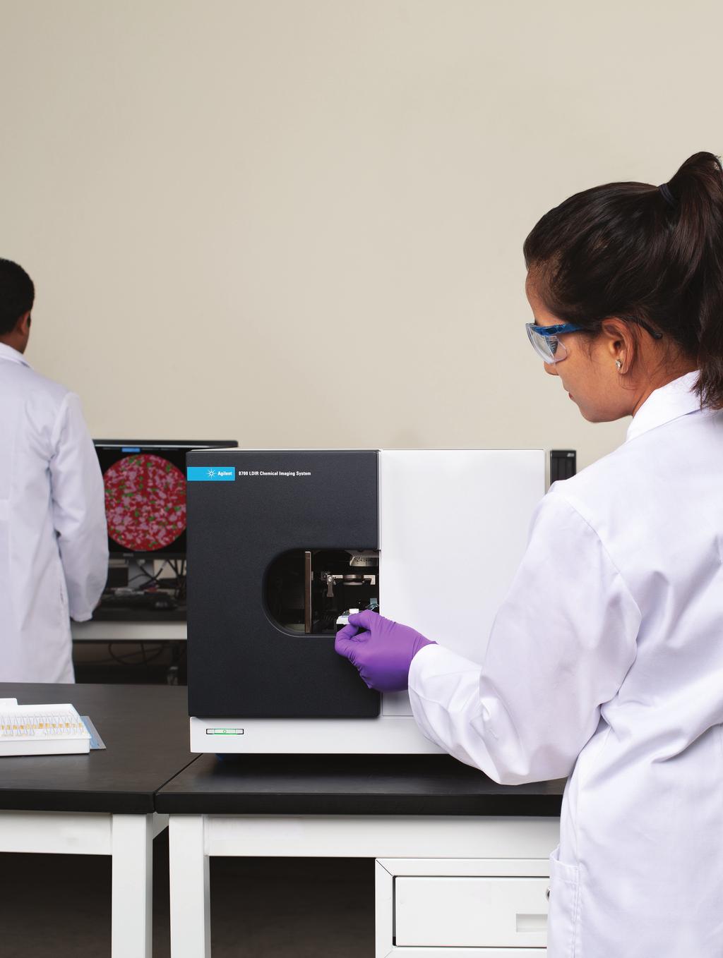
3 What if you could save time and achieve better results? The Agilent 8700 Laser Direct Infrared (LDIR) chemical imaging system provides a sophisticated new approach to chemical imaging and spectral analysis. Designed to be used by both experts and nonexperts alike, the 8700 LDIR provides a simple highly automated approach for obtaining reliable high-definition chemical images of constituents on a surface. The 8700 LDIR uses the latest Quantum Cascade Laser (QCL) technology coupled with rapidly scanning optics to provide fast, clear, high-quality images and spectral data. This technology is combined with intuitive Agilent Clarity software, for rapid and detailed imaging of large sample areas with minimal instrument interaction via a simple load and go method. Using the 8700 LDIR, you can analyze more samples, in greater detail, in less time. This robust solution provides you with more statistical data than ever to aid in the compositional analysis of tablets, laminates, tissues, polymers, and fibers. With more meaningful information available, you can make more informed, faster decisions in product development reducing both costs and analysis time. (From Left to right) Agilent Sample Planer, Agilent 8700 LDIR chemical imaging system and Agilent Clarity software analysis window 3
4 LDIR Spectroscopy How it works The 8700 LDIR works in either reflectance or Attenuated Total Reflectance (ATR) mode, automatically switching between these two modes by directing the incident beam to the appropriate objective. The movement of the sample relative to the beam is fully automated. The 8700 LDIR has two visible channels: a large field of view camera to obtain an entire view of the sample and a microscope grade objective to capture high magnification detail. Scanning Mirror Thermoelectrically Cooled MCT Detector Beam Splitter Flip Mirror Fixed ATR Objective Quantum Cascade Laser Mirror Sample Scanning Fast Scanning Objective Sample Infrared light from the QCL (shown in red) is directed to the sample. Infrared light reflected by the sample is then directed to the detector via either of the selected optical paths (shown in dark blue). Figure LDIR instrument optics In reflectance mode (solid line), infrared light from the laser is focused by the fast scanning objective system that is rapidly scanned back and forth. Concurrently, the sample is automatically moved in a perpendicular plane, and the infrared light reflected by the sample is directed back to the thermoelectrically cooled mercury cadmium telluride (MCT) detector. This process yields a high-quality two-dimensional molecular image in a remarkably short time period. In ATR mode (dashed line), infrared light from the laser is directed onto a scanning mirror that rapidly moves the light across the fixed ATR element, which is in contact with the sample. Totally internally reflected light is directed to the thermoelectrically cooled MCT detector. The key benefits Automated sample analysis. Ability to survey large sample areas and then explore smaller areas of interest in more detail without changing any optics. Full software control allows changing the field of view from microns to centimeters or the pixel size from 1 to 40 µm. Acquire ATR imaging data with pixel size as small as 0.1 μm for unmatched image detail and spectral quality. Rapidly identify unknowns using either commercial or custom libraries via ATR capabilities. Obtain relative quantitative information of sample constituents without complex method development. No requirement for liquid nitrogen reduces operating costs and simplifies maintenance. 4


5 The Agilent 8700 LDIR Chemical Imaging System handles both your routine and challenging applications The 8700 LDIR is suitable for a range of applications including pharmaceutical, material science, polymer analysis and life science research. Pharmaceutical Tablet content distribution image the spatial distribution of Active Pharmaceutical Ingredients (APIs) and excipients to ensure consistency, quality, and to aid in formulation development and troubleshooting. Investigation of factors affecting polymorphism, crystallization and salt exchange. Analysis of multi-layer tablets monitor inter- and intra-layer consistency. Analysis of single and multilayer coatings for consistency. Correlation of drug formulations (chemical and physical structure) with dissolution studies. Identification of extraneous particles and impurities. Counterfeit drug analysis create spectral and image databases of drug tablets to support anti-counterfeiting efforts. Accelerate pharmaceutical drug development In the pharmaceutical industry, time is a critical factor when releasing products. Problems that arise during drug formulation take time and resources. With the 8700 LDIR, an entire tablet can be quickly and easily analyzed, speeding up your troubleshooting process % Acetaminophen 41.46% Aspirin 9.28% Caffeine 1.78% Cellulose 1.30% Sodium Lauryl Sulfate 1.26% Starch 0.16% Hypomellose Figure 2. A high spatial resolution chemical image of a generic headache tablet consisting of three APIs (acetaminophen, aspirin and caffeine) and four excipients. All seven components were imaged across the entire tablet (11 mm diameter) with 10 micron pixel size in only 1 hour. Formulation and batch release testing are complex and critical processes. In addition, consistency is required, batch after batch. The 8700 LDIR system provides high sensitivity chemical composition analysis. With this system, you can now quickly and easily obtain qualitative and semi-quantitative information about APIs (polymorphs, salts), excipients, and impurities. 4.33% Carbamazepine form I 11.05% Carbamazepine form III 84.62% Cellulose Figure 3. Polymorph analysis Carbamazepine form I (red) form III (blue) The 13 mm tablet was analyzed in 27 minutes at 10 µm pixel size. 5

 at 1 µm pixel measured in 1 hour.")
6 Biomedical/Life science research High-quality imaging and infrared spectra of cells, tissues, cartilage, bone, and other biological materials. Rapidly survey specimens to find and then interrogate areas of interest. Analysis of biopolymer surfaces to further understand activity and support quality assurance. Find and identify defects, impurities, and extraneous particles in biopolymer matrices. Figure 4. A chemical image of a mouse brain slice showing lipid distribution (12mm x 7mm) at 1 µm pixel measured in 1 hour. Materials science/polymer analysis Packaging/laminates analysis rapidly image and determine layer identity and thickness for functional and tie (adhesive) layers, down to ~ 3 µm. Rapidly identify defects within polymers and multilayer films. Analysis of extraneous surface particles and impurities on materials including semiconductors and electronic components. Determine and identify the authenticity of components. Figure 5. A chemical image showing layers of the laminate sample (120 µm width) consisting of polyethylene (PE), polyamide (PA), poly (ethylene vinyl alcohol) (P(EVOH)), polypropylene (PP) and polyurethane (PU). The thinnest layer observed was only 2.6 µm thick. 6


")
 detectors, the 8700 LDIR employs a single-element electrically cooled detector to eliminate laser coherence")

7 Automated. Intuitive. Fast. Sample preparation and automated analysis is now accessible The instrument control and software tools of the 8700 LDIR allow both expert spectroscopists and trained technicians to analyze and characterize samples rapidly and accurately. Simply load the sample in the instrument and allow Agilent s Clarity software to reveal complex statistical data in a rapid and intuitive manner. Hours Vs Minutes Prepare your sample using Agilent Sample Planer Sample loading and automated initialization 5 minutes Automated method creation 2 minutes A whole tablet can be chemically imaged in 2-60 minutes 5 minutes 5 min 5 min 2 min minutes = Hours of time savings Breakthrough IR Technology Agilent s innovative design uses Quantum Cascade Laser (QCL) light, high spatial imaging, and intuitive Agilent Clarity software to create detailed chemical images. Unlike other QCL imaging systems that use 2D Focal Plane Array (FPA) detectors, the 8700 LDIR employs a single-element electrically cooled detector to eliminate laser coherence artifacts from images and spectra. Agilent Clarity software Redefining chemical imaging software Innovative Agilent Clarity software, built from the ground up and designed with the user experience at the forefront, is simple and easy to use. This intuitive visualization software facilitates complex data interrogation and reporting. The software totally redefines the chemical imaging software user experience by providing high spatial resolution compositional analysis together with spectral library matching. Key software analysis features include: Fast, easy method creation. Spectral analysis including mathematical functions (e.g. variance, addition, averaging) and spectrum transformations. Create and search libraries which enables compound identification. Simple report generation 7

8 Agilent Sample Planer The Agilent Sample Planer is used to prepare samples for analysis using the Agilent 8700 LDIR Chemical Imaging System. Preparing a flat surface has never been easier. Prepares flat sample surfaces. Simple manual adjustment to control sample thickness. Requires no power supply enabling portability. Maintenance free. Learn more: Buy online: U.S. and Canada agilent_inquiries@agilent.com Europe info_agilent@agilent.com Asia Pacific inquiry_lsca@agilent.com For Research Use Only. Not for use in diagnostic procedures This information is subject to change without notice. Agilent Technologies, Inc Published in the USA, September 25, EN
Agilent Cary 610/620 FTIR microscopes and imaging systems RESOLUTION FOR EVERY APPLICATION
 Agilent Cary 610/620 FTIR microscopes and imaging systems RESOLUTION FOR EVERY APPLICATION AGILENT CARY 610/620 FTIR MICROSCOPES ADVANCING FTIR MICROSCOPY AND IMAGING Agilent s 610/620 FTIR microscopes
Material analysis by infrared mapping: A case study using a multilayer
 Material analysis by infrared mapping: A case study using a multilayer paint sample Application Note Author Dr. Jonah Kirkwood, Dr. John Wilson and Dr. Mustafa Kansiz Agilent Technologies, Inc. Introduction
FT-IR IMAGING THAT'S CLEARLY MEASURABLY AMAZING. Spotlight 400 FT-IR and 400N FT-NIR Imaging Systems
 FT-IR IMAGING THAT'S CLEARLY MEASURABLY AMAZING Spotlight 400 FT-IR and 400N FT-NIR Imaging Systems YOUR CHALLENGES COME IN ALL SHAPES AND SIZES ONE SYSTEM CAN HANDLE THEM ALL It s been called the most
Development and Applications of a Sample Compartment FTIR Microscope
 Application Note Development and Applications of a Sample Since the early to mid-1940 s, scientists using infrared spectroscopy have been trying to obtain spectral data from ever smaller samples. Starting
Spectrum 400. FT-IR and FT-NIR Spectrometer. There is only one answer.
 Spectrum 400 FT-IR and FT-NIR Spectrometer There is only one answer. The latest innovation in PerkinElmer s long history of IR technology leadership For over 60 years, PerkinElmer has been the world leader
FT-IR.
 FT-IR varian, inc. 610/620-IR ft-ir MICROSCOPY AND IMAGING SoLUTIONS www.varianinc.com VARIAN, INC. Setting the Standard Again When Only the Best Will Do The world leader in molecular spectroscopy innovation
More Detail. Faster. Easier. The results will inspire you. Spotlight 400
 More Detail. Faster. Easier. The results will inspire you. Spotlight 400 FT-IR and 400N FT-NIR Imaging Systems Raising the Level of Lab Productivity to an Art From SPOTLIGHT 400 FT-IR AND 400N FT-NIR IMAGING
Advancing FT-IR Imaging Synchrotron IR Imaging in Your Lab. Dr. Mustafa Kansiz FTIR Microscopy & Imaging Product Manager
 Advancing FT-IR Imaging Synchrotron IR Imaging in Your Lab Dr. Mustafa Kansiz FTIR Microscopy & Imaging Product Manager How can FTIR microscopy imaging help me? An FTIR microscope has two essential purposes:
Characteristics and Applications of Imaging Microscope FT-IR. Teo Wei Boon PerkinElmer Singapore
 Characteristics and Applications of Imaging Microscope FT-IR Teo Wei Boon PerkinElmer Singapore Infrared (IR) Spectroscopy. IR Spectroscopy measures the radiation absorbed by materials due to molecular
FTIR Microscopy and Imaging
 FTIR Microscopy and Imaging A world of applications and solutions Dr. Mustafa Kansiz FTIR Imaging and Microscopy Product Manager Agilent Technologies Email: mustafa.kansiz@agilent.com FTIR Spectroscopy
Raman Imaging: Unlocking Solid Dosage Form Evaluation. Robert Heintz, Ph.D. Senior Applications Specialist
 Raman Imaging: Unlocking Solid Dosage Form Evaluation Robert Heintz, Ph.D. Senior Applications Specialist Agenda Brief overview of Raman spectroscopy & Raman imaging Introducing the Thermo Scientific DXR
INFRARED ANALYSIS OF SINGLE AND MULTILAYER FILMS IN THE PRODUCTION AREA
 INFRARED ANALYSIS OF SINGLE AND MULTILAYER FILMS IN THE PRODUCTION AREA Sandy Rintoul Wilks Enterprise, Inc. South Norwalk, CT Scott Cobranchi Sealed Air Corporation Duncan, SC Nina Tani Sealed Air Corporation
FTIR microscopy and imaging for failure analysis in electronics manufacturing
 FTIR microscopy and imaging for failure analysis in electronics manufacturing Application Note Author Steven M. Barnett, Ellen V. Miseo, and Wayne Jalenak Agilent Technologies, Inc. Introduction The electronics
Damage-free failure/defect analysis in electronics and semiconductor industries using micro-atr FTIR imaging
 Damage-free failure/defect analysis in electronics and semiconductor industries using micro-atr FTIR imaging Application note Electronics and Semiconductor Authors Dr. Mustafa Kansiz and Dr. Kevin Grant
Approachable Raman Solutions The Shortest Path from Problem to Answer
 Approachable Raman Solutions The Shortest Path from Problem to Answer Michael S. Bradley The world leader in serving science Thermo Scientific Raman Spectroscopy: Discover. Solve. Assure. Raman Spectroscopy
Fastest high definition Raman imaging. Fastest Laser Raman Microscope RAMAN
 Fastest high definition Raman imaging Fastest Laser Raman Microscope RAMAN - 11 www.nanophoton.jp Observation A New Generation in Raman Observation RAMAN-11 developed by Nanophoton was newly created by
Thermo Scientific Nicolet in10 FT-IR Microscope. Infrared microscopy. with confidence. intuitive innovative integrated
 Thermo Scientific Nicolet in10 FT-IR Microscope Infrared microscopy with confidence intuitive innovative integrated Thermo Scientific Nicolet in10 FT-IR Microscope The Infrared Microscope that Delivers
Applications of Steady-state Multichannel Spectroscopy in the Visible and NIR Spectral Region
 Feature Article JY Division I nformation Optical Spectroscopy Applications of Steady-state Multichannel Spectroscopy in the Visible and NIR Spectral Region Raymond Pini, Salvatore Atzeni Abstract Multichannel
Fast Laser Raman Microscope RAMAN
 Fast Laser Raman Microscope RAMAN - 11 www.nanophoton.jp Fast Raman Imaging A New Generation of Raman Microscope RAMAN-11 developed by Nanophoton was created by combining confocal laser microscope technology
Infrared Microscope AIM-9000 C103-E103B
 Infrared Microscope C103-E103B Wide-Field Camera Automatic Recognition Automatic Qualitative High Sensitivity Finally, a wide view on micro sample analysis Surface Micro SHIMADZU AIMs to provide analysis
Fast Laser Raman Microscope RAMAN
 Fast Laser Raman Microscope RAMAN - 11 www.nanophoton.jp Fast Raman Imaging A New Generation of Raman Microscope RAMAN-11 developed by Nanophoton was created by combining confocal laser microscope technology
Thermo Scientific Nicolet in10 FT-IR Microscope
 m o l e c u l a r s p e c t r o s c o p y Thermo Scientific Nicolet in10 FT-IR Microscope Infrared Microscopy with Confidence intuitive innovative integrated Part of Thermo Fisher Scientific Thermo Scientific
Infrared Microscope. Dedicated AIMsolution Software. Hisato Fukuda. 1. Introduction. 2. Automatic Contaminant Recognition Function
 C103-E120 Vol. 28 Infrared Microscope Dedicated AIMsolution Software ------- 02 Infrared Microscope Using Imaging Analysis ------- 05 EDXIR-Analysis EDX-FTIR Contaminant Finder/Material Inspector -------
Near-IR cameras... R&D and Industrial Applications
 R&D and Industrial Applications 1 Near-IR cameras... R&D and Industrial Applications José Bretes (FLIR Advanced Thermal Solutions) jose.bretes@flir.fr / +33 1 60 37 80 82 ABSTRACT. Human eye is sensitive
Agilent 4300 Handheld FTIR Spectrometer AT-SITE. IMMEDIATE RESULTS. TRUE NON-DESTRUCTIVE ANALYSIS.
 Agilent 4300 Handheld FTIR Spectrometer AT-SITE. IMMEDIATE RESULTS. TRUE NON-DESTRUCTIVE ANALYSIS. BRING THE POWER OF FTIR SPECTROSCOPY OUT OF THE LAB AND TO THE SAMPLE From improving composite bonding
A Laser-Based Thin-Film Growth Monitor
 TECHNOLOGY by Charles Taylor, Darryl Barlett, Eric Chason, and Jerry Floro A Laser-Based Thin-Film Growth Monitor The Multi-beam Optical Sensor (MOS) was developed jointly by k-space Associates (Ann Arbor,
SENTERRA II. Innovation with Integrity. The Next Level of Compact Raman Microscopy. Raman
 SENTERRA II The Next Level of Compact Raman Microscopy Innovation with Integrity Raman Research-grade spectroscopic performance Next Level Compact Raman Microscopy The SENTERRA II defines a new level of
Improving the Collection Efficiency of Raman Scattering
 PERFORMANCE Unparalleled signal-to-noise ratio with diffraction-limited spectral and imaging resolution Deep-cooled CCD with excelon sensor technology Aberration-free optical design for uniform high resolution
OCT Spectrometer Design Understanding roll-off to achieve the clearest images
 OCT Spectrometer Design Understanding roll-off to achieve the clearest images Building a high-performance spectrometer for OCT imaging requires a deep understanding of the finer points of both OCT theory
Spotlight 150 and 200 FT-IR Microscopy Systems
 S P E C I F I C A T I O N S Spotlight 150 and 200 FT-IR Microscopy Systems FT-IR Microscopy Spotlight 200 with Frontier FT-IR Spectrometer Introduction PerkinElmer Spotlight FT-IR Microscopy Systems are
SENTERRA II. Innovation with Integrity. The Next Level of Compact Raman Microscopy. Raman
 SENTERRA II The Next Level of Compact Raman Microscopy Innovation with Integrity Raman Next Level Compact Raman Microscopy The SENTERRA II defines a new level of spectroscopic performance and user friendliness
Chemical Imaging. Whiskbroom Imaging. Staring Imaging. Pushbroom Imaging. Whiskbroom. Staring. Pushbroom
 Chemical Imaging Whiskbroom Chemical Imaging (CI) combines different technologies like optical microscopy, digital imaging and molecular spectroscopy in combination with multivariate data analysis methods.
THE ULTIMATE DOCUMENT EXAMINATION SYSTEM STATE-OF-THE-ART SPECTRAL ANALYSIS FORENSIC LABS SECURITY PRINTERS IMMIGRATION AUTHORITIES
 THE ULTIMATE DOCUMENT EXAMINATION SYSTEM STATE-OF-THE-ART SPECTRAL ANALYSIS FORENSIC LABS SECURITY PRINTERS IMMIGRATION AUTHORITIES WHEN DETAILS MATTER PROJECTINA SPECTRA PRO The Ultimate Document Examination
Thermo Scientific DXRxi Raman Imaging Microscope. Accelerate your work Visualize your answers
 Thermo Scientific DXRxi Raman Imaging Microscope Accelerate your work Visualize your answers Raman imaging evolved Walk-up-and-run ease of use Visually driven image acquisition A microscopy-first approach
Leading in Desktop SEM Imaging and Analysis
 Leading in Desktop SEM Imaging and Analysis Fast. Outstanding. Reliable SEM imaging and analysis. The Phenom: World s Fastest Scanning Electron Microscope With its market-leading Phenom desktop Scanning
Terahertz Spectroscopic/ Imaging Analysis Systems
 Terahertz Spectroscopic/ Series Non-Destructive Analysis of Pharmaceuticals, Chemicals, Communication Materials, etc. Compact, High-Speed Terahertz Spectroscopic/ High-speed measurement functionality Compact,
LVEM 25. Low Voltage Electron Microscope Fast Compact Powerful.... your way to electron microscopy
 LVEM 25 Low Voltage Electron Microscope Fast Compact Powerful... your way to electron microscopy INTRODUCING THE LVEM 25 High Contrast & High Resolution Unmatched contrast of biologic and light material
TriVista. Universal Raman Solution
 TriVista Universal Raman Solution Why choose the Princeton Instruments/Acton TriVista? Overview Raman Spectroscopy systems can be derived from several dispersive components depending on the level of performance
BaySpec SuperGamut OEM
 BaySpec SuperGamut OEM Spectrographs & Spectrometers RUGGED SOLID STATE HIGH RESOLUTION OPTIMIZED COOLING COST EFFECTIVE HIGH THROUGHPUT www.bayspec.com Specifications Model UV-NIR VIS-NIR NIR 900-1700nm
Novel laser power sensor improves process control
 Novel laser power sensor improves process control A dramatic technological advancement from Coherent has yielded a completely new type of fast response power detector. The high response speed is particularly
Light Microscopy. Upon completion of this lecture, the student should be able to:
 Light Light microscopy is based on the interaction of light and tissue components and can be used to study tissue features. Upon completion of this lecture, the student should be able to: 1- Explain the
NanoSpective, Inc Progress Drive Suite 137 Orlando, Florida
 TEM Techniques Summary The TEM is an analytical instrument in which a thin membrane (typically < 100nm) is placed in the path of an energetic and highly coherent beam of electrons. Typical operating voltages
Introduction of New Products
 Field Emission Electron Microscope JEM-3100F For evaluation of materials in the fields of nanoscience and nanomaterials science, TEM is required to provide resolution and analytical capabilities that can
Bringing Answers to the Surface
 3D Bringing Answers to the Surface 1 Expanding the Boundaries of Laser Microscopy Measurements and images you can count on. Every time. LEXT OLS4100 Widely used in quality control, research, and development
MR-i. Hyperspectral Imaging FT-Spectroradiometers Radiometric Accuracy for Infrared Signature Measurements
 MR-i Hyperspectral Imaging FT-Spectroradiometers Radiometric Accuracy for Infrared Signature Measurements FT-IR Spectroradiometry Applications Spectroradiometry applications From scientific research to
The FTNIR Myths... Misinformation or Truth
 The FTNIR Myths... Misinformation or Truth Recently we have heard from potential customers that they have been told that FTNIR instruments are inferior to dispersive or monochromator based NIR instruments.
MR-i. Hyperspectral Imaging FT-Spectroradiometers Radiometric Accuracy for Infrared Signature Measurements
 MR-i Hyperspectral Imaging FT-Spectroradiometers Radiometric Accuracy for Infrared Signature Measurements FT-IR Spectroradiometry Applications Spectroradiometry applications From scientific research to
Scanning Electron Microscopy. EMSE-515 F. Ernst
 Scanning Electron Microscopy EMSE-515 F. Ernst 1 2 Scanning Electron Microscopy Max Knoll Manfred von Ardenne Manfred von Ardenne Principle of Scanning Electron Microscopy 3 Principle of Scanning Electron
LMT F14. Cut in Three Dimensions. The Rowiak Laser Microtome: 3-D Cutting and Imaging
 LMT F14 Cut in Three Dimensions The Rowiak Laser Microtome: 3-D Cutting and Imaging The Next Generation of Microtomes LMT F14 - Non-contact laser microtomy The Rowiak laser microtome LMT F14 is a multi-purpose
WHITE PAPER MINIATURIZED HYPERSPECTRAL CAMERA FOR THE INFRARED MOLECULAR FINGERPRINT REGION
 WHITE PAPER MINIATURIZED HYPERSPECTRAL CAMERA FOR THE INFRARED MOLECULAR FINGERPRINT REGION Denis Dufour, David Béland, Hélène Spisser, Loïc Le Noc, Francis Picard, Patrice Topart January 2018 Low-cost
High-sensitivity. optical molecular imaging and high-resolution digital X-ray. In-Vivo Imaging Systems
 High-sensitivity optical molecular imaging and high-resolution digital X-ray In-Vivo Imaging Systems In vivo imaging solutions available in several packages Carestream Molecular Imaging offers a selection
Vol. Validation of FTIR Systems AIM-9000 Infrared Microscope Infrared Microscope. Convenience of a Wide-View Camera
 C103-E119 Vol. 27 Validation of FTIR Systems ------- 02 Infrared Microscope Convenience of a Wide-View Camera ------- 06 AIM-9000 Infrared Microscope ------- 10 Validation of FTIR Systems Spectroscopy
PB T/R Two-Channel Portable Frequency Domain Terahertz Spectrometer
 Compact, Portable Terahertz Spectroscopy System Bakman Technologies versatile PB7220-2000-T/R Spectroscopy Platform is designed for scanning complex compounds to precise specifications with greater accuracy
Rapid Quantification of the A:B mixratio of a 2K Industrial OEM PU paint prior to autoclave thermal activation
 Rapid Quantification of the A:B mixratio of a 2K Industrial OEM PU paint prior to autoclave thermal activation Authors Leung Tang Agilent Technologies Introduction Modern industrial paints are complex
Minimizes reflection losses from UV to IR; No optical losses due to multiple optical surfaces; Optional AR coating and wedge windows available.
 SOPHIA: 2048B The SOPHIA : 2048B camera from Princeton Instruments (PI) is fully integrated, ultra-low noise 2048 x 2048, 15 µm pixel CCD camera designed expressly for the most demanding quantitative scientific
Nikon Instruments Europe
 Nikon Instruments Europe Recommendations for N-SIM sample preparation and image reconstruction Dear customer, We hope you find the following guidelines useful in order to get the best performance out of
Add CLUE to your SEM. High-efficiency CL signal-collection. Designed for your SEM and application. Maintains original SEM functionality
 Add CLUE to your SEM Designed for your SEM and application The CLUE family offers dedicated CL systems for imaging and spectroscopic analysis suitable for most SEMs. In addition, when combined with other
Bruker Optics Inc. Bruker Hong Kong Ltd. Billerica, MA USA Phone +1 (978) Fax +1 (978)
 Fax +1 (978)") Bruker Optics is ISO 9001 certified. Laser class 1 Technologies used are protected by one or more of the following patents: US 5309217; DE 4212143; DE 104025448; DE 19940981; US 5923422; DE 19704598 www.brukeroptics.com
Bruker Optics is ISO 9001 certified. Laser class 1 Technologies used are protected by one or more of the following patents: US 5309217; DE 4212143; DE 104025448; DE 19940981; US 5923422; DE 19704598 www.brukeroptics.com
Measurement of Surface Profile and Layer Cross-section with Wide Field of View and High Precision
 Hitachi Review Vol. 65 (2016), No. 7 243 Featured Articles Measurement of Surface Profile and Layer Cross-section with Wide Field of View and High Precision VS1000 Series Coherence Scanning Interferometer
PB T/R Two-Channel Portable Frequency Domain Terahertz Spectrometer
 PB7220-2000-T/R Two-Channel Portable Frequency DATASHEET MA 2015 Compact, Portable Terahertz Spectroscopy System Bakman Technologies versatile PB7220-2000-T/R Spectroscopy Platform is designed for scanning
Choosing the Right Accelerating Voltage for SEM (An Introduction for Beginners)
") Microscopy101 Choosing the Right Accelerating Voltage for SEM (An Introduction for Beginners) V.M. Dusevich*, J.H. Purk, and J.D. Eick University of Missouri Kansas City, School of Dentistry, 650 E. 25
Microscopy101 Choosing the Right Accelerating Voltage for SEM (An Introduction for Beginners) V.M. Dusevich*, J.H. Purk, and J.D. Eick University of Missouri Kansas City, School of Dentistry, 650 E. 25
OPTICAL COHERENCE TOMOGRAPHY: OCT supports industrial nondestructive depth analysis
 OPTICAL COHERENCE TOMOGRAPHY: OCT supports industrial nondestructive depth analysis PATRICK MERKEN, RAF VANDERSMISSEN, and GUNAY YURTSEVER Abstract Optical coherence tomography (OCT) has evolved to a standard
Parameter Selection and Spectral Optimization Using the RamanStation 400
 Parameter Selection and Spectral Optimization Using the RamanStation 400 RAMAN SPECTROSCOPY A P P L I C A T I O N N O T E In modern dispersive Raman spectroscopy, good quality spectra can be obtained from
Foreign Particulate Matter testing using the Morphologi G3
 Foreign Particulate Matter testing using the Morphologi G3 Introduction The Morphologi G3 with its Foreign Particle Detection capabilities allows the detection, enumeration and size classification of foreign
Mercury Cadmium Telluride Detectors
 Mercury Cadmium Telluride Detectors ISO 9001 Certified J15 Mercury Cadmium Telluride Detectors (2 to 26 µm) General HgCdTe is a ternary semiconductor compound which exhibits a wavelength cutoff proportional
Get the full picture of your sample. Applications
 Follow the Experts Get the full picture of your sample The new generation of confocal Raman microscopes offers a non-destructive and non-contact method of sample analysis at the sub-micron level. More
PERFORMANCE CHARACTERIZATION OF AMORPHOUS SILICON DIGITAL DETECTOR ARRAYS FOR GAMMA RADIOGRAPHY
 12 th A-PCNDT 2006 Asia-Pacific Conference on NDT, 5 th 10 th Nov 2006, Auckland, New Zealand PERFORMANCE CHARACTERIZATION OF AMORPHOUS SILICON DIGITAL DETECTOR ARRAYS FOR GAMMA RADIOGRAPHY Rajashekar
In-Vivo IMAGING SYSTEMS. A complete line of high resolution optical & X-ray systems for pre-clinical imaging
 In-Vivo IMAGING SYSTEMS A complete line of high resolution optical & X-ray systems for pre-clinical imaging In-Vivo Imaging Systems Carestream is a strong, successful, multi-billion dollar, international
Moving from biomedical to industrial applications: OCT Enables Hi-Res ND Depth Analysis
 Moving from biomedical to industrial applications: OCT Enables Hi-Res ND Depth Analysis Patrick Merken a,c, Hervé Copin a, Gunay Yurtsever b, Bob Grietens a a Xenics NV, Leuven, Belgium b UGENT, Ghent,
Dynamic Phase-Shifting Microscopy Tracks Living Cells
 from photonics.com: 04/01/2012 http://www.photonics.com/article.aspx?aid=50654 Dynamic Phase-Shifting Microscopy Tracks Living Cells Dr. Katherine Creath, Goldie Goldstein and Mike Zecchino, 4D Technology
Keysight Technologies Why Magnification is Irrelevant in Modern Scanning Electron Microscopes. Application Note
 Keysight Technologies Why Magnification is Irrelevant in Modern Scanning Electron Microscopes Application Note Introduction From its earliest inception, the Scanning Electron Microscope (SEM) has been
Operation Guide for the Leica SP2 Confocal Microscope Bio-Imaging Facility Hunter College October 2009
 Operation Guide for the Leica SP2 Confocal Microscope Bio-Imaging Facility Hunter College October 2009 Introduction of Fluoresence Confocal Microscopy The first confocal microscope was invented by Princeton
Low Voltage Electron Microscope
 LVEM5 Low Voltage Electron Microscope Nanoscale from your benchtop LVEM5 Delong America DELONG INSTRUMENTS COMPACT BUT POWERFUL The LVEM5 is designed to excel across a broad range of applications in material
MS260i 1/4 M IMAGING SPECTROGRAPHS
 MS260i 1/4 M IMAGING SPECTROGRAPHS ENTRANCE EXIT MS260i Spectrograph with 3 Track Fiber on input and InstaSpec IV CCD on output. Fig. 1 OPTICAL CONFIGURATION High resolution Up to three gratings, with
THE VERSATILE TERAHERTZ-SPECTROMETERS T-SPECTRALYZER. HÜBNER Photonics Coherence Matters.
 THE VERSATILE TERAHERTZ-SPECTROMETERS T-SPECTRALYZER HÜBNER Photonics Coherence Matters. TERAHERTZ TECHNOLOGY VISUALIZING THE INVISIBLE Due to its non-invasive and non-ionizing properties, terahertz (THz)
High-performance MCT Sensors for Demanding Applications
 Access to the world s leading infrared imaging technology High-performance MCT Sensors for www.sofradir-ec.com High-performance MCT Sensors for Infrared Imaging White Paper Recent MCT Technology Enhancements
Dark Field Technologies In-Situ Defect Detection Practical Considerations and Results
 Dark Field Technologies In-Situ Defect Detection Practical Considerations and Results June 21, 2017 In-Situ Defect Detection The need for In-Situ Defect Detection Solid State Laser Reflection Practical
Introduction. Chapter 16 Diagnostic Radiology. Primary radiological image. Primary radiological image
 Introduction Chapter 16 Diagnostic Radiology Radiation Dosimetry I Text: H.E Johns and J.R. Cunningham, The physics of radiology, 4 th ed. http://www.utoledo.edu/med/depts/radther In diagnostic radiology
Why and How? Daniel Gitler Dept. of Physiology Ben-Gurion University of the Negev. Microscopy course, Michmoret Dec 2005
 Why and How? Daniel Gitler Dept. of Physiology Ben-Gurion University of the Negev Why use confocal microscopy? Principles of the laser scanning confocal microscope. Image resolution. Manipulating the
Compatible with Windows 8/7/XP, and Linux; Universal programming interfaces for easy custom programming.
 NIRvana: 640LN The NIRvana: 640LN from Princeton Instruments is a scientific-grade, deep-cooled, large format InGaAs camera for low-light scientific SWIR imaging and spectroscopy applications. The camera
Maya2000 Pro Spectrometer
 now with triggering! Maya2000 Pro Our Maya2000 Pro Spectrometer offers you the perfect solution for applications that demand low light-level, UV-sensitive operation. This back-thinned, 2D FFT-CCD, uncooled
Sensors & Applications Glass Industry. More Precision
 Sensors & Applications Glass Industry More Precision Sensors and measuring systems for glass production Modern glass production is increasingly determined by maximum efficiency. Therefore, rapid access
FTIR Spectrophotometer
 FTIR Spectrophotometer www.labocon.com NOTE: The color of the actual product may differ from the color pictured in this catalog due to printing limitation. FTIR SPECTROPHOTOMETER LFTIR-101 The flagship
Examination, TEN1, in courses SK2500/SK2501, Physics of Biomedical Microscopy,
 KTH Applied Physics Examination, TEN1, in courses SK2500/SK2501, Physics of Biomedical Microscopy, 2009-06-05, 8-13, FB51 Allowed aids: Compendium Imaging Physics (handed out) Compendium Light Microscopy
Philippa Eshun. Lincoln Leadership Academy Charter School (Allentown, PA)
") Outreach Program: Plastics are all around us. Support: NSF Polymer Program NSF-1308617 (PI: Chang Ryu)/RPI Polymer Center Understanding FTIR and Polymer Plastics Philippa Eshun Lincoln Leadership Academy
Outreach Program: Plastics are all around us. Support: NSF Polymer Program NSF-1308617 (PI: Chang Ryu)/RPI Polymer Center Understanding FTIR and Polymer Plastics Philippa Eshun Lincoln Leadership Academy
LC/MS/MS. Page Header. triple quadrupole mass spectrometer.
 LC/MS/MS VARIAN, INC. 320-MS Page Header triple quadrupole mass spectrometer www.varianinc.com VARIAN, INC. 320-MS Unsurpassed commitment to innovation Varian, Inc. is an innovator and leader in mass spectrometry
LUMOS. Innovation with Integrity. FTIR Microscopy made easy FTIR
 LUMOS FTIR Microscopy made easy Innovation with Integrity FTIR LUMOS: A Clear Vision LUMOS is a stand-alone FTIR microscope with full automation. It is designed to combine best performance for visible
Resolution. Diffraction from apertures limits resolution. Rayleigh criterion θ Rayleigh = 1.22 λ/d 1 peak at 2 nd minimum. θ f D
 Microscopy Outline 1. Resolution and Simple Optical Microscope 2. Contrast enhancement: Dark field, Fluorescence (Chelsea & Peter), Phase Contrast, DIC 3. Newer Methods: Scanning Tunneling microscopy (STM),
Bandpass Edge Dichroic Notch & More
 Edmund Optics BROCHURE Filters COPYRIGHT 217 EDMUND OPTICS, INC. ALL RIGHTS RESERVED 1/17 Bandpass Edge Dichroic Notch & More Contact us for a Stock or Custom Quote Today! USA: +1-856-547-3488 EUROPE:
Application Note AN M102. FTIR Microscopic Identification of Fibers
 Application Note AN M102 FTIR Microscopic Identification of Fibers Introduction The identification of fibers is of hih importance in forensic science as it can yield trace evidence in a criminal case.
Multifluorescence The Crosstalk Problem and Its Solution
 Multifluorescence The Crosstalk Problem and Its Solution If a specimen is labeled with more than one fluorochrome, each image channel should only show the emission signal of one of them. If, in a specimen
Minimizes reflection losses from UV-IR; Optional AR coatings & wedge windows are available.
 Now Powered by LightField PyLoN:2K 2048 x 512 The PyLoN :2K is a controllerless, cryogenically-cooled CCD camera designed for quantitative scientific spectroscopy applications demanding the highest possible
Compact Dual Field-of-View Telescope for Small Satellite Payloads
 Compact Dual Field-of-View Telescope for Small Satellite Payloads James C. Peterson Space Dynamics Laboratory 1695 North Research Park Way, North Logan, UT 84341; 435-797-4624 Jim.Peterson@sdl.usu.edu
Put your best ideas forward.
 Improve the way people view your brand. High-performance optical polymers and films for the electronics market Put your best ideas forward. The world is increasingly connected by technology that uses electronic
Components of Optical Instruments
 Components of Optical Instruments General Design of Optical Instruments Sources of Radiation Wavelength Selectors (Filters, Monochromators, Interferometers) Sample Containers Radiation Transducers (Detectors)
STEM Spectrum Imaging Tutorial
 STEM Spectrum Imaging Tutorial Gatan, Inc. 5933 Coronado Lane, Pleasanton, CA 94588 Tel: (925) 463-0200 Fax: (925) 463-0204 April 2001 Contents 1 Introduction 1.1 What is Spectrum Imaging? 2 Hardware 3
Unit Two Part II MICROSCOPY
 Unit Two Part II MICROSCOPY AVERETT 1 0 /9/2013 1 MICROSCOPES Microscopes are devices that produce magnified images of structures that are too small to see with the unaided eye Humans cannot see objects
Supplementary Materials
 Supplementary Materials In the supplementary materials of this paper we discuss some practical consideration for alignment of optical components to help unexperienced users to achieve a high performance
Micro-sensors - what happens when you make "classical" devices "small": MEMS devices and integrated bolometric IR detectors
 Micro-sensors - what happens when you make "classical" devices "small": MEMS devices and integrated bolometric IR detectors Dean P. Neikirk 1 MURI bio-ir sensors kick-off 6/16/98 Where are the targets
Maria Smedh, Centre for Cellular Imaging. Maria Smedh, Centre for Cellular Imaging
 Nonlinear microscopy I: Two-photon fluorescence microscopy Multiphoton Microscopy What is multiphoton imaging? Applications Different imaging modes Advantages/disadvantages Scattering of light in thick