ARTHROSCOPIC ROTATOR CUFF REPAIR SURGICAL TECHNIQUE. The following techniques are described by Vernon J. Cooley, M.D.
|
|
|
- Gyles Lane
- 5 years ago
- Views:
Transcription
1
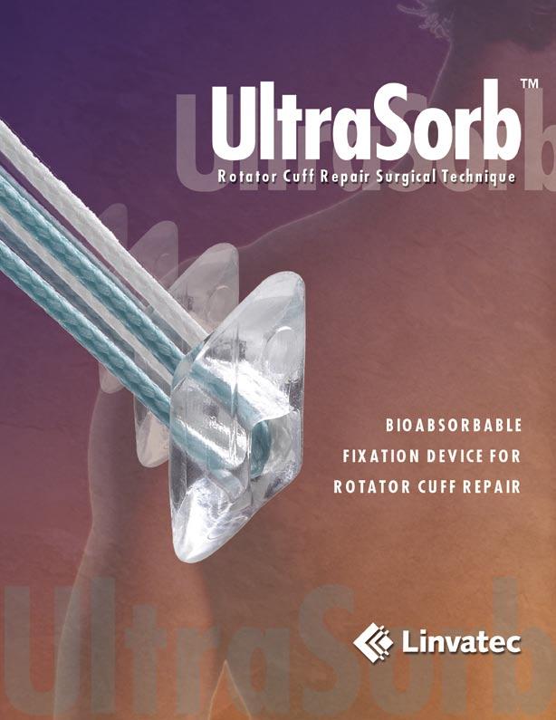
 UltraSorb Implant is pre-loaded on a sterile disposable inserter. The unique deployment mechanism allows for a simple and reproducible insertion of the anchor into bone.")

2 The UltraSorb Rotator Cuff Suture Anchor offers superior performance, reliability, and reproducibility for mini-open or arthroscopic repairs. The Poly-L-Lactide Acid (PLLA) UltraSorb Implant is pre-loaded on a sterile disposable inserter. The unique deployment mechanism allows for a simple and reproducible insertion of the anchor into bone. The Independent Suture Sliding (I.S.S. ) Eyelet design allows for the two pre-threaded sutures to independently move when passing through soft tissue or during knot tying. The sutures slide with relative ease, which allows for sliding knots to be tied without worry of locking the other strand and minimizes suture abrasion. In addition, The UltraSorb Implant is designed for improved pull-out strength in all types of bone quality. The UltraSorb Suture Anchor has additional bioabsorbable benefits, which include radiolucency and a lengthy history of clinical effectiveness and safety. The implant will retain its strength during the important healing period, but will slowly resorb into the body over a two to three year period. The Linvatec UltraSorb Suture Anchor System is another example of Linvatec s commitment to the design, manufacture, sale and service of quality products and instruments for least invasive orthopaedic surgery. The surgeon must have an excellent understanding of the technique and must practice suture passing and knot tying before attempting the operation. The following outline highlights the important steps in a typical rotator cuff repair. Linvatec Corporation will be happy to provide more comprehensive videotape instructions. Information can be obtained by calling your local Linvatec representative or Customer Service at (800) The rotator cuff tear is carefully evaluated with an arthroscope on both the articular and bursal sides, and the frayed edges of the cuff are debrided. The best view of the rotator cuff is usually the 50 yard line view with the arthroscope in a lateral subacromial portal which is located at the center point of the rotator cuff tear. 4 5 The bone is lightly decorticated at the anatomical neck of the humerus, adjacent to the articular cartilage, using a high speed bur and/or shaver. The rotator cuff is mobilized to minimize tension on the repair. 2 The UltraSorb Awl or Drill Bit is inserted through a small puncture wound adjacent to the lateral border of the acromion to create pilot holes for the anchors. The Awl is directed to enter the bone in a medial direction below the subchondral bone at approximately a 45 angle. An optional Drill Guide and Obturator can be used to enter the subacromial space to assist in the creation of the pilot hole and aid in the delivery of the implant. The pilot holes are angled away from the center of the trough in a fan-like pattern. (To ensure proper anchor placement, the Awl can be left in place until ready to place the inserter.)


3 The following techniques are described by Vernon J. Cooley, M.D., Park City, Utah ARTHROSCOPIC ROTATOR CUFF REPAIR SURGICAL TECHNIQUE Steps in repair technique 2 3 A Spectrum Crescent Suture Hook with a Shuttle Relay or a Blitz suture passer is used to perform a side-to-side repair of longitudinal tears in the rotator cuff tendon. After passing the curved suture hook across the tear, a strong, long lasting suture is carried across the tear and the suture limbs tied together. 6 The UltraSorb inserter, with the pre-loaded anchor, is inserted directly through the percutaneous wound (no cannula is needed to insert this anchor). 7 Push the inserter into the pilot hole until the wider shoulder touches the bone. A circular depth mark is located at the shoulder of the inserter. The vertical orientation mark, the solid line which indicates the direction the anchor eyelet is facing, is aligned toward the cuff edge. This ensures that the sutures pass in a direct line from the eyelet to the cuff without forming a twist.
4 8 9 Maintain inserter contact with bone and maintain the same insertion angle by forward pressure, and squeeze the trigger fully until it cannot move any farther. An audible click in the handle should be noticeable when fully squeezed. 12 The arthroscope can be positioned in the anterior or posterior portal but most often the overall visualization is best from the lateral acromial portal. 13 A crochet hook or suture retrieval forceps is inserted through the anterior portal (Linvatec 6mm operating cannula) and retrieves the strand of the green suture that exits the anchor closest to the cuff. The retriever must pass behind (medial to) the suture limbs. 4
5 ARTHROSCOPIC ROTATOR CUFF REPAIR SURGICAL TECHNIQUE Steps in repair technique 10 Withdraw the inserter from the pilot hole and subacromial space allowing the suture pack and needles to come out of the handle. 11 After insertion, test the anchor security by applying nominal tension to the suture limbs. (In soft bone, expect the anchor to withdraw slightly as the suture security is tested.) 14 A Spectrum Crescent Suture Hook is inserted into the posterior cannula and through the bursal side of the posterior edge of the torn rotator cuff 5mm posterior to the anchor. The Shuttle Relay suture passer is sent through the hook and retrieved with a grasping forceps out the anterior cannula. Care must be taken to insure that the grasping forceps follows the same path as the green suture when retrieving the Shuttle Relay to avoid causing twists in the strands. 15 The green suture strand is loaded into the eyelet of the Shuttle Relay suture passer outside the anterior cannula. The suture is then carried through the cuff from the articular side to the bursal side by withdrawing the opposite end of the suture passer out the posterior cannula. 5


6 16 A crochet hook is used to retrieve the other limb of green suture into the posterior cannula. A switching stick is then inserted through the posterior cannula and the cannula is removed from the joint. 17 The cannula is reinserted over the switching stick leaving the sutures outside the cannula where they will be less likely to be tangled during stitching with the white sutures. 20 The white suture strand is loaded into the eyelet of the Shuttle Relay suture passer outside the anterior cannula. The suture is carried through the cuff from the articular side to the bursal side by withdrawing the opposite end of the suture passer out the posterior portal. 18 a ALTERNATIVE METHOD (MODIFIED CASPARI SUTURE PUNCH): A crochet hook is used to retrieve the limb of white suture that is closest to the cuff. The suture is pulled out through the lateral cannula.

7 ARTHROSCOPIC ROTATOR CUFF REPAIR SURGICAL TECHNIQUE Steps in repair technique 18 A crochet hook or suture retrieval forceps is used to retrieve the limb of white suture that exits the anchor eyelet closest to the rotator cuff. The suture is pulled through the anterior cannula. 19 The Spectrum Suture Hook is passed through the torn rotator cuff from top to bottom approximately 5mm anterior to the anchor site. If a crescent suture hook is used again, it may be inserted through the posterior cannula. If a more angled suture hook is used, the posterior cannula can be removed and the hook passed directly through the portal without a cannula. The Shuttle Relay suture passer is passed through the hook and retrieved with a grasping forceps through the anterior cannula. 19 a MODIFIED CASPARI SUTURE PUNCH CONTINUED: With the scope viewing from the anterior portal, a modified Caspari Suture Punch can be inserted through a 6.5mm ClearFlex Cannula in the lateral portal to pass a Shuttle Relay suture passer from the bottom to top through the cuff. The suture passer is carried out the posterior cannula with a grasping forceps. 20 a MODIFIED CASPARI SUTURE PUNCH CONTINUED: The eyelet of the Shuttle Relay suture passer is loaded with the suture outside the lateral cannula and carried through the cuff from bottom to top by pulling on the opposite end.
8 21 The posterior cannula is reinserted and the remaining white suture limb is retrieved using a crochet hook or suture retrieval forceps. ARTHROSCOPIC KNOT TYING TECHNIQUE 22 The ring handled knot pusher is threaded on to the green suture exiting the top of the cuff. It is passed into the joint to ensure there are no twists or obstructing soft tissue. The green and white suture limbs associated with the posterior anchor are first tied using a knot of choice. The second anchor is placed in a similar fashion, suture limbs passed, and tied down. REVO KNOT The arthroscopic Revo knot is an extremely important knot for all surgeons performing advanced shoulder reconstruction procedures. This knot can be used in any and all situations, whether or not the suture material slides freely through the tissue and anchor eyelet. If a complex stitch such as a figure-of-eight or double-pass mattress stitch is used, this knot is preferable to any sliding knot. In addition, when capsular plication is performed, it is important not to use a sliding knot because of the possible trauma to the labrum as the suture is pulled through. 1. Both suture tails are the same length and the loophandled knot pusher is threaded onto the suture which has been passed through the soft tissue. This original post is positioned on the left side, shown as the darker tail for illustration purposes. The knot pusher is passed down the original post suture to ensure that there are no twists or soft tissue obstructions. 2. An underhand halfhitch is placed around the original post and advanced into position on the edge of the soft tissue.
 anchor is placed in the same fashion and suture limbs passed through the cuff, usually suturing from the anterior portal.")
9 ARTHROSCOPIC ROTATOR CUFF REPAIR SURGICAL TECHNIQUE Steps in repair technique 23 The arthroscope may be moved to the posterior cannula for The visualization. The third (anterior) anchor is placed in the same fashion and suture limbs passed through the cuff, usually suturing from the anterior portal. 24 illustration of the final repair shows three UltraSorb anchors in place. Each anchor has two fixation points through the rotator cuff oriented 45 from the anchor. Notice the final side-to-side repair. At the completion of the repair, the torn end of the rotator cuff is tightly opposed to the bone to promote strong rotator cuff tendon healing. 3. Tension is held on the post suture while a second underhand half-hitch is worked down the post suture to reinforce the first hitch. 4. An overhand halfhitch is next placed on the same initial post and worked down into position on the other two throws. 5. The knot pusher and clamp are changed to the opposite suture and after checking for twists and soft tissue, an underhanded throw is advanced down onto the knot stack. 6. The knot pusher is advanced to past point to lock the halfhitch securely. 7. A fifth overhand half-hitch is placed over the second post and worked down into position on the knot stack. 8. Sometimes a sixth half-hitch can be used as the surgeon prefers, and the suture tails are cut with microscissors.
10 ARTHROSCOPIC KNOT TYING TECHNIQUE SMC KNOT The SMC knot* is a unique sliding knot that utilizes a self-locking loop to achieve good initial knot security. The SMC knot is low profile and there is minimal or no slack once the knot is secured. The SMC Knot cannot be used if the sutures do not easily slide through the soft tissues. If there is any doubt about the freedom of suture passage, then the Revo knot should be used. 1. Thread the knot pusher on the post strand (held in the left hand) and place a clamp on the post. Pass the knot pusher into the joint to ensure that there are no twists or obstructing soft tissue. Arrange the suture so that the original post suture is short, with only 10cm of the suture outside of the cannula. 2. Pinch the two strands together between the thumb and index finger, crossing the loop strand over the post Pass the loop suture under and then over both strands. 4. Pass the loop strand under the post strand between the two sutures and over the top of the post strand in a direction away from the pinching fingers. There will be a triangular interval formed between the two previous loops over the post strand (red arrow). 5. Feed the free end of the loop strand from bottom to top through this interval under the post strand. As the suture is pulled through, a "locking loop" is created (blue arrow). *Developed by Seung-Ho Kim, M.D., Samsung Medical Center, Seoul, Korea 10
11 Do not pull on the loop strand 6. Release the thumb and index finger and place the left index finger into the "locking loop" from bottom to top to keep it open. Remove all slack (dress the knot) from the sutures with the index finger in place to avoid tightening the "locking loop" prematurely. 7. Pull on the post strand and use the knot pusher to guide the knot down to the tissue. Do not pull on the loop strand until the knot is seated. Maintain tension on the post strand and back off the knot pusher to assess the knot. 8. Once satisfied that the knot is well seated, tighten the "locking loop" by pulling on the loop strand while maintaining pressure on the knot with the knot pusher. 9. The "locking loop" will slide over the knot pusher and secure the knot. For further security, an underhand half-hitch is worked down the post suture. 10. An overhand halfhitch is next placed on the post and worked down into position onto the knot stack. 11. Suture tails are cut with microscissors. 11
 UltraSorb Instrument Set UltraSorb Drill Bit, 4.0mm... 10402 UltraSorb Awl, 4.")

12 ORDERING INFORMATION THE ULTRASORB SHOULDER FIXATION SYSTEM UltraSorb Implant UltraSorb Bioabsorbable Rotator Cuff Kit (pre-loaded on a disposable inserter, pre-threaded with two #2 braided sutures - green and white, tapered needles) UltraSorb Instrument Set UltraSorb Drill Bit, 4.0mm UltraSorb Awl, 4.3mm UltraSorb Drill Guide (6 point) UltraSorb Drill Guide Obturator UltraSorb Sterilization Tray Suture Passing Instruments Slotted Jaw, Suture Punch, 4.0mm needle Spectrum Instrument Set: Suture Hook Handle Suture Hook, Straight Suture Hook, 45 Left Curve Suture Hook, 45 Right Curve Suture Hook, 90 Left Curve Suture Hook, 90 Right Curve Crescent Suture Hook, Small Curve, 3.0 x 15.0mm... C8740 Crescent Suture Hook, Medium Curve, 4.0 x 20.0mm... C8741 Crescent Suture Hook, Large Curve, 6.0 x25.0mm... C8742 Shuttle-Relay Suture Passer (10 per box)... C6004 Blitz Suture Retriever, Straight (6 per box)... C6111 Blitz Suture Retriever, 45 Left (6 per box)... C6211 Blitz Suture Retriever, 45 Right (6 per box)... C6311 Hawkeye Suture Needle (6 per box)... C6001 Accessories Loop Handle Knot Pusher... C6112 Crochet Hook... C6105 Teaser Knot Tier... C8004 Microscissors, 2.75mm Diameter, Straight Grasping Forceps, 3.4mm Diameter, Straight with Ratchet Suture Retrieval Forceps, 3.4mm Diameter Liberator Knife Entry Systems (Disposable) 6.5mm x 73.0mm Clear Flexible Cannula with Disposable Conical Obturator... C mm x 50.0mm Cannula with Disposable Conical Obturator Threaded Body... C7322 Smooth Body... C mm x 75.0mm Cannula with Disposable Conical Obturator Threaded Body... C7332 Smooth Body... C mm x 90.0mm Cannula with Disposable Conical Obturator Threaded Body... C7342 Smooth Body... C mm x 50.0mm Cannula with Disposable Conical Obturator Threaded Body... C7352 Smooth Body... C mm x 75.0mm Cannula with Disposable Conical Obturator Threaded Body... C7362 Smooth Body... C mm x 90.0mm Cannula with Disposable Conical Obturator Threaded Body... C7372 Smooth Body... C mm Universal Cannula Set with Fenestrations mm Universal Cannula Set without Fenestrations Patent Pending UltraSorb Sterilization Tray with instruments UltraSorb Implant and Disposable Inserter Concept Boulevard, Largo, Florida Phone: (727) Customer Service: (800) USA Fax: (727) International Fax: (727) Linvatec Corporation, a subsidiary of CONMED Corporation. CST 3020R 11/01
Super Revo. Surgical Technique. Super Revo Shoulder Fixation System. The Ideal Fixation Device For Rotator Cuff Repair. Self-drilling, titanium anchor
 Super Revo Shoulder Fixation System The Ideal Fixation Device For Rotator Cuff Repair Super Revo Surgical Technique INCREASED Strength and Suture Management Self-drilling, titanium anchor Independent Suture
Dr. S.D. Gerber Double Row Method Surgical Technique
 Dr. S.D. Gerber Double Row Method Surgical Technique Dr. S.D. Gerber Double Row Method Surgical Technique Introduction There has been a rapid proliferation of techniques for arthroscopic rotator cuff tear
LabraLock P for Labrum Repair, LabraFix System. The Reinforcement Technique Guide
 LabraLock P for Labrum Repair, Part of the OPUS LabraFix System A revolutionary new system specifically designed for labrum repair surgery System Includes: SpeedStitch Suturing Device LabraLock P Implant
Double Row Rotator Cuff Repair. Surgical Protocol by Frank Bonnarens, M.D.
 Double Row Rotator Cuff Repair Surgical Protocol by Frank Bonnarens, M.D. This brochure is presented to demonstrate the surgical technique utilized by Frank Bonnarens, M.D. Biomet Sports Medicine, as the
The OPUS AutoCuff System for Rotator Cuff Repair
 The OPUS AutoCuff System for Rotator Cuff Repair A revolutionary new system specifically designed for rotator cuff repair surgery System Includes: SmartStitch Suturing Device The Magnum PI Implant with
VERSALOK. Suture Anchor. Rotator Cuff Repair Surgical Technique
 VERSALOK Suture Anchor Rotator Cuff Repair Surgical Technique Introduction VERSALOK Suture Anchor, available in Titanium and PEEK from DePuy Synthes Mitek Sports Medicine, were designed to allow for versatile
Double Row Rotator Cuff Repair. Surgical Protocol by Frank Bonnarens, M.D.
 Double Row Rotator Cuff Repair Surgical Protocol by Frank Bonnarens, M.D. Features Lateral row knotless anchor fixation Lower knot profile medially and completely knotless laterally No arthroscopic knot
Knotless SutureTak Instability Repair. Surgical Technique
 Knotless SutureTak Instability Repair Surgical Technique Knotless SutureTak Instability Repair Load-to-Failure (lbf) Knotless SutureTak Instability Repair The Knotless SutureTak suture anchor simplifies
Champion Instrumentation. Shoulder Repair Made Simpler
 Champion Instrumentation Shoulder Repair Made Simpler Champion Instruments Simplicity & Versatility The Champion Shoulder instrumentation system was designed with simplicity in mind. Clever instruments
Arthroscopic Protector Meniscus Suturing Surgical Techniques
 Arthroscopic Protector Meniscus Suturing Surgical Techniques Protector Meniscus Suturing Protector Meniscus Suturing Technique I For the relatively rare meniscus tears located sufficiently anterior, to
Knotless Bankart Repair Using the Labral SwiveLock. and FiberStick. Surgical Technique. Labral SwiveLock
 Knotless Bankart Repair Using the Labral SwiveLock and FiberStick Surgical Technique Labral SwiveLock SwiveLock Bankart and SLAP Repair Versatility, Speed and Security in Knotless Instability Repair Knotless
Rotator Cuff Repair ICONN Answer PEEK Suture Anchor 4.75/5.5. Surgical Protocol by Geoffrey Connor, 4.75/5.5 MD of D1 Sports Medicine
 Rotator Cuff Repair ICONN Answer PEEK Suture Anchor 4.75/5.5 Surgical Protocol by Geoffrey Connor, 4.75/5.5 MD of D1 Sports Medicine ICONN s revolutionary approach is to drive value into every aspect of
Arthroscopic Rotator Cuff Repair
 Bio-Corkscrew Suture Anchor Rotator Cuff Repair Surgical Technique Arthroscopic Rotator Cuff Repair 1 2 The patient may be positioned in the beach chair position using the Beach Chair Lateral Traction
Techniques of the hand tie and instrument tie
 Techniques of the hand tie and instrument tie 1. The Anatomy of a Square Knot A square knot consists of two "throws". Throws are constructed by crossing the ends of the suture to form a loop and then wrapping
Technique Guide. Modular Sternal Cable System. Flexibility and strength in sternal closure and repair.
 Technique Guide Modular Sternal Cable System. Flexibility and strength in sternal closure and repair. Table of Contents Introduction Overview 2 Indications and Contraindications 3 Surgical Technique A.
BioPlug 5.2 SPS Secure Portal System
 ART 27 11.1 11/2017-E BioPlug 5.2 SPS Secure Portal System For use in reconstructive shoulder surgery on the humeral head BioPlug 5.2 SPS Secure Portal System For use in reconstructive shoulder surgery
Technique Guide. Modular Sternal Cable System. Flexibility and strength in sternal closure and repair.
 Technique Guide Modular Sternal Cable System. Flexibility and strength in sternal closure and repair. Table of Contents Introduction Modular Sternal Cable System 2 Indications 4 Modular Sternal Closure
Ankle Fracture System. Surgical Technique STRENGTH FROM WITHIN
 Ankle Fracture System Surgical Technique STRENGTH FROM WITHIN Ankle Fracture System The Sonoma FibuLock nail is the first intramedullary device that has the same indications as plates and delivers anatomic
PARS Achilles Jig System
 PARS Achilles Jig System Surgical Technique PARS Achilles Jig System PARS Achilles Jig System Achilles tendon ruptures are common in the elite and recreational athlete and most often occur in the non-insertional
High polish finish. Complete line of biters, tissue graspers
 with the Introducing the Precision II Series, a complete line of hand instrumentation that has been developed based on surgeons input to have the most tactile and most advanced instrumentation for arthroscopic
Revolutionizing Orthopaedic Surgery. Braided Composite Suture
 Revolutionizing Orthopaedic Surgery Braided Composite Suture Revolutionizing Orthopaedic Surgery FiberWire suture is constructed of a multi-stranded long chain ultra-high molecular weight polyethylene
Product Pamphlet. Parcus Medical s Value Proposition Program is designed to reduce cost per case by:
 Product Pamphlet Parcus Medical designs, manufactures, and distributes implants and instrumentation used by orthopedic surgeons in sports medicine procedures to repair the shoulder, knee, hip, and distal
Arthrex. Bio-Compression. Screw System. Knee OCD. Hand, Wrist & Elbow. Foot & Ankle
 Arthrex Bio-Compression Screw System Knee OCD Hand, Wrist & Elbow Foot & Ankle Bio-Compression Screw System Bio-Compression Screws are versatile and may be used to treat a broad range of indications in
Prostar XL Percutaneous Vascular Surgical Device
 Clinical leader for percutaneous large access site closure Close Access Sites Up to 24F Secure Closure Prostar XL Percutaneous Vascular Surgical Device Improved Outcomes 2009 Abbott Laboratories. All rights
Suture anchors for VeterinarY use
 Suture anchors for VeterinarY use For Joint Stabilization and Ligamentous and Tendon Injury Repairs in Cats and Dogs Mini QUICKANCHOR Plus Anchor GII QUICKANCHOR Plus Anchor Super QUICKANCHOR Plus Dual
Universal Humeral Nail
 990210009 INDEX Indications Preoperative Planning Patient Position Surgical Technique - Step 1 Open Humerus - Step 2 Calibrate The Nail - Step 3 Insert Nail - Step 4 Proximal Locking - Step 5 Assemble
Simple Interrupted Suture (using a tea towel)
") (using a tea towel) Disclaimer A series of booklets has been developed by the Clinical Skills Lab team (staff, recent graduates and students) from the School of Veterinary Sciences, University of Bristol,
(using a tea towel) Disclaimer A series of booklets has been developed by the Clinical Skills Lab team (staff, recent graduates and students) from the School of Veterinary Sciences, University of Bristol,
Anterior Cervical Plate SURGICAL TECHNIQUE GUIDE. Surgeon Driven Innovation
 Anterior Cervical Plate SURGICAL TECHNIQUE GUIDE Surgeon Driven Innovation 1 The Snowmass Anterior Cervical Plate System is intended for the surgical treatment and correction of traumatic and pathologic
Simple Interrupted Suture (using a silicon skin pad)
") (using a silicon skin pad) Disclaimer A series of booklets has been developed by the Clinical Skills Lab team (staff, recent graduates and students) from the School of Veterinary Sciences, University of
(using a silicon skin pad) Disclaimer A series of booklets has been developed by the Clinical Skills Lab team (staff, recent graduates and students) from the School of Veterinary Sciences, University of
The use of sutures and ligatures is an integral part of surgical procedures. Being proficient in suturing and ligating will not only make your
 1 The use of sutures and ligatures is an integral part of surgical procedures. Being proficient in suturing and ligating will not only make your treatments more effective but also shorten the time of the
ACLP Anterior Cervical Locking Plate System TECHNIQUE GUIDE
 ACLP Anterior Cervical Locking Plate System TECHNIQUE GUIDE Instruments and implants approved by the AO Foundation ACLP Anterior Cervical Locking Plate System The ACLP System is designed to reduce the
Orthopedic Bone Nail System Universal Humeral Nail
 Orthopedic Bone Nail System Universal Humeral Nail Surgical Technique Manual Note: The surgical procedures should be performed under the guidance of qualified skilled orthopedic surgeons, and this surgical
Lag Screw Device TECHNIQUE GUIDE. Indicated for symphyseal fracture fixation of the mandible. Instruments and implants approved by the AO Foundation
 Lag Screw Device TECHNIQUE GUIDE Indicated for symphyseal fracture fixation of the mandible Instruments and implants approved by the AO Foundation Lag Screw Device Indicated for symphyseal fracture fixation
Aesculap Spine S 4 Spinal System. Instrumentation Guide
 Aesculap Spine S 4 Spinal System Instrumentation Guide S 4 Spinal System S 4 From initial conception, the S 4 Spinal System was developed to meet the spine surgeon s need for an extremely low profile and
Bio-Compression. Arthrex. Screw System
 Bio-Compression Arthrex Screw System Ordering Information 3 mm Bio-Compression Screw Instrumentation Set (AR-5025S) includes: Bio-Compression Screw Driver, noncannulated, 2.7 mm AR-5025DB Small Handle
Cerclage Passer. For minimally invasive application of cerclage cables.
 Cerclage Passer. For minimally invasive application of cerclage cables. Handling Technique Cable application This publication is not intended for distribution in the USA. Instruments and implants approved
PARS Achilles Jig System Surgical Technique
 PARS Achilles Jig System Surgical Technique PARS Achilles Jig System PARS Achilles Jig System Achilles tendon ruptures are common in the elite and recreational athlete and most often occur in the noninsertional
MEDICAL ADVANCED TECHNOLOGY EMERGENCY REMOVAL UNIVERSAL EXTRACTION SET. for Intramedullary Nail System
 MEDICAL ADVANCED TECHNOLOGY EMERGENCY REMOVAL UNIVERSAL EXTRACTION SET for Intramedullary Nail System introducing ourselve Manufacturer of surgical implants and medical devices. Solutions for patient orthopaedic
ISO Plate SURGICAL TECHNIQUE
 MINI MAXLOCK EXTREME ISO Plate SURGICAL TECHNIQUE Contents Table of Contents Key Design Features 2 Surgical Technique 3 Implants and Instruments 8 Key Design Features The MINI MAXLOCK EXTREME ISO (Intraosseous
5th Metatarsal Fracture System Surgical Technique
 5th Metatarsal Fracture System Surgical Technique 5th Metatarsal Fracture System 5th Metatarsal Fracture System The 5th Metatarsal Fracture System (AR-8956S) is a uniquely designed screw and plate system
The Percutaneous Reduction Forceps Technique Guide
 The Percutaneous Reduction Forceps Technique Guide Indications + Product Overview Introduction The Percutaneous Reduction Forceps The Percutaneous Reduction Forceps facilitate standard technique for fixation
Small Plate and Screw System SURGICAL TECHNIQUE
 MINI MAXLOCK EXTREME Small Plate and Screw System SURGICAL TECHNIQUE Contents Key Design Features 2 Surgical Technique 3 Implants and Instruments 9 Proper surgical procedures and techniques are the responsibility
Basic Instructions. The Ring: Fill the shuttle with thread from a ball DMC or Coats 20, but DO NOT CUT from the ball.
 Basic Instructions Fill the shuttle with thread from a ball DMC or Coats 20, but DO NOT CUT from the ball. The Ring: Wrap the center of uncut thread around the finger tips of the left hand and cross the
Technique Guide. 7.0 mm Cannulated Screws. Part of the Synthes Cannulated Screw System.
 Technique Guide 7.0 mm Cannulated Screws. Part of the Synthes Cannulated Screw System. Table of Contents Introduction 7.0 mm Cannulated Screws 2 AO Principles 3 Indications 4 Surgical Technique Surgical
TORNIER MAXLOCK EXTREME. Clavicle Plating System SURGICAL TECHNIQUE
 TORNIER MAXLOCK EXTREME Clavicle Plating System SURGICAL TECHNIQUE 2 Table of Contents: Key Design Features...4 Surgical Technique...5 Implants & Instruments...9 3 Key Design Features There are 7 anatomically
EP A1 (19) (11) EP A1 (12) EUROPEAN PATENT APPLICATION. (43) Date of publication: Bulletin 2011/04
 (11) EP A1 (12) EUROPEAN PATENT APPLICATION. (43) Date of publication: Bulletin 2011/04") (19) (12) EUROPEAN PATENT APPLICATION (11) EP 2 277 457 A1 (43) Date of publication: 26.01.2011 Bulletin 2011/04 (51) Int Cl.: A61B 17/04 (2006.01) (21) Application number: 10251328.0 (22) Date of filing:
(19) (12) EUROPEAN PATENT APPLICATION (11) EP 2 277 457 A1 (43) Date of publication: 26.01.2011 Bulletin 2011/04 (51) Int Cl.: A61B 17/04 (2006.01) (21) Application number: 10251328.0 (22) Date of filing:
Basic Bearmaking Instructions
 Laying out the Pattern Basic Bearmaking Instructions We recommend that you read the instructions right through before commencing. This will help you to understand the steps you need to take. Copying your
Surgical Technique. Customer Service:
 Patent Pending CAUTION: Federal Law (USA) restricts this device to sale by or on the order of a physician. Notes This page is blank INDICATIONS FOR USE The Extremity Medical Hallu X Intramedullary Fusion
MTP Fusion Surgical Technique
 MTP Fusion Surgical Technique Patent and Patent Pending CAUTION: Federal Law (USA) restricts this device to sale by or on the order of a physician. INDICATIONS FOR USE The Omni Foot Plating System is intended
For Minimally Invasive Application of Cerclage Wires. Cerclage Passer. Surgical Technique
 For Minimally Invasive Application of Cerclage Wires Cerclage Passer Surgical Technique Table of Contents Introduction Cerclage Passer 2 Surgical Technique Preparation 4 Insert Cerclage Passer 5 Connect
ExpresSew II. Intuition redefined.
 ExpresSew II Surgical Technique Intuition redefined. Enhanced ergonomics & functionality provide unparalleled simplicity, flexibility and reliability in suture management Intuitive Simple Flexible Reliable
MatrixMANDIBLE Preformed Reconstruction Plates. Preshaped to the mandibular anatomy.
 MatrixMANDIBLE Preformed Reconstruction Plates. Preshaped to the mandibular anatomy. Technique Guide CMF Matrix Table of Contents Introduction MatrixMANDIBLE Preformed Reconstruction Plates 2 AO Principles
NOTE: Top section pole (Q) is packed INSIDE bottom section pole (S)
 is packed INSIDE bottom section pole (S)") Form 0905-0 Instructions and Parts List TM- Mini Castle (modified) MARTIN SAFETY SYSTEM NOTES: () A complete system is packed in two boxes post box and house box. House box contains hardware for both post
Form 0905-0 Instructions and Parts List TM- Mini Castle (modified) MARTIN SAFETY SYSTEM NOTES: () A complete system is packed in two boxes post box and house box. House box contains hardware for both post
Interlagos Retractor System Surgical Technique
 Interlagos Retractor System Surgical Technique TABLE OF CONTENTS Instructions for Use Design Rationale Surgical Technique 1. Pre-Operative Preparation 2. Pedicle Preparation 3. Primary Retraction 4. Secondary
Perclose ProGlide. Deployment Steps. Suture-Mediated Closure System Abbott. All rights reserved. ANZ /11
 Deployment Steps SUTURE TRIMMER Handle Needle Plunger Collar Lever Marker Lumen Body Foot QuickCut Link Foot (Deployed) Proximal Guide Guide Wire Exit Port Distal Guide Marker Port Sheath posterior side
Digital Compression Screw
 Digital Compression Screw Surgical Technique Contents Product The BioPro Digital Compression Screw is a stainless steel lag screw designed for digital fusions. Table of contents Indications & Contraindications
Integra. Capture Screw System SURGICAL TECHNIQUE
 Integra Capture Screw System SURGICAL TECHNIQUE Table of Contents Indications... 2 Contraindications... 2 System Description... 2 System Features... 2 Cannulated Low-Profile Screws (AC-Series) Overview...
SURGICAL TECHNIQUE. SpineTune THORACO-LUMBAR POSTERIOR OSTEOSYNTHESIS SYSTEM
 SURGICAL TECHNIQUE SpineTune TM TL THORACO-LUMBAR POSTERIOR OSTEOSYNTHESIS SYSTEM SURGICAL TECHNIQUE SpineTune TM TL Table of Contents page Step 1 - Site preparation.......................................................................................
Integra. Stainless Headed Compression Screw System SURGICAL TECHNIQUE
 Integra Stainless Headed Compression Screw System SURGICAL TECHNIQUE Table of Contents Design Rationale...2 Indications...2 Contraindications...2 Surgical Technique Step 1: Inserting Guide Wire... 3 Step
Electric Skein Winder
 Electric Skein Winder Assembly and Use Package Contents 1 - Triangular Body (w/ motor) 1 - Cross Arm 1 - Left Foot (w/ yarn guide) 1 - Right Foot 1 - Adjustable Finger (w/ yarn clip) 3 - Adjustable Fingers
OPERATIVE TECHNIQUE RIVAL REDUCE FRACTURE PLATING SYSTEM. foot & ankle trauma procedures
 OPERATIVE TECHNIQUE RIVAL REDUCE FRACTURE PLATING SYSTEM foot & ankle trauma procedures INTRODUCTION 3 SYSTEM DESCRIPTION 3 TECHNICAL DETAILS 4 SALES AND MARKETING CONFIGURATION 5 OPERATIVE TECHNIQUE 7
Surgical Technique 1
 Surgical Technique 1 D-RAD SMART PACK Single-Use Volar Distal Radius Plating System Surgical Technique Table of Contents Indications... 3 Contraindications... 3 D-RAD SMART PACK product overview... 4 Instrumentation...
Revolutionizing Orthopaedic Surgery. Braided Composite Suture
 Revolutionizing Orthopaedic Surgery Braided Composite Suture Revolutionizing Orthopaedic Surgery FiberWire suture is constructed of a multi-stranded long chain ultrahigh molecular weight polyethylene core
To register your machine warranty and receive Baby Lock product updates and offers, go to If you have questions with
 To register your machine warranty and receive Baby Lock product updates and offers, go to www.babylock.com/profile. If you have questions with registration, visit your Authorized Baby Lock Retailer. CONTENTS
Technique Guide. Cable System. For Orthopaedic Trauma Surgery.
 Technique Guide Cable System. For Orthopaedic Trauma Surgery. Table of Contents Introduction Overview 2 AO Principles 4 Indications and Contraindications 5 Surgical Technique Standard Cerclage Technique
Procedure Guide. Eliminate Big Problems Safely and Quickly Close Your Patients Port Sites
 NEW! Procedure Guide Eliminate Big Problems Safely and Quickly Close Your Patients Port Sites Following the trocar track helps prevent loss of pneumoperitoneum Ergonomically engineered handle with ribbed
Back to health. Back to work. Back to life.
 TECHNIQUE Back to health. Back to work. Back to life. U PLUS 90 INSTRUMENTATION OVERVIEW W&H IMPLANTMED POWER UNIT OVERVIEW Low-profile Primary Guides Compresses the U-clip to match rib thickness Clamps
Instructions for Use. LCP Locking Compression Plate. Combine without Compromise.
 Instructions for Use LCP Locking Compression Plate. Combine without Compromise. Table of Contents LCP: Combine without Compromise 2 AO ASIF Principles of Osteosynthesis 4 Indications and Contraindications
STILLE Micro Instruments. Solid Performance - Outstanding Precision
 STILLE Micro Instruments Solid Performance - Outstanding Precision STILLE introduces the highest quality handmade instruments for the world s best surgeons The new and expanded line of STILLE Micro Instruments
FLIP RIGID HEDDLE LOOM
 FLIP RIGID HEDDLE LOOM SL2013, SL2014 SL2015, SL2016 Warping and Weaving Instructions Flip Loom shown with optional accessories Trestle Floor Stand and Flip Trap Find out more at schachtspindle.com Schacht
ACCS Anterior Cervical Compression System TECHNIQUE GUIDE
 ACCS Anterior Cervical Compression System TECHNIQUE GUIDE Original Instruments and Implants of the Association for the Study of Internal Fixation AO ASIF ACCS Anterior Cervical Compression System The Anterior
Objectives. xxx00.#####.ppt 5/10/17 9:42 AM
 Objectives Understand and demonstrate proper suturing techniques Learn how to properly handle instruments Know types of suture and appropriate use Hand-tying Learn how to trouble-shoot complications after
LCP Pilon Plate 2.7/3.5
 Surgical Technique LCP Locking Compression Plate Original Instruments and Implants of the Association for the Study of Internal Fixation AO/ASIF Table of contents Indications 3 Implants 4 Instruments 5
University of Alabama at Birmingham. ObGyn Residency. Laparoscopy Training Lab PGY 1-4. Individual Pelvic Trainer Tasks
 University of Alabama at Birmingham ObGyn Residency Laparoscopy Training Lab PGY 1-4 Individual Pelvic Trainer Tasks 2010-2011 Skill 1: Peg Board (Blue Board) Goal: Pick up various rings and move them
Instruments for Removing DePuy Synthes Screws. Screw Removal Set
 Instruments for Removing DePuy Synthes Screws Screw Removal Set Surgical Technique Table of Contents Introduction Screw Removal Set 2 Surgical Technique Preoperative Planning and Preparation 6 Removal
MTP Set SURGICAL TECHNIQUE
 MINI MAXLOCK EXTREME MTP Set SURGICAL TECHNIQUE Contents Table of Contents Key Design Features 3 Surgical Technique Standard MTP Plate 4 MTP Plate with POCKETLOCK Technology 10 Implants and Instruments
OsteoBridge IKA Intramedullary Knee Arthrodesis Fixation System. From the «BioBall Company» OsteoBridge Family
 From the «BioBall Company» OsteoBridge Family OsteoBridge IKA Intramedullary Knee Arthrodesis Fixation System The modular system for the fixation of the knee joint 01. OsteoBridge IKA The OsteoBridge IKA
Learn to weave. on the Knitters Loom
 Learn to weave on the Knitters Loom Welcome to the wonderful world of weaving... YOUR FIRST PROJECT A SCARF You will need: A pair of sharp scissors A measuring tape Cardboard strips 20cm (8ins) wide Two
Vortex TRAUMATOLOGY. Vortex Distal Femur
 Vortex TRAUMATOLOGY Vortex Distal Femur 1 Content 1. Introduction 4 4. Implant list 16-17 The following surgical description contains general outlines for Vortex Distal Femur plating. However, the operating
ORTHOPEDICS Extracapsular Repair
 ORTHOPEDICS Extracapsular Repair Monofilament Nylon Suture Veterinary Instrumentation Nylon Suture Packs are supplied sterile, unless otherwise noted. Weight Kgs (lbs.)* Strength of Nylon Nylon Line Monofilament
POLYAXIAL SPINE SYSTEM SURGICAL TECHNIQUE
 POLYAXIAL SPINE SYSTEM SURGICAL TECHNIQUE P O L Y A X I A L S P I N E S Y S T E M POLYAXIALITY STABILITY EFFICIENCY All the implants offer the Polyaxiality, including Hooks, Claws, Sacral Plates and of
HCS 2.4/3.0. The countersinkable compression screw.
 Technique Guide HCS 2.4/3.0. The countersinkable compression screw. Table of Contents Introduction Features and Benefits 2 Functional Principle 3 Indications 4 Surgical Technique Hand Scaphoid 5 Foot
A free-extending two part cannulated screw that will elongate with growth. SURGICAL TECHNIQUE
 A free-extending two part cannulated screw that will elongate with growth. SURGICAL TECHNIQUE The Free-Gliding SCFE Screw System, designed to treat the most common hip problem in growing children, SLIPPED
Distal Medial Tibia Plate Surgical Technique
 Locking Compression Technology by aap 1 Disclaimer This surgical technique is exclusively intended for medical professionals, especially physicians, and therefore may not be regarded as a source of information
Emilia. The Folding Rigid Heddle Loom. Learning to weave on your Emilia loom by Joanne Hall
 Emilia The Folding Rigid Heddle Loom Learning to weave on your Emilia loom by Joanne Hall Heddle Handle Heddle Bracket Rigid Heddle Warping peg The Emilia Loom and Accessories Warp beam Texsolv cord Folding
LCP Pilon Plate 2.7/3.5
 LCP Pilon Plate 2.7/3.5 Surgical Technique This publication is not intended for distribution in the USA. Instruments and implants approved by the AO Foundation. Table of contents Indications 2 Implants
TwinFix Surgical Protocol. 3.2mm Cannulated Compression Screw System
 TwinFix Surgical Protocol Compression Screw System TwinFix Compression Screw System Indications for Use: The Stryker TwinFix Interfragmentary Compression Screw System is intended to be used for fractures
edge of the section wound, probably from opening of the wound
 CORNEO-SCLERAL SUTURE IN CATARACT EXTRACTION 269 A CORNEO-SCLERAL SUTURE IN CATARACT EXTRACTION. ITS TECHNIQUE AND ADVANTAGES BY H. B. STALLARD LONDON THE use of a corneo-scleral suture in the operation
Surgical Technique Guide
 Surgical Technique Guide Patented - www.flow-fx.net Flow-FX, LLC 9301 W 191st Street Mokena, IL 60448 P. 815.531.4424 by Flow-FX, LLC. 2017 Products referenced with TM are trademarks of Flow-Fx. STG-101
Baby Grande or Grande Crank Shade with Cables and Housing Installation Instructions
 Baby Grande or Grande Crank Shade with Cables and Housing Installation Instructions Tools Needed Drill 3/8 Metal Drill Bit Screwdriver (Flat & Phillips) Measuring Tape Pencil 4 Level Plumb Line ¼ Masonry
Technique Guide. LCP Pilon Plate 2.7/3.5
 Technique Guide LCP Pilon Plate 2.7/3.5 LCP Pilon Plate 2.7/3.5 Table of contents Indications 3 Implants 4 Instruments 5 Surgical technique 6 Implant removal 12 Image intensifier control Warning This
For a flat or four-sided reticule:
 Page 1 of 8 DRAWSTRINGS When purchasing, one fabric store meter is the minimum to make two drawstrings of a nice length for one reticule in most designs. If you like longer drawstrings, increase the length.
Technique Guide. Variable Angle LCP Opening Wedge Plates 2.4/2.7. Part of the Variable Angle LCP Forefoot / Midfoot System 2.4 / 2.7.
 Technique Guide Variable Angle LCP Opening Wedge Plates 2.4/2.7. Part of the Variable Angle LCP Forefoot / Midfoot System 2.4 / 2.7. Table of Contents Introduction Variable Angle LCP Opening Wedge Plates
Technique Guide. Variable Angle LCP 1 st MTP Fusion Plates 2.4/2.7. Part of the Variable Angle LCP Forefoot / Midfoot System 2.4 / 2.7.
 Technique Guide Variable Angle LCP 1 st MTP Fusion Plates 2.4/2.7. Part of the Variable Angle LCP Forefoot / Midfoot System 2.4 / 2.7. Table of Contents Introduction Variable Angle LCP 1 st MTP Fusion
Central New York Rocket Team Challenge 2018 Rocket Assembly Instructions
 Central New York Rocket Team Challenge 2018 Rocket Assembly Instructions Note: These instructions vary from those provided by the manufacturer of the rocket kits. There is also considerable varying discussion
OPERATIVE TECHNIQUE RIVAL VIEW PLATING SYSTEM. foot & ankle reconstruction procedures
 OPERATIVE TECHNIQUE RIVAL VIEW PLATING SYSTEM foot & ankle reconstruction procedures INTRODUCTION 3 SYSTEM DESCRIPTION 3 TECHNICAL DETAILS 4 SALES AND MARKETING CONFIGURATION 5 OPERATIVE TECHNIQUE 7 OPERATIVE
Variable Angle LCP Forefoot/Midfoot System 2.4/2.7. Procedure specific plates for osteotomies, arthrodeses and fractures of the foot.
 Instruction for Use Variable Angle LCP Forefoot/Midfoot System 2.4/2.7. Procedure specific plates for osteotomies, arthrodeses and fractures of the foot. Table of Contents Introduction VA-LCP Forefoot/Midfoot
Desktop Trebuchet Kit Assembly Instructions
 Desktop Trebuchet Kit Assembly Instructions Contents of package (drawings are not to scale for clarity, parts that have duplicates are indicated with total number of that part to be found, example: 2X
MIS TECHNOLOGY GUIDE READ PRODUCT INSERT THOROUGHLY BEFORE USE
 COR-KNOT FIG. 1 MIS TECHNOLOGY GUIDE READ PRODUCT INSERT THOROUGHLY BEFORE USE 1 3 COR-KNOT QUICK LOAD UNIT DESCRIPTION Each COR-KNOT QUICK LOAD UNIT provides one sterile COR-KNOT FASTENER 1 held in a
SECTION 7. SAFETYING
 9/8/98 AC 43.13-1B SECTION 7. SAFETYING 7-122. GENERAL. The word safetying is a term universally used in the aircraft industry. Briefly, safetying is defined as: Securing by various means any nut, bolt,