OPHTHALMIC SURGICAL MODELS
|
|
|
- Winfred McDowell
- 5 years ago
- Views:
Transcription


1 OPHTHALMIC SURGICAL MODELS

2 BIONIKO designs innovative surgical models, task trainers and teaching tools for the ophthalmic industry. Our surgical models present the user with dexterity and coordination challenges of common surgical scenarios, allowing fundamental skills to be practiced using real surgical instruments. The models aim to replicate consistency, proportions and continuous interfaces between different ocular sub-structures, providing high fidelity tissue simulators that can be cut, dissected and sutured. Surgical models allow faculty to demonstrate surgical technique. Users gain proficiency and perfect surgical skills in a safe, realistic, and non-stressful environment. Furthermore, synthetic models enable repeatable and standardized assessment of surgical technique. Our surgical models can be easily incorporated into any training program to help develop and perfect: Spatial Perception: Surgical approach, orientation, depth and scale Motor Skills: Instrument control, muscle memory and hand-eye coordination Technical Understanding: Instrumentation, surgical sequence and best practices
3 FEATURES AND BENEFITS AVAILABLE Training on demand for individual pace and needs PORTABLE Teach in the classroom, train in the OR and practice at home REPEATABLE Standardize training and assessment without model variability AFFORDABLE Surgical simulation tools at textbook cost SIMPLE Ready to use with minimum setup and assembly SYNTHETIC No refrigeration or special disposal required
4 KERATO Suturing Task Suturing is a fundamental skill in ophthalmic surgery. However, it is a common weakness among aspiring surgeons due to the lack of operative experience and suitable training models. This skill is absent in virtual reality tools, and animal models are not accurate or repeatable. By presenting the main challenges of a penetrating keratoplasty (PKP) scenario, the KERATO task allows users to learn, train and perfect the skills required to perform precise suturing under a microscope. The KERATO task consists in suturing a corneal graft to a host limbus using real surgical instruments. The user will need to properly handle forceps, needle holder, scissors and 10-0 suture to complete the task. With practice the user will gain confidence, reduce time to completion, improve suture radiality and spacing, maintain even and safe distance to tissue edge and gain better feel for suture tension during knot creation. Pat. Pending


5 KERATO 1 - CORNEA 2 - LIMBUS 3 - SCLERA 4 - GRAFT EDGE 5 - RECIPIENT EDGE 6 - SUTURE FEATURES AND BENEFITS Accurate tissue proportions Realistic tissue feel Repeatable and available Encourages awareness of tissue hydration Practice microsuturing techniques (continous/interrupted) Improve confidence and decrease fatigue Improve time to completion (Decrease OR time) Self asses execution by checking for: - Suture radiality - Safe and even distance to tissue edge - Knot tension - Even spacing between sutures
6 RHEXIS Instrument Control Task Instrument control through ports is a fundamental skill in ophthalmic surgery. However, it is a common weakness among aspiring surgeons due to the lack of operative experience and suitable feedback from available models. By presenting the main challenges of a capsulorhexis scenario, the RHEXIS task allows users to learn, train and perfect the fine motor skills required to properly use the wound as a fulcrum point for instrument movement. The task consists in performing a capsulorhexis by manipulating instruments through an incision on a delicate limbus rim. Improper instrument control will cause stress and damage to the limbus, providing feedback to the user. In addition to a proper capsulorhexis, minimizing damage to the limbus rim is key to complete the task succesfully. With practice the user will increase confidence, reduce time to completion, improve rhexis shape, size and centration, and minimize stress on the wound. Pat. Pending


7 RHEXIS 1 - LENS CAPSULE 2 - LIMBUS RIDGE 3 - SCLERA 4 - WOUND 5 - INSTRUMENT FEATURES AND BENEFITS Repeatable and available Limbus ridge designed to provide instrument handling feedback Encourages good practices: - posterior incisions - regrasping - wound awareness Improve surgical skills confidence Develop instrument control Decrease reliance on donor tissue for training Decrease wound size Improve time to completion Asses instrument control by checking wound integrity Assess execution by measuring rhexis size and centration

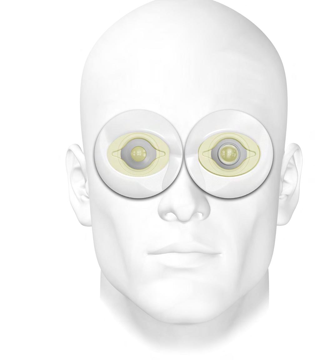
8 ORBIT Model The ORBIT is the holder for all BIONIKO anterior segment models. It provides an anatomical frame of reference and adds realism to the surgical scenario by challenging the user to manipulate instruments according to the facial structures around the eye. The ORBIT can be secured to any smooth surface (horizontal or vertical) with its integrated suction cup and will still retain a realistic degree of freedom that simulates head and eye movement. There are right and left ORBIT models to practice all approaches: Right-Superior, Right-Temporal, Left-Superior and Left-Temporal Pat. Pending


9 ORBIT 1 - BROW / SUPERIOR 2 - BRIDGE / NASAL 3 - ZYGOMATIC / INFERIOR 4 - TEMPORAL 5 - EYELID / SOCKET 6 - EYE MODEL 7 - SUCTION CUP FEATURES AND BENEFITS Provides anatomical frame of reference to anterior segment models Practice superior (1) or temporal (4) approaches on both left (L) and right (R) eyes Suction-cup firmly attaches to any smooth surface while retaining realistic movement Compact and portable design
, while providing the user with an anatomical frame of reference.")
10 FLEX-ORBIT Modular Training Platform SOCKET ADAPTER The FLEX-ORBIT modular training platform enhances and facilitates the use of ex-vivo and synthetic eye models for training and R&D purposes. It can position, secure and pressurize ex-vivo eyes of different sizes (18-26mm Ø), while providing the user with an anatomical frame of reference. The FLEX-ORBIT is designed to receive and complement the entire line of BIONIKO synthetic models. Whole globe models like OJOS and EXOS fit right in. With the included socket adapter, the FLEX-ORBIT can receive all anterior segment models and task trainers, such as the RHEXIS and KERATO. This makes the FLEX-ORBIT a versatile tool for any surgical training program. This compact platform is ideal for use in the classroom, research lab, wet-lab and even at the office to communicate with colleagues and patients. Pat. Pending


11 FLEX-ORBIT 1 - SUPERIOR (BROW) 2 - NASAL (BRIDGE) 3 - INFERIOR 4 - TEMPORAL 5 - ANTERIOR SCREW 6 - POSTERIOR SCREW 7 - SUCTION CUP 8 - DRAIN/PORT 9 - SOCKET ADAPTER 10 - ADAPTER GROOVE 11 - SNAP RING 12 - SNAP RING GAP 13 - EYELID 14 - SOCKET BASE FEATURES AND BENEFITS Use with both synthetic and biological tissue models* Adjust eye position with posterior screws (6) Adjust intra-ocular pressure with anterior screws (5) Suction-cup firmly attaches to any smooth surface Socket adapter for anterior segment models * For research use only

12 OJOS Model Eyes The OJOS Extraocular model simulates the external anatomical features of the eye, including conjunctiva, sclera, cornea and rectus muscles. This model allows demonstration and practice of numerous techniques requiring dissection and manipulation of the conjunctiva and sclera. Procedures encountered in glaucoma surgery, such as shunt implants and trabeculectomies, can be demonstrated and practiced on the model. The OJOS model can be used in conjunction with the FLEX- ORBIT platform for added challenge and realism. Pat. Pending

13 OJOS 1 - CORNEA 2 - CONJUNTIVA 3 - SCLERA 4 - MUSCLE INSERTION 5 - RECTUS MUSCLE 6 - OPTIC NERVE 7 - BASE FEATURES AND BENEFITS Loose and continuous conjunctiva/tenon s layer for realistic dissections Standardize your instruction and assessment around a consistent model Long shelf life No refrigeration needed No biohazard handling or disposal required Available on demand Decrease reliance on donor/animal tissue for training

14 Recovery Simulator The CORDELIA recovery simulator is based on an in-situ excision scenario. Eye bank technicians must master this technique to successfully recover delicate corneal tissue from donors in the field. CORDELIA is a task trainer that allows users to learn and practice the tissue recovery technique without using valuable donor tissue, in a realistic yet simple manner. Its repeatability and availability makes it ideal for developing standardized methods of instruction and assessment. The ORBIT (Sold separately) serves a holder for the CORDELIA models and provides support, frame of reference and the challenges posed by facial features surrounding the eye. The CORDELIA recovery model was made possible by the valuable guidance and support of LIONS Vision Gift

15 CORDELIA 1 - SCLERAL LAYER 2 - CHOROID LAYER 3 - LIMBUS 4 - SUPRA-CHOROIDAL SPACE 5 - SPUR 6 - CORNEA 7 - IRIS 8 - STRUCTURAL RING 9 - NOTCH DETAIL VIEW FEATURES AND BENEFITS Practice corneal detachment technique Refine sequence and manipulation of surgical instruments Decrease reliance on donor tissue for training Available on demand Standardize your instruction and assessment around a consistent model


 serves as a holder for the EXOS model and provides reference and realism by challenging the user to manipulate")
16 EXOS Enucleation Simulator The EXOS model focuses on simulating the challenging steps of an enucleation and corneal excision procedures. Learning to hook and transect the muscles and optic nerve is a fundamental skill for eye-bank technicians. The EXOS is coupled to our CORDELIA recovery simulator, allowing the practice of corneal excision with greater realism. Users can demonstrate and practice the sequence and manipulation of surgical instruments around anatomical landmarks. The EXOS model serves as a tool for introductory instruction and competency reviews. The FLEX-ORBIT (Sold separately) serves as a holder for the EXOS model and provides reference and realism by challenging the user to manipulate instruments according to the orbit cavity and facial structures around the eye. The EXOS Enucleation Simulator was made possible by the valuable guidance and support of Florida Lions Eye Bank

17 EXOS 1 - CORDELIA MODEL 2 - MUSCLE INSERTION 3 - RECTUS MUSCLE 4 - GLOBE 5 - OPTIC NERVE 6 - BASE FEATURES AND BENEFITS Realistic muscle traction and feel Practice enucleation and excision in one model Practice sequence and manipulation of surgical instruments Decrease reliance on donor tissue for training Available on demand Standardize your instruction and assessment around a consistent model
18 Phone: (507) BIONIKO Copyright 2015 BIONIKO
OPHTHALMIC SURGICAL MODELS
 OPHTHALMIC SURGICAL MODELS BIONIKO designs innovative surgical models, task trainers and teaching tools for the ophthalmic industry. Our surgical models present the user with dexterity and coordination
edge of the section wound, probably from opening of the wound
 CORNEO-SCLERAL SUTURE IN CATARACT EXTRACTION 269 A CORNEO-SCLERAL SUTURE IN CATARACT EXTRACTION. ITS TECHNIQUE AND ADVANTAGES BY H. B. STALLARD LONDON THE use of a corneo-scleral suture in the operation
Health Science 1110 Module 9 Sensations LAB 9. View the Film on Cornea Transplant and answer the questions on your laboratory worksheet.
 Health Science 1110 Module 9 Sensations LAB 9 View the Film on Cornea Transplant and answer the questions on your laboratory worksheet. Webpage Activities o Open Internet Explorer o Go to the Health Sciences
EYE ANATOMY. Multimedia Health Education. Disclaimer
 Disclaimer This movie is an educational resource only and should not be used to manage your health. The information in this presentation has been intended to help consumers understand the structure and
Sheep Eye Dissection
 Sheep Eye Dissection Question: How do the various parts of the eye function together to make an image appear on the retina? Materials and Equipment: Preserved sheep eye Scissors Dissection tray Tweezers
Contents. Corneal Markers, Scleral Depressors
 Corneal Markers, Scleral Depressors Contents Ring Markers... 4-1 LASIK Markers... 4-2 LRI/Astigmatism Markers... 4-3 Miscellaneous Markers... 4-4 Scleral Depressors... 4-5 4-B www.storzeye.com 800-338-2020
PRE-PLACED VERSUS POST-PLACED CORNEO-SCLERAL
 Brit. J. Ophthal. (1963) 47, 116. PRE-PLACED VERSUS POST-PLACED CORNEO-SCLERAL SUTURES IN CATARACT SURGERY* BY DHANWANT SINGH Department of Ophthalmology, Government Medical College, Patiala, Punjab, India
1. Introduction to Anatomy of the Eye and its Adnexa
 1. Introduction to Anatomy of the Eye and its Adnexa Fig 1: A Cross section of the human eye. Let us imagine we are traveling with a ray of light into the eye. The first structure we will encounter is
Surgical robot simulation with BBZ console
 Review Article on Thoracic Surgery Surgical robot simulation with BBZ console Francesco Bovo 1, Giacomo De Rossi 2, Francesco Visentin 2,3 1 BBZ srl, Verona, Italy; 2 Department of Computer Science, Università
FEMTO LDV Z8 IMAGE-GUIDED SURGERY ENHANCE YOUR SURGICAL PLANNING
 FEMTO LDV Z8 IMAGE-GUIDED SURGERY ENHANCE YOUR SURGICAL PLANNING FEMTO LDV Z8 OCT applications OCT APPLICATION: CATARACT Enhanced planning and customized individual treatments High resolution OCT images
GETTING STARTED WITH DMEK
 ORBIS WEBINAR MARCH 2018 GETTING STARTED WITH DMEK JAMES LEHMANN, MD SAN ANTONIO, TX GETTING STARTED WITH DMEK DISCLOSURES JAMES LEHMANN, MD Private Practice, Focal Point Vision Associate Instructor, University
Materials Cow eye, dissecting pan, dissecting kit, safety glasses, lab apron, and gloves
 Cow Eye Dissection Guide Introduction How do we see? The eye processes the light through photoreceptors located in the eye that send signals to the brain and tells us what we are seeing. There are two
TrHE CORNEO-SCLERAL SUTURE*
 Brit. J. Ophihal. (1954) 38, 232. TrHE CORNEO-SCLERAL SUTURE* A TECHNICAL MODIFICATION BY H. B. STALLARD London THE corneo-scleral suture is to-day widely recognized as important in the prevention of serious
2 Reusable Titanium. Single Piece Specula. Page 4-6
 Duckworth & Kent titanium surgical instrument manufacturer at the leading edge Specula Opening the eyes of the world Closed Blades Open Blades Solid Blades Find us on Single Piece Specula Page 4-6 Made
PRODUCT CATALOGUE Sterile Single Use Instruments for Cataract and Vitreoretinal Surgery
 PRODUCT CATALOGUE Sterile Single Use Instruments for Cataract and Vitreoretinal Surgery Safety. Consistency. Convenience. CONTENTS CONTENTS CONTENTS INTRODUCTION Page Single Use Instruments 4 5 Per Procedure
Dr. S.D. Gerber Double Row Method Surgical Technique
 Dr. S.D. Gerber Double Row Method Surgical Technique Dr. S.D. Gerber Double Row Method Surgical Technique Introduction There has been a rapid proliferation of techniques for arthroscopic rotator cuff tear
The Eye. (We ll leave the Lord Sauron jokes to you.)
") The Eye (We ll leave the Lord Sauron jokes to you.) When you look in the mirror, you only see a very small part of your eyes. In reality, they are incredibly complex organs with a pretty big job: enabling
The Eye (We ll leave the Lord Sauron jokes to you.) When you look in the mirror, you only see a very small part of your eyes. In reality, they are incredibly complex organs with a pretty big job: enabling
EYE: THE PHOTORECEPTOR SYSTEM. Prof. Dr. Huda Al Khateeb
 EYE: THE PHOTORECEPTOR SYSTEM Prof. Dr. Huda Al Khateeb Lecture 1 The eye ball Objectives By the end of this lecture the student should: 1. List the layers and chambers of the eye ball 2. Describe the
Basic Principles of the Surgical Microscope. by Charles L. Crain
 Basic Principles of the Surgical Microscope by Charles L. Crain 2006 Charles L. Crain; All Rights Reserved Table of Contents 1. Basic Definition...3 2. Magnification...3 2.1. Illumination/Magnification...3
Virtual Reality Phacoemulsification: a Comparison Between Skilled Surgeons and Students Naive To Cataract Surgery
 Virtual Reality Phacoemulsification: a Comparison Between Skilled Surgeons and Students Naive To Cataract Surgery Per Söderberg* anf, Carl-Gustaf Laurell af, Wamidh Simawi* anf, Per Nordqvist bf, Eva Skarman
Handout G: The Eye and How We See
 Handout G: The Eye and How We See Prevent Blindness America. (2003c). The eye and how we see. Retrieved July 31, 2003, from http://www.preventblindness.org/resources/howwesee.html Your eyes are wonderful
FEA of Prosthetic Lens Insertion During Cataract Surgery
 Visit the SIMULIA Resource Center for more customer examples. FEA of Prosthetic Lens Insertion During Cataract Surgery R. Stupplebeen, C. Liu, X. Qin Bausch + Lomb, SIMULIA, SIMULIA Abstract: Cataract
EXCHANGE. Financial Disclosure. Clinical pearls In advanced anterior segment surgery being able to do a IOL exchange is a must. Why Do an Exchange
 Financial Disclosure D. Ayres, MD Cornea Service IOLBrandon Wills Eye Hospital EXCHANGE Alcon Allergan AMO Bausch and Lomb TearScience BioTissue Why Do an Exchange Refractive surprise after cataract surgery
VR for Microsurgery. Design Document. Team: May1702 Client: Dr. Ben-Shlomo Advisor: Dr. Keren Website:
 VR for Microsurgery Design Document Team: May1702 Client: Dr. Ben-Shlomo Advisor: Dr. Keren Email: med-vr@iastate.edu Website: Team Members/Role: Maggie Hollander Leader Eric Edwards Communication Leader
Visual Optics. Visual Optics - Introduction
 Visual Optics Jim Schwiegerling, PhD Ophthalmology & Optical Sciences University of Arizona Visual Optics - Introduction In this course, the optical principals behind the workings of the eye and visual
Sensory receptors External internal stimulus change detectable energy transduce action potential different strengths different frequencies
 General aspects Sensory receptors ; respond to changes in the environment. External or internal environment. A stimulus is a change in the environmental condition which is detectable by a sensory receptor
Objectives. 3. Visual acuity. Layers of the. eye ball. 1. Conjunctiva : is. three quarters. posteriorly and
 OCULAR PHYSIOLOGY (I) Dr.Ahmed Al Shaibani Lab.2 Oct.2013 Objectives 1. Review of ocular anatomy (Ex. after image) 2. Visual pathway & field (Ex. Crossed & uncrossed diplopia, mechanical stimulation of
da Vinci Skills Simulator
 da Vinci Skills Simulator Introducing Simulation for the da Vinci Surgical System Skills Practice in an Immersive Virtual Environment Portable. Practical. Powerful. The da Vinci Skills Simulator contains
4Basic anatomy and physiology
 Hene_Ch09.qxd 8/30/04 6:51 AM Page 348 348 4Basic anatomy and physiology The eye is a highly specialized organ with an average axial length of 24 mm and a volume of 6.5 ml. Except for its anterior aspect,
Techniques of the hand tie and instrument tie
 Techniques of the hand tie and instrument tie 1. The Anatomy of a Square Knot A square knot consists of two "throws". Throws are constructed by crossing the ends of the suture to form a loop and then wrapping
OPTI-201/202 Geometrical and Instrumental Optics Copyright 2018 John E. Greivenkamp. Section 16. The Eye
 16-1 Section 16 The Eye The Eye Ciliary Muscle Iris Pupil Optical Axis Visual Axis 16-2 Cornea Right Eye Horizontal Section Zonules Crystalline Lens Vitreous Sclera Retina Macula And Fovea Optic Nerve
Microsurgical Suturing Techniques: Closure of the Cataract Wound
 Microsurgical Suturing Techniques: Closure of the Cataract Wound 4 Scott A. Uttley and Stephen S. Lane Key Points Surgical Indications The placement of a suture in a cataract wound should be considered
Medical Robotics. Part II: SURGICAL ROBOTICS
 5 Medical Robotics Part II: SURGICAL ROBOTICS In the last decade, surgery and robotics have reached a maturity that has allowed them to be safely assimilated to create a new kind of operating room. This
Section 22. The Eye The Eye. Ciliary Muscle. Sclera. Zonules. Macula And Fovea. Iris. Retina. Pupil. Optical Axis.
 Section 22 The Eye 22-1 The Eye Optical Axis Visual Axis Pupil Iris Cornea Right Eye Horizontal Section Ciliary Muscle Zonules Crystalline Lens Vitreous Sclera Retina Macula And Fovea Optic Nerve 22-2
Simple Interrupted Suture (using a silicon skin pad)
") (using a silicon skin pad) Disclaimer A series of booklets has been developed by the Clinical Skills Lab team (staff, recent graduates and students) from the School of Veterinary Sciences, University of
(using a silicon skin pad) Disclaimer A series of booklets has been developed by the Clinical Skills Lab team (staff, recent graduates and students) from the School of Veterinary Sciences, University of
Objectives. xxx00.#####.ppt 5/10/17 9:42 AM
 Objectives Understand and demonstrate proper suturing techniques Learn how to properly handle instruments Know types of suture and appropriate use Hand-tying Learn how to trouble-shoot complications after
Vision. By: Karen, Jaqui, and Jen
 Vision By: Karen, Jaqui, and Jen Activity: Directions: Stare at the black dot in the center of the picture don't look at anything else but the black dot. When we switch the picture you can look around
ADVANCED CLINICAL APPLICATIONS AND TROUBLESHOOTING IN SCLERAL LENSES
 ADVANCED CLINICAL APPLICATIONS AND TROUBLESHOOTING IN SCLERAL LENSES Langis Michaud, OD Jason Jedlicka, OD Disclosures Langis Honorarium and research grants Alcon Cooper Allergan Bausch*Lomb Johnson *
Anterior and Posterior Axial Lens Displacement and Human Aqueous Outflow Facility
 Investigative Ophthalmology & Visual Science, Vol. 29, No. 7, July 1988 Copyright Association for Research in Vision and Ophthalmology Anterior and Posterior Axial Lens Displacement and Human Aqueous Outflow
Instruments Commonly Used For Examination of the Eye
 Instruments Commonly Used For Examination of the Eye There are many instruments that the eye doctor might use to evaluate the eye and the vision system. This report presents some of the more commonly used
Biology 9 Senses Lab
 Biology 9 Senses Lab Objectives: To understand the anatomy and physiology of several of our senses both through observation and by means of some simple experiments and examinations. PART 1: The Eye 1.
The Eye. Nakhleh Abu-Yaghi, M.B.B.S Ophthalmology Division
 The Eye Nakhleh Abu-Yaghi, M.B.B.S Ophthalmology Division Coats of the Eyeball 1- OUTER FIBROUS COAT is made up of : Posterior opaque part 2-THE SCLERA the dense white part 1- THE CORNEA the anterior
Latest Product Releases
 Latest Product Releases Max360 Series Max360 Three Mirror Universal Max360 Magna View Latina 1X Indexing PSLT Latina 5 Bar Indexing SLT Hwang-Latina 5.0 Indexing SLT Hill Open Access Surgical Gonio Upright
Special Senses- THE EYE. Pages
 Special Senses- THE EYE Pages 548-569 Accessory Structures Eyebrows Eyelids Conjunctiva Lacrimal Apparatus Extrinsic Eye Muscles EYEBROWS Deflect debris to side of face Facial recognition Nonverbal communication
EYE. The eye is an extension of the brain
 I SEE YOU EYE The eye is an extension of the brain Eye brain proxomity Can you see : the optic nerve bundle? Spinal cord? The human Eye The eye is the sense organ for light. Receptors for light are found
Eye. Eye Major structural layer of the wall of the eye is a thick layer of dense C.T.; that layer has two parts:
 General aspects Sensory receptors ; External or internal environment. A stimulus is a change in the environmental condition which is detectable by a sensory receptor 1 Major structural layer of the wall
CATARACT SUTURES* During the last 15 years a great deal has been written on the subject of
 Brit. J. Ophthal. (1954) 38, 345. CATARACT SUTURES* BY W. L. HUGHES New York MOST eye surgeons used no sutures in routine cataract operations 30 years ago, but to-day some type of suturing is almost always
EXPAND YOUR VIEW. SUSTAIN YOUR ACCESS. OASIS IRIS EXPANDER DESIGNED FOR SAFETY & EFFICIENCY. PREMIER EDGE SAFETY SCALPELS SAFETY CRESCENT SCALPEL
 EXPAND YOUR VIEW. SUSTAIN YOUR ACCESS. OASIS IRIS EXPANDER PATENT NO. 8,439,833 B2 A polypropylene ring that gently expands the iris due to insufficiently dilated pupils and sustains visibility throughout
Laser Glasses
 Laser Glasses 2 Laser Glasess is a disposable product. It has %99 UV protection It is desinged to protect eye from light, wind or any external impacts Com24 hours continuous use due to its flexibility.
PRODUCT CATALOGUE Sterile Single Use Instruments for Cataract and Vitreoretinal Surgery
 PRODUCT CATALOGUE Sterile Single Use Instruments for Cataract and Vitreoretinal Surgery CONTENTS CONTENTS CONTENTS INTRODUCTION Page Single Use Instruments 4 5 Per Procedure Trays 7 CATARACT SURGERY Specula
Horizon. Your innovative partner in medicine technology.
 Horizon Valve Channel - Minimally Invasive Valve Surgery Original, Advanced, The Next Generation, Accessories, Sterilisation Container Your innovative partner in medicine technology. micvision.indd 1 08.10.2009
HUMAN Robot Cooperation Techniques in Surgery
 HUMAN Robot Cooperation Techniques in Surgery Alícia Casals Institute for Bioengineering of Catalonia (IBEC), Universitat Politècnica de Catalunya (UPC), Barcelona, Spain alicia.casals@upc.edu Keywords:
Sutureless, Glueless, Scleral Fixation of Single-Piece and Toric Intraocular Lens: A Novel Technique
 Published online: July 21, 2015 1663 2699/15/0062 0239$39.50/0 This is an Open Access article licensed under the terms of the Creative Commons Attribution-NonCommercial 3.0 Unported license (CC BY-NC)
implantation Marvin L Kwitko, MD Artificial lens
 Marvin L Kwitko, MD Artificial lens implantation Cataract formation is one of the major causes of blindness. More than 400,000 persons in the United States and about 10% of that number in Canada, most
The complete choice in refractive lens solutions
 The complete choice in refractive lens solutions The VeriFlex phakic IOL is part of Advanced Medical Optics range of high-contrast, high-quality refractive lens solutions. In addition to excellent training
Scleral Fixated Intraocular Lens
 Intraocular Lenses Scleral Fixated Intraocular Lens Vijay Kumar Sharma MS Vijay Kumar Sharma MS, Tarun Arora MD, Rajesh Sinha MD Dr. Rajendra Prasad Centre for Ophthalmic Sciences, All India Institute
THE EYE. People of Asian descent have an EPICANTHIC FOLD in the upper eyelid; no functional difference.
 THE EYE The eye is in the orbit of the skull for protection. Within the orbit are 6 extrinsic eye muscles, which move the eye. There are 4 cranial nerves: Optic (II), Occulomotor (III), Trochlear (IV),
Simple Interrupted Suture (using a tea towel)
") (using a tea towel) Disclaimer A series of booklets has been developed by the Clinical Skills Lab team (staff, recent graduates and students) from the School of Veterinary Sciences, University of Bristol,
(using a tea towel) Disclaimer A series of booklets has been developed by the Clinical Skills Lab team (staff, recent graduates and students) from the School of Veterinary Sciences, University of Bristol,
Trouble Shooting Guide for Ortho-K lenses
 Trouble Shooting Guide for Ortho-K lenses The basic design of the third generation e Lens for Orthokeratology 1. Optic Zone (Base curve, Compression zone, BC) width 5.6 to 6.4mm 2. Fitting curve (second
Sense Organs (Eye) The eye is the sense organ of sight. The eye is shaped like a ball and is located in bony
 The eye is the sense organ of sight. The eye is shaped like a ball and is located in bony") Sense Organs (Eye) The eye is the sense organ of sight. The eye is shaped like a ball and is located in bony sockets in the skull. It is held in place by six muscles which are joined to the outside of
Sense Organs (Eye) The eye is the sense organ of sight. The eye is shaped like a ball and is located in bony sockets in the skull. It is held in place by six muscles which are joined to the outside of
Technique Guide. Modular Sternal Cable System. Flexibility and strength in sternal closure and repair.
 Technique Guide Modular Sternal Cable System. Flexibility and strength in sternal closure and repair. Table of Contents Introduction Overview 2 Indications and Contraindications 3 Surgical Technique A.
SCANLAN LEGACY. High Precision Titanium Instruments
 SCANLAN LEGACY High Precision Titanium Instruments SCANLAN LEGACY Titanium Instruments Since 1921, Scanlan International has been dedicated to working hand-in-hand with surgeons to provide the highest
CONTACT LENSES Discover a different world
 CONTACT LENSES Discover a different world Tradition and innovation Since 1858, visionary thinking and a fascination with technology have guided us to develop innovative products of outstanding reliability:
THE BEST OF BOTH WORLDS Dual-Scheimpflug and Placido Reaching a new level in refractive screening
 THE BEST OF BOTH WORLDS Dual-Scheimpflug and Placido Reaching a new level in refractive screening Clinical Applications Corneal Implant Planning The comes with a licensable corneal inlay software module
BSS Training Kit (5th Ed)
") User Guide BSS Training Kit (5th Ed) Part No: 50070 (Day Kit) Part No: 5007 (Day Kit) BSS Day Kit BSS Day Kit Part No: 065-094 Issue, February 0 0 Limbs & Things For more Basic Surgical Skills training
User Guide BSS Training Kit (5th Ed) Part No: 50070 (Day Kit) Part No: 5007 (Day Kit) BSS Day Kit BSS Day Kit Part No: 065-094 Issue, February 0 0 Limbs & Things For more Basic Surgical Skills training
[Chapter 2] Ocular Geometry and Topography. Elements of Ocular Structure
![[Chapter 2] Ocular Geometry and Topography. Elements of Ocular Structure](/thumbs/95/124174711.jpg "[Chapter 2] Ocular Geometry and Topography. Elements of Ocular Structure") [Chapter 2] Ocular Geometry and Topography Before Sam Clemens became Mark Twain, he had been, among other things, a riverboat pilot, a placer miner, and a newspaper reporter, occupations in which success
[Chapter 2] Ocular Geometry and Topography Before Sam Clemens became Mark Twain, he had been, among other things, a riverboat pilot, a placer miner, and a newspaper reporter, occupations in which success
EnFocus Your Upgrade Path to High Performance Intrasurgical OCT
 Your Upgrade Path to High Performance Intrasurgical OCT is FDA 510(k) Cleared > Ultra HD OCT extends your microscope s potential with intrasurgical OCT BRILLIANT IMAGES, SUB-SURFACE KNOWLEDGE is an intrasurgical
Champion Instrumentation. Shoulder Repair Made Simpler
 Champion Instrumentation Shoulder Repair Made Simpler Champion Instruments Simplicity & Versatility The Champion Shoulder instrumentation system was designed with simplicity in mind. Clever instruments
DIAGNOSIS OF INTRA-OCULAR FOREIGN BODIES*
 Brit. J. Ophthal. (1959) 43, 744. USE OF THE ECHOGRAM IN THE LOCATION AND DIAGNOSIS OF INTRA-OCULAR FOREIGN BODIES* BY ARVO OKSALA AND ANTTI LEHTINEN From the Ophthalmic Department of the Central Finland
EYE STRUCTURE AND FUNCTION
 Name: Class: Date: EYE STRUCTURE AND FUNCTION The eye is the body s organ of sight. It gathers light from the environment and forms an image on specialized nerve cells on the retina. Vision occurs when
Wide range in small dimensions
 Wide range in small dimensions The complete solution for Minilaparoscopy The complete solution for Minilaparoscopy The use of 3.5 mm instruments and appropriate trocars leaves behind only very small scars
November 14, 2017 Vision: photoreceptor cells in eye 3 grps of accessory organs 1-eyebrows, eyelids, & eyelashes 2- lacrimal apparatus:
 Vision: photoreceptor cells in eye 3 grps of accessory organs 1-eyebrows, eyelids, & eyelashes eyebrows: protection from debris & sun eyelids: continuation of skin, protection & lubrication eyelashes:
Eyeball Model Lab Date Block
 Science 8 Name Eyeball Model Lab Date Block Problem: Identify the twelve key parts of the eye and describe their function. Materials: dissecting scissors ping pong ball transparent plastic ordinary scissors
Methods for Haptic Feedback in Teleoperated Robotic Surgery
 Young Group 5 1 Methods for Haptic Feedback in Teleoperated Robotic Surgery Paper Review Jessie Young Group 5: Haptic Interface for Surgical Manipulator System March 12, 2012 Paper Selection: A. M. Okamura.
Newsletter Spring 2015
 NEWSTM WORLD S LEADING CATARACT TECHNOLOGY FOR SMALL INCISION 015 SPRING VISION NEWS Vol. I Page : Micro Incision Cataract Innovations Page : Femtosecond Cataract Toric Marking Simplified Technology Page
ifs 150 khz IntraLase Technology
 How to create a perfect LASIK flap with Intralase Dr Lim Li, FRCS Head(Clinical & Education) Senior Consultant Corneal and External Eye Disease Service Singapore National Eye Centre Financial Interest:
What s Fundus photography s purpose? Why do we take them? Why do we do it? Why do we do it? Why do we do it? 11/3/2014. To document the retina
 What s Fundus photography s purpose? To document the retina Photographers role to show the retina Document other ocular structures Why do we take them? Why do we do it? We as photographers help the MD
Ezrif a. (12) United States Patent US 7,135,041 B2 V-CHOROID. Nov. 14, (45) Date of Patent: (10) Patent No.:
 United States Patent US 7,135,041 B2 V-CHOROID. Nov. 14, (45) Date of Patent: (10) Patent No.:") US007135041B2 (12) United States Patent Tashiro et al. (10) Patent No.: (45) Date of Patent: US 7,135,041 B2 Nov. 14, 2006 (54) ARTIFICIAL VISION SYSTEM (75) Inventors: Hiroyuki Tashiro, Aichi (JP): Yasuo
US007135041B2 (12) United States Patent Tashiro et al. (10) Patent No.: (45) Date of Patent: US 7,135,041 B2 Nov. 14, 2006 (54) ARTIFICIAL VISION SYSTEM (75) Inventors: Hiroyuki Tashiro, Aichi (JP): Yasuo
Introducing a lens that can take your scleral fitting to a higher plane.
 Introducing a lens that can take your scleral fitting to a higher plane. Alden Optical s newest specialty lens has arrived: Zenlens, a mini-scleral, fully vaulting lens co-designed with Jason Jedlicka,
T i t a n i u m I n s t r u m e n t s
 TITANIUM INSTRUMENTS T i t a n i u m I n s t r u m e n t s Titanium is a rare, lustrous metallic element which in its natural form is found in combination with other elements. The complex process of converting
THE XTRAFOCUS IS AN ELEGANT SOLUTION TO COMPLEX CASES.
 XtraFocus THE XTRAFOCUS IS AN ELEGANT SOLUTION TO COMPLEX CASES. CONTENT Based on the well-established principle of pinhole optics, this intraocular implant represents an innovative alternative for the
AHEAD Superior technology, thoughtfully designed with you in mind for an intelligent approach to cataract surgery.
 ALWAYS THINKING AHEAD Superior technology, thoughtfully designed with you in mind for an intelligent approach to cataract surgery. The LENSAR Laser System was designed with your efficiency in mind, so
INTRODUCING THE VIRTUAL REALITY FLIGHT SIMULATOR FOR SURGEONS
 INTRODUCING THE VIRTUAL REALITY FLIGHT SIMULATOR FOR SURGEONS SAFE REPEATABLE MEASUREABLE SCALABLE PROVEN SCALABLE, LOW COST, VIRTUAL REALITY SURGICAL SIMULATION The benefits of surgical simulation are
TORNIER MAXLOCK EXTREME. Clavicle Plating System SURGICAL TECHNIQUE
 TORNIER MAXLOCK EXTREME Clavicle Plating System SURGICAL TECHNIQUE 2 Table of Contents: Key Design Features...4 Surgical Technique...5 Implants & Instruments...9 3 Key Design Features There are 7 anatomically
Fitting Manual Use with kerasofttraining.com
 Fitting Manual Use with Fitting Manual: Contents This fitting manual is best used in conjunction with KeraSoft IC online training. To register, please visit www. 01 Kerasoft IC Design - Outlines the KeraSoft
Sutureless Trocar-Cannula Based Transconjunctival Flanged Intrascleral Intraocular Lens Fixation
 1:35 PM Sutureless Trocar-Cannula Based Transconjunctival Flanged Intrascleral Intraocular Lens Fixation Mark K. Walsh, MD, PhD OBJECTIVE To teach attendees a novel sutureless scleral intraocular lens
Optical Coherence Tomography Retina Scan Duo
 Optical Coherence Tomography Retina Scan Duo High Definition OCT & Fundus Imaging in One Compact System The Retina Scan Duo is a combined OCT and fundus camera system that is a user friendly and versatile
Arthroscopic Protector Meniscus Suturing Surgical Techniques
 Arthroscopic Protector Meniscus Suturing Surgical Techniques Protector Meniscus Suturing Protector Meniscus Suturing Technique I For the relatively rare meniscus tears located sufficiently anterior, to
Lab #11 - Nervous System II Senses
 Page1 Nervous System II Lab #11 - Nervous System II Senses Objectives: Dissect a sheep eye and identify the structures Observe a human eye model and identify the structures Observe a human ear model and
MatrixMANDIBLE Preformed Reconstruction Plates. Preshaped to the mandibular anatomy.
 MatrixMANDIBLE Preformed Reconstruction Plates. Preshaped to the mandibular anatomy. Technique Guide CMF Matrix Table of Contents Introduction MatrixMANDIBLE Preformed Reconstruction Plates 2 AO Principles
Epona Medical simulation products catalog Version 1.0
 Epona Medical simulation products catalog Version 1.0 Simulator for laparoscopic surgery Simulator for Arthroscopic surgery Simulator for infant patient critical care Simulator for vascular procedures
HSC Biology. Published Feb 9, 2017 HSC BIOLOGY OPTION: COMMUNICATION. By Sahar (99.1 ATAR)
") HSC Biology Year 2014 Mark 92.00 Pages 11 Published Feb 9, 2017 HSC BIOLOGY OPTION: COMMUNICATION By Sahar (99.1 ATAR) Your notes author, Sahar. Sahar achieved an ATAR of 99.1 in 2014 while attending Carlingford
HSC Biology Year 2014 Mark 92.00 Pages 11 Published Feb 9, 2017 HSC BIOLOGY OPTION: COMMUNICATION By Sahar (99.1 ATAR) Your notes author, Sahar. Sahar achieved an ATAR of 99.1 in 2014 while attending Carlingford
Most open angle glaucomas result from the retention of
 Glaucoma Ex Vivo Optical Coherence Tomography Imaging of Collector Channels with a Scanning Endoscopic Probe Jian Ren, 1 Henrick K. Gille, 2 Jigang Wu, 1 and Changhuei Yang 1,3 PURPOSE. To achieve high-fidelity
A M E D S MART SOLUTIONS FAMILY MEMBER
 A M E D S MART SOLUTIONS FAMILY MEMBER Based in the United Kingdom, Medsmart Solutions is a dynamic 100% British owned company which is dedicated to the manufacture and supply of an extensive range of
Eyes. Inspection Visual Acuity Visual Fields Pupillary Response Fundoscopic Exam
 Eyes Inspection Visual Acuity Visual Fields Pupillary Response Fundoscopic Exam Eye Examination Inspection 11.Inspects external ocular (eye) structures (lids, conjunctiva, iris, cornea, pupils) 12.Gently
IOVS. Ex vivo Optical Coherence Tomography Imaging of Collector Channels with. a Scanning Endoscopic Probe
 Papers in Press. Published on February 25, 2011 as Manuscript iovs.10-6744 IOVS Title Page: Ex vivo Optical Coherence Tomography Imaging of Collector Channels with a Scanning Endoscopic Probe Jian Ren
Please feel to contact Sonomed for any suggestions, comments or request about this bulletin.
 Sonomed Quarterly Bulletin Volume One, January 2008 Dear Sonomed Colleagues: Sonomed is proud to announce the VuMAX II Ultrasound Biomicroscope is experiencing huge success in the global ophthalmic markets.
INTRODUCING OPTICS CONCEPTS TO STUDENTS THROUGH THE OX EYE EXPERIMENT
 INTRODUCING OPTICS CONCEPTS TO STUDENTS THROUGH THE OX EYE EXPERIMENT Marcela L. Redígolo redigolo@univap.br Leandro P. Alves leandro@univap.br Egberto Munin munin@univap.br IP&D Univap Av. Shishima Hifumi,
Title: Live volumetric (4D) visualization and guidance of in vivo human ophthalmic surgery with intraoperative optical coherence tomography
 visualization and guidance of in vivo human ophthalmic surgery with intraoperative optical coherence tomography") Title: Live volumetric (4D) visualization and guidance of in vivo human ophthalmic surgery with intraoperative optical coherence tomography Authors: O. M. Carrasco-Zevallos 1, B. Keller 1, C. Viehland
Title: Live volumetric (4D) visualization and guidance of in vivo human ophthalmic surgery with intraoperative optical coherence tomography Authors: O. M. Carrasco-Zevallos 1, B. Keller 1, C. Viehland
HIP Solutions by Tornier MEIJE DUO 12/14 TAPER SURGICAL TECHNIQUE. surgical technique
 HIP Solutions by Tornier MEIJE DUO 12/14 TAPER SURGICAL TECHNIQUE surgical technique MEIJE DUO 12/14 TAPER SURGICAL TECHNIQUE Meije duotm H I P P R O S T H E S I S 1. Locating the pelvic horizontal line