OCULUS Keratograph 5M Topographer
|
|
|
- Roy Lewis
- 5 years ago
- Views:
Transcription


1 OCULUS Keratograph 5M Topographer


 Marc Schulze, PhD, Dipl. Eng.")


2 OCULUS Keratograph 5M Topographer The multi-purpose topographer has become an integral part of the ophthalmological and optometric practice. Examiner-independent measurements provide reliable data, clear analyses and full documentation. Clear and easy-to-understand representations facilitate communication with your patients and ensure a time-saving workflow. The Keratograph 5M is one of the most versatile instruments that we have in our practice. It is highly valuable and efficient for a very busy and technologydriven eye care practice such as ours. Barry Eiden, O.D., USA The Keratograph with easy handling when it comes to performing meibography and excellent quality images really won me over! Elisabeth Messmer, M.D.,Germany I use the R-Scan for contact lens fitting and documentation of ocular changes what a helpful visual consultation tool! (FH) Marc Schulze, PhD, Dipl. Eng., Canada In my clinic we use the automated pupillometry of the Keratograph for more accurate diagnosis of mild concussions. The examination takes one minute to complete. One minute for clinicians to reduce neuropsychological problems among athletes. Rolando Toyos, M.D., USA The information that I get from this instrument plays a very important role in the fitting of all forms of rigid gas-permeable contact lenses, as well as, the simple fits of everyday soft lenses. Chris Eksteen, DipOptom, South Africa I use the Keratograph imaging tool to assess the fit of contact lenses without any additional fluorescein! Sebastian Marx, Dipl. Eng.,Germany

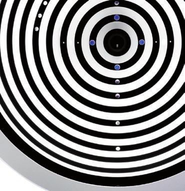
3 OCULUS Keratograph 5M The Allrounder Measurements With Placido Ring Illumination White ring illumination is used to measure thousands of points on the entire corneal surface. Infrared ring illumination is also available for analyzing the tear fi lm in order to prevent reflex tear secretion caused by glare. LED Measurements The Keratograph 5M proudly offers the perfect illumination for each function: White diodes for tear film dynamics, blue diodes for fluorescein images and infrared diodes for meibography. > white illumination > infrared illumination > blue diodes Where to find? Precise measurement of the corneal shape Extensive analyses and graphics Automatic keratoconus detection Course of disease displays Image and video documentation Measuring instruments Selection of contact lenses Fluorescein image simulation OxiMap Tear film analysis Meibography Classification of redness Technical data Network connection ability Software overview Topography Documentation Contact Lens Fitting Dry Eye Screening Technology & Software


4 Topography Quick, precise and clear Aside from topography and automatic keratoconus detection, the Keratograph 5M provides a large contact lens data base and many analyses for daily practice. The built-in keratometer and automatic measurement ensure the utmost accuracy and reproducibility. After completing the measurement, the overview display provides a detailed outline. Keratometric data, diameter of the cornea and pupil, K-values and index for keratokonus detection, size of the analyzed surface Built-in measuring instrument for measurements in the camera image 3D view can be selected and displayed directly beside the camera image Display as sagittal or tangential curvature, elevation data or refractive power, overlay of apex position, pupil centre and contour, numerical values and major meridians


5 Detailed Display of the Cornea The Keratograph software includes a reliable screening package for corneal disease detection, lens fitting and refractive surgery. The complex corneal surface structure is measured by means of mathematical analyses, which serves as the basis for accurate detection of irregularities like keratoconus. In addition, optical properties of the front surface of the cornea are exactly characterized. Fourier Analysis Zernike Analysis Keratoconus Detection The refractive power of the front surface of the cornea consists of different components. The Fourier Analysis identifies four of them which are shown in the following displays: Spherical component Decentration Regular astigmatism Irregularities Zernike polynomials are adapted to the elevation data of the cornea, which is crucial for locating the apex. The apex position is labelled with a cross. This display shows you if a rear surface toric lens is applicable to the particular case. Zernike polynomials and the aberration coefficient give you important indications of the imaging quality of the corneal surface. Abnormal values are marked in colour. Keratoconus classification is based on numerous parameters. The indices display merges these parameters. The coloured label illustrates abnormal values. Temporal changes of the parameters are shown side by side in a table, to facilitate your follow-ups. The Amsler classification system is applied to the keratoconus domains. Pathological changes can be quantified and possible effects on visual acuity can be explained. Topography Documentation Contact Lens Fitting Dry Eye Screening Technology & Software

6 Complete Documentation Follow-ups provide reliability Follow-ups require comparison of several examinations. In doing so, changes can be easily detected and fully documented. Regular follow-up examinations provide reliability and increase the trusting relationship between you and your patient. The Keratograph software contains both data and image documentation. Comparing Examinations The comparing three examinations display shows changes over a certain period of time, e.g. the progressive course of disease of keratoconus. Choose between sagittal and tangential curvature and between elevation data and refractive power. Use the comparing two examinations display for a right/left or before/after comparison. The easy-tounderstand displays help you describe even complex contexts to your patient. Course of Disease display showing three examinations Selection of examination from the patient data base Graphic display of differences between individual examinations. Display as sagittal or tangential curvature, elevation data or refractive power.


7 A Picture Is Worth a Thousand Words. The Keratograph 5M contains features that offer optimal conditions for your image documentation such as the high-resolution colour camera and different illumination options. An image aids in communication with education of your patients, thus eliminating the need for long explanations. You save time with only one mouse click. Precise Measurements Instead of Rough Guesses The Keratograph 5M is the ideal device for your professional documentation. The imaging software includes features such as magnification function hand tool measuring tool angle measurement High-Resolution Images You can evaluate the wettability of contact lenses, without fluorescein application and determine the exact rotating of toric lenses. It is also possible to detect lipids and deposits on the lens surface, as well as corneal staining or vascularization. Show your patients images they have never seen before. Reliable Diagnosis Documentation The resulting classifi cation from corneal staining requires well-trained examiners. It is diffi cult to estimate the number of hyper-fluorescent dots on the corneal surface, but the integrated JENVIS grading scale facilitates this evaluation. Every image taken can be compared with a sample image on the screen. Vessel injections can also be evaluated and documented in this way. Pathological changes can be exactly localized, and changes in size can be determined. This ensures that all of your patients questions will be answered. Topography Documentation Contact Lens Fitting Dry Eye Screening Technology & Software

8 Contact Lens Fitting Professionalism through innovation An ideal lens is chosen from the large lens data base and is then suggested in the lens fitting display. Based on this topographic data, a simulated fluorescein image of this particular lens is created. You can then take real fluorescein images with the Keratograph 5M and compare them with the simulated images. Selection between RGP and soft lenses Contact lens suggestion from the large data base Subjective refraction data and CVD conversion Keratometric data, diameter of the cornea and pupil, fixation deviation Distance of major meridians of the cornea from the lens Simulated fluorescein image of a toric RGP lens Eccentricity values for both major meridians


9 Multifocal, Bifocal, Toric With the Keratograph 5M you can quickly and precisely measure all of the data needed for multifocal, bifocal and toric contact lenses. These measurements also facilitate the fitting of multifocal or bifocal lenses. Furthermore the Keratograph 5M software can be linked to fitting programs of various contact lens manufacturers. Pupillometry Using the Pupillometry option is a quick and easy way to measure the pupil size of your patients under different illumination conditions. This option not only supports you when fitting multifocal lenses, but also when measuring the optical zone before refractive or cataract surgery. Near-Portion Height Measurement The near-portion height of RGP bifocal lenses can be simulated and precisely determined with this software, even before ordering the first-fitting lens. This also facilitates the complex fitting of multifocal lenses. Palpebral Angle Measurement The imaginable angle of the nasal side of the lower eyelid can be measured to determine the expected nasal rotation when fitting lenses for astigmatism. Topography Documentation Contact Lens Fitting Dry Eye Screening Technology & Software


10 OxiMap Visualizing the oxygen transmissibility of soft lenses An intact tear film and good oxygen supply to the cornea are essential for comfortable lens wear. The OxiMap displays the oxygen transmissibility of soft lenses in different colours depending on the optical power and is easy to understand even for your patients. oxygen transmissibility (Dk/t value) wearing contact lenses up to 8 hours Dk/t > 35 wearing contact lenses up to 30 days Dk/t > 125 wearing contact lenses up to 7 days Dk/t > 87 Recommended lens wearing time Influence of Contact Lens Wearing Time The oxygen transmissibility is an important quality criterion of soft lenses. It is indicated as Dk/t value, and has a significant influence on the recommended lens wearing time. The higher the Dk/t value, the more oxygen gets through the lens to the cornea. Oxygen transmissibility changes depending on the material and the optical power of the lens. Only measurements of oxygen transmissibility in the centre of a lens with dpt have been demonstrated thus far. For the first time, the OxiMap integrated in the Keratograph 5M displays Dk/t values over the entire surface depending on the contact lens power. You choose the lens type and the respective power. The OxiMap is projected onto your patient's eye and you can immediately see if the selected lens is suitable for wearing overnight, for example. Explain to your patient the advantages of modern contact lenses. cm/sec The recommendations and Dk/t values stated [ x 10 9 ] refer to: ml O 2 /ml x mm Hg Harvitt & Bonanno et al.: Re-Evaluation of the Oxygen Diffusion Model for Predicting Minimum Contact Lens Dk/t Values Needed to Avoid Corneal Anoxia. Optom. Vis. Sci. 1999; 76:

11 Topography Documentation Contact Lens Fitting Dry Eye Screening Technology & Software


12 JENVIS Dry Eye Report Dry eye screening made easy Find the cause of dry eye quickly and reliably. The JENVIS Dry Eye Report is a unique tool for doing this. After measurements are taken using the Keratograph 5M and the slit lamp, your customer/patient receives an easy-to-grasp print-out. The Dry Eye Report combines screening and consultancy. Your personal logo on the Dry Eye Report Your personal recommendation for the customer/patient Real image of the patient s eye after use of the R-Scan on the Keratograph 5M Easy-to-grasp Radar chart containing 6 essential measurements for reliable assessment of the screening Abbreviations and technical terms are comprehensibly explained to your customer/patient Individual measurements and results are presented in a clear-cut fashion

 What good would a screening be without history taking? Commonly used questionnaires are incorporated into the Dry Eye Report on the Keratograph 5M.")
13 3 Tools for the JENVIS Dry Eye Report Here s How it Works Drawing up a comprehensive Dry Eye Report entails filling out a questionnaire, measuring the LIPCOF using the slit lamp and taking four measurements using the Keratograph 5M. Other nationally and internationally approved screening methods can be added with ease numerous supplementary procedures ranging from eyelid blink frequency to staining are incorporated into the Keratograph 5M. Dry Eye Questionnaire (DEQ) What good would a screening be without history taking? Commonly used questionnaires are incorporated into the Dry Eye Report on the Keratograph 5M. OSDI (Ocular Surface Disease Index) McM (McMonnies). You can choose your favourite one. A look through the slit lamp The slit lamp is the number one instrument when it comes to eye diagnostics. To draw up a comprehensive Dry Eye Report it is necessary to assess the LIPCOF (lid-parallel conjunctival folds). You enter the results into the Dry Eye Report on the Keratograph 5M. Non-invasive measurements The practical tools provided by the Keratograph 5M help you perform comprehensive Dry Eye analysis. Analyzing the level of redness using R-Scan, measurement of tear meniscus height, tear film break-up time and meibography can all be performed with ease using the Keratograph 5M. The result? A comprehensive Dry Eye Report. Topography Documentation Contact Lens Fitting Dry Eye Screening Technology & Software

14 TF-Scan Evaluation of non-invasive tear film break-up time The non-invasive tear film break-up time (NIKBUT) measures tear film stability. The NIKBUT is automatically measured within seconds, without fluorescein application. Human eyes are not able to perceive infrared illumination. Glare and reflex tear secretion are therefore avoided during the examination. The TF-Scan visualizes the results in an easy and understandable way for you and your patients. The Tear Map shows the affected areas: The respective break-up time is graphically illustrated for each segment in seconds and according to the principle of a traffic light. The graph shows percent of the examined area that is affected during the measuring period. Data field showing tear film break-up time (NIKBUT) in seconds and the corresponding classification. You can watch the video after the measurement. The break-up areas detected by the software are highlighted accordingly.


15 Quantity and Quality of the Tear Film The high-resolution colour camera makes the smallest structures visible. This enables you to measure the tear meniscus height and evaluate the lipid layer, as well as analyze the tear fi lm dynamics. Not only do you gain very important fi ndings about tear fi lm break-up time, but also those about the quantity and quality of the tear fi lm. Tear Meniscus Height Never has a precise measurement been so easy. You can evaluate the course of the tear meniscus along the eyelid by means of the new infrared illumination and precisely measure the tear meniscus height with the built-in ruler. Different magnification levels facilitate measurement and the resulting value is automatically saved in the patient file. Evaluation of Lipid Layer Hyper-evaporative dry eye is easily overlooked when using conventional tests. Thus evaluating the lipid layer of the tear film is even more important. With the Keratograph 5M you can record videos of interference patterns of the lipid layer. Distribution characteristics, morphology and thickness of the lipid film can be continuously evaluated. Tear Film Dynamics The tear film contains numerous particles. These can be made visible using a specific light source. These particles are distributed in the tear fluid from bottom to top during each blink. The velocity of these particles provides information on tear film viscosity. You can quickly and easily evaluate the quantity and movement of these tear film particles using the TF-Scan. Topography Documentation Contact Lens Fitting Dry Eye Screening Technology & Software


16 Meibo-Scan Meibography of the upper and lower eyelid The new multi-functional Keratograph 5M easily and efficiently integrates difficult examinations such as meibography. The dysfunction of meibomian glands is the most frequent cause of dry eye. Morphological changes in the gland tissue are made visible using the Meibo-Scan. Easy Operation Through Optimum Working Distance The Keratograph 5M enables a greater working distance in the examination of the eyelids. This makes it easy to evert the upper and lower eyelid and to assess the meibomian glands. Convincing Images for Reliable Evaluation Different views can be selected for a precise analysis of the meibomian glands. Even untrained examiners can easily perform this evaluation due to the labelling of the individual examination field and the high-contrast display.

17 R-Scan Automatic classification of conjunctival redness Previously conjunctival redness evaluation has been carried out subjectively, and the results have varied according to the examiner s qualification. Now for the first time it is possible to objectively classify bulbar and limbal redness completly and automatically using the R-Scan. The R-Scan detects vessels in the conjunctiva and evaluates the degree of redness. Automatic classification eliminates the need for time-consuming comparison and provides greater reliability during evaluation. Bulbar and Limbal Redness Different display options help to classify the degree of redness. Choose between the camera image, view of fine vessels in the conjunctiva, red-free or contrast-enhanced display options. Bulbar and limbal redness are evaluated in the temporal and nasal areas, and all results are saved automatically. JENVIS Grading Scale The degree of redness is based on the JENVIS grading scale. The comparison of your examination results with the actual-scale images of the JENVIS grading scale facilitates the conversation when consulting with your patient. Further information on possible causes of redness, the normal condition as well as practical notes for capturing an image are provided below the actual-scale images. Topography Documentation Contact Lens Fitting Dry Eye Screening Technology & Software
18 All Features at a Glance Customize the OCULUS Keratograph 5M to your own requirements! Software included Topography Lens rear surface measurement Overview Display Colour maps 4 maps selectable Camera image 3D view Fourier Analysis Zernike Analysis Indices Elevation map Corneal asphericity Contact lens fitting Two examination display Two examination comparison Three examination comparison Optional examination functions TF-Scan Evaluation of lipid layer and tear film dynamics, measurement of tear meniscus height and non-invasive tear film break-up time (NIKBUT) R-Scan Automatic classification of bulbar and limbal redness Meibo-Scan Meibography of upper and lower eyelid Pupillometry Examination of pupillary response using the pupillometer, asymmetry test and manual measuring mode Imaging Image and video documentation with fluorescein imaging, near-portion height measurement and eyelid angle measurement Optional evaluation functions Keratoconus package Includes Indices and Zernike Analysis Contact lens fitting Simulation of fluorescein images of RGP lenses OxiMap Graphic display of oxygen transmissibility (Dk/t value) of soft lenses JENVIS Dry Eye Report Comprehensive summary display of all available dry eye tests My wish list My wish list Topography Documentation Contact Lens Fitting Dry Eye Screening Technology & Software
19 Floating License Key More flexibility with the OCULUS license model Patient check-in Activate Functions Exactly as You Need Them The choice is yours in how you use the Keratograph 5M and which examination and evaluation functions you desire. You can order additional functions of optional evaluation functions, according to your modular design principle. After purchase, licenses for the respective evaluation functions are activated on the OCULUS Floating License Key and are provided in your network. It is possible to call and view previously performed examinations for free on all workstations within the network. Optional examination function TF-Scan R-Scan Meibo-Scan Pupillometry Imaging Optional evaluation functions Keratoconus package Contact lens fitting OxiMap JENVIS Dry Eye Report Examination room 1 Examination room 2 Server (with/without DICOM) You can decide which additional functions to allocate to each device. Examination room 3 Efficiency Through Networking Consultation The OCULUS patient data management system enables you to merge all OCULUS devices in a local network. It allows you to collaborate with external data management systems (EMR) to optimize your workflows. DICOM interface is not necessary for device connection. Topography Documentation Contact Lens Fitting Dry Eye Screening Technology & Software
 Imaging illumination: blue diodes (465 nm) Meibography: infrared")
 Weight 3.2 kg (7.1 lbs) (measuring equipmentl) 6.1 kg (13.5 lbs) (with xy base) Max.")
 in accordance with Medical Device Directive 93/42/EEC WWW.OCULUS.")

20 Technical Data OCULUS Keratograph 5M General Information Precision ± 0.1 dpt Reproducibility ± 0.1 dpt Number of rings 22 Working distance 78 / 100 mm Number of evaluated data points Camera Digital CCD camera Illumination source Placido illumination: white diodes Placido illumination: infrared diodes (880 nm) Imaging illumination: blue diodes (465 nm) Meibography: infrared diodes (840 nm) Tear film dynamics: white diodes Pupillometry illumination: infrared diodes (880 nm) Technical specifications Dimensions (W x D x H) 275 x x mm (10.8 x x in) Weight 3.2 kg (7.1 lbs) (measuring equipmentl) 6.1 kg (13.5 lbs) (with xy base) Max. power consumption 25 W Voltage V AC Frequency Hz Minimum PC requirements Processor: Intel Core i3 or better, 4GB main memory, Hard disk: 500GB and more, graphics card: Intel HD Graphics 2000 or better, recommended screen resolution: 1920 x 1080 (full HD) in accordance with Medical Device Directive 93/42/EEC OCULUS is certified by TÜV according to DIN EN ISO OCULUS Optikgeräte GmbH Postfach Wetzlar GERMANY Tel Fax export@oculus.de OCULUS USA, sales@oculususa.com OCULUS Asia, info@oculus.hk OCULUS Czechia, oculus@oculus.cz OCULUS Iberia, info@oculus.es OCULUS Poland, biuro@oculus.pl OCULUS Turkey, info@oculus-turkey.com.tr mm in 250 mm 9.8 in 275 mm 10.8 in mm in The availability of products and features may vary by country. OCULUS reserves the right to change product specifications and design. All information is valid at the time of printing (04/15) 23/0415/EN/HA P/77000/EN
OCULUS Keratograph 5M Topographer
 OCULUS Keratograph 5M Topographer OCULUS Keratograph 5M Topographer The multi-purpose topographer has become an integral part of the ophthalmology and optometry practices. User independent measurements
OCULUS Keratograph 4. Topographer. We focus on progress
 OCULUS Keratograph 4 Topographer We focus on progress Ophthalmologist Versatile and precise For me the Keratograph 4 is an indispensable device for diagnosis and surgical planning. Its automatic measurement
OCULUS Easygraph. So Small and Yet a Topographer. Small, but Efficient As a Big One. All Important Parameters at a Glance
 OCULUS Easygraph So Small and Yet a Topographer Small, but Efficient As a Big One Wanting to work with a video keratometer in spite of limited space? Then the OCULUS Easygraph is what you re looking for.
OCULUS Easygraph. Topographer. We focus on progress
 OCULUS Easygraph Topographer We focus on progress The Compact Corneal Topographer at a Glance Corneal topographer with built-in keratometer Comfortable, hygienic working distance reduces the influence
OCULUS Pentacam AXL Always an Axial Length Ahead
 OCULUS Pentacam AXL Always an Axial Length Ahead EFFICIENCY AND BETTER WORKFLOW Your Cataract Workstation! The new Pentacam AXL is an alliance of the time-tested Pentacam technology with high-precision
The New Corvis ST. Evaluation of corneal biomechanical response, tonometry and pachymetry. Biomechanical Response
 The New Corvis ST Evaluation of corneal biomechanical response, tonometry and pachymetry. The revolutionary Corvis ST records the reaction of the cornea to a defined air pulse using a newly developed high-speed
OCULUS ImageCam 2. Slit lamp documentation. We focus on progress
 OCULUS ImageCam 2 Slit lamp documentation We focus on progress OCULUS ImageCam 2 Slit lamp documentation at its best Versatile applications The OCULUS ImageCam 2 can easily document the results of your
OCULUS Binoptometer 4P
 OCULUS Binoptometer 4P Professional Vision Testing Device We focus on progress Innovative Test presentation on a high-resolution color display Visual acuity test Landolt rings conform to DIN EN ISO 8596
Refractive Power / Corneal Analyzer. OPD-Scan III
 Refractive Power / Corneal Analyzer OPD-Scan III Comprehensive Vision Analysis and NIDEK, a global leader in ophthalmic and optometric equipment, has created the OPD-Scan III, the third generation aberrometer
Sirius TOMOGRAPH AND CORNEAL TOPOGRAPHER
 Sirius TOMOGRAPH AND CORNEAL TOPOGRAPHER EN Sirius TOMOGRAPH AND CORNEAL TOPOGRAPHER Combines placido disk topography with Sheimpflug tomography of the anterior segment. Sirius provides information on
OCULUS Twinfield 2. Perimeter. We focus on progress
 OCULUS Twinfield 2 Perimeter We focus on progress Ophthalmologist Definitely my all-round favourite! For glaucoma care, macular examinations and neurological cases as well as for formulation of expert
ATLAS Corneal Topography System
 ATLAS Corneal Topography System Simply accurate for maximum productivity Model 9000 The New ATLAS Take your practice to the next level Carl Zeiss Meditec has taken the world s leading corneal topography
Dynamic Ophthalmic Software for today s market. Automated Grading System
 Dynamic Ophthalmic Software for today s market Automated Grading System AOS Anterior is a new device agnostic software designed to analyse digital images of the eye and automate the grading scale for (1)
Optometrist Private ophthalmologists
 Public hospital Post-operative follow up Specialist diagnosis Screening service Optometrist Private ophthalmologists Ophthalmic diagnostics anywhere, anytime TEARSCOPE (Tear Film Screening) The scattered
FITTING GUIDE PRACTITIONER S ROSE K2 KC ROSE K2 NC ROSE K2 IC ROSE K2 PG NIPPLE CONE IRREGULAR CORNEA POST GRAFT
 Keratoconus Nipple Cone Irregular Cornea Post Graft PRACTITIONER S FITTING GUIDE NIPPLE CONE IRREGULAR CORNEA POST GRAFT Four lens designs... One simple systematic approach to fitting Featuring Easy-to-fit
OCULUS Easyfield. Perimeter. We focus on progress
 OCULUS Easyfield Perimeter We focus on progress OCULUS Easyfield The right choice for all your needs The OCULUS Easyfield is a full-fledged compact perimeter capable of performing standard automated perimetry
(495) (495)
 (495)") МЕДТЕХНИКА-СТОЛИЦА (495) 902-59-26 (495) 518-55-99 127 238, г. Москва, Дмитровское ш. 85 ATLAS Corneal Topography Product Overview Model 9000 ATLAS Model 9000 Overview Next-generation corneal topography
МЕДТЕХНИКА-СТОЛИЦА (495) 902-59-26 (495) 518-55-99 127 238, г. Москва, Дмитровское ш. 85 ATLAS Corneal Topography Product Overview Model 9000 ATLAS Model 9000 Overview Next-generation corneal topography
Wave Front Topography. ReSeeVit Evolution Topography Module for Modi Topographer
 Wave Front Topography ReSeeVit Evolution Topography Module for Modi Topographer Introduction The aberrations in the central optical zone have a greater effect than those closer to the edge. From an optical
SOFT (HYDROPHILIC) CONTACT LENSES DAILY WEAR FOR PLANNED REPLACEMENT OR DAILY DISPOSABLE. PRACTITIONER FITTING GUIDE July 2009
 CONTACT LENSES DAILY WEAR FOR PLANNED REPLACEMENT OR DAILY DISPOSABLE. PRACTITIONER FITTING GUIDE July 2009") BIOMEDICS 55 (ocufilcon D) BIOMEDICS 55 Toric (ocufilcon D) BIOMEDICS 55 Multifocal (ocufilcon D) SOFT (HYDROPHILIC) CONTACT LENSES DAILY WEAR FOR PLANNED REPLACEMENT OR DAILY DISPOSABLE PRACTITIONER FITTING
BIOMEDICS 55 (ocufilcon D) BIOMEDICS 55 Toric (ocufilcon D) BIOMEDICS 55 Multifocal (ocufilcon D) SOFT (HYDROPHILIC) CONTACT LENSES DAILY WEAR FOR PLANNED REPLACEMENT OR DAILY DISPOSABLE PRACTITIONER FITTING
THE BEST OF BOTH WORLDS Dual-Scheimpflug and Placido Reaching a new level in refractive screening
 THE BEST OF BOTH WORLDS Dual-Scheimpflug and Placido Reaching a new level in refractive screening Clinical Applications Corneal Implant Planning The comes with a licensable corneal inlay software module
 Página 1 de 9 TopPage > Eye Care > Diagnostic > Wave-Front Analyzer KR-1W Wave-Front Analyzer KR-1W Perfection for Professionals : KR-1W Topcon, with its wealth of experience in designing and manufacturing
An Interesting Use of Bausch and Lomb s KeraSoft IC Lens
 An Interesting Use of Bausch and Lomb s KeraSoft IC Lens Nate Schlotthauer, OD 2012 Michigan College of Optometry Cornea and Contact Lens Resident Introduction: The KeraSoft IC lens, introduced to the
Aberrometry in Clinical Practice
 Aberrometry in Clinical Practice Aravind Roy, M.S L V Prasad Eye Institute KVC Campus, Vijayawada, India No financial disclosures No conflicts of interest What is your position? Poll Question 1 1. Ophthalmologist
KERATOCONUS. In the most advances cases, the corneal deformation can be easy observed fig. 1. Fig. 1
 Mario Giovanzana Milano, 14 nd october 01 KERATOCONUS INTRODUCTION The keratocunus is a deformation of the cornea that tends to assume the shape of a cono. The genesis is substantially uncertain. It is
Lecture 2 Slit lamp Biomicroscope
 Lecture 2 Slit lamp Biomicroscope 1 Slit lamp is an instrument which allows magnified inspection of interior aspect of patient s eyes Features Illumination system Magnification via binocular microscope
What s a Corneal GP Lens?
 Slide 1 What s a Corneal GP Lens? Richard Dorer NCLEC Blanchard Contact Lens Inc. 800-367-4009 x 131 richarddorer@gmail.com www.blanchardlab.com Slide 2 Endorsements I am a paid representative and consultant
DesiGneD and ManUFaCTUreD in italy The sharpest vision.
 The sharpest vision. DESIGNED AND MANUFACTURED in ItalY 2 A complete diagnostic station used in clinical practice and research to analyze the optical environment of ocular aberration. Its functions are:
We focus on progress OCULUS CENTERFIELD 2
 We focus on progress OCULUS CENTERFIELD 2 Oculus Centerfield 2 Projection perimeter for visual field tests up to 70 Our know-how to your benefit Take advantage of the more than 50 years experience of Oculus
Experience with correcting myopia with different types of contact lenses
 Experience with correcting myopia with different types of contact lenses Edward BENNETT Refer this article as: Bennett, E., Experience with correcting myopia with different types of contact lenses, Points
THE SHARPEST VISION. DESIGNED AND MANUFACTURED IN ITALY
 THE SHARPEST VISION. DESIGNED AND MANUFACTURED IN ITALY 2 A complete diagnostic station used in clinical practice and research to analyze the optical environment of ocular aberration. Its functions are:
Principles and clinical applications of ray-tracing aberrometry (Part II)
") UPDATE/REVIEW Principles and clinical applications of ray-tracing aberrometry (Part II) Alfredo Castillo Gómez, MD, PhD 1 ; Antonio Verdejo del Rey, OD 2 ; Carlos Palomino Bautista, MD 3 ; Ana Escalada
UPDATE/REVIEW Principles and clinical applications of ray-tracing aberrometry (Part II) Alfredo Castillo Gómez, MD, PhD 1 ; Antonio Verdejo del Rey, OD 2 ; Carlos Palomino Bautista, MD 3 ; Ana Escalada
FOR FREQUENT REPLACEMENT SOFT HYDROPHILIC CONTACT LENSES PRACTITIONER FITTING GUIDE
 AVAIRA (enfilcon A) AVAIRA Toric (enfilcon A) & AVAIRA Multifocal (enfilcon A) FOR FREQUENT REPLACEMENT SOFT HYDROPHILIC CONTACT LENSES PRACTITIONER FITTING GUIDE Part Number: PFG01012 Page 1 of 13 Table
OPH 260 BASIC CONTACT LENS CONCEPTS
 OPH 260 BASIC CONTACT LENS CONCEPTS COURSE DESCRIPTION: Prerequisites: OPH 121 and OPH 141 Corequisites: None This course introduces the theory of contact lens fitting. Emphasis is on rigid and soft contact
Trouble Shooting Guide for Ortho-K lenses
 Trouble Shooting Guide for Ortho-K lenses The basic design of the third generation e Lens for Orthokeratology 1. Optic Zone (Base curve, Compression zone, BC) width 5.6 to 6.4mm 2. Fitting curve (second
family of lens designs fitting guide ICD is Exclusively Manufactured In
 TM family of lens designs fitting guide ICD is Exclusively Manufactured In paragon 1 Select Initial Diagnostic Lens: Identify the Corneal Condition Normal Depth Eyes Normal Shapes Median Flat KReading
ADVANCED CLINICAL APPLICATIONS AND TROUBLESHOOTING IN SCLERAL LENSES
 ADVANCED CLINICAL APPLICATIONS AND TROUBLESHOOTING IN SCLERAL LENSES Langis Michaud, OD Jason Jedlicka, OD Disclosures Langis Honorarium and research grants Alcon Cooper Allergan Bausch*Lomb Johnson *
The Aberration Structure of the Keratoconic Eye
 The Aberration Structure of the Keratoconic Eye Geunyoung Yoon, Ph.D. Department of Ophthalmology Center for Visual Science Institute of Optics Department of Biomedical Engineering University of Rochester
Optical Path Difference Scanning System OPD-Scan II ARK-10000
 Optical Path Difference Scanning System OPD-Scan II ARK-10000 Optical Path Difference Scanning System OPD-Scan II ARK-10000 Accurate and Reliable Data for Optic Diagnostics The OPD-Scan II provides information
Introduces The OK -EX -Total Tear Layer Control- Features. What is the OK-EX? Multi-Axis Toric Option.
 Features Optimum Control and Results for High Myopia Hyperbolic Treatment Zones for Maximum Flattening Dual Aspheric Reverse and Alignment Zones for Improved Lens Centering Topographical / Empirical /
Soft CL Multifocals Design and Fitting. Soft Multifocal Lens Designs. Issues Surrounding Multifocals. Blur Interpretation. Simultaneous Vision Designs
 Soft CL Multifocals Design and Fitting Mark Andre, FAAO Associate Professor of Optometry Pacific University Mark Andre, FAAO is affiliated with CooperVision, as a consultant. Issues Surrounding Multifocals
FITTING GUIDE. Applications. Design Options THE CRISP, CLEAR VISUAL ACUITY OF A GP LENS MEETS THE HYDRATING COMFORT OF A SOFT LENS
 THE CRISP, CLEAR VISUAL ACUITY OF A GP LENS MEETS THE HYDRATING COMFORT OF A SOFT LENS FITTING GUIDE Applications Design Options NORMAL PROLATE CORNEAS PRESBYOPIA ASTIGMATISM EMERGENT OR FRUSTE KERATOCONUS
The Unique Mu l t i f o c a l S i l i c o n e H y d r o g e l
 C2 MULTIFOCAL The Unique Mu l t i f o c a l S i l i c o n e H y d r o g e l C o n t a c t L e n s UNIQUE DESIGN E X C E L L E N T V I S I O N F O R A L L P R E S B Y O P E S VISUAL EXCELLENCE IN SIMULTANEOUS
PROFESSIONAL FITTING GUIDE
 PROFESSIONAL FITTING GUIDE FluoroPerm 92 (paflufocon A) Rigid Gas Permeable Contact Lenses for Daily Wear with Tangible TM Hydra-PEG FluoroPerm 60 (paflufocon B) Rigid Gas Permeable Contact Lenses for
Auto Ref/ Keratometer / Auto Refractometer ARK-1 / AR-1
 Auto Ref/ Keratometer / Auto Refractometer ARK-1 / AR-1 The Superior Auto Ref / Keratometer and Auto Refractometer What is the superior auto ref / keratometer and auto refractometer? The ARK-1 / AR-1 series
Auto Ref/ Keratometer / Auto Refractometer ARK-1 / AR-1
 Auto Ref/ Keratometer / Auto Refractometer ARK-1 / AR-1 The Superior Auto and Auto What is the superior auto ref / The ARK-1 / AR-1 series speak for themselves, and auto refractometer generating greater
Auto Ref/ Keratometer / Auto Refractometer ARK-1 / AR-1
 Auto Ref/ Keratometer / Auto Refractometer ARK-1 / AR-1 The Superior Auto and Auto What is the superior auto ref / The ARK-1 / AR-1 series speak for themselves, and auto refractometer generating greater
Instruments Commonly Used For Examination of the Eye
 Instruments Commonly Used For Examination of the Eye There are many instruments that the eye doctor might use to evaluate the eye and the vision system. This report presents some of the more commonly used
The soft approach to RGPs
 CET CONTINUING EDUCATION & TRAINING Sponsored by 1 CET POINT The soft approach to RGPs Part 3: don t let torics put you in a spin 46 Mark Tomlinson BSc (Hons), MCOptom, FBDO (Hons) Most practitioners acknowledge
Customized intraocular lenses
 Customized intraocular lenses Challenges and limitations Achim Langenbucher, Simon Schröder & Timo Eppig Customized IOL what does this mean? Aspherical IOL Diffractive multifocal IOL Spherical IOL Customized
Fitting Manual Use with kerasofttraining.com
 Fitting Manual Use with Fitting Manual: Contents This fitting manual is best used in conjunction with KeraSoft IC online training. To register, please visit www. 01 Kerasoft IC Design - Outlines the KeraSoft
Choices and Vision. Jeffrey Koziol M.D. Thursday, December 6, 12
 Choices and Vision Jeffrey Koziol M.D. How does the eye work? What is myopia? What is hyperopia? What is astigmatism? What is presbyopia? How the eye works How the Eye Works 3 How the eye works Light rays
Quality Testing of Intraocular Lenses. OptiSpheric IOL Family and WaveMaster IOL 2
 Quality Testing of Intraocular Lenses OptiSpheric IOL Family and WaveMaster IOL 2 LEADING TO THE FUTURE OF OPTICS Optical systems have changed the world. And they will continue to do so. TRIOPTICS is significantly
Irregular Cornea. ROSE K2 Soft TM. Practitioner s Fitting Guide
 Irregular Cornea ROSE K2 Soft TM Practitioner s Fitting Guide ROSE K2 Soft Applications Design ROSE K2 Soft is a daily wear soft lens for irregular corneas. ROSE K2 Soft is a 3 month replacement lens when
Choices and Vision. Jeffrey Koziol M.D. Friday, December 7, 12
 Choices and Vision Jeffrey Koziol M.D. How does the eye work? What is myopia? What is hyperopia? What is astigmatism? What is presbyopia? How the eye works Light rays enter the eye through the clear cornea,
Siesta 100. Training + Certification Guide. Bringing eye care professionals the finest quality, custom GP lens designs.
 o r t h o k er a t o l o g y Training + Certification Guide Bringing eye care professionals the finest quality, custom GP lens designs. 800-792-1095 tfoptics.com Training and Certification Corneal Shape
Optical Connection, Inc. and Ophthonix, Inc.
 Optical Connection, Inc. and Ophthonix, Inc. Partners in the delivery of nonsurgical vision optimization www.opticonnection.com www.ophthonix.com The human eye has optical imperfections that can not be
Multifocal Contact Lenses. Steps for Success. Disclosures. Patient Selection. Presbyopic Soft Contact Lenses: Options for Success
 Disclosures Outside Consultant Presbyopic Soft Contact Lenses: Options for Success Precilens Coopervision Research Funds Bausch and Lomb Brooke Messer, OD, FAAO, FSLS Cornea and Contact Lens Institute
A Study of the Effectiveness of CK One-Step Lenses for Correcting Myopia
 13 A Study of the Effectiveness of CK One-Step Lenses for Correcting Myopia Ann M. Foss Faculty Sponsor: Margaret A. Maher, Departments of Biology/Microbiology Clinical Sponsor: Richard L. Foss, O.D. ABSTRACT
G.P. MULTIFOCAL LENSES: A FITTING WORKSHOP
 G.P. MULTIFOCAL LENSES: A FITTING WORKSHOP Susan J. Gromacki, OD, MS, FAAO, FSLS Daniel G. Fuller, OD, FAAO, FSLS Cornea, Contact Lenses and Refractive Technologies Section The American Academy of Optometry
FITTING GUIDE. Applications. Design Options OPTIMUM OXYGENATION FOR LONG-TERM CORNEAL HEALTH
 OPTIMUM OXYGENATION FOR LONG-TERM CORNEAL HEALTH FITTING GUIDE Applications Design Options NORMAL PROLATE CORNEAS PRESBYOPIA ASTIGMATISM EMERGENT OR FRUSTE KERATOCONUS SOFT CONTACT LENS INTOLERANT SMALL
10/25/2017. Financial Disclosures. Do your patients complain of? Are you frustrated by remake after remake? What is wavefront error (WFE)?
?") Wavefront-Guided Optics in Clinic: Financial Disclosures The New Frontier November 4, 2017 Matthew J. Kauffman, OD, FAAO, FSLS STAPLE Program Soft Toric and Presbyopic Lens Education Gas Permeable Lens
Wavefront-Guided Optics in Clinic: Financial Disclosures The New Frontier November 4, 2017 Matthew J. Kauffman, OD, FAAO, FSLS STAPLE Program Soft Toric and Presbyopic Lens Education Gas Permeable Lens
Slide 1. Slide 2. Slide 3. Richard Dorer NCLE
 Slide 1 Heart of America 2014 Richard Dorer NCLE richarddorer@gmail.com Slide 2 I am a paid representative and consultant of Blanchard Contact Lens. I represent Blanchard Contact Lens in the Mid-Western
Quality Testing of Intraocular Lenses. OptiSpheric IOL Family and WaveMaster IOL 2
 Quality Testing of Intraocular Lenses OptiSpheric IOL Family and WaveMaster IOL 2 LEADING TO THE FUTURE OF OPTICS Optical systems have changed the world. And they will continue to do so. TRIOPTICS is significantly
The War against Corneal Warpage
 Page 1 of 5 I-site Amsterdam Netherlands i-site@netherlens.com Home Archive April 2010 Downloads The War against Corneal Warpage Key words: Corneal Warpage, Corneal Topography, Soft Lens Fit, (R)GP lens
Advanced Fitting Guide
 Advanced Fitting Guide Keratoconus Pellucid Marginal Corneal Degeneration Corneal Ectasia Corneal Graft Irregular Cornea Post Refractive Surgery Astigmatism Chronic Dry Eye Sports 1 Innovators of Software,
OptiSpheric IOL. Integrated Optical Testing of Intraocular Lenses
 OptiSpheric IOL Integrated Optical Testing of Intraocular Lenses OPTICAL TEST STATION OptiSpheric IOL ISO 11979 Intraocular Lens Testing OptiSpheric IOL PRO with in air tray on optional instrument table
DESIGNED FOR SUCCESS...GUARANTEED TO FIT
 This is a supplement to the main Flexlens fitting guide and does not contain all the warnings, precautions and directions for use found in the main fitting guide. Please refer to the main fitting guide
Sets distance refraction. Moves camera forward / backward. Moves camera up / down. Moves camera left / right. Starts image acquisition (HRT 3 only)
") The perfect Image General workflow Do not conduct any examination beforehand that can disturb the tear film (e.g., examination using a contact glass, applanation tonometry). Explain the examination process
The perfect Image General workflow Do not conduct any examination beforehand that can disturb the tear film (e.g., examination using a contact glass, applanation tonometry). Explain the examination process
Using Orthotool Software
 Using Orthotool Software OrthoTool 101010 (2008) Original Design with 5 Curves. You can just use the new version 2013 Toric RC, AC & PC only with OT 2008 so far Hyperopic ortho-k GP lens design Aspheric
Customized Correction of Wavefront Aberrations in Abnormal Human Eyes by Using a Phase Plate and a Customized Contact Lens
 Journal of the Korean Physical Society, Vol. 49, No. 1, July 2006, pp. 121 125 Customized Correction of Wavefront Aberrations in Abnormal Human Eyes by Using a Phase Plate and a Customized Contact Lens
Keratoconus contact lenses
 Indic ations All Keratoconus. Daily wear. Recommended replacement: < 2 years. Design Aberration control aspheric optics providing outstanding acuity, reduced flare and glare. Unique design that changes
NIMO TR1504 CONTACT US. Lambda-X s.a. Av. Robert Schuman 102 B-1400 NIVELLES Belgium
 CONTACT US Lambda-X s.a. Av. Robert Schuman 102 B-1400 NIVELLES Belgium NIMO TR1504 Phone : +32 67 79 40 80 Fax : +32 67 55 27 91 Email : info@lambda-x.com www.lambda-x.com 3 CONTENTS 3 www.lambda-x.com
HARD TORIC CONTACT LENSES ASTIGMATISM DEFINITION AND OPTIC BASIS
 Mario Giovanzana Milano 20.06.01 HARD TORIC CONTACT LENSES ASTIGMATISM DEFINITION AND OPTIC BASIS An astigmatism, according to Whevell (1817) has been defined as astigmatism or astigmatic ametropia; the
Small lens. Great freedom.
 thezeisslens.com Contact Day 1, the comfortable daily lens. For active moments without glasses. Small lens. Great freedom. thezeisslens.com I like to be active and on the move during my free time. I will
Wave-Front Analyzer KR-1W
 Wave-Front Analyzer Perfection for Professionals: Topcon, with its wealth of experience in designing and manufacturing refractometers and other diagnostic equipment for over 50 years, introduces a new
The First True Color Confocal Scanner
 The First True Color Confocal Scanner White color and infrared confocal images: the advantages of white color and confocality together for better fundus images. The infrared to see what our eye is not
Technicians & Nurses Program
 ASCRS ASOA Symposium & Congress Technicians & Nurses Program April 17-21, 2015 San Diego, California What Do All These Colors Mean And who s paying for all the ink? BLUF: Red is bad. Frank W. Scribbick,
A CA system for RGP contact lens design
 International conference on Innovative Methods in Product Design June 15 th 17 th, 2011, Venice, Italy A CA system for RGP contact lens design L. D Angelo (a), M. Rizzi (a) (a) University of Ferrara, Engineering
MEDICAL & LIFE SCIENCES
 MEDICAL & LIFE SCIENCES Basler cameras - the power of sight for medical and life science technology Broad industrial camera portfolio for digital imaging -year warranty, long-term availability Trust in
CA-800 Corneal Analyzer
 CA-800 Corneal Analyzer CA-800 - Fully featured Ease of use The CA-800 is extremely easy to handle and use. From image acquisition to analysis, the on-board software is intuitive and user-friendly and
History of SCL. What is with all these Soft Contacts!? Krystle Kennedy, O.D. In 1999, PureVision, the world s first silicone hydrogel is introduced
 History of SCL What is with all these Soft Contacts!? Publication about SCL first appeared in 1960s, by Czech doctors. Best VA 20/40 Krystle Kennedy, O.D. In March 1971, B&L had the entire soft lens system
SCHWIND AMARIS. We have redefined perfection for you
 SCHWIND AMARIS We have redefined perfection for you 2 SCHWIND AMARIS the TotalTech Laser Not only can it do anything it can do it outstandingly well, too. The SCHWIND AMARIS is a TotalTech Laser. It is
Corneal Mapping over the Contact Lens. Challenge: Getting the Most out of Soft Contact Lens Multifocals
 Contact Lens Management of the Challenging Patient Disclosures: Alcon Bausch + Lomb SpecialEyes Valley Contax Vistakon Contact Lens Challenges Matthew J. Lampa, OD, FAAO lampa@pacificu.edu Challenge: Getting
Fitting Manual Use with
 Fitting Manual Use with The KeraSoft IC Lens for and Other Irregular Corneas The KeraSoft IC is a front surface asphere or aspheric toric prism ballasted lens with balanced overall thickness and wavefront
11/10/2015. Haag Streit Topcon Zeiss Kowa Add On Systems- OIS/Escalon and Others. The Original Design. Photo Slit lamp Systems. Who Makes Them?
 The Original Design Photo Slit lamp Systems Who Makes Them? 1862-1930 Alvar Gullstrand Inventor of the Slit lamp illuminator - 1911 Swedish ophthalmologist, recipient of the 1911 Nobel Prize for Physiology
FITTING GUIDE. Duette Hybrid Contact Lenses Duette Progressive Hybrid Contact Lenses - Center Distance - Center Near
 FITTING GUIDE Duette Hybrid Contact Lenses Duette Progressive Hybrid Contact Lenses - Center Distance - Center Near P R O G R E S S I V E The unique advanced-technology Duette hybrid contact lenses are
ClearKone Restoring vision. Changing lives.
 ClearKone Restoring vision. Changing lives. For additional information on fitting ClearKone, please visit www.fitsynergeyes.com to view fitting videos, case studies, NaFL pictures and more. FITTING GUIDE
The First True Color Confocal Scanner on the Market
 The First True Color Confocal Scanner on the Market White color and infrared confocal images: the advantages of white color and confocality together for better fundus images. The infrared to see what our
Mehrstärken- Kontaktlinsen Michael Wyss
 Mehrstärken- Kontaktlinsen Michael Wyss dipl. Augenoptiker FAAO mwyss@kontaktlinsenstudio.ch kontaktlinsenstudio baertschi, Bern, Switzerland Situation on the market Blind-Date Get in contact with your
Patient information. Your options for cataract treatment Enjoy clear vision at all distances with multifocal IOLs
 Patient information Your options for cataract treatment Enjoy clear vision at all distances with multifocal IOLs Bring your vision into focus Good vision is a major contributor to the quality of life.
Our vision is foresight
 Our vision is foresight iseries OCT Systems The Optovue iseries Improving OCT performance with ease Who ever said advanced OCT scanning had to be complicated? When an OCT design puts user experience first,
CA-800 Corneal Analyzer
 CA-800 Corneal Analyzer CA-800 - Fully featured CA-800 - Corneal Analyzer Ease of use The CA-800 is extremely easy to handle and use. From image acquisition to analysis, the on-board software is intuitive
Smart-Device Ophthalmic Photography
 Smart-Device Ophthalmic Photography Austin Holmes, M.P.H. Clifford M. Terry, M.D. Financial Interest Disclosure: Unrestricted Grant From Terry Eye Institute Overview Creation of Smart-Device Photography
Transferring wavefront measurements to ablation profiles. Michael Mrochen PhD Swiss Federal Institut of Technology, Zurich IROC Zurich
 Transferring wavefront measurements to ablation profiles Michael Mrochen PhD Swiss Federal Institut of Technology, Zurich IROC Zurich corneal ablation Calculation laser spot positions Centration Calculation
Showcasing the Innovative Lens Design Portfolio that will Maximize Your Patient s Vision. Carri Russell, FCLSA, COT
 Showcasing the Innovative Lens Design Portfolio that will Maximize Your Patient s Vision Carri Russell, FCLSA, COT Family business that was established in 1976 Headquarters in Dallas/Ft. Worth, Texas Long
Impressive Wide Field Image Quality with Small Pupil Size
 Impressive Wide Field Image Quality with Small Pupil Size White color and infrared confocal images: the advantages of white color and confocality together for better fundus images. The infrared to see
Lumenis Array LaserLink Pattern Scanning Laser Technology RETINA
 Lumenis Array LaserLink Pattern Scanning Laser Technology RETINA Array LaserLink Pattern Scanning Laser Technology Pattern Scanning Laser can reduce photocoagulation treatment time by as much as 60% Pattern
Part 1: Ophthalmic optics Contact lenses. Vocabulary, classification system and recommendations for labelling specifications
 Provläsningsexemplar / Preview INTERNATIONAL STANDARD ISO 18369-1 Second edition 2017-08 Corrected version 2017-10 Ophthalmic optics Contact lenses Part 1: Vocabulary, classification system and recommendations
Introducing an enlightened scleral lens designed specifically for regular corneas.
 ZEN TM RC scleral lenses Introducing an enlightened scleral lens designed specifically for regular corneas. NEW ZEN RC ALL THE BENEFITS OF ZENLENS SCLERAL LENSES IN A SMALLER DIAMETER. The Zen RC lens
case report Scleral lens fit based on OCT data
 Page 1 of 5 I-site Amsterdam Netherlands i-site@netherlens.com Home Archive September 2009 Downloads case report Scleral lens fit based on OCT data Key words: scleral lens, ocular coherence tomography,