Lecture 2 Slit lamp Biomicroscope
|
|
|
- Darrell Poole
- 6 years ago
- Views:
Transcription

1 Lecture 2 Slit lamp Biomicroscope 1

2 Slit lamp is an instrument which allows magnified inspection of interior aspect of patient s eyes Features Illumination system Magnification via binocular microscope 3 Slit lamp is used to evaluated the health of anterior segment of the eye it also conjunction with auxiliary lenses to view the anterior champers and retina 2
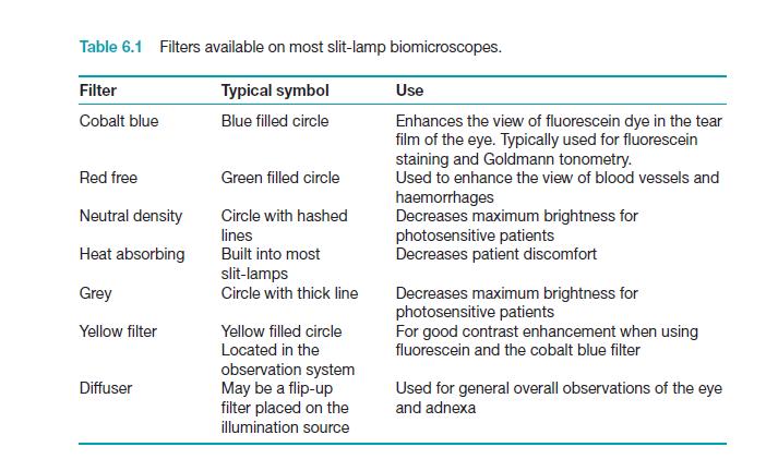
3 Basic components of slit lamp 1- Illumination arm contain the illumination system and the angle can be change from It is has A- Slit controls to control the light width and orientations B- Click stop it is change the position of the reflecting mirror to change the angle of the beam with respect to the viewing system C- Filters used to change the appearance of the beam like cobalt blue filter green or red free filter 3

4 Basic Components: illumination Bulb Filters Slit height control Slit rotator Mirror Slit width control 5 Filters 1.Unfiltered 2. Heat absorbing 3. 10% Grey 4. Red free 5. Cobalt blue
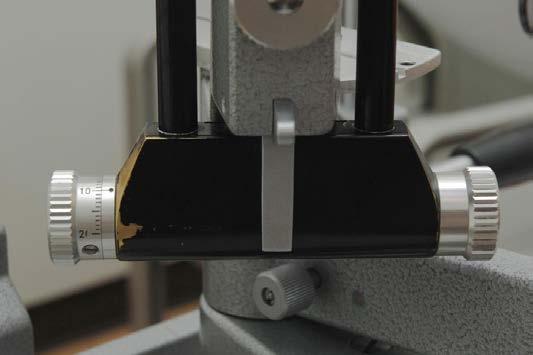
5 Width 8 5
6 Microscope arm A- Oculars : it can adjust to the examiner refractive error the distance between them can be adjust as the examiner IPD B-Magnification changer Slit lamp position controls - Joystick ( elevation knops ) to control the movement of the slit lamp forward and right left movement to have sharp focus 6
7 Basic Components: magnification Eye pieces Magnification changer Joy stick Lock Base 10 7
 2 eye piece options (10x and 16x) Total magnification ranges thus from")
8 Magnification Most slit lamps have: 2 objective settings (1 and 1.6) 2 eye piece options (10x and 16x) Total magnification ranges thus from 10x-25x


9 Use of the Slit Lamp Seat patient comfortabl y Adjust table, chair Position patient s head 13 Focus Patient s Eye Microscope straight Light column degrees from side Microscope moves via joystick Move laterally Move in and out 14 9

10 Adjust the Illumination Brightness: filters Width: slit vs broad beam Height: long vs pinpoint 15 10
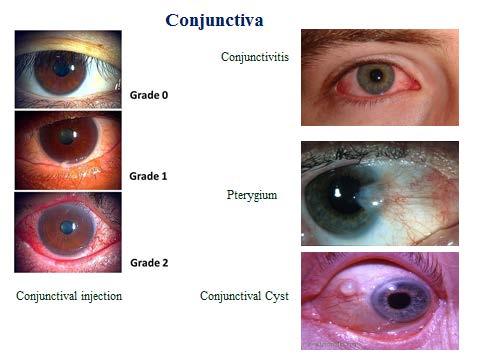
11 Illumination techniques Diffuse illumination Direct illumination Parallelepiped Optic section Conical(pinpoint) Tangential Specular reflection Indirect illumination Retro-illumination Sclerotic scatter Transillumination 11


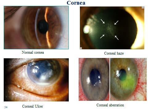
12 Diffuse illumination Angle between microscope and illumination system should be degree. Slit width should be widest. Filter to be used is diffusing filter. Magnification: low to medium Illumination: medium to high. Applications: General view of anterior of eye: lids,lashes,sclera,cornea,iris,pupil, Gross pathology and media opacities Contact lens fitting. Assessment of lachrymal reflex. Optics of diffuse illumination Diffuse illumination with slit beam and background illumination 12
13 Direct illumination Involves placing the light source at an angle of about degree from microscope. This arrangement permits both light beam and microscope to be sharply focused on the ocular tissue being observed. Wide beam direct illumination is commonly used as a preliminary technique to evaluate large area It is particularly suitable for assessment of cataracts, scars, nerves and vessels. It is also important for the determination of stabilization of axis of toric contact lens Parallelepiped: Constructed by narrowing the beam to 1-2mm in width to illuminate a rectangular area of cornea. Microscope is placed directly in front of patients cornea. Light source is approximately 45 degree from straight ahead position. 13


14 Applications: Used to detect and examine corneal structures and defects. Used to detect corneal striae that develop when corneal edema occurs with hydrogel lens wear and in keratoconus. Higher magnification than that used with wide beam illumination is preferred to evaluate both depth and extent of corneal, scarring or foreign bodies. 14
15 Conical beam (pinpoint) Produced by narrowing the vertical height of a parallelepiped to produce a small circular or square spot of light. Light source is degree temporally and directed into pupil. Biomicroscope: directly in front of eye. Magnification: high(16-25x) Intensity of light source to highest setting. Focusing: Beam is focused between cornea and anterior lens surface and dark zone between cornea and anterior lens observed. Principle is same as that of beam of sun light streaming through a room,illuminating airborne dust particles. Most useful when examining the transparency of anterior chamber for evidence of floating cells and flare seen in anterior uveitis. 15

16 Optic section Optic section is a very thin parallelepiped and optically cuts a very thin slice of the cornea. Axes of illuminating and viewing path intersect in the area of anterior eye media to be examined e.g. the individual corneal layers. Angle between illuminating and viewing path is 45 degree. Slit length should be kept small to minimize dazzling the patient. 16
 can be resolved more easily.")


17 With narrow slit the depth and portion of different objects(penetration depth of foreign bodies, shape of lens etc) can be resolved more easily. With wider slit their extension and shape are visible more clearly. Magnification: maximum. Examination of AC depth is performed by wider slit width.1-.3mm. Used to localize: Nerve fibers Blood vessels Infiltrates Cataracts AC depth. Optical section of lens 1.Corneal scar with wide beam illumination 2.optical section through scar indicating scar is with in superficial layer of cornea. 17
18 Examination methods A- direct illumination : angle between the light source and microscope is about and both microscope and light will direct to the focusing area, different type of direct illumination can be used 1- wide-beam direct illumination use to evaluate large area 2- parallelepiped it is constructed beam occur by narrowing the beam to 1-2 mm in width use for examined the layered of cornea and lens especially in the depth and extent of the corneal abrasions, scarring and foreign bodies 3- optic section it is when the parallelepiped reduced in width to an extremely thin, it is use for evaluated the layer of cornea and the depth of the foreign body 4- conical beam produce by narrowing the vertical height of a parallelepiped to produce a small circular or square spot of light, used to examined the transparency of the anterior chamber for floating cells 5- Specular reflection Established by separating the microscope and slit beam by equal angles from normal to cornea. Position of illuminator about 30 degree to one side and the microscope 30 degree to other side. Angle of illuminator to microscope must be equal and opposite. Angle of light should be moved until a very bright reflex obtained from corneal surface which is called zone of specular reflection. 18

19 Schematic of specular reflection. Reflection from front surface endothelium 19
20 Observe: Anterior and posterior cornea Iris is best viewed without dilation by this method. Anterior lens (especially useful for viewing pseudoexfolation). Indirect illumination The beam is focused in an area adjacent to ocular tissue to be observed. Main application: Examination of objects in direct vicinity of corneal areas of reduced transparency e,g, infiltrates,corneal scars,deposits,epithelial and stromal defects 20

21 Indirect illumination Illumination: Narrow to medium slit beam Decentred beam Magnification: approx. m=12x (depending upon object size 21

22 Retroillumination Formed by reflecting light of slit beam from a structure more posterior than the structure under observation. A vertical slit beam 1-4mm wide can be used. Purpose: Place object of regard against a bright background allowing object to appear dark or black. Used most often in searching for keratic precipitates and other debris on corneal endothelium. The crystalline lens can also be retroilluminated for viewing of water clefts and vacuoles of anterior lens and posterior subcapsular cataract Slitlamp photo shows keratic precipitates inside the eye. These opacities are collections of inflammatory cells that collect on the inner surface of the cornea. 22

23 Direct retroillumination from iris: Used to view corneal pathology. A moderately wide slit beam is aimed towards the iris directly behind the corneal anomaly. Use magnification of 16x to 25x and direct the light from 45 degree. Microscope is directed straight ahead. Schematic of direct retroillumination from the iris. direct retroillumination from the iris. 23

24 Sclerotic scatter It is formed by focusing a bright but narrow slit beam on the limbus and using microscope on low magnification. Such an illumination technique causes cornea to take on total internal reflection. The slit beam should be placed approximately degree from the microscope. When properly positioned this technique will produce halo glow of light around the limbus as the light is transmitted around the cornea. Corneal changes or abnormalities can be visualized by reflecting the scattered light. Used to observe: Central corneal epithelial edema Corneal abrasions Corneal nebulae and maculae. Depending on the density of the abnormality, the light from behind may reflect through, allowing detailed examination of the internal structure of the pathology. Observe: corneal opacities (edema, infiltrates, vessels, foreign bodies), lens, iris 24
25 Procedure: - Patient will examined without glasses - Room illumination is dim - Adjust the height of the slit lamp table to the comfort position for patient and examiner - Instruct the patient to place his chin on chin rest and his forehead against forehead rest - Adjust the chin rest to align the patient canthus - Set the magnification in low setting,remove all filters - Open the both eyes of you ( examiner) and set the IPD - Use one hand to use the joystick and the other hand to control the angle between the microscope and light Fluorescein staining: Fluorescein is an orange colored dye, it is instilled into the eye and a fine film over the corneal surface, it will appear by using ultra violet light as green color, it use in detected F.B and corneal abrasion 25
26 Ocular structure Type of slit lamp beam Angle of illumination arm magnification Lids / lashes diffuse 30 low conjunctiva parallelepiped 30 low cornea Anterior chamber Angle depth aqueous Iris Lens Narrow medium parallelepiped Optic section 60 medium Conical beam 30 high Wide parallelepiped Narrow parallelepiped medium medium 26

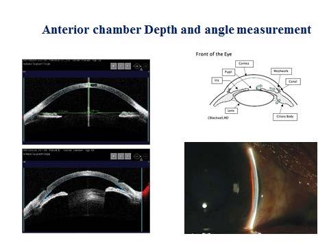
27 Eye Anatomy 27

28 Anterior segment disease evaluation Lids and lashes Conjunctiva and cornea Instillation of fluorescein and BUT measurement Anterior chamber Depth and angle measurement Iris Crystalline lens 28
29 29
30 30
11/10/2015. Haag Streit Topcon Zeiss Kowa Add On Systems- OIS/Escalon and Others. The Original Design. Photo Slit lamp Systems. Who Makes Them?
 The Original Design Photo Slit lamp Systems Who Makes Them? 1862-1930 Alvar Gullstrand Inventor of the Slit lamp illuminator - 1911 Swedish ophthalmologist, recipient of the 1911 Nobel Prize for Physiology
Eyes. Inspection Visual Acuity Visual Fields Pupillary Response Fundoscopic Exam
 Eyes Inspection Visual Acuity Visual Fields Pupillary Response Fundoscopic Exam Eye Examination Inspection 11.Inspects external ocular (eye) structures (lids, conjunctiva, iris, cornea, pupils) 12.Gently
Smart-Device Ophthalmic Photography
 Smart-Device Ophthalmic Photography Austin Holmes, M.P.H. Clifford M. Terry, M.D. Financial Interest Disclosure: Unrestricted Grant From Terry Eye Institute Overview Creation of Smart-Device Photography
What s Fundus photography s purpose? Why do we take them? Why do we do it? Why do we do it? Why do we do it? 11/3/2014. To document the retina
 What s Fundus photography s purpose? To document the retina Photographers role to show the retina Document other ocular structures Why do we take them? Why do we do it? We as photographers help the MD
OCULAR MEDIA* PHOTOGRAPHIC RECORDING OF OPACITIES OF THE. development by the control of diabetes, the supply of a deficient hormone
 Brit. J. Ophthal. (1955) 39, 85. PHOTOGRAPHIC RECORDING OF OPACITIES OF THE OCULAR MEDIA* BY E. F. FINCHAM Institute of Ophthalmology, University of London THE value of photography for recording pathological
EYE ANATOMY. Multimedia Health Education. Disclaimer
 Disclaimer This movie is an educational resource only and should not be used to manage your health. The information in this presentation has been intended to help consumers understand the structure and
Digital Slit Lamp Photography. A User s Primer
 Digital Slit Lamp Photography A User s Primer Table of Contents Table of Contents Table of Contents Acknowledgements i Introduction Basic Premises 1 Slit Lamp Components 3 Microscope 4 Illumination Source
2 The First Steps in Vision
 2 The First Steps in Vision 2 The First Steps in Vision A Little Light Physics Eyes That See light Retinal Information Processing Whistling in the Dark: Dark and Light Adaptation The Man Who Could Not
Television slit-lamp biomicroscopy
 British Journal of Ophthalmology, 1978, 62, 644-650 Television slit-lamp biomicroscopy A. J. BRON, D. V. KAUFMAN, AND D. HARWOOD From the Eye Hospital, Oxford SUMMARY The basic requirements for performing
1. Introduction to Anatomy of the Eye and its Adnexa
 1. Introduction to Anatomy of the Eye and its Adnexa Fig 1: A Cross section of the human eye. Let us imagine we are traveling with a ray of light into the eye. The first structure we will encounter is
Wide Angle Ophthalmoscope Instructions
 Wide Angle Ophthalmoscope Instructions PLEASE READ AND FOLLOW THESE INSTRUCTIONS CAREFULLY Contents 1. Symbols 2. Warnings & Cautions 3. Description of Product 4. Getting Started 5. Apertures & Filters
Gonioscopy Wet-Lab. About Me. About Me Gonioscopy. Indications for Gonioscopy. Billing Gonioscopy 12/13/2012. Code: 92020
 About Me Gonioscopy Wet-Lab Marcus Gonzales, OD, FAAO Cedar Springs Eye Clinic (formerly BridgeBuilders Eye Clinic) About Me Gonioscopy Evaluates the anterior chamber angle responsible for the outflow
Basic Principles of the Surgical Microscope. by Charles L. Crain
 Basic Principles of the Surgical Microscope by Charles L. Crain 2006 Charles L. Crain; All Rights Reserved Table of Contents 1. Basic Definition...3 2. Magnification...3 2.1. Illumination/Magnification...3
Retinal stray light originating from intraocular lenses and its effect on visual performance van der Mooren, Marie Huibert
 University of Groningen Retinal stray light originating from intraocular lenses and its effect on visual performance van der Mooren, Marie Huibert IMPORTANT NOTE: You are advised to consult the publisher's
Uses of the Slit Lamp. Parts of the Slit Lamp
 2 14 Chapter I 0: Slit-Lamp Biomicroscopy for performing Goldmann applanation tonometry with the slit lamp appear in Chapter 12; instructions for indirect slit-lamp biomicroscopy of the posterior segment
Instruments Commonly Used For Examination of the Eye
 Instruments Commonly Used For Examination of the Eye There are many instruments that the eye doctor might use to evaluate the eye and the vision system. This report presents some of the more commonly used
11 Human Eye & colourful world IMPORTANT NOTES ANIL TUTORIALS
 11 Human Eye & colourful world IMPORTANT NOTES 1. Parts of the Human Eye : (i) Sclerotic is the outermost white fibrous covering of the eye. (ii) Cornea is the transparent front bulging portion of the
ensory System III Eye Reflexes
 ensory System III Eye Reflexes Quick Review from Last Week Eye Anatomy Inside of the Eye choroid Eye Reflexes Eye Reflexes A healthy person has a number of eye reflexes: Pupillary light reflex Vestibulo-ocular
HEINE QUALITY. :- HEINE Hand-held Ophthalmic Instruments
 HEINE QUALITY M A D E I N G E R M A N Y :- HEINE Hand-held Ophthalmic Instruments :- HEINE HSL150 Hand-held Slit Lamp Uncompromisingly compact. HEINE QUALITY M A D E I N G E R M A N Y 003 [ HEINE HSL150
Refraction, Lenses, and Prisms
 CHAPTER 16 14 SECTION Sound and Light Refraction, Lenses, and Prisms KEY IDEAS As you read this section, keep these questions in mind: What happens to light when it passes from one medium to another? How
EYE STRUCTURE AND FUNCTION
 Name: Class: Date: EYE STRUCTURE AND FUNCTION The eye is the body s organ of sight. It gathers light from the environment and forms an image on specialized nerve cells on the retina. Vision occurs when
GIST OF THE UNIT BASED ON DIFFERENT CONCEPTS IN THE UNIT (BRIEFLY AS POINT WISE). RAY OPTICS
. RAY OPTICS") 209 GIST OF THE UNIT BASED ON DIFFERENT CONCEPTS IN THE UNIT (BRIEFLY AS POINT WISE). RAY OPTICS Reflection of light: - The bouncing of light back into the same medium from a surface is called reflection
209 GIST OF THE UNIT BASED ON DIFFERENT CONCEPTS IN THE UNIT (BRIEFLY AS POINT WISE). RAY OPTICS Reflection of light: - The bouncing of light back into the same medium from a surface is called reflection
Keeler Direct Ophthalmoscopes
 Keeler Direct Ophthalmoscopes Direct Ophthalmoscopes Introduction Direct Ophthalmoscopes A combination of optical perfection, superb ergonomics and versatile features make Keeler direct ophthalmoscopes
Chapter 36. Image Formation
 Chapter 36 Image Formation Image of Formation Images can result when light rays encounter flat or curved surfaces between two media. Images can be formed either by reflection or refraction due to these
In the following diagram the parts of the eye are visualized and labeled for you.
 Investigation 3.12B: The Eye In the preceding case study marker of the problem of greatest concern to you lay in finding the pupils fixed in a dilated position. But what is the pupil and what makes it
By Dr. Abdelaziz Hussein
 By Dr. Abdelaziz Hussein Light is a form of radiant energy, consisting of electromagnetic waves a. Velocity of light: In air it is 300,000 km/second. b. Wave length: The wave-length of visible light to
Physics Chapter Review Chapter 25- The Eye and Optical Instruments Ethan Blitstein
 Physics Chapter Review Chapter 25- The Eye and Optical Instruments Ethan Blitstein The Human Eye As light enters through the human eye it first passes through the cornea (a thin transparent membrane of
Sense Organs (Eye) The eye is the sense organ of sight. The eye is shaped like a ball and is located in bony
 The eye is the sense organ of sight. The eye is shaped like a ball and is located in bony") Sense Organs (Eye) The eye is the sense organ of sight. The eye is shaped like a ball and is located in bony sockets in the skull. It is held in place by six muscles which are joined to the outside of
Sense Organs (Eye) The eye is the sense organ of sight. The eye is shaped like a ball and is located in bony sockets in the skull. It is held in place by six muscles which are joined to the outside of
Macrophotography of the anterior
 Brit. J. Ophthal. (I970) 54, 697 Macrophotography of the anterior segment of the eye NICHOLAS BROWN Moorfields Eye Hospital, City Road, London Existing photo-slit lamps are used at magnifications of X
Chapter 29/30. Wave Fronts and Rays. Refraction of Sound. Dispersion in a Prism. Index of Refraction. Refraction and Lenses
 Chapter 29/30 Refraction and Lenses Refraction Refraction the bending of waves as they pass from one medium into another. Caused by a change in the average speed of light. Analogy A car that drives off
Instructional Resources/Materials: Light vocabulary cards printed (class set) Enough for each student (See card sort below)
 Enough for each student (See card sort below)") Grade Level/Course: Grade 7 Life Science Lesson/Unit Plan Name: Light Card Sort Rationale/Lesson Abstract: Light vocabulary building, students identify and share vocabulary meaning. Timeframe: 10 to 20
Grade Level/Course: Grade 7 Life Science Lesson/Unit Plan Name: Light Card Sort Rationale/Lesson Abstract: Light vocabulary building, students identify and share vocabulary meaning. Timeframe: 10 to 20
Chapter Introduction. Chapter Wrap-Up. and the Eye
 Chapter Introduction Lesson 1 Lesson 2 Lesson 3 Sound Light Chapter Wrap-Up Mirrors, Lenses, and the Eye How do sound and light waves travel and interact with matter? What do you think? Before you begin,
Macula centred, giving coverage of the temporal retinal. Disc centred. Giving coverage of the nasal retina.
 3. Field positions, clarity and overall quality For retinopathy screening purposes in England two images are taken of each eye. These have overlapping fields of view and between them cover the main area
Objectives. 3. Visual acuity. Layers of the. eye ball. 1. Conjunctiva : is. three quarters. posteriorly and
 OCULAR PHYSIOLOGY (I) Dr.Ahmed Al Shaibani Lab.2 Oct.2013 Objectives 1. Review of ocular anatomy (Ex. after image) 2. Visual pathway & field (Ex. Crossed & uncrossed diplopia, mechanical stimulation of
Chapter 6 Human Vision
 Chapter 6 Notes: Human Vision Name: Block: Human Vision The Humane Eye: 8) 1) 2) 9) 10) 4) 5) 11) 12) 3) 13) 6) 7) Functions of the Eye: 1) Cornea a transparent tissue the iris and pupil; provides most
Chapter 36. Image Formation
 Chapter 36 Image Formation Notation for Mirrors and Lenses The object distance is the distance from the object to the mirror or lens Denoted by p The image distance is the distance from the image to the
Image Modeling of the Human Eye
 Image Modeling of the Human Eye Rajendra Acharya U Eddie Y. K. Ng Jasjit S. Suri Editors ARTECH H O U S E BOSTON LONDON artechhouse.com Contents Preface xiiii CHAPTER1 The Human Eye 1.1 1.2 1. 1.4 1.5
Light Microscopy. Upon completion of this lecture, the student should be able to:
 Light Light microscopy is based on the interaction of light and tissue components and can be used to study tissue features. Upon completion of this lecture, the student should be able to: 1- Explain the
30 Lenses. Lenses change the paths of light.
 Lenses change the paths of light. A light ray bends as it enters glass and bends again as it leaves. Light passing through glass of a certain shape can form an image that appears larger, smaller, closer,
Chapter 25. Optical Instruments
 Chapter 25 Optical Instruments Optical Instruments Analysis generally involves the laws of reflection and refraction Analysis uses the procedures of geometric optics To explain certain phenomena, the wave
Ocular Lenses. Head. Arm. Objective Lenses. Slide Holder Stage. On / Off Switch. Condenser with Iris Diaphragm. Light Intensity Control
 BIOLOGY 211: HUMAN ANATOMY & PHYSIOLOGY ********************************************************************************************************* USE OF THE LIGHT MICROSCOPE **********************************************************************************************************
Telemedical diagnosis of anterior segment eye diseases: validation of digital slit-lamp still images
 (2009) 23, 652 660 & 2009 Macmillan Publishers Limited All rights reserved 0950-222X/09 $32.00 www.nature.com/eye CLINICAL STUDY Telemedical diagnosis of anterior segment eye diseases: validation of digital
Sensory receptors External internal stimulus change detectable energy transduce action potential different strengths different frequencies
 General aspects Sensory receptors ; respond to changes in the environment. External or internal environment. A stimulus is a change in the environmental condition which is detectable by a sensory receptor
The microscope is useful in making observations and collecting data in scientific experiments. Microscopy involves three basic concepts:
 AP BIOLOGY Chapter 6 NAME DATE Block MICROSCOPE LAB PART I: COMPOUND MICROSCOPE OBJECTIVES: After completing this exercise you should be able to: Demonstrate proper care and use of a compound microscope.
Class 10 Science NCERT Exemplar Solutions Human Eye and Colourful World
 Class 10 Science NCERT Exemplar Solutions Human Eye and Colourful World Short Answer Questions Question 1. A student sitting at the back of the classroom cannot read clearly the letters written on the
Reports. \ $mm>-j \ksiiimj
 Reports A scanning slit optical microscope. DAVID M. MAURICE. A transparent tissue is illuminated with a narrow slit of light which is formed by light transmitted down one-half of a microscope objective,
Vocabulary. Unit 9 Forms of Energy. ENERGY: The capacity for doing work.
 Unit 9 Forms of Energy Main Idea: There are many forms of energy, including radiant energy and chemical energy. Energy can change form. ENERGY: The capacity for doing work. Heat, Light and Radiant Energy
Life Science Chapter 2 Study Guide
 Key concepts and definitions Waves and the Electromagnetic Spectrum Wave Energy Medium Mechanical waves Amplitude Wavelength Frequency Speed Properties of Waves (pages 40-41) Trough Crest Hertz Electromagnetic
4Basic anatomy and physiology
 Hene_Ch09.qxd 8/30/04 6:51 AM Page 348 348 4Basic anatomy and physiology The eye is a highly specialized organ with an average axial length of 24 mm and a volume of 6.5 ml. Except for its anterior aspect,
Lenses- Worksheet. (Use a ray box to answer questions 3 to 7)
") Lenses- Worksheet 1. Look at the lenses in front of you and try to distinguish the different types of lenses? Describe each type and record its characteristics. 2. Using the lenses in front of you, look
Lenses- Worksheet 1. Look at the lenses in front of you and try to distinguish the different types of lenses? Describe each type and record its characteristics. 2. Using the lenses in front of you, look
Person s Optics Test KEY SSSS
 Person s Optics Test KEY SSSS 2017-18 Competitors Names: School Name: All questions are worth one point unless otherwise stated. Show ALL WORK or you may not receive credit. Include correct units whenever
Sheep Eye Dissection
 Sheep Eye Dissection Question: How do the various parts of the eye function together to make an image appear on the retina? Materials and Equipment: Preserved sheep eye Scissors Dissection tray Tweezers
MAKING SENSE OF SLIT LAMP SERVICING
 MAKING SENSE OF SLIT LAMP SERVICING HOW TO LOOK AFTER AND CARE FOR A SLIT LAMP The slit lamp is an essential and oftenused diagnostic instrument in ophthalmology. It provides illumination and magnification
LAB 12 Reflection and Refraction
 Cabrillo College Physics 10L Name LAB 12 Reflection and Refraction Read Hewitt Chapters 28 and 29 What to learn and explore Please read this! When light rays reflect off a mirror surface or refract through
Dr. Magda Rau Eye Clinic Cham, Germany
 3 and 6 Months clinical Results after Implantation of OptiVis Diffractive-refractive Multifocal IOL Dr. Magda Rau Eye Clinic Cham, Germany Refractive zone of Progressive power for Far to Intermediate
Use these words to complete the sentences about light: absorb different diffuse focus prism refraction same slower specula transmit
 Aims In the activity you will learn more about how we see, how light interacts with materials, and how we see colour. Task 1: Light Use these words to complete the sentences about light: absorb different
Chapter Human Vision
 Chapter 6 6.1 Human Vision How Light Enters the Eye Light enters the eye through the pupil. The pupil appears dark because light passes through it without reflecting back Pupil Iris = Coloured circle of
Health Science 1110 Module 9 Sensations LAB 9. View the Film on Cornea Transplant and answer the questions on your laboratory worksheet.
 Health Science 1110 Module 9 Sensations LAB 9 View the Film on Cornea Transplant and answer the questions on your laboratory worksheet. Webpage Activities o Open Internet Explorer o Go to the Health Sciences
Introduction. Chapter Aim of the Thesis
 Chapter 1 Introduction 1.1 Aim of the Thesis The main aim of this investigation was to develop a new instrument for measurement of light reflected from the retina in a living human eye. At the start of
Biology 70 Slides for Lecture 1 Fall 2007
 Biology 70 Part II Sensory Systems www.biology.ucsc.edu 1 2 intensity vs spatial position (image formation) color 3 4 motion depth (monocular) 5 6 1 depth (binocular) 1. In the lectures on perception we
Handout G: The Eye and How We See
 Handout G: The Eye and How We See Prevent Blindness America. (2003c). The eye and how we see. Retrieved July 31, 2003, from http://www.preventblindness.org/resources/howwesee.html Your eyes are wonderful
OPTICAL SYSTEMS OBJECTIVES
 101 L7 OPTICAL SYSTEMS OBJECTIVES Aims Your aim here should be to acquire a working knowledge of the basic components of optical systems and understand their purpose, function and limitations in terms
Section 1: Sound. Sound and Light Section 1
 Sound and Light Section 1 Section 1: Sound Preview Key Ideas Bellringer Properties of Sound Sound Intensity and Decibel Level Musical Instruments Hearing and the Ear The Ear Ultrasound and Sonar Sound
ABO Certification Training. Part I: Anatomy and Physiology
 ABO Certification Training Part I: Anatomy and Physiology Major Ocular Structures Centralis Nerve Major Ocular Structures The Cornea Cornea Layers Epithelium Highly regenerative: Cells reproduce so rapidly
Mastery. Chapter Content. What is light? CHAPTER 11 LESSON 1 C A
 Chapter Content Mastery What is light? LESSON 1 Directions: Use the letters on the diagram to identify the parts of the wave listed below. Write the correct letters on the line provided. 1. amplitude 2.
Notes: Light and Optics. Reflection. Refraction. Law of Reflection. Light goes straight 12/13/2012
 Notes: Light and Optics Light goes straight Light travels in a straight line unless it interacts with a medium. The material through which a wave travels is called a medium. Light can be reflected, refracted
Light and Applications of Optics
 UNIT 4 Light and Applications of Optics Topic 4.1: What is light and how is it produced? Topic 4.6: What are lenses and what are some of their applications? Topic 4.2 : How does light interact with objects
User Manual. Digital Compound Binocular LED Microscope. MicroscopeNet.com
 User Manual Digital Compound Binocular LED Microscope Model MD82ES10 MicroscopeNet.com Table of Contents i. Caution... 1 ii. Care and Maintenance... 2 1. Components Illustration... 3 2. Installation...
family of lens designs fitting guide ICD is Exclusively Manufactured In
 TM family of lens designs fitting guide ICD is Exclusively Manufactured In paragon 1 Select Initial Diagnostic Lens: Identify the Corneal Condition Normal Depth Eyes Normal Shapes Median Flat KReading
3. Study the diagram given below and answer the questions that follow it:
 CH- Human Eye and Colourful World 1. A 14-year old student is not able to see clearly the questions written on the blackboard placed at a distance of 5 m from him. (a) Name the defect of vision he is suffering
Yokohama City University lecture INTRODUCTION TO HUMAN VISION Presentation notes 7/10/14
 Yokohama City University lecture INTRODUCTION TO HUMAN VISION Presentation notes 7/10/14 1. INTRODUCTION TO HUMAN VISION Self introduction Dr. Salmon Northeastern State University, Oklahoma. USA Teach
Introduction. Strand F Unit 3: Optics. Learning Objectives. Introduction. At the end of this unit you should be able to;
 Learning Objectives At the end of this unit you should be able to; Identify converging and diverging lenses from their curvature Construct ray diagrams for converging and diverging lenses in order to locate
ECEN 4606, UNDERGRADUATE OPTICS LAB
 ECEN 4606, UNDERGRADUATE OPTICS LAB Lab 2: Imaging 1 the Telescope Original Version: Prof. McLeod SUMMARY: In this lab you will become familiar with the use of one or more lenses to create images of distant
Chapter 23. Mirrors and Lenses
 Chapter 23 Mirrors and Lenses Mirrors and Lenses The development of mirrors and lenses aided the progress of science. It led to the microscopes and telescopes. Allowed the study of objects from microbes
Basics of Light Microscopy and Metallography
 ENGR45: Introduction to Materials Spring 2012 Laboratory 8 Basics of Light Microscopy and Metallography In this exercise you will: gain familiarity with the proper use of a research-grade light microscope
Visual Optics. Visual Optics - Introduction
 Visual Optics Jim Schwiegerling, PhD Ophthalmology & Optical Sciences University of Arizona Visual Optics - Introduction In this course, the optical principals behind the workings of the eye and visual
12.1. Human Perception of Light. Perceiving Light
 12.1 Human Perception of Light Here is a summary of what you will learn in this section: Focussing of light in your eye is accomplished by the cornea, the lens, and the fluids contained in your eye. Light
VISULAS Trion. Treatment flexibility to the power of three. Multicolor Photocoagulation Laser
 VISULAS Trion Treatment flexibility to the power of three Multicolor Photocoagulation Laser Carl Zeiss: A pioneer in retinal therapy For many years, Carl Zeiss has fostered a culture of highest precision,
General Physics Experiment 5 Optical Instruments: Simple Magnifier, Microscope, and Newtonian Telescope
 General Physics Experiment 5 Optical Instruments: Simple Magnifier, Microscope, and Newtonian Telescope Objective: < To observe the magnifying properties of the simple magnifier, the microscope and the
Lenses. A lens is any glass, plastic or transparent refractive medium with two opposite faces, and at least one of the faces must be curved.
 PHYSICS NOTES ON A lens is any glass, plastic or transparent refractive medium with two opposite faces, and at least one of the faces must be curved. Types of There are two types of basic lenses. (1.)
ECEN 4606, UNDERGRADUATE OPTICS LAB
 ECEN 4606, UNDERGRADUATE OPTICS LAB Lab 3: Imaging 2 the Microscope Original Version: Professor McLeod SUMMARY: In this lab you will become familiar with the use of one or more lenses to create highly
The Eye. Nakhleh Abu-Yaghi, M.B.B.S Ophthalmology Division
 The Eye Nakhleh Abu-Yaghi, M.B.B.S Ophthalmology Division Coats of the Eyeball 1- OUTER FIBROUS COAT is made up of : Posterior opaque part 2-THE SCLERA the dense white part 1- THE CORNEA the anterior
The Human Eye and a Camera 12.1
 The Human Eye and a Camera 12.1 The human eye is an amazing optical device that allows us to see objects near and far, in bright light and dim light. Although the details of how we see are complex, the
EnFocus Your Upgrade Path to High Performance Intrasurgical OCT
 Your Upgrade Path to High Performance Intrasurgical OCT is FDA 510(k) Cleared > Ultra HD OCT extends your microscope s potential with intrasurgical OCT BRILLIANT IMAGES, SUB-SURFACE KNOWLEDGE is an intrasurgical
EYE. The eye is an extension of the brain
 I SEE YOU EYE The eye is an extension of the brain Eye brain proxomity Can you see : the optic nerve bundle? Spinal cord? The human Eye The eye is the sense organ for light. Receptors for light are found
Chapter 16 Light Waves and Color
 Chapter 16 Light Waves and Color Lecture PowerPoint Copyright The McGraw-Hill Companies, Inc. Permission required for reproduction or display. What causes color? What causes reflection? What causes color?
Vision 1. Physical Properties of Light. Overview of Topics. Light, Optics, & The Eye Chaudhuri, Chapter 8
 Vision 1 Light, Optics, & The Eye Chaudhuri, Chapter 8 1 1 Overview of Topics Physical Properties of Light Physical properties of light Interaction of light with objects Anatomy of the eye 2 3 Light A
VISUAL PHYSICS ONLINE DEPTH STUDY: ELECTRON MICROSCOPES
 VISUAL PHYSICS ONLINE DEPTH STUDY: ELECTRON MICROSCOPES Shortly after the experimental confirmation of the wave properties of the electron, it was suggested that the electron could be used to examine objects
Mirrors and Lenses. Images can be formed by reflection from mirrors. Images can be formed by refraction through lenses.
 Mirrors and Lenses Images can be formed by reflection from mirrors. Images can be formed by refraction through lenses. Notation for Mirrors and Lenses The object distance is the distance from the object
Using a Microscope. Year Group: BVSc1 + Document number: CSL_L07
 Year Group: BVSc1 + Document number: CSL_L07 Equipment list: Equipment for this station: Microscope Power supply and a level surface to work on Gloves The sample to examine Marker or pencil for labelling
Practice Problems for Chapter 25-26
 Practice Problems for Chapter 25-26 1. What are coherent waves? 2. Describe diffraction grating 3. What are interference fringes? 4. What does monochromatic light mean? 5. What does the Rayleigh Criterion
Figure 1. Relative intensity of solar energy of different wavelength at the earth's surface.
 Spectrum of light from the sun: Fig.1 Figure 1. Relative intensity of solar energy of different wavelength at the earth's surface. Properties of light 1-The speed of light changes when it goes from one
Physics 9 Wednesday, February 1, 2012
 Physics 9 Wednesday, February 1, 2012 learningcatalytics.com class session ID: 542970 Today: repeat soap bubble; measure λ for laser Today: telescope, human eye Friday: first of 3 days on fluids (liquids,
Vision. The eye. Image formation. Eye defects & corrective lenses. Visual acuity. Colour vision. Lecture 3.5
 Lecture 3.5 Vision The eye Image formation Eye defects & corrective lenses Visual acuity Colour vision Vision http://www.wired.com/wiredscience/2009/04/schizoillusion/ Perception of light--- eye-brain
Topic 4: Lenses and Vision. Lens a curved transparent material through which light passes (transmit) Ex) glass, plastic
 Ex) glass, plastic") Topic 4: Lenses and Vision Lens a curved transparent material through which light passes (transmit) Ex) glass, plastic Double Concave Lenses Are thinner and flatter in the middle than around the edges.
Topic 4: Lenses and Vision Lens a curved transparent material through which light passes (transmit) Ex) glass, plastic Double Concave Lenses Are thinner and flatter in the middle than around the edges.
Chapter 3 Optical Systems
 Chapter 3 Optical Systems The Human Eye [Reading Assignment, Hecht 5.7.1-5.7.3; see also Smith Chapter 5] retina aqueous vitreous fovea-macula cornea lens blind spot optic nerve iris cornea f b aqueous
Chapter Ray and Wave Optics
 109 Chapter Ray and Wave Optics 1. An astronomical telescope has a large aperture to [2002] reduce spherical aberration have high resolution increase span of observation have low dispersion. 2. If two
HEINE Direct Ophthalmoscopes
 [ 036 ] 02 HEINE Direct Ophthalmoscopes BETA 200 S BETA 200 / BETA 200 M2 Opt. 1 Opt. 2 K 180 Opt. 1 Opt. 2 mini 3000 mini 3000 LED Optical System Aspherical Conventional Illumination LED-Illumination
Unit Two Part II MICROSCOPY
 Unit Two Part II MICROSCOPY AVERETT 1 0 /9/2013 1 MICROSCOPES Microscopes are devices that produce magnified images of structures that are too small to see with the unaided eye Humans cannot see objects
Lecture 8. Lecture 8. r 1
 Lecture 8 Achromat Design Design starts with desired Next choose your glass materials, i.e. Find P D P D, then get f D P D K K Choose radii (still some freedom left in choice of radii for minimization