Review Scope Operator s Manual
|
|
|
- Felicia Craig
- 6 years ago
- Views:
Transcription


1
2 ThinPrep Imaging System Review Scope Operator s Manual HOLOGIC, INC. 250 CAMPUS DRIVE MARLBOROUGH, MA USA TEL: FAX: WEB: For Use With Version 6.x.y Software MAN
3 The ThinPrep Imaging System is a PC-based and automated review system for use with ThinPrep. cervical cytology sample slides. The ThinPrep. Imaging System is intended to help a cytotechnologist or pathologist highlight areas of a slide for further manual review. The Product is not a replacement for manual review. Determination of slide adequacy and patient diagnosis is at the sole discretion of the cytotechnologists and pathologists trained by Hologic to evaluate ThinPrep. prepared slides. If and only if it is finally determined by a court of competent jurisdiction that the Product sold to Customer thereunder was defective in design or contained a manufacturing defect and that such defect was solely responsible for an error in diagnosis that caused harm to a patient, Hologic shall indemnify Customer for the compensatory damages paid by Customer to discharge the personal injury judgment with respect to Product. Caution: Federal law restricts this device to sale by or on the order of a physician, or any other practitioner licensed by the law of the State in which the practitioner practices to use or order the use of the device and are trained and experienced in the use of the ThinPrep Imaging System. Hologic, Inc., All rights reserved. No part of this publication may be reproduced, transmitted, transcribed, stored in a retrieval system, or translated into any language or computer language, in any form, or by any means, electronic, mechanical, magnetic, optical, chemical, manual, or otherwise, without the prior written permission of Hologic, 250 Campus Drive, Marlborough, Massachusetts, 01752, United States of America. Although this guide has been prepared with every precaution to ensure accuracy, Hologic assumes no liability for any errors or omissions, nor for any damages resulting from the application or use of this information. This product may be covered by one or more U.S. patents identified at Hologic, PreservCyt and ThinPrep are registered trademarks in the United States and other countries. All other trademarks are the property of their respective companies. Changes or modifications to this unit not expressly approved by the party responsible for compliance could void the user s authority to operate the equipment. Document Number: AW Rev. 005
4 The ThinPrep Imaging System The ThinPrep Imaging System

5 Operation Summary and Clinical Information The ThinPrep Imaging System
6 A. INTENDED USE The Hologic ThinPrep Imaging System (Imager) is a device that uses computer imaging technology to assist in primary cervical cancer screening of ThinPrep Pap Test slides for the presence of atypical cells, cervical neoplasia, including its precursor lesions (Low Grade Squamous Intraepithelial Lesions, High Grade Squamous Intraepithelial Lesions), and carcinoma as well as all other cytologic criteria as defined by 2001 Bethesda System: Terminology for Reporting Results of Cervical Cytology 1. B. SUMMARY AND EXPLANATION OF THE SYSTEM The ThinPrep Imaging System is an automated imaging and review system for use with ThinPrep Pap Test slides. It combines imaging technology to identify microscopic fields of diagnostic interest with automated stage movement of a microscope in order to locate these fields. In routine use, the ThinPrep Imaging System selects 22 fields of view for a Cytotechnologist to review. Following review of these fields, the Cytotechnologist will either complete the diagnosis if no abnormalities are identified or review the entire slide if any abnormalities are identified. The ThinPrep Imaging System also allows the physical marking of locations of interest for the Cytopathologist. C. PRINCIPLES OF OPERATION The ThinPrep Imaging System consists of an Image Processor and one, or more, Review Scopes. The system makes use of computer imaging to select fields of view for presentation to a Cytotechnologist on a Review Scope. Slides used with this system must first be prepared on a ThinPrep 2000 or 3000 Processor, and stained with ThinPrep Stain. The Imaging Processor acquires and processes image data from the slides to identify diagnostically relevant cells or cell groups based on an imaging algorithm that considers cellular features and nuclear darkness. During slide imaging, the alphanumeric slide accession identifier is recorded and the x and y coordinates of 22 fields of interest are stored in the computer database. This computer also coordinates the communication of information between the Image Processor and the Review Scopes. After image processing, slides are distributed to Cytotechnologists for review utilizing the Review Scopes. The Review Scope is a microscope with an automated stage to facilitate the locating of the 22 fields containing the cells of interest. Additionally, the Review Scope provides a method for automated marking of objects for further review. Slides are individually loaded onto the Review Scope stage, the alphanumeric slide accession identifier is automatically scanned and the stored x and y coordinates representing fields of interest for that slide are electronically downloaded from the computer to the Review Scope. The Cytotechnologist then uses a keypad to step through each of the fields of interest (Autolocate). If the Cytotechnologist identifies any of these fields as containing abnormal objects, that field may be marked electronically. The Review Scope will guide the Cytotechnologist to conduct a review of the entire cell spot for any slide that has had fields electronically marked (Autoscan). The Cytotechnologist determines specimen adequacy and the presence of infections during the review of the 22 fields of view presented by the ThinPrep Imaging System. Either of two methods can be used to determine specimen adequacy. The first method is to count cells and determine the average number of cells in the 22 fields of view presented by the Imager. The second method is to count and determine the average number of cells in 10 fields of view across the diameter of the cell spot. Either method will enable the Cytotechnologist to determine if the minimum cells, as recommended by Bethesda System 2001 criteria, are present on the slide. At the conclusion of the slide review electronically marked objects are automatically ink marked. Any x and y coordinates representing marked locations along with a slide completion status are then electronically transmitted back to the computer for storage. MAN Rev. 002 page 2 of 23
7 D. LIMITATIONS Only personnel who have been appropriately trained should operate the ThinPrep Imaging System Image Processor or Review Scope. All slides that undergo primary automated screening with the Image Processor require manual rescreening of the selected fields of view by a Cytotechnologist using a Review Scope. The ThinPrep Imaging System is only indicated for use with the ThinPrep Pap Test. The laboratory Technical Supervisor should establish individual workload limits for personnel using the ThinPrep Imaging System. The maximum daily limit specified is only an upper limit and should never be used as an expectation for daily productivity or as a performance target. The ThinPrep Imaging System has not been proven to be safe or effective at workload levels which exceed product labeling. ThinPrep slides with fiducial marks must be used. Slides must be stained using the ThinPrep Stain according to the applicable ThinPrep Imaging System slide staining protocol. Slides should be clean and free of debris before being placed on the system. The slide coverslip should be dry and located correctly. Slides that are broken or poorly coverslipped should not be used. Slides used with the ThinPrep Imaging System must contain properly formatted accession number identification information as described in the operator s manual. Slides once successfully imaged on the Image Processor cannot be imaged again. The performance of the ThinPrep Imaging System using slides prepared from reprocessed sample vials has not been evaluated; therefore it is recommended that these slides be manually reviewed. E. WARNINGS The Imager generates, uses, and can radiate radio frequency energy and may cause interference to radio communications. A Hologic authorized service representative must install the ThinPrep Imaging System. F. PRECAUTIONS Caution should be used when loading and unloading glass slides on the ThinPrep Imaging System to prevent slide breakage and/or injury. Care should be taken to assure that slides are correctly oriented in the ThinPrep Imaging System cassettes to prevent rejection by the system. Partially processed slide cassettes should not be removed from the Image Processor, as data may be lost. The Image Processor should be placed on a flat, sturdy surface away from any vibrating machinery to assure proper operation. MAN Rev. 002 page 3 of 23
8 G. PERFORMANCE CHARACTERISTICS A multi-center, two-armed clinical study was performed over an eleven (11) month period at four (4) cytology laboratory sites within the United States. The objective of the study entitled Multi- Center Trial Evaluating the Primary Screening Capability of the ThinPrep Imaging System was to show that routine screening of ThinPrep Pap Test slides using the ThinPrep Imaging System is equivalent to a manual review of ThinPrep slides for all categories used for cytologic diagnosis (specimen adequacy and descriptive diagnosis) as defined by the Bethesda System criteria 2. The two-arm study approach allowed a comparison of the cytologic interpretation (descriptive diagnosis and specimen adequacy) from a single ThinPrep prepared slide, screened first using standard laboratory cervical cytology practices (Manual Review) and then after a 48 day time lag were screened with the assistance of the ThinPrep Imaging System (Imager Review). A subset of slides from the study were reviewed and adjudicated by a panel of three (3) independent Cytopathologists to determine a consensus diagnosis. The consensus diagnosis was used as a gold standard for truth to evaluate the results of the study. G.1 LABORATORY AND PATIENT CHARACTERISTICS Of the 10,359 subjects in the study, 9,550 met the requirements for inclusion in the descriptive diagnosis analysis. During the study, 7.1% (732/10,359) slides could not be read on the Imager and required a manual review during the Imager Review arm. Excessive number of air bubbles on the slides was the leading contributor. Additional factors included focus problems, slide density, slide identification read failures, slides detected out of position, multiple slides seated within a cassette slot and slides that had already been imaged. The cytology laboratories participating in the study were comprised of four centers. All sites selected had extensive experience in the processing and evaluation of gynecologic ThinPrep slides, and were trained in the use of the ThinPrep Imaging System. The study population represented diverse geographic regions and subject populations of women who would undergo cervical screening with the ThinPrep Imaging System in normal clinical use. These sites included both women being routinely screened (screening population) and patients with a recent previous cervical abnormality (referral population). The characteristics of the study sites are summarized in Table 1. Table 1: Site Characteristics Site Low Risk Population 88% 82% 90% 94% High Risk Population 12% 18% 10% 6% HSIL+ prevalence 1.1% 0.7% 0.4% 0.6% ThinPrep Pap Tests Per Year 120,000 70, , ,000 Number of Cytotechnologists Number of Cytotechnologists in Study Number of Cytopathologists Number of Cytopathologists in Study MAN Rev. 002 page 4 of 23
9 G.2 DESCRIPTIVE DIAGNOSIS SENSITIVITY AND SPECIFICITY ESTIMATES A panel of three independent Cytopathologists adjudicated slides from all discordant (one-grade or higher cytologic difference) descriptive diagnosis cases (639), all concordant positive cases (355) and a random 5% subset of the 8550 negative concordant cases (428). The Cytopathologists on the adjudication panel were board-certified, all of whom had a subspecialty certification in Cytopathology. Their experience levels in Cytopathology ranged from 6 to 12 years. Two of the adjudicators were from university practices and one adjudicator was from a private medical center. The volumes for the adjudicator s institutions ranged from 12,000 to 30,000 ThinPrep Pap Tests annually. A consensus diagnosis was defined as agreement by at least 2 of 3 Cytopathologists. All slides sent to the panel of Cytopathologists were not identified by site nor ordered in any fashion. When a consensus diagnosis could not be obtained by at least 2 of 3 Cytopathologists, the full panel of Cytopathologists reviewed each case simultaneously using a multi-headed microscope to determine a consensus diagnosis. The adjudicated results were used as a gold standard to define the following major true descriptive diagnosis classifications of the Bethesda System: Negative, ASCUS, AGUS, LSIL, HSIL, Squamous Cell Carcinoma (SQ CA) and Glandular Cell Carcinoma (GL CA). Estimates of sensitivity and specificity together with 95% confidence intervals were calculated for the Manual Review and Imager Review arms of the study. The differences in sensitivity and specificity between the two arms, together with their 95% confidence intervals were also calculated. Among the random 5% subset of 8,550 cases (428 slides) that were found to be negative by both arms and adjudicated, there were 425 true negative and 3 true ASCUS slides. A multiple imputation technique was used to adjust the numbers of true positives and true negatives for the 8,550 negative concordant cases based on the 5% of cases that were adjudicated 3. Tables 2-4 below summarize the descriptive diagnosis sensitivity and specificity estimates with 95% confidence intervals for each of the four sites and all sites combined for true ASCUS+, LSIL+ and HSIL+. Table 2: Adjudicated Review Versus Imager And Manual Reviews ASCUS+ Descriptive Diagnosis Summary. Sensitivity is a percent of true ASCUS+ (combined ASCUS, AGUS, LSIL, HSIL, SQ CA and GL CA) slides classified in either study arm as ASCUS+ and specificity is a percent of true Negative slides classified in either study arm as Negative. Site/ Number Cases Site Site Site Site All 692 Sensitivity Manual Imager Difference 77.2% (70.4, 83.1) 63.1% (56.5, 69.3) 80.6% (71.6, 87.7) 87.2% (81.4, 91.7) 75.6% (72.2, 78.8) 78.3% (71.6, 84.1) 77.5% (71.4, 82.6) 94.2% (87.8, 97.8) 84.4% (78.2, 89.4) 82.0% (78.8, 84.8) +1.1% (-5.8, 8.0) +14.4% (8.2, 20.5) +13.6% (4.3, 22.9) -2.8% (-10.6, 5.0) +6.4% (2.6, 10.0) Numbers in parentheses represent 95% confidence intervals. Site/ Number Cases Site Site Site Site All 8851 Specificity Manual Imager Difference 98.7% (98.1, 99.1) 95.8% (94.9, 96.6) 98.5% (97.9, 99.0) 97.3% (96.6, 97.9) 97.6% (97.2, 97.9) 99.2% (98.7, 99.5) 96.1% (95.2, 96.9) 98.8% (98.3, 99.2) 97.0% (96.2, 97.7) 97.8% (97.4, 98.1) +0.4% (-0.1, 1.0) +0.3% (-0.7, 1.3) +0.4% (-0.3, 1.0) -0.3% (-1.1, 0.5) +0.2% (-0.2, 0.6) The results presented in Table 2 show that for ASCUS+, the increase in sensitivity of the Imager Review over the Manual Review was statistically significant with the lower limit of the 95% confidence interval being 2.6% for all sites combined. The observed difference between sensitivities for ASCUS+ varied among the sites from 2.8% with a 95% confidence interval of ( 10.6%; 5.0%) to +14.4% with a 95% confidence interval of (8.2%; 20.5%). The difference in specificity results between the Imager Review and the Manual Review was not statistically significant with a 95% confidence interval of -0.2% to +0.6%. The observed differences between specificities varied among the sites from 0.3% to +0.4%. MAN Rev. 002 page 5 of 23
10 Table 3: Adjudicated Review Versus Imager Review LSIL+ Descriptive Diagnosis Summary for Each Site and All Sites Combined. Sensitivity is a percent of true LSIL+ (combined LSIL, HSIL, SQ CA and GL CA) slides classified in either study arm as LSIL+ and specificity is a percent of true Non-LSIL+ (combined Negative, ASCUS, AGUS) slides classified in either study arm as Non-LSIL+. Site/ Number Cases Site Site 2 98 Site 3 62 Site All 375 Sensitivity Manual Imager Difference 84.6% (76.2, 90.9) 70.4% (60.3, 79.2) 77.4% (65.0, 87.1) 84.7% (98.1, 99.1) 79.7% (75.3, 83.7) 82.7% (74.0, 89.4) 72.4% (62.5, 81.0) 85.5% (74.2, 93.1) 78.4% (76.6, 90.8) 79.2% (74.7, 83.2) -1.9% (-9.5, 5.6) +2.0% (-6.9, 11.0) +8.1% (-4.0, 20.1) -6.3% (-14.7, 2.1) -0.5% (-5.0, 4.0) Numbers in parentheses represent 95% confidence intervals. Site/ Number Cases Site Site Site Site All 9168 Specificity Manual Imager Difference 98.7% (98.1, 99.1) 99.3% (98.8, 99.6) 99.2% (98.7, 99.5) 98.7% (98.2,.99.2) 99.0% (98.8, 99.2) 99.3% (98.9, 99.6) 98.9% (98.4, 99.3) 99.5% (99.1, 99.8) 98.7% (98.1, 99.1) 99.1% (98.9, 99.3) +0.6% (0.1, 1.0) -0.4% (-0.8,.001) +0.3% (-0.1, 0.6) -0.08% (-0.6, 0.4) +0.09% (-0.1, 0.3) The results presented in Table 3 show that the difference between sensitivities of the Imager Review and Manual Review arms for LSIL+ for all sites combined was not statistically significant with a 95% confidence interval of 5.0% to +4.0%. The observed difference between sensitivities for LSIL+ varied among the sites from 6.3% with a 95% confidence interval of ( 14.7%; 2.1%) to +8.1% with a 95% confidence interval of ( 4.0%; 20.1%). The difference in specificity results between the Imager Review and the Manual Review was not statistically significant with a 95% confidence interval of -0.1% to +0.3%. The observed differences between specificities varied among the sites from 0.4% to +0.6%. Table 4: Adjudicated Review Versus Imager Review HSIL+ Descriptive Diagnosis Summary for Each Site and All Sites Combined. Sensitivity is a percent of true HSIL+ (combined HSIL, SQ CA and GL CA) slides classified in either study arm as HSIL+ and specificity is a percent of true Non-HSIL+ (combined Negative, ASCUS, AGUS, LSIL) slides classified in either study arm as Non-HSIL+. Site/ Number Cases Site 1 38 Site 2 40 Site 3 22 Site 4 39 All 139 Sensitivity Manual Imager Difference 89.5% (75.2, 97.1) 72.5% (56.1, 85.4) 72.7% (49.8, 89.3) 61.5% (44.6, 76.6) 74.1% (66.0, 81.2) 92.1% (78.6, 98.3) 70.0% (53.4, 83.4) 86.4% (65.1, 97.1) 74.4% (57.9, 87.0) 79.9% (72.2, 86.2) 2.6% (-8.9, 14.1) -2.5% (-15.4, 10.4) +13.6% (-0.7, 28.0) +12.8% (-1.7, 27.4) +5.8% (-1.1, 12.6) Site/ Number Cases Site Site Site Site All 9404 Numbers in parentheses represent 95% confidence intervals. Specificity Manual Imager Difference 98.8% (98.3, 99.2) 99.8% (99.5, 99.9) 99.7% (99.4, 99.9) 99.5% (99.2, 99.8) 99.4 % (99.2, 99.6) 99.5% (99.1, 99.8) 99.6% (99.2, 99.8) 99.7% (99.4, 99.9) 99.8% (99.5, 99.9) 99.6% (99.5, 99.7) +0.7% (0.2, 1.2) -0.1% (-0.3,.09) 0% (-0.2, 0.2) +0.3% (-0.003, 0.6) +0.2% (0.06, 0.4) MAN Rev. 002 page 6 of 23
11 The results presented in Table 4 show that the difference between sensitivities of the Imager Review and Manual Review arms for HSIL+ for all sites combined was not statistically significant with a 95% confidence interval of -1.1% to +12.6%. The observed difference between sensitivities for HSIL+ varied among the sites from 2.5% with a 95% confidence interval of ( 15.4%; 10.4%) to +13.6% with a 95% confidence interval of ( 0.7%; 28.0%). The increase in specificity of the Imager Review over the Manual Review was statistically significant with a 95% confidence interval of +0.06% to +0.4%. The observed differences between specificities varied among the sites from 0.1% to +0.7%. Tables 5-9 show the performance of the Imager Review and Manual Review compared to the final consensus diagnosis made by the adjudication panel (truth) for the following major descriptive diagnosis classifications of the Bethesda System: Negative, ASCUS, AGUS, LSIL, HSIL, Cancer* (CA) *Includes SQ CA and GL CA. Abbreviations for Diagnoses: NEG = Normal or negative, ASCUS = Atypical Squamous Cells of Undetermined Significance, AGUS = Atypical Glandular Cells of Undetermined Significance, LSIL = Low-grade Squamous Intraepithelial Lesion, HSIL = High-grade Squamous Intraepithelial Lesion, SQ CA = Squamous Cell Carcinoma, GL CA = Glandular Cell Adenocarcinoma. Table 5: 6x6 True Negative Contingency Table For All Sites Combined All 786 Cases Determined To Be Negative By Adjudication Unadjudicated Imager Review Arm Diagnosis Unadjudicated Manual Review Arm Diagnosis NEG ASCUS AGUS LSIL HSIL CA TOTAL NEG ASCUS AGUS LSIL HSIL CA TOTAL Among the 786 cases determined by the adjudication panel to be Negative, 587 (74.7%) cases in the Imager Review arm and 570 (72.5%) cases in the Manual Review arm were diagnosed as Negative and 21 (2.7%) cases in the Imager Review arm and 26 (3.3%) cases in the Manual Review arm were diagnosed as LSIL+. Table 6: 6x6 True ASCUS Contingency Table For All Sites Combined All 251 Cases Determined To Be ASCUS By Adjudication Unadjudicated Imager Review Arm Diagnosis Unadjudicated Manual Review Arm Diagnosis NEG ASCUS AGUS LSIL HSIL CA TOTAL NEG ASCUS AGUS LSIL HSIL CA TOTAL Among the 251 cases determined by the adjudication panel to be ASCUS, 142 (56.6%) cases in the Imager Review arm and 103 (41.0%) cases in the Manual Review arm were diagnosed as ASCUS and 45 (17.9%) cases in the Imager Review arm and 83 (33.1%) cases in the Manual Review arm were diagnosed as Negative. MAN Rev. 002 page 7 of 23
12 Table 7: 6x6 True AGUS Contingency Table For All Sites Combined All 10 Cases Determined To Be AGUS By Adjudication Unadjudicated Imager Review Arm Diagnosis Unadjudicated Manual Review Arm Diagnosis NEG ASCUS AGUS LSIL HSIL CA TOTAL NEG ASCUS AGUS LSIL HSIL CA TOTAL Among the 10 cases determined by the adjudication panel to be AGUS, 4 (40.0%) cases in the Imager Review arm and 3 (30.0%) cases in the Manual Review arm were diagnosed as AGUS and 4 (40.0%) cases in the Imager Review arm and 2 (20.0%) cases in the Manual Review arm were diagnosed as Negative. Table 8: 6x6 True LSIL Contingency Table For All Sites Combined All 236 Cases Determined To Be LSIL By Adjudication Unadjudicated Imager Review Arm Diagnosis Unadjudicated Manual Review Arm Diagnosis NEG ASCUS AGUS LSIL HSIL CA TOTAL NEG ASCUS AGUS LSIL HSIL CA TOTAL Among the 236 cases determined by the adjudication panel to be LSIL, 155 (65.6%) cases in the Imager Review arm and 152 (64.4%) cases in the Manual Review arm were diagnosed as LSIL and 17 (7.2%) cases in the Imager Review arm and 21 (8.9%) cases in the Manual Review arm were diagnosed as Negative. Table 9: 6x6 True HSIL Contingency Table For All Sites Combined All 138 Cases Determined To Be HSIL By Adjudication Unadjudicated Imager Review Arm Diagnosis Unadjudicated Manual Review Arm Diagnosis NEG ASCUS AGUS LSIL HSIL CA TOTAL NEG ASCUS AGUS LSIL HSIL CA TOTAL MAN Rev. 002 page 8 of 23
13 Among the 138 cases determined by the adjudication panel to be HSIL, 108 (78.3%) cases in the Imager Review arm and 100 (72.5%) cases in the Manual Review arm were diagnosed as HSIL and 2 (1.4%) cases in the Imager Review arm and 6 (4.3%) cases in the Manual Review arm were diagnosed as Negative. There was one (1) squamous cell carcinoma (SQ CA) case resulting from adjudication. It was diagnosed as HSIL in the Imager Review arm and SQ CA in the Manual Review arm. Table 10 shows the study subjects unadjudicated descriptive diagnosis marginal frequencies for benign cellular changes for all sites combined. Table 10: Unadjudicated Marginal Frequencies Summary of Descriptive Diagnosis for Benign Cellular Changes All Sites Combined. Manual Review Imager Review Number of Patients: Descriptive Diagnosis N % N % Benign Cellular Changes: Infection: Trichomonas Vaginalis Fungal organisms consistent with Candida spp Predominance of coccobacilli Bacteria consistent with Actinomyces spp Cellular Changes associated with Herpes virus Other Infection Reactive Cellular Changes Associated with: Inflammation Atrophic with inflammation (atrophic vaginitis) Radiation Intrauterine contraceptive device (IUD) Other Reactive Cellular Change Note: Some patients had more than one diagnostic subcategory. The Manual Review showed a higher rate of Benign Cellular Changes (405) than the Imager Review cases (293). G.3 ANALYTICAL PERFORMANCE OF THINPREP IMAGING SYSTEM FOR DETECTION OF CERVICAL CANCER USING THINPREP PAP TEST SLIDES FRESHLY PREPARED FROM ARCHIVAL SAMPLES This analytical study was conducted to compare the Bethesda System 2001 results, obtained by a Cytotechnologist and a Cytopathologist, when their review was limited to 22 fields that were selected by the ThinPrep Imaging System, to their diagnostic results obtained from their independent blinded review of the entire cell spot on the ThinPrep Pap Test slides. All of the reviews were performed in an independent and blinded manner. The test materials consisted of 33 archival PreservCyt-preserved cervical samples that had been previously diagnosed as AGUS or cancer. One ThinPrep Pap Test slide was freshly prepared from each of the 33 original PreservCyt vials. All of the ThinPrep slides used in the study were made on the TP-2000 processor and stained with ThinPrep Stain. Based on the current cervical cancer prevalence rate in the United States, 33 cases of cervical cancer would represent the number of invasive cervical cancer cases in a population of approximately 275,000 women 4. Initially, a board-certified Cytopathologist manually reviewed all of the fields on the ThinPrep Pap Test slides and identified and recorded the number of individual cancer cells and clusters of cancer cells that were present. For this part of the study, the Cytopathologist was not required to record any other cells with other Bethesda System 2001 diagnoses. The 33 cases included slides that represented both rare numbers of cancer cells (5-20 per slide), and numerous cancer cells (>20/slide). Cancer cells were categorized according to Bethesda System 2001 criteria for Glandular Cancer, Adenocarcinoma-in-situ and Squamous Cell Cancer. Each slide was then processed on a ThinPrep Imaging System. The Cytotechnologist then reviewed only the 22 fields of view presented by the Autolocate mode of the Review Scope. No review outside of the selected 22 fields of view was permitted. For each field of view, the Cytotechnologist counted and recorded all abnormal cell types based on the following seven Bethesda System classifications: ASCUS, LSIL, HSIL, AGUS, Glandular Cancer, Squamous Cell Carcinoma and Adenocarcinoma-In-Situ. MAN Rev. 002 page 9 of 23
14 Finally, the same Cytopathologist who had conducted the manual review of the entire ThinPrep Pap Test slide, independently re-reviewed the slides using the identical method used by the Cytotechnologists. The Cytopathologist was blinded from the original manual review results. For each of the 22 fields of view selected by the ThinPrep Imaging System, the Cytopathologist verified and recorded the number of individual cancer cells, clusters of cancer cells, and any other abnormalities present. Table 11 summarizes the results from this study: Table 11: Summary of Results From Restricted Analytical Cancer Study Cytopathologist Full Manual Review 10 Glandular Cancer Cytotechnologist Review of Imager Identified 22 Fields of View * 5 Glandular Carcinoma 1 Squamous Cell Carcinoma 1 Adenocarcinoma In-situ 2 HSIL/AGUS 1 ASC-H/ASC-US Cytopathologist Review of Imager Identified 22 Fields of View ** 7 Glandular Carcinoma 1 Squamous Cell Carcinoma 1 AGUS 1 HSIL 1 Adenocarcinoma In-situ 1 Adenocarcinoma In-Situ 1 Adenocarcinoma In-Situ 22 Squamous Cell Carcinoma 3 Glandular Carcinoma 12 Squamous Cell Carcinoma 1 Squamous/Glandular Carcinoma 2 Adenocarcinoma In-situ 4 HSIL 21 Squamous Cell Carcinoma 1 Adenocarcinoma In-situ Total = 33 Total = 33 Total = 33 * In the intended use of the ThinPrep Imaging System (Imager), the Cytotechnologist would perform a full manual slide review of each of these cases and pass them on to a Cytopathologist for further review. * *In the intended use of the ThinPrep Imaging System (Imager), the Cytopathologist would perform a full manual slide review of each of these cases. The results in Table 11 demonstrate the ability of the ThinPrep Imaging System to successfully identify abnormalities in the 22 fields of view presented during the Autolocate mode of slide review. In all 33 cases in this study, the ThinPrep Imaging System identified and presented cells among the 22 fields of view that were categorized as Cancer, HSIL, AGUS or ASCUS. In addition, the Cytopathologists confirming review of the identical 22 fields of view showed consistent, but slightly improved results with all cases being categorized as Cancer, HSIL or AGUS. Consistent with the intended use of the ThinPrep Imaging System, the Cytotechnologists diagnoses in every one of these 33 cases would have invoked the full slide Autoscan mode that would require a Cytotechnologist to screen the entire slide before making a final diagnosis. The results of this study indicate that ThinPrep Imaging System will accurately lead to a full manual slide review for the detection of cervical cancer or its precursor lesions. G.4 SPECIMEN ADEQUACY STUDY Of the 10,359 subjects in the study, 9627 met the requirements for inclusion in the specimen adequacy analysis. MAN Rev. 002 page 10 of 23
15 Table 12: Unadjudicated Marginal Frequencies Summary of Specimen Adequacy Results All Sites Combined. Manual Review Imager Review Number of Patients: Descriptive Diagnosis N % N % Satisfactory for Evaluation Satisfactory but Limited by Endocervical Component Absent Scant Squamous Epithelial Component Obscuring Blood Obscuring Inflammation No Clinical History Cytolysis Other Unsatisfactory for Evaluation Endocervical Component Absent Scant Squamous Epithelial Component Obscuring Blood Obscuring Inflammation No Clinical History Cytolysis Other Note: Some patients had more than one diagnostic subcategory. For SAT cases, there was agreement between the Manual Review cases (7375) and the Imager Review cases (7346). For SBLB cases, there is agreement between the Manual Review cases (2186) and the Imager Review cases (2252). Unsatisfactory cases were greater in the Manual Review cases (66) versus the Imager Review cases (29). The adjudicated results were used as a gold standard to define true specimen adequacy classifications of the Bethesda System: SAT/SBLB and UNSAT. There were 58 true UNSAT cases and 9569 true SAT/SBLB cases. Table 13 below summarizes specimen adequacy performance for the Imager Review and Manual Review arms for all four sites and all sites combined using the Bethesda System 1991 criteria. Table 13: Adjudicated Review Versus Imager Review Specimen Adequacy Summary for All Sites and All Sites Combined. Sensitivity is a percent of true UNSAT slides classified in either study arm as UNSAT and specificity is a percent of true SAT/SBLB slides classified in either study arm as SAT/SBLB. Site/ Number Cases Site 1 21 Site 2 6 Site 3 5 Site 4 26 All 58 CI* Sensitivity Manual Imager Difference 0% (0/21) 100% (6/6) 80.0% (4/5) 30.8% (8/26) 29.3% (17/58) (18.1, 42.7) *95% Confidence Interval 0% (0/21) 16.7% (1/6) 60.0% (3/5) 19.2% (5/26) 13.8% (8/58) (6.1, 25.4) 0.0% (0/21) -83.3% (-5/6) -20.0% (-1/5) -11.5% (-3/26) -15.5% (-9/58) (-25.9, -5.0) Site/ Number Cases Site Site Site Site All 9569 CI* Specificity Manual Imager Difference 100% (2292/2292) 98.9% (2449/2476) 99.2% (2304/2323) 99.9% (2475/2478) 99.5% (9520/9569) (99.3, 99.6) 100% (2292/2292) 99.6% (2465/2476) 99.7% (2315/2323) 99.9% (2476/2478) 99.8% (9548/9569) (99.7, 99.9) 0.0% (0/2292) +0.6% (16/2476) +0.5% (11/2323) +0.04% (1/2478) +0.3% (28/9569) (0.2, 0.4) All ThinPrep slides that produced discordant unsatisfactory determinations (Manual Review arm MAN Rev. 002 page 11 of 23
16 vs. Imager Review arm) during the clinical study were assessed in an additional clinical support study to compare the method used for specimen adequacy in the clinical study with a control cell count of the slides and 3 different methods as follows: (1) Manual assessment of specimen adequacy on the entire microscope slide based on ThinPrep Bethesda System 1991 criteria; (2) Using the diameter method of Bethesda System 2001, which requires that the Cytotechnologist counts cells in 10 fields of view along the diameter of the cell spot and calculate the number of cells on the slide; (3) Having the Cytotechnologist count the cells in the 22 fields of view presented by the system and calculate the number of cells on the slide. This additional support study demonstrated that the Bethesda System 1991 estimation methods, including the method used in the clinical study, do not generate similar specimen adequacy determinations when compared against each other or with the control method. Therefore, the recommended methods for determining specimen adequacy on the ThinPrep Imaging System are (1) the Bethesda System 2001 count of fields along a diagonal of the cell spot or (2) counting the cells in the 22 fields-of-view selected by the ThinPrep Imager System. Refer to the ThinPrep Imaging System Review Scope Operator s Manual for instructions on the proper use of these methods. G.5 CYTOTECHNOLOGIST SCREENING RATES Daily Cytotechnologist screening rates were recorded throughout the duration of the clinical study. The study was conducted in a manner designed to reproduce actual clinical conditions. Eight (8) Cytotechnologists participated in the study; two (2) at each clinical site. The experience levels of the Cytotechnologists ranged from 5 to 23 years. During the study the Cytotechnologist s screening times for the Imager Review arm included automated screening of the 22 fields of view with subsequent full side review of abnormal slides. A full slide review consists of approximately 120 fields of view. The number of hours each Cytotechnologist screened slides per day varied due to logistical issues and scheduling. With the ThinPrep Imaging System, Cytotechnologist screening rates were uniformly faster than the Manual Review method. Table 14 summarizes the Cytotechnologist screening rates for both the Imager Review and the Manual Review methods. The total number of slides reviewed in the study and the average number of hours screened per day are presented for each Cytotechnologist and site. Screening rates (extrapolated to an 8 hour workday) are presented as the low, average and high daily screening rates achieved by each Cytotechnologist and site. The low and high daily rates were selected from the lowest and highest daily hourly rates, respectively, and are extrapolated to 8 hours. MAN Rev. 002 page 12 of 23
17 Table 14: Cytotechnologist Screening Rates Site/CT Review Methods Total Number of Slides Evaluated Average Number of Hours Screened Per Day Low Day Extrapolated Daily Rates (8-hour workday) Average Day Site 1 Manual Imager Manual Imager Manual Imager Site 2 Manual Imager Manual Imager Manual Imager Site 3 Manual Imager Manual Imager Manual Imager Site 4 Manual Imager Manual Imager Manual Imager High Day Table 15 summarizes the Manual Review versus the Imager Review for ASCUS+ and HSIL+ sensitivity and specificity by site. The table also presents the prevalence of ASCUS+, LSIL+, and HSIL+ among the reviewed slides and the respective screening daily rates of each review method. The daily screening rates are extrapolated to an 8-hour workday and are presented as the low, average and high daily screening rates by site. Table 15: Screening Rates, Prevalence of ASCUS+, LSIL+, HSIL+, and Respective Performance for ASCUS+ and HSIL+. Site % of ASCUS+ % of LSIL+ % of HSIL+ Site 1 7.7% 4.5% 1.6% Site2 9.2% 4.0% 1.6% Site 3 4.4% 2.7% 1.0% Site 4 7.2% 4.5% 1.6% Review Methods Extrapolated Daily Rates (8-hour workday) Low Day Average Day High Day Performance for ASCUS+ Performance for HSIL+ Sensitivity Specificity Sensitivity Specificity Manual % 98.7% 89.5% 98.8% Imager % +1.1% 99.2% +0.4% 92.1% +2.6% 99.5% Manual % 95.8% 72.5% 99.8% Imager % +14.4% 96.1% +0.3% 70.0% -2.5% 99.6% Manual % 98.5% 64.3% 99.7% Imager % +13.6% 98.8% +0.4% 78.6% +13.6% 99.7% Manual % 97.3% 61.5% 99.5% Imager % -2.8% 97.0% -0.3% 74.4% +12.8% 99.8% +0.7% -0.1% 0% +0.3% The clinical study data show that the screening rates achieved with the ThinPrep Imaging System resulted in sensitivity or specificity values that fall within acceptable limits. MAN Rev. 002 page 13 of 23
18 Laboratorians should use the following method when calculating workload: All slides with Fields of View (FOV) only review count as 0.5 or ½ slide All slides with full manual review (FMR) using the Autoscan feature count as 1 slide (as mandated by CLIA 88 for manual screening) Then, slides with both FOV and FMR count as 1.5 or 1½ slides Use these values to count workload, not exceeding the CLIA maximum limit of 100 slides in no less than an 8-hour day. FMR = 1 slide FOV = 0.5 slide FMR + FOV = 1.5 slides Upper Limit = 100 slides The ThinPrep Imaging System limit of 100 slides in an 8-hour workday includes the following: Screening 22 Fields of View Full manual slide review using the Autoscan feature Review clinical history Record results and triage appropriately An example of workload scenario for ThinPrep Pap slides using the ThinPrep Imaging System: 100 FOV review only = 50 slides (100 x 0.5 = 50) 30 FOV review + FMR = 45 slides (30 x 1.5 = 45) Total number of slides screened = 95 (50 FOV only and 45 FOV + FMR) Note: ALL laboratories should have a clear standard operation procedure for documentation of their method of workload counting and for establishing workload limits. It is the responsibility of the Technical Supervisor to evaluate and set workload limits for individual cytotechnologists based on laboratory clinical performance. According to CLIA 88, these workload limits should be reassessed every six months. For less than an 8-hour workday, the following formula must be applied to determine the maximum number of slides to be reviewed during that workday: The manual workload limit does not supercede the CLIA requirement of 100 slides in a 24-hour period in no less than an 8-hour day. Manual review includes the following types of slides: Slides reviewed on the ThinPrep Imaging System using the Autoscan feature Slides reviewed without the ThinPrep Imaging System Non gynecologic slides. When conducting manual review, refer to the CLIA requirements for calculating workload limits. G.6 THINPREP IMAGING SYSTEM USE WITH THINPREP 5000 PROCESSOR A study was conducted to estimate the Positive Percent Agreement (PPA) and Negative Percent Agreement (NPA) for Imager-assisted review as compared with manual review of specimens processed on the ThinPrep 5000 processor. MAN Rev. 002 page 14 of 23
19 Clinical Study Design The study was a prospective, multi-center, blinded evaluation of ThinPrep slides of known diagnoses generated from residual cytological specimens which were prepared, reviewed and adjudicated in a previous study. One thousand two hundred sixty (1260) slides were prepared on a ThinPrep 5000 processor and were reviewed independently by a Cytotechnologist and confirmed by a Pathologist. All cytologic diagnoses were determined in accordance with the Bethesda System 2001 criteria for all slides 1. The study was conducted at Hologic, Inc., Marlborough, MA and at two external laboratories in the United States. Table 16: Laboratory Imager-Assisted Review Diagnosis vs. Laboratory Manual Review Diagnosis by one Pair of Cytotechnologist/Pathologist (Combined Sites) Lab Imager- Lab Manual Review Diagnosis Assisted Review Diagnosis UNSAT NILM ASC-US AGUS LSIL ASC-H HSIL Cancer Total UNSAT NILM ASC-US AGUS LSIL ASC-H HSIL Cancer Total Reference Diagnosis by Adjudication Review All slides were subject to an adjudication review. Adjudication was done at a facility that was not one of the study sites conducting the study. Slides for adjudication were evenly divided between three (3) adjudication panels each consisting of one (1) Cytotechnologist and three (3) independent Pathologists. Each adjudication panel was blinded to the original review diagnosis for all slides and each independent Pathologist within each panel was also blinded to other adjudicator s diagnoses for all slides. Adjudication consensus agreement was obtained for each slide reviewed. Consensus agreement was achieved when at least two (2) of the three (3) Pathologists from a panel rendered an identical diagnosis. In cases where consensus agreement was not achieved the panel members were brought together at a multi-head microscope to review the slides together and come to a consensus diagnosis. In the study, there were 18 Cancer, 92 HSIL, 37 ASC-H, 180 LSIL, 18 AGUS, 122 ASC-US, 770 NILM, and 23 UNSAT specimens. Clinical sensitivity and specificity (e.g., with reference to a histological diagnosis) cannot be measured in this study which relied on cytological examination alone. Instead, laboratory positive and negative diagnoses by both methods, Imager-assisted and manual review, for the specimens with Reference Diagnosis of ASC-US+ (combined ASC-US, AGUS, LSIL, ASC-H, HSIL, and Cancer), LSIL+ (combined LSIL, ASC-H, HSIL, and Cancer), ASC-H+ (combined ASC-H, HSIL, and Cancer) and HSIL+ (combined HSIL and Cancer) were compared. MAN Rev. 002 page 15 of 23
20 Clinical Study Results Tables 17 through 20 present the comparison of Laboratory true positive and negative rates for ASC-US+, LSIL+, ASC-H+, and HSIL+. Table 17: Laboratory Imager-Assisted Review Results vs. Laboratory Manual Review Results for the Specimens with Reference Diagnosis of ASC-US+ In the study, there were 467 specimens with Reference Diagnosis of ASC-US+ (combined ASC-US, AGUS, LSIL, ASC-H, HSIL, and Cancer) and 770 specimens with Reference Diagnosis of NILM. In this table, Positive means ASC-US+ or UNSAT, and Negative means NILM. All percentages are rounded to the nearest 0.1%. ASC-US+ Positive Percent Agreement Negative Percent Agreement Lab CT/ Pathologist Imager-Assisted Manual Difference Imager-Assisted Manual Difference (95% CI) (95% CI) (95% CI) (95% CI) (95% CI) (95% CI) 93.8% 95.1% -1.3% 84.4% 81.9% 2.5% #1 (287/306) (291/306) (-4/306) (434/514) (421/514) (13/514) (90.5% to 96.0%) (92.1% to 97.0%) (-4.2% to 1.5%) (81.0% to 87.3%) (78.3% to 85.0%) (-0.2% to 5.3%) 91.6% 92.3% -0.6% 84.8% 85.2% -0.4% #2 (428/467) (431/467) (-3/467) (653/770) (656/770) (-3/770) (88.8% to 93.8%) (89.5% to 94.4%) (-3.3% to 1.9%) (82.1% to 87.2%) (82.5% to 87.5%) (-2.9% to 2.1%) 91.9% 91.4% 0.4% 83.0% 83.4% -0.4% #3 (429/467) (427/467) (2/467) (639/770) (642/770) (-3/770) (89.0% to 94.0%) (88.5% to 93.6%) (-2.1% to 3.0%) (80.2% to 85.5%) (80.6% to 85.8%) (-2.9% to 2.1%) 92.3% 92.7% -0.4% 84.0% 83.7% 0.3% Combined (1144/1240) (1149/1240) (-5/1240) (1726/2054) (1719/2054) (7/2054) (90.6% to 93.6%) (91.1% to 94.0%) (-1.9% to 1.1%) (82.4% to 85.6%) (82.0% to 85.2%) (-1.1% to 1.8%) MAN Rev. 002 page 16 of 23
21 Table 18: Laboratory Imager-Assisted Review Results vs. Laboratory Manual Review Results for the Specimens with Reference Diagnosis of LSIL+ In the study, there were 327 specimens with Reference Diagnosis of LSIL+ (combined LSIL, ASC-H, HSIL, and Cancer) and 910 specimens with Reference Diagnosis of (combined NILM, ASC-US, and AGUS). In this table, Positive means LSIL+ or UNSAT, and Negative means NILM or ASC-US/AGUS. All percentages are rounded to the nearest 0.1%. LSIL+ Positive Percent Agreement Negative Percent Agreement Lab CT/ Pathologist Imager-Assisted Manual Difference Imager-Assisted Manual Difference (95% CI) (95% CI) (95% CI) (95% CI) (95% CI) (95% CI) 93.9% 90.0% 3.9% 86.1% 85.3% 0.8% #1 (215/229) (206/229) (9/229) (509/591) (504/591) (5/591) (90.0% to 96.3%) (85.4% to 93.2%) (-0.5% to 8.5%) (83.1% to 88.7%) (82.2% to 87.9%) (-1.7% to 3.5%) 85.0% 88.1% -3.1% 87.4% 87.7% -0.3% #2 (278/327) (288/327) (-10/327) (795/910) (798/910) (-3/910) (80.7% to 88.5%) (84.1% to 91.2%) (-7.0% to 0.8%) (85.0% to 89.4%) (85.4% to 89.7%) (-2.3% to 1.6%) 93.9% 87.5% 6.4% 84.3% 84.6% -0.3% #3 (307/327) (286/327) (21/327) (767/910) (770/910) (-3/910) (90.7% to 96.0%) (83.4% to 90.6%) (3.2% to 10.0%) (81.8% to 86.5%) (82.1% to 86.8%) (-2.4% to 1.7%) 90.6% 88.3% 2.3% 85.9% 85.9% 0.0% Combined (800/883) (780/883) (20/883) (2071/2411) (2072/2411) (-1/2411) (88.5% to 92.4%) (86.0% to 90.3%) (0.1% to 4.5%) (84.5% to 87.2%) (84.5% to 87.3%) (-1.3% to 1.2%) MAN Rev. 002 page 17 of 23
22 Table 19: Laboratory Imager-Assisted Review Results vs. Laboratory Manual Review Results for the Specimens with Reference Diagnosis of ASC-H+ In the study, there were 147 specimens with Reference Diagnosis of ASC-H+ (combined ASC-H, HSIL, and Cancer) and 1,090 specimens with Reference Diagnosis of (combined NILM, ASC-US/AGUS, and LSIL). In this table, Positive means ASC-H+ or UNSAT, and Negative means NILM, ASC-US/AGUS, or LSIL. All percentages are rounded to the nearest 0.1%. ASC-H+ Positive Percent Agreement Negative Percent Agreement Lab CT/ Pathologist Imaged Manual Difference Imaged Manual Difference (95% CI) (95% CI) (95% CI) (95% CI) (95% CI) (95% CI) 93.7% 88.3% 5.4% 86.7% 86.7% 0.0% #1 (104/111) (98/111) (6/111) (615/709) (615/709) (0/709) (87.6% to 96.9%) (81.0% to 93.0%) (-0.6% to 12.0%) (84.0% to 89.0%) (84.0% to 89.0%) (-2.2% to 2.2%) 86.4% 86.4% 0.0% 89.4% 89.4% -0.1% #2 (127/147) (127/147) (0/147) (974/1090) (975/1090) (-1/1090) (79.9% to 91.0%) (79.9% to 91.0%) (-6.8% to 6.8%) (87.4% to 91.1%) (87.5% to 91.1%) (-1.8% to 1.6%) 95.2% 89.8% 5.4% 88.2% 87.4% 0.7% #3 (140/147) (132/147) (8/147) (961/1090) (953/1090) (8/1090) (90.5% to 97.7%) (83.8% to 93.7%) (-0.1% to 11.4%) (86.1% to 90.0%) (85.3% to 89.3%) (-1.0% to 2.5%) 91.6% 88.1% 3.5% 88.3% 88.0% 0.2% Combined (371/405) (357/405) (14/405) (2550/2889) (2543/2889) (7/2889) (88.5% to 93.9%) (84.6% to 90.9%) (0.0% to 7.0%) (87.0% to 89.4%) (86.8% to 89.2%) (-0.8% to 1.3%) MAN Rev. 002 page 18 of 23
23 Table 20: Laboratory Imager-Assisted Review Results vs. Laboratory Manual Review Results for the Specimens with Reference Diagnosis of HSIL+ In the study, there were 110 specimens with Reference Diagnosis of HSIL+ (combined HSIL and Cancer) and 1,127 specimens with Reference Diagnosis of (combined NILM, ASC-US/AGUS, LSIL, and ASC-H). In this table, Positive means HSIL+ or UNSAT, and Negative means NILM, ASC-US/AGUS, LSIL, or ASC-H. All percentages are rounded to the nearest 0.1%. HSIL+ Positive Percent Agreement Negative Percent Agreement Lab CT/ Pathologist Imaged Manual Difference Imaged Manual Difference (95% CI) (95% CI) (95% CI) (95% CI) (95% CI) (95% CI) 90.7% 80.2% 10.5% 86.8% 89.1% -2.3% #1 (78/86) (69/86) (9/86) (637/734) (654/734) (-17/734) (82.7% to 95.2%) (70.6% to 87.3%) (2.9% to 18.8%) (84.1% to 89.0%) (86.6% to 91.2%) (-4.6% to -0.1%) 80.9% 74.5% 6.4% 92.1% 92.3% -0.2% #2 (89/110) (82/110) (7/110) (1038/1127) (1040/1127) (-2/1127) (72.6% to 87.2%) (65.7% to 81.8%) (-2.0% to 14.7%) (90.4% to 93.5%) (90.6% to 93.7%) (-1.7% to 1.4%) 90.9% 82.7% 8.2% 89.0% 89.7% -0.7% #3 (100/110) (91/110) (9/110) (1003/1127) (1011/1127) (-8/1127) (84.1% to 95.0%) (74.6% to 88.7%) (0.7% to 16.0%) (87.0% to 90.7%) (87.8% to 91.3%) (-2.4% to 0.9%) 87.3% 79.1% 8.2% 89.6% 90.5% -0.9% Combined (267/306) (242/306) (25/306) (2678/2988) (2705/2988) (-27/2988) (83.1% to 90.5%) (74.2% to 83.3%) (3.7% to 12.7%) (88.5% to 90.7%) (89.4% to 91.5%) (-1.9% to 0.1%) In the study, there were 1.83% (23/1260) ThinPrep 5000 slides with UNSAT results by Adjudication. MAN Rev. 002 page 19 of 23
24 Agreement among Laboratory Cytotechnologists/Pathologists The following tables indicate the extent to which the laboratory Cytotechnologists/Pathologists at a given site agreed amongst themselves on the diagnosis, comparing the Imager-assisted review to the manual review. Tables are provided for ASC-US+ and ASC-H+. Note that since one site had only two CT/Pathologist pairs, the three-way agreement analysis is available for just two sites, with 840 total specimens. In Table 21 for ASC-H+, the number of specimens is shown for which various levels of agreement among the CTs occurred. Either all three CTs rated the slide as positive (ASC-H+), two out of three rated it positive, one out of three, or none of them. Table 21: Laboratory Cytotechnologist/Pathologist Agreement, All Results, ASC-H+ Imager- Assisted Review Three lab CTs have read the same slide ASC-H+ Three CTs had ASC-H+ Two CTs had ASC-H+ and one had <ASC-H One CT had ASC-H+ and two had <ASC-H Three CTs had ASC-H+ Manual Review Three lab CTs have read the same slide Two CTs had ASC-H+ & one had <ASC-H One CT had ASC-H+ & two had <ASC-H Three CTs had <ASC-H Totals Three CTs had <ASC-H Totals Manual Review Three lab CTs have read the same slide ASC-H+ Three or two CTs had ASC-H+ Three or two CTs had <ASC-H Totals Imager- Assisted Review Three lab CTs have read the same slide Three or two CTs had ASC-H+ Three or two CTs had <ASC-H Totals MAN Rev. 002 page 20 of 23
25 The rate of agreement between the Imager-assisted review result and the manual review result from the previous table is presented below. PPA is the positive percent agreement, percent of specimens of ASC-H+ diagnosis with Imager-assisted review by a majority of laboratory CT/Pathologists among all specimens of ASC-H+ diagnosis with manual review by a majority of laboratory CT/Pathologists. NPA is the negative percent agreement, percent of specimens of <ASC-H diagnosis with Imager-assisted review by a majority of laboratory CT/Pathologists among all specimens of <ASC-H diagnosis with manual review by a majority of laboratory CT/Pathologists. Table 22: Rate of CT/Pathologist Agreement, ASC-H+ ASC-H+ PPA 89.0% (147/164) (83.3% to 92.9%) NPA 96.6% (653/676) (95.0% to 97.7%) In Table 23 for ASCUS+, the number of specimens is shown for which various levels of agreement among the CTs occurred. Either all three CTs rated the slide as positive (ASCUS+), two out of three rated it positive, one out of three, or none of them. Table 23: CT Agreement, All Results, ASCUS+ Imager- Assisted Review Three lab CTs have read the same slide ASCUS+ Three CTs had ASCUS+ Two CTs had ASCUS+ and one had <ASCUS One CT had ASCUS+ and two had <ASCUS Three CTs had <ASCUS Totals Three CTs had ASCUS+ Manual Review Three lab CTs have read the same slide Two CTs had ASCUS+ & one had <ASCUS One CT had ASCUS+ & two had <ASCUS Three CTs had <ASCUS Totals Manual Review Three lab CTs have read the same slide ASCUS+ Three or two CTs had ASCUS+ Three or two CTs had <ASCUS Totals Imager- Assisted Review Three lab CTs have read the same slide Three or two CTs had ASCUS+ Three or two CTs had <ASCUS Totals MAN Rev. 002 page 21 of 23
26 The rate of agreement between the Imager-assisted review result and the manual review result from the previous table is presented below. PPA is the positive percent agreement, percent of specimens of ASCUS+ diagnosis with Imager-assisted review by a majority of laboratory CT/Pathologists among all specimens of ASCUS+ diagnosis with manual review by a majority of laboratory CT/Pathologists. NPA is the negative percent agreement, percent of specimens of <ASCUS diagnosis with Imager-assisted review by a majority of laboratory CT/Pathologists among all specimens of <ASCUS diagnosis with manual review by a majority of laboratory CT/Pathologists. Table 24: Rate of CT Agreement, ASCUS+ ASCUS+ PPA 93.7% (325/347) (90.6% to 95.8%) NPA 95.7% (472/493) (93.6% to 97.2%) H. Clinical Investigation Conclusions For all sites combined for ASCUS+, the improvement in sensitivity of the Imager Review method over the Manual Review method is statistically significant. This increase is 6.4% with a 95% confidence interval of 2.6% to 10.0% for all sites combined. The differences in sensitivity varied among the sites from 2.8% to +14.4%. For LSIL+ and HSIL+ the sensitivity of the Imager Review method is equivalent to the Manual Review method. For all sites combined for HSIL+, the improvement in specificity of the Imager Review method over the Manual Review method is statistically significant. This increase is 0.2% with a 95% confidence interval of 0.06% to 0.4% for all sites combined. The differences in specificity varied among the sites from 0.1% to +0.7%. For ASCUS+ and LSIL+ the specificity of the Imager Review method is equivalent to the Manual Review method. Specimen adequacy can be determined using the method described in Bethesda System 2001 or by having the Cytotechnologist count the cells in the 22 fields of view presented by the Imager. The workload limit for the ThinPrep Imaging System has been established at 200 slides in no less than an 8-hour workday. This workload limit of 200 slides includes the time spent for manual review of slides that is not to exceed 100 slides in an 8 hour workday. For these clinical sites and these study populations, the data from the clinical trial and clinical support studies demonstrate that the use of the ThinPrep Imaging System to assist during primary screening of ThinPrep Pap Test slides for all cytologic interpretations, as defined by the Bethesda System, is safe and effective for the detection of cervical abnormalities. Performance may vary from site to site as a result of differences in patient populations and reading practices. As a result each laboratory using this device should employ quality assurance and control systems to ensure proper use and selection of appropriate workload limits. MAN Rev. 002 page 22 of 23
27 I. Bibliography 1. Solomon D., Davey D, Kurman R, Moriarty A, O Connor D, Prey M, Raab S, Sherman M, Wilbur D, Wright T, Young N, for the Forum Group Members and the 2001 Bethesda Workshop. The 2001 Bethesda System Terminology for Reporting Results of Cervical Cancer. JAMA. 2002;287: Kurman RJ, Solomon D. The Bethesda System for Reporting Cervical/Vaginal Cytologic Diagnoses. Springer-Verlag Schafer, J.L. Multiple imputation: a primer. Statistical Methods in Medical Research, 1999, 8: National Cancer Institute. SEER Cancer Statistics Review Available at: Accessed February Hologic, Inc. 250 Campus Drive Marlborough, MA USA www. hologic.com AW Rev Hologic, Inc. All rights reserved. MAN Rev. 002 page 23 of 23
28 Table of Contents Table of Contents
29 Table of Contents Chapter One INTRODUCTION SECTION A: Overview and Function of the Review Scope 1.1 SECTION B: The ThinPrep Imaging System Process 1.4 SECTION C: Specimen Preparation and Processing 1.6 SECTION D: Review Scope Technical Specifications 1.6 SECTION E: Internal Quality Control 1.13 SECTION F: Review Scope Hazards 1.13 SECTION G: Disposal 1.18 Chapter Two INSTALLATION SECTION A: General 2.1 SECTION B: Action Upon Delivery 2.1 SECTION C: Preparation Prior to Installation 2.1 SECTION D: Moving the Review Scope 2.2 SECTION E: Connecting Review Scope Components 2.3 SECTION F: Power On the Review Scope 2.7 SECTION G: Storage and Handling - Post Installation 2.10 Chapter Three OPERATION OF THE REVIEW SCOPE SECTION A: Overview 3.1 SECTION B: Materials Required Prior to Operation 3.5 SECTION C: Auto Review 3.6 SECTION D: Subsequent Review 3.16 SECTION E: Manual Slide Review 3.18 SECTION F: Shutting Down the Review Scope 3.24 Review Scope Operator s Manual i
30 Chapter Four OPERATION OF THE SOFTWARE MENU SECTION A: Introduction 4.1 SECTION B: Menu Option 1 Login/Logout 4.3 SECTION C: Menu Option 2 Joystick Calibration 4.4 SECTION D: Menu Option 3 Setup Marker 4.6 SECTION E: Menu Option 4 Preferences 4.13 SECTION F: Menu Option 5 Open Error Log 4.27 SECTION G: Menu Option 6 Usage Count 4.28 ThinPrep Review Scope Preferences Worksheet Chapter Five REVIEW SCOPE MAINTENANCE SECTION A: Daily 5.1 SECTION B: General Cleaning 5.1 SECTION C: Replacing the Illuminator Light Bulb 5.2 SECTION D: Replacing the Fuses 5.5 SECTION E: Koehler Alignment 5.8 Chapter Six TROUBLESHOOTING SECTION A: Operation Errors 6.1 SECTION B: User-Correctable Errors 6.4 SECTION C: Recoverable System Errors 6.13 SECTION D: Miscellaneous 6.13 Chapter Seven DEFINITIONS AND ABBREVIATIONS 7.1 Chapter Eight SERVICE INFORMATION 8.1 Chapter Nine ORDERING INFORMATION 9.1 INDEX ii Review Scope Operator s Manual
31 1. Introduction 1. Introduction
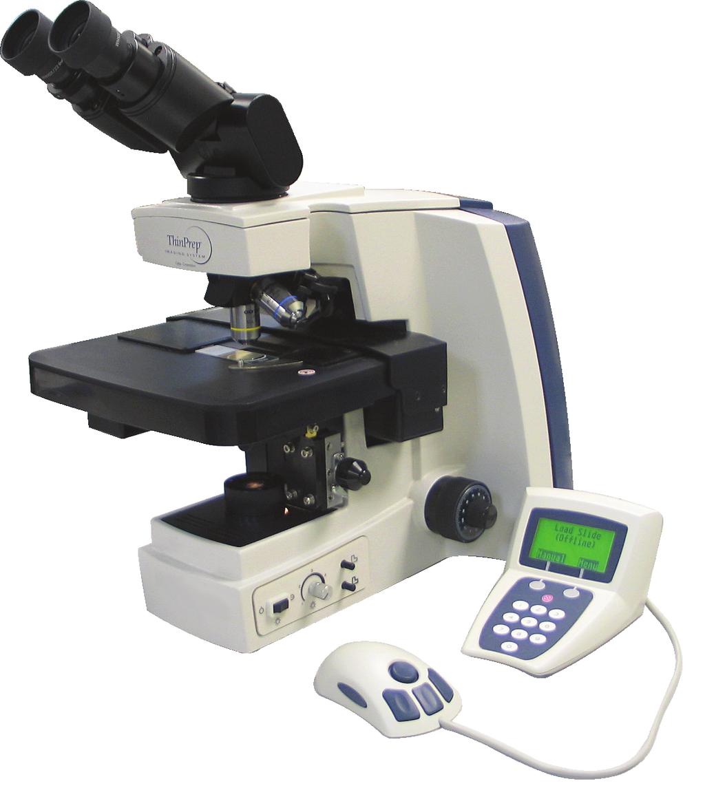
32 INTRODUCTION 1 Chapter One Introduction SECTION A OVERVIEW AND FUNCTION OF THE REVIEW SCOPE The Review Scope is an automated microscope to be used by a cytotechnologist (CT) to screen Thin- Prep Pap Test slides that have been imaged by a ThinPrep Image Processor. The microscope uses standard microscope optics enhanced with automated features that facilitate review of the slide. The CT views the slide and by means of automatic stage movement, is presented with fields of view containing objects of interest identified by the Imaging System. A motorized nosepiece allows the CT to change magnification via a pod device, without interrupting visual observation of the slide. An automated slide marking system allows the CT to mark objects for further review by a pathologist. The Review Scope is networked to the imaging system and slide data is retrieved from and later updated and returned to a slide database maintained by the imaging system. Review Scope Operator s Manual 1.1
33 1 INTRODUCTION Figure 1-1 Review Scope 1.2 Review Scope Operator s Manual
34 INTRODUCTION 1 ThinPrep Imaging System: Laboratory Flow Figure 1-2 Lab Flow Review Scope Operator s Manual 1.3
35 1 INTRODUCTION SECTION B THE THINPREP IMAGING SYSTEM PROCESS Slides that have been prepared for screening are loaded into cassettes which are placed into the Imaging Station. The operator uses a PC keyboard, mouse and monitor to interact with the instrument via a graphic, menu driven interface. A slide ID reader scans the slide accession ID and then the Imaging Station scans the entire ThinPrep cell spot. The system identifies objects of interest found on the slide, based on integrated optical density. (Refer to Figure 1-3, ThinPrep Imaging Process.) The coordinates of 22 of those objects are recorded and the slide is returned to its cassette. Following processing of each cassette of slides, the numeric slide ID and associated data record are sent to the Server. The Server acts as the central data manager for the ThinPrep Imaging System. As slides are imaged by the Image Processor and reviewed at the Review Scope, the Server stores, retrieves and transmits information based on the slide ID. The Cytotechnologist (CT) reviews slides at the Review Scope (RS). The RS consists of elements of a standard microscope, augmented with automated capabilities for viewing and marking the microscope slides. The RS contains an optical scanner which reads the slide ID when a slide is loaded on the stage. When a valid slide accession ID has been identified at the RS, the Server sends the object of interest coordinates for that ID and the CT is presented with the 22 fields of view determined for that slide. It is required that the CT review each of these fields of view before completing a slide review. As each field of view is being reviewed, the CT has the option to electronically mark the contents of the field for subsequent physical marking at completion of the slide review. The CT always has the option to control the position of the stage/slide manually, which provides complete freedom to move any portion of the ThinPrep cell spot into the field of view for examination. Note: The object of interest is typically placed in the center of the field of view, however the CT must screen the entire field of each of the 22 fields of view presented. Before completing the review, the CT may revisit any locations and mark or unmark fields of view, as desired. At the conclusion of electronically marking the slide, physical marks are applied at those locations with a semi-permanent translucent marker. A slide reviewed mark is placed on the slide at the end of the review, whether or not any physical marks were placed. 1.4 Review Scope Operator s Manual

36 INTRODUCTION 1 Figure 1-3 ThinPrep Imaging Process Review Scope Operator s Manual 1.5
NEMA XR X-ray Equipment for Interventional Procedures User Quality Control Mode
 NEMA XR 27-2012 X-ray Equipment for Interventional Procedures User Quality Control Mode Published by: National Electrical Manufacturers Association 1300 North 17th Street, Suite 1752 Rosslyn, Virginia
AN0509 swarm API Country Settings
 1.0 NA-15-0356-0002-1.0 Version:1.0 Author: MLA Document Information Document Title: Document Version: 1.0 Current Date: 2015-04-16 Print Date: 2015-04-16 Document ID: Document Author: Disclaimer NA-15-0356-0002-1.0
Using Iterative Automation in Utility Analytics
 Using Iterative Automation in Utility Analytics A utility use case for identifying orphaned meters O R A C L E W H I T E P A P E R O C T O B E R 2 0 1 5 Introduction Adoption of operational analytics can
4.1. Accurate: The information is a true reflection of the original observation.
 SOP #: DOC-101 Page: 1 of 7 Effective Date: 1. POLICY STATEMENT: The Principal Investigator and research team members are required to prepare and maintain adequate and accurate case histories designed
QwikCheck Beads Precision and Linearity Kit Instructions QwikCheck GOLD Analyzer
 Medical Electronic Systems www.mes-global.com service@mes-llc.com QwikCheck Beads Precision and Linearity Kit Instructions QwikCheck GOLD Analyzer OVERVIEW The QwikCheck Beads Precision and Linearity Kit
Positive Pixel Count Algorithm. User s Guide
 Positive Pixel Count Algorithm User s Guide Copyright 2004, 2006 2008 Aperio Technologies, Inc. Part Number/Revision: MAN 0024, Revision B Date: December 9, 2008 This document applies to software versions
Quality and GLP for Histology and Pathology of Drug Safety Studies
 Quality and GLP for Histology and Pathology of Drug Safety Studies Roger Alison BVSc MRCVS DiplECVP Consultant Toxicological Pathologist What is Quality Histology? It depends upon the purpose - Answer
SPECIFICATIONS FOR A QUALITY LABEL FOR DECORATION OF COATED ALUMINIUM USED IN ARCHITECTURAL APPLICATIONS Edition
 SPECIFICATIONS FOR A QUALITY LABEL FOR DECORATION OF COATED ALUMINIUM USED IN ARCHITECTURAL APPLICATIONS Master version ratified by the QUALIDECO Committee on 27 April 2017 Effective from 1 July 2017 Published
MIRAX SCAN The new way of looking at pathology
 Microscopy from Carl Zeiss MIRAX SCAN The new way of looking at pathology Greater reliability. Greater efficiency. Plus points for your diagnostics Better. More efficient. Quality as a factor for success
SolarEdge Export Limitation Application Note
 SolarEdge Export Limitation Application Note Europe and APAC Version 2.3 Disclaimers Disclaimers Important Notice Copyright SolarEdge Inc. All rights reserved. No part of this document may be reproduced,
Language Standardization for Mortality Coding A German Approach Stefanie Weber, Orlando Özer
 MEETING OF WHO COLLABORATING CENTRES FOR THE FAMILY OF INTERNATIONAL CLASSIFICATIONS Tunis, Tunisia 29 Oct. - 4 Nov. 2006 A German Approach Stefanie Weber, Orlando Özer Abstract In Germany 16 counties
Multi-resolution Cervical Cell Dataset
 Report 37 Multi-resolution Cervical Cell Dataset Patrik Malm December 2013 Centre for Image Analysis Swedish University of Agricultural Sciences Uppsala University Uppsala 2013 Multi-resolution Cervical
TERMS AND CONDITIONS. for the use of the IMDS Advanced Interface by IMDS-AI using companies
 TERMS AND CONDITIONS for the use of the IMDS Advanced Interface by IMDS-AI using companies Introduction The IMDS Advanced Interface Service (hereinafter also referred to as the IMDS-AI ) was developed
X9 REGISTRY FOR CHECK IMAGE TESTS
 X9 REGISTRY FOR CHECK IMAGE TESTS FSTC Horizontal Streaks Present In The Image #015.00 Check Image Test Status: A Where: A = Active (approved for use) W = Withdrawn (not for use) S = Superseded (not for
THEORY AND APPROACHES TO AUTOMATED IMAGE ANALYSIS IN DIGITAL PATHOLOGY
 THEORY AND APPROACHES TO AUTOMATED IMAGE ANALYSIS IN DIGITAL PATHOLOGY Kyle Takayama, MS Charles River Laboratories EVERY STEP OF THE WAY EVERY STEP OF THE WAY MORPHOMETRY Measurements or counts performed
High Dynamic Range Microscopy for Color Selective Virtual De-Staining of Immunocytological Specimens
 High Dynamic Range Microscopy for Color Selective Virtual De-Staining of Immunocytological Specimens David Friedrich 1, André Bell 1, Kraisorn Chaisaowong 1, Till Braunschweig 2, Ruth Knüchel-Clarke 2,
WirelessUSB LS Radio Module FCC Testing & Verification - AN4006
 WirelessUSB LS Radio Module FCC Testing & Verification - AN4006 Introduction One of the bottlenecks that many product developers encounter in incorporating any radio communication device is facing the
Surveying and QC of Stereotactic Breast Biopsy Units for ACR Accreditation
 Surveying and QC of Stereotactic Breast Biopsy Units for ACR Accreditation AAPM Annual Clinical Meeting Indianapolis, IN August 5, 2013 Learning Objectives Become familiar with the recommendations and
GUITAR PRO SOFTWARE END-USER LICENSE AGREEMENT (EULA)
") GUITAR PRO SOFTWARE END-USER LICENSE AGREEMENT (EULA) GUITAR PRO is software protected by the provisions of the French Intellectual Property Code. THIS PRODUCT IS NOT SOLD BUT PROVIDED WITHIN THE FRAMEWORK
GUITAR PRO SOFTWARE END-USER LICENSE AGREEMENT (EULA) GUITAR PRO is software protected by the provisions of the French Intellectual Property Code. THIS PRODUCT IS NOT SOLD BUT PROVIDED WITHIN THE FRAMEWORK
(R) Aerospace First Article Inspection Requirement FOREWORD
 Aerospace First Article Inspection Requirement FOREWORD") AEROSPACE STANDARD AS9102 Technically equivalent to AECMA pren 9102 Issued 2000-08 Revised 2004-01 REV. A Supersedes AS9012 (R) Aerospace First Article Inspection Requirement FOREWORD In December 1998,
AEROSPACE STANDARD AS9102 Technically equivalent to AECMA pren 9102 Issued 2000-08 Revised 2004-01 REV. A Supersedes AS9012 (R) Aerospace First Article Inspection Requirement FOREWORD In December 1998,
X9 REGISTRY FOR CHECK IMAGE TESTS
 X9 REGISTRY FOR CHECK IMAGE TESTS FSTC Excessive Spot Noise In The Image #014.00 Check Image Test Status: A Where: A = Active (approved for use) W = Withdrawn (not for use) S = Superseded (not for use
(Non-legislative acts) DECISIONS
 DECISIONS") 4.12.2010 Official Journal of the European Union L 319/1 II (Non-legislative acts) DECISIONS COMMISSION DECISION of 9 November 2010 on modules for the procedures for assessment of conformity, suitability
4.12.2010 Official Journal of the European Union L 319/1 II (Non-legislative acts) DECISIONS COMMISSION DECISION of 9 November 2010 on modules for the procedures for assessment of conformity, suitability
RFTX-1 Installation Manual
 RFTX-1 Installation Manual complete control Universal Remote Control RFTX-1 Installation Manual 2009-2014 Universal Remote Control, Inc. The information in this Owner s Manual is copyright protected. No
DICOM Correction Proposal
 Tracking Information - Administration Use Only DICOM Correction Proposal Correction Proposal Number Status CP-1713 Letter Ballot Date of Last Update 2018/01/23 Person Assigned Submitter Name David Clunie
Planishing hammer stand For use with SKU Planishing hammer
 Planishing hammer stand For use with SKU 94847 Planishing hammer Model 96300 Assembly And Operation Instructions Please Note: Planishing Hammer not included with Stand. Due to continuing improvements,
AUTOMATED BEARING WEAR DETECTION. Alan Friedman
 AUTOMATED BEARING WEAR DETECTION Alan Friedman DLI Engineering 253 Winslow Way W Bainbridge Island, WA 98110 PH (206)-842-7656 - FAX (206)-842-7667 info@dliengineering.com Published in Vibration Institute
Biomedical Equipment Technician
 Biomedical Equipment Technician Occupational Skill Stards Texas Skill Stards Board Recognized Critical Work Function 1. Install Biomedical Equipment 1.1 Receive, inspect inventory upon delivery 1.2 Deliver
MISSISSIPPI STATE UNIVERSITY Office of Planning Design and Construction Administration
 SECTION 01 340 - SHOP DRAWINGS, PRODUCT DATA AND SAMPLES PART 1 - GENERAL 1.1 RELATED DOCUMENTS A. Drawings and general provisions of the Contract, including General and Supplementary Conditions and other
ORiNOCO AP-4000MR-LR and AP-4900MR-LR Access Points Safety and Regulatory Compliance Information
 IMPORTANT! Visit http://support.proxim.com for the latest safety and regulatory compliance information for this product. ORiNOCO AP-4000MR-LR and AP-4900MR-LR Access Points Safety and Regulatory Compliance
SIR-WRR1. User's Guide SIRIUS Echo Antenna. Signal Repeater System Accessory
 SIR-WRR1 User's Guide SIRIUS Echo Antenna Signal Repeater System Accessory Desktop SIRIUS Docking Echo Station Antenna FCC NOTICE: This device complies with part 15 of the FCC Rules and with RSS-210 of
GENERAL DESCRIPTION OF THE CMC SERVICES
 STANDARD FOR CERTIFICATION No.1.1 GENERAL DESCRIPTION OF THE CMC SERVICES MAY 2007 FOREWORD (DNV) is an autonomous and independent foundation with the objectives of safeguarding life, property and the
Regulatory Forum. Society of Toxicologic Pathology Position Paper on Pathology Image Data: Compliance with 21 CFR Parts 58 and 11
 Regulatory Forum Toxicologic Pathology, 35:450 455, 2007 Copyright C by the Society of Toxicologic Pathology ISSN: 0192-6233 print / 1533-1601 online DOI: 10.1080/01926230701284509 Society of Toxicologic
igeacom User Guide V2.0
 Quality Care through innovative technology igeacom User Guide V2.0 IgeaCare Systems Inc. 5650 Tomken Road, Unit #9, Mississauga, Ontario, L4W 4P1, Canada Tel: 905.361.6225 Fax: 905.361.6209 www.igeacare.com
Engineering Policy & Procedure
 FPD > Engineering > Global Standards Engineering Policy & Procedure Revision History Number: G2-4 Section: G Subject: Radiographic Examination Procedure 1.0 SCOPE This procedure specifies the requirements
Issues in Emerging Health Technologies Bulletin Process
 Issues in Emerging Health Technologies Bulletin Process Updated: April 2015 Version 1.0 REVISION HISTORY Periodically, this document will be revised as part of ongoing process improvement activities. The
INFORMATION FOR RELATIVES
 ST. JAMES S HOSPITAL DUBLIN INFORMATION FOR RELATIVES THE AUTOPSY OR POST-MORTEM EXAMINATION Based on Faculty of Pathology Guidelines Information for Relatives The Autopsy or Post-Mortem Examination INTRODUCTION:
9 PIECE TUNGSTEN CARBIDE HOLE SAW KIT. Model 90721
 9 PIECE TUNGSTEN CARBIDE HOLE SAW KIT Model 90721 Set up And Operating Instructions Diagrams within this manual may not be drawn proportionally. Due to continuing improvements, actual product may differ
This document is a preview generated by EVS
 TECHNICAL REPORT IEC/TR 80002-1 Edition 1.0 2009-09 colour inside Medical device software Part 1: Guidance on the application of ISO 14971 to medical device software IEC/TR 80002-1:2009(E) THIS PUBLICATION
A POLICY in REGARDS to INTELLECTUAL PROPERTY. OCTOBER UNIVERSITY for MODERN SCIENCES and ARTS (MSA)
") A POLICY in REGARDS to INTELLECTUAL PROPERTY OCTOBER UNIVERSITY for MODERN SCIENCES and ARTS (MSA) OBJECTIVE: The objective of October University for Modern Sciences and Arts (MSA) Intellectual Property
A POLICY in REGARDS to INTELLECTUAL PROPERTY OCTOBER UNIVERSITY for MODERN SCIENCES and ARTS (MSA) OBJECTIVE: The objective of October University for Modern Sciences and Arts (MSA) Intellectual Property
Goals, progress and difficulties with regard to the development of German nuclear standards on the example of KTA 2000
 Goals, progress and difficulties with regard to the development of German nuclear standards on the example of KTA 2000 Dr. M. Mertins Gesellschaft für Anlagen- und Reaktorsicherheit (GRS) mbh ABSTRACT:
Virtual Microscopy: Potential Applications in Medical Education and Telemedicine in Countries with Developing Economies
 Virtual Microscopy: Potential Applications in Medical Education and Telemedicine in Countries with Developing Economies Paul Fontelo 1, Ernest DiNino 2, Krista, Johansen 2, Ashraf Khan 2, Michael Ackerman
SCANNING ELECTRON MICROSCOPE (SEM) INSPECTION OF SEMICONDUCTOR DICE. ESCC Basic Specification No
 INSPECTION OF SEMICONDUCTOR DICE. ESCC Basic Specification No") Page 1 of 24 SCANNING ELECTRON MICROSCOPE (SEM) INSPECTION OF SEMICONDUCTOR DICE ESCC Basic Specification Issue 2 February 2014 Document Custodian: European Space Agency see https://escies.org PAGE 2 LEGAL
Page 1 of 24 SCANNING ELECTRON MICROSCOPE (SEM) INSPECTION OF SEMICONDUCTOR DICE ESCC Basic Specification Issue 2 February 2014 Document Custodian: European Space Agency see https://escies.org PAGE 2 LEGAL
Confidently Assess Risk Using Public Records Data with Scalable Automated Linking Technology (SALT)
") WHITE PAPER Linking Liens and Civil Judgments Data Confidently Assess Risk Using Public Records Data with Scalable Automated Linking Technology (SALT) Table of Contents Executive Summary... 3 Collecting
WHITE PAPER Linking Liens and Civil Judgments Data Confidently Assess Risk Using Public Records Data with Scalable Automated Linking Technology (SALT) Table of Contents Executive Summary... 3 Collecting
MECOS-C2 microscopy systems
 MECOS-C2 microscopy systems Microscopy systems of the MECOS-C2 family production LLC "Medical computer Systems (MECOS)" belong to a class of scanning microscopes-analyzers and are intended for: Increase
2-Slot Desktop Chassis (DC) Extended Temperature
 Extended Temperature") APRIL 2008 LMC5202A 2-Slot Desktop Chassis (DC) Extended Temperature Copyright 2008. Black Box Corporation. All rights reserved 50 80105BB 01 A0 1000 Park Drive Lawrence, PA 35055 1018 724 746 5500 Fax
APRIL 2008 LMC5202A 2-Slot Desktop Chassis (DC) Extended Temperature Copyright 2008. Black Box Corporation. All rights reserved 50 80105BB 01 A0 1000 Park Drive Lawrence, PA 35055 1018 724 746 5500 Fax
Appendix 6.1 Data Source Described in Detail Vital Records
 Appendix 6.1 Data Source Described in Detail Vital Records Appendix 6.1 Data Source Described in Detail Vital Records Source or Site Birth certificates Fetal death certificates Elective termination reports
ACT-IR220Li/220LN IrDA Serial Port Adapter
 ACT-IR220Li/220LN IrDA Serial Port Adapter Product Specification Summary ACTiSYS Corp. 48511 Warm Springs Blvd, Suite 206 Fremont, CA 94539, USA TEL: (510) 490-8024, FAX: (510) 623-7268 E-Mail: irda-support@actisys.com
Standard MOD Area Interchange Methodology
 A. Introduction 1. Title: Area Interchange Methodology 2. Number: MOD-028-2 3. Purpose: To increase consistency and reliability in the development and documentation of Transfer Capability calculations
User s Guide ASSISTIVE LISTENING SYSTEMS
 User s Guide ASSISTIVE LISTENING SYSTEMS 2 Digital-1 User s Guide Contents How to use Digital-1...3 Tuning...6 Frequency Chart...8 Correcting Interference...9 Recharging...10 Specifications...12 Notice...13
Technical Support, End User License & Warranty Information
 Technical Support, End User License & Warranty Information How to get Technical Support Pazzles provides free Technical Support for your Inspiration Vūe for a period of 1 year from the date of purchase.
Powered by. For further installation assistance: prxperformance.com/pages/murphy-rack
 Powered by The 90 Fold-in Murphy Rack is made by the creators of the original Profile Folding Rack at PRx Performance and is Patent Pending. An up-to-date record of patents and patent pending items can
INTERNATIONAL TELECOMMUNICATION UNION DATA COMMUNICATION NETWORK: INTERFACES
 INTERNATIONAL TELECOMMUNICATION UNION CCITT X.21 THE INTERNATIONAL (09/92) TELEGRAPH AND TELEPHONE CONSULTATIVE COMMITTEE DATA COMMUNICATION NETWORK: INTERFACES INTERFACE BETWEEN DATA TERMINAL EQUIPMENT
SinglFuse SF-1206HHxxM Series Features
 *RoHS COMPLIANT & **HALOGEN FREE X SinglFuse SF-1206HHxxM Series Features n Single blow fuse for overcurrent protection n 3216 (EIA 1206) footprint n High current rating applications n High inrush withstand
Active Medical Implants Operating in the MHz Band
 Issue 2 November 2005 Spectrum Management and Telecommunications Radio Standards Specification Active Medical Implants Operating in the 402-405 MHz Band Aussi disponible en français - CNR-243 Preface Radio
SolarEdge Export Limitation Application Note
 SolarEdge Export Limitation Application Note North America Version 2.3 Disclaimers Disclaimers Important Notice Copyright SolarEdge Inc. All rights reserved. No part of this document may be reproduced,
GenePix Application Note
 GenePix Application Note Biological Relevance of GenePix Results Shawn Handran, Ph.D. and Jack Y. Zhai, Ph.D. Axon Instruments, Inc. 3280 Whipple Road, Union City, CA 94587 Last Updated: Aug 22, 2003.
Preparing for the new Regulations for healthcare providers
 Preparing for the new Regulations for healthcare providers Cathal Brennan, Medical Device Assessor HPRA Information Day on Medical Devices 23 rd October 2014 Brussels, 26.9.2012 COM(2012) 542 final 2012/0266
1. Redistributions of documents, or parts of documents, must retain the SWGIT cover page containing the disclaimer.
 Disclaimer: As a condition to the use of this document and the information contained herein, the SWGIT requests notification by e-mail before or contemporaneously to the introduction of this document,
Technology: Lighting Units
 Triple E Register Eligibility Criteria Category: Lighting Technology: Lighting Units Lighting units are products that are specifically designed to provide high efficiency interior or exterior illumination.
Using a Microscope. Year Group: BVSc1 + Document number: CSL_L07
 Year Group: BVSc1 + Document number: CSL_L07 Equipment list: Equipment for this station: Microscope Power supply and a level surface to work on Gloves The sample to examine Marker or pencil for labelling
DEPARTMENT OF PUBLIC SAFETY DIVISION OF FIRE COLUMBUS, OHIO. SOP Revision Social Media Digital Imagery
 DEPARTMENT OF PUBLIC SAFETY DIVISION OF FIRE COLUMBUS, OHIO 17-007 SUBJECT: TITLE: Administration SOP Revision-04-05-07 Social Media 04-05-08 Digital Imagery Implementation Office of the Chief PURPOSE:
Elo Touch Solutions Wallmounting Kit for the 7001L IDS Touchmonitors
 Installation Manual Elo Touch Solutions Wallmounting Kit for the 7001L IDS Touchmonitors SW602083 Rev E Table of Contents Chapter 1: Safety Warning... 3 Chapter 2: Kit Contents... 4 Included in Kit...
Standard Test Method for Grading Spun Yarns for Appearance 1
 Designation: D 2255 02 Standard Test Method for Grading Spun Yarns for Appearance 1 This standard is issued under the fixed designation D 2255; the number immediately following the designation indicates
AT&T INDIANA GUIDEBOOK. PART 2 - General Terms and Conditions 1st Revised Sheet 1 SECTION 9 - Connections
 PART 2 - General Terms and Conditions 1st Revised Sheet 1 EXCHANGE SERVICES 1. General Provisions A. General Terminal equipment and communications systems provided by the customer may be connected at the
CATALOG FOR WOODCO USA Brand 150 TON SPIDER. API ROTARY TAPER TRADEMARK WOODCO BUY THE BRAND REG. U.S. PATENT OFFICE MANUFACTURER
 CATALOG FOR WOODCO USA Brand 150 TON SPIDER. API ROTARY TAPER Hinged Casing and Tubing Spider, includes an assembly drawing, dimensions, weight and parts list. TRADEMARK WOODCO USA BUY THE BRAND REG. U.S.
Standard Practice for Qualification of Radioscopic Systems 1
 Designation: 95 An American National Standard Standard Practice for Qualification of Radioscopic Systems 1 This standard is issued under the fixed designation ; the number immediately following the designation
Instruction also available on
 TERA Radon Program EN TCR3 Central Unit Technical Specifications & Operation Manual v.2 2016 Table of Contents 1 Introduction...2 2 Description and Utilization...2 3 Scope of Delivery...4 4 Product Specification...5
ivu Plus Quick Start Guide P/N rev. A -- 10/8/2010
 P/N 154721 rev. A -- 10/8/2010 Contents Contents 1 Introduction...3 2 ivu Plus Major Features...4 2.1 Demo Mode...4 2.2 Sensor Types...4 2.2.1 Selecting a Sensor Type...5 2.3 Multiple Inspections...6 2.3.1
Agilent G1888 Network Headspace Sampler
 Agilent G1888 Network Headspace Sampler Safety and Regulatory Information Agilent Technologies Notices Agilent Technologies, Inc. 2004 No part of this manual may be reproduced in any form or by any means
ISO INTERNATIONAL STANDARD. Non-destructive testing of welds Radiographic testing Part 1: X- and gamma-ray techniques with film
 INTERNATIONAL STANDARD ISO 17636-1 First edition 2013-01-15 Non-destructive testing of welds Radiographic testing Part 1: X- and gamma-ray techniques with film Contrôle non destructif des assemblages soudés
J O I N T I N D U S T R Y S T A N D A R D. Requirements for Soldering Pastes J Standard 005A. December 2011
 J O I N T I N D U S T R Y S T A N D A R D Requirements for Soldering Pastes J Standard 005A December 2011 Requirements for Soldering Pastes 1.0 SCOPE 1.1 Scope This standard prescribes general requirements
Pickens Savings and Loan Association, F.A. Online Banking Agreement
 Pickens Savings and Loan Association, F.A. Online Banking Agreement INTERNET BANKING TERMS AND CONDITIONS AGREEMENT This Agreement describes your rights and obligations as a user of the Online Banking
Eidgenössisches Departement für Umwelt, Verkehr, Energie und Kommunikation UVEK. Bundesamt für Umwelt BAFU Abteilung Luftreinhaltung und Chemikalien
 Eidgenössisches Departement für Umwelt, Verkehr, Energie und Kommunikation UVEK Bundesamt für Umwelt BAFU Abteilung Luftreinhaltung und Chemikalien Is it acceptable to destroy the paper originals of raw
STATISTIC ANALYZER S21
 STATISTIC ANALYZER S21 PRESENTATION, PERFORMANCE AND CAPACITY OF THE EQUIPMENT Figure 1 Analyzer S21 The analyzer S21 is an inspector of cereal grains that through image processing and subsequent statistical
Radio Remote Controls Manual K Series
 Radio Remote Controls Manual K Series PN 52764 2010.12.20 Rev. 2 K Series radio control manual 1 Conductix Incorporated The technical data and images which appear in this manual are for informational purposes
Projects Connector User Guide
 Version 4.3 11/2/2017 Copyright 2013, 2017, Oracle and/or its affiliates. All rights reserved. This software and related documentation are provided under a license agreement containing restrictions on
ISO 4090 INTERNATIONAL STANDARD. Photography Medical radiographic cassettes/screens/films and hard-copy imaging films Dimensions and specifications
 INTERNATIONAL STANDARD ISO 4090 Third edition 2001-08-15 Photography Medical radiographic cassettes/screens/films and hard-copy imaging films Dimensions and specifications Photographie Cassettes/écrans/films
Sense. 3D Scanner. User Guide. See inside for use and safety information.
 Sense 3D Scanner User Guide See inside for use and safety information. 1 CONTENTS INTRODUCTION.... 3 IMPORTANT SAFETY INFORMATION... 4 Safety Guidelines....4 SENSE 3D SCANNER FEATURES AND PROPERTIES....
SF Certified International Shipping Customer Agreement V1.0
 SF Certified International Shipping Customer Agreement V1.0 SF Certified International Shipping (further referred as SF CIS ) is a service provided by S.F. Express Co., Ltd. (further referred as SF ) to
SECTION I - CHAPTER 2 DIGITAL IMAGING PROCESSING CONCEPTS
 RADT 3463 - COMPUTERIZED IMAGING Section I: Chapter 2 RADT 3463 Computerized Imaging 1 SECTION I - CHAPTER 2 DIGITAL IMAGING PROCESSING CONCEPTS RADT 3463 COMPUTERIZED IMAGING Section I: Chapter 2 RADT
SPOT PathSuite Solutions
 SPOT PathSuite Solutions The Perfect Fit PathStation TM Imaging system for the gross dissection in hood PathStand TM 40 imaging station for medium to large specimens PathSuite Is Easy To Use A turnkey
TAKING DIAGNOSTICS TO THE NEXT LEVEL ENDRESS+HAUSER
 TAKING DIAGNOSTICS TO THE NEXT LEVEL ENDRESS+HAUSER The FOUNDATION fieldbus specification was created from the ground up to allow suppliers to add their own competitive advantage to the technology. At
eskbook Emerging Life Sciences Companies second edition Chapter 8 Checklist for Planning and Conducting an Effective FTO Search
 eskbook Emerging Life Sciences Companies second edition Chapter 8 Checklist for Planning and Conducting an Effective FTO Search Chapter 8 CHECKLIST FOR PLANNING AND CONDUCTING AN EFFECTIVE FTO SEARCH The
MIL-STD-883H METHOD ULTRASONIC INSPECTION OF DIE ATTACH
 * ULTRASONIC INSPECTION OF DIE ATTACH 1. PURPOSE. The purpose of this examination is to nondestructively detect unbonded regions, delaminations and/or voids in the die attach material and at interfaces
Transmitter. User Manual. Firmware version 1.0 and greater
 ProRF SPC Transmitter User Manual Firmware version 1.0 and greater FCC NOTICE This equipment has been tested and found to comply with the limits for a class B digital device, pursuant to part 15 of the
TOSHIBA Fast Recovery Diode Silicon Diffused Type CMF01
 TOSHIBA Fast Recovery Diode Silicon Diffused Type Switching Mode Power Supply Applications DC/DC Converter Applications Unit: mm Repetitive peak reverse voltage: V RRM = 6 V Average forward current: I
DEVELOPING THE WORKFORCE
 DEVELOPING THE WORKFORCE Assessing the quality of death certification: Instructions for the online assessment tool Resources and Tools 3 November 2016 About this series Capacity-building resources and
CALIBRATION OF MICROSCOPE EYEPIECE GRATICULE
 CALIBRATION OF MICROSCOPE EYEPIECE GRATICULE A typical eyepiece graticule looks like this: It is 10mm in length and each mm is divided into 10 parts So each small division = 0.1mm = 100µm The eyepiece
ISO INTERNATIONAL STANDARD. Imaging materials Ammonia-processed diazo photographic film Specifications for stability
 INTERNATIONAL STANDARD ISO 18905 First edition 2002-11-01 Imaging materials Ammonia-processed diazo photographic film Specifications for stability Matériaux pour l'image Film photographique diazoïque traité
Category: ELECTRICITY Requirement: EL-ENG Page: 1 of 13. Document(s): S-E-01, S-E-04 Issue Date: Effective Date:
: S-E-01, S-E-04 Issue Date: Effective Date:") Requirements for Certification Category: ELECTRICITY Requirement: EL-ENG-11-02 Page: 1 of 13 Requirements for Certification and Use of Portable Measuring Apparatus for Reverification and Dispute Meter
Requirements for Certification Category: ELECTRICITY Requirement: EL-ENG-11-02 Page: 1 of 13 Requirements for Certification and Use of Portable Measuring Apparatus for Reverification and Dispute Meter
Product Guide Verizon Delaware LLC. Section 31 Delaware LLC Original Sheet 1. Connection With Certain Facilities of Others
 Delaware LLC Original Sheet 1 A. GENERAL Part 68 of the Federal Communications Commission's Rules and Regulations applies to customer premises equipment, with specified exceptions. Accordingly, regulations
INTERNATIONAL STANDARD
 Provläsningsexemplar / Preview INTERNATIONAL STANDARD ISO 23713 First edition 2005-07-01 Pulps Determination of fibre coarseness by automated optical analysis Polarized light method Pâtes Détermination
DNVGL-CP-0338 Edition October 2015
 CLASS PROGRAMME DNVGL-CP-0338 Edition October 2015 The electronic pdf version of this document, available free of charge from http://www.dnvgl.com, is the officially binding version. FOREWORD DNV GL class
ACT-IR220L/LE IrDA Serial Port Adapter
 ACT-IR220L/LE IrDA Serial Port Adapter Product Specification Summary ACTiSYS Corp. 48511 Warm Springs Blvd, Suite 206 Fremont, CA 94539, USA TEL: (510) 490-8024, FAX: (510) 623-7268 E-Mail: irda-support@actisys.com
Owner s Manual & Safety Instructions
 Owner s Manual & Safety Instructions Save This Manual Keep this manual for the safety warnings and precautions, assembly, operating, inspection, maintenance and cleaning procedures. Write the product s
ISO INTERNATIONAL STANDARD. Non-destructive testing of welds Radiographic testing of fusionwelded
 INTERNATIONAL STANDARD ISO 17636 First edition 2003-09-15 Non-destructive testing of welds Radiographic testing of fusionwelded joints Contrôle non destructif des assemblages soudés Contrôle par radiographie
View Terms and Conditions: Effective 12/5/2015 Effective 6/17/2017
 View Terms and Conditions: Effective 12/5/2015 Effective 6/17/2017 Comerica Mobile Banking Terms and Conditions - Effective 12/5/2015 Thank you for using Comerica Mobile Banking combined with your device's
automatic embosser & die cutter USER MANUAL
 TM TM automatic embosser & die cutter USER MANUAL CREATE A BEAUTIFUL LIFE IN THE BOX Cut n Boss machine (7) Embossing Folders (12) Cutting Dies Platforms (2) (1) Platform B (1) Platform D Magnetic Shim
30 Bending Brake. Model Assembly and Operating Instructions. Distributed exclusively by Harbor Freight Tools.
 30 Bending Brake Model 41311 Assembly and Operating Instructions Distributed exclusively by Harbor Freight Tools. 3491 Mission Oaks Blvd., Camarillo, CA 93011 Copyright 1999 by Harbor Freight Tools. All
A-16D A-Net Distributor
 A-16D A-Net Distributor For use with the Personal Monitor Mixing System Information in this document is subject to change. All rights reserved. Copyright 2003 Aviom, Inc. Printed in USA Document Rev. 1.03