Supplemental Figure 1: High-resolution AO images of the cone mosaics in Figure 5 in normal
|
|
|
- Monica Hunter
- 5 years ago
- Views:
Transcription
1 1
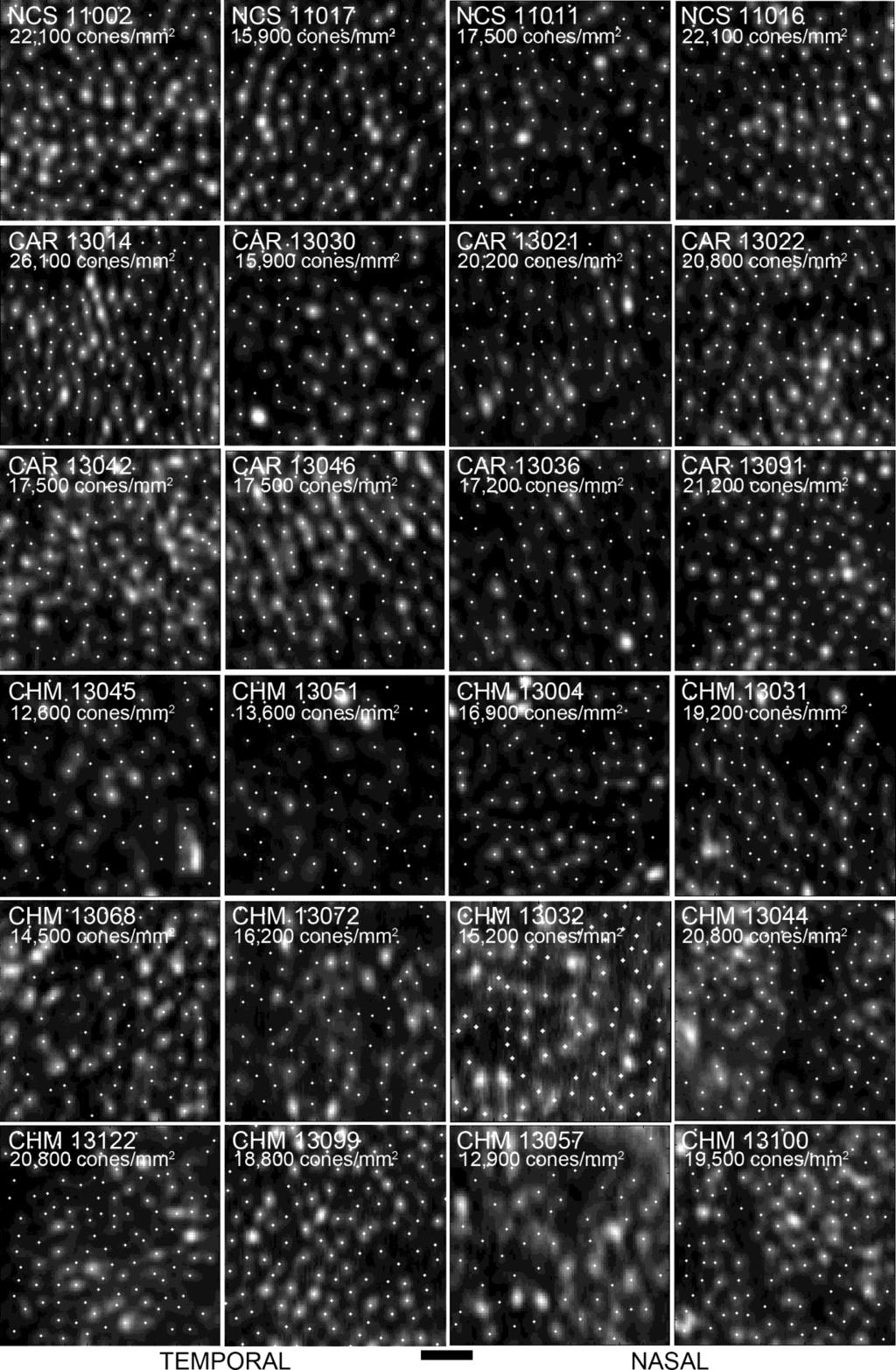
2 Supplemental Figure 1: High-resolution AO images of the cone mosaics in Figure 5 in normal control subjects (NCS), carriers of choroideremia (CAR) and choroideremia patients (CHM) showing the locations of each cone determined by direct counting and used for measurements of cone density. Cones were identified by author JM through a semi-automated custom Matlab 26, 27 program. Cone densities are shown for each image, and all densities measured fell within the previously reported normal range. Manual cone identification still leaves room for error through misidentifying or missed cones. This underscores the need for well-designed studies to test the repeatability, reliability, and variability of cone analysis with multiple observers and for more accurate, automated cone identification programs to alleviate the need for manual intervention. 2
.")
3 Supplemental Figure 2: High-resolution AO images of the cone mosaic at approximately 0.5 mm along each meridian in choroideremia patients (CHM). Cone densities for each of the images in the two left columns fell within the previously reported normal range (Supplemental Figure 3). 32 Cone densities for each of the images in the two right columns fell below the previously reported normal range. 32 Scale bar 20 um. 3
 showing the locations of each cone determined by direct counting and used for")
4 Supplemental Figure 3: AO images of the cone mosaics in Supplemental Figure 2 at 0.5mm in choroideremia patients (CHM) showing the locations of each cone determined by direct counting and used for measurements of cone density. 4

5 Supplemental Figure 4: IR and OCT images from the left eye of symptomatic carrier showing large regions of RPE atrophy in the macula, including the foveal region. The atrophic border exhibits the classic scalloped edges characteristic of choroideremia. OCT shows loss of outer retinal layers in regions of atrophy, thinned RPE and indistinguishable interdigitation layer 5
6 in regions of relatively intact retina, abrupt borders at the edge of atrophy, interlaminar bridges, and outer retinal tubulation. Supplemental video 1: High resolution AOSLO images recorded at various focus levels in patient where bubble-like features are found adjacent to the centrally intact retina. This is the same location as shown in Figure 10 patient By adjusting the focus of the AO system, the bubble-like features came into best focus approximately 0.4D posterior to the plane of best focus for the cone photoreceptors. This image series was obtained using a custom built AOSLO as previously described. 40 The OCT for in Figure 10 shows that the bubble-like features co-locate with the hypo-reflective space in the en face translational directions; this through focus video this gives further evidence that the bubble-like features also co-locate with the hypo-reflective space in the choroid in depth. Supplemental Table 1: Normal Control Patient Data * Number of retinal locations at which cone density was measured (Number of locations where cone density was within/higher/lower than the normal range reported by Song et al. 32 ) Supplemental Table 2: Choroideremia Carrier Patient Data * Eye was excluded from study due to AOSLO imaging artifact Number of retinal locations at which cone density was measured (Number of locations where cone density was within/higher/lower than the normal range reported by Song et al. 32 ) Carrier was symptomatic 6
7 Supplementary Table 1: Normal Control Patient Data Patient ID Age Axial Length (mm) Cone Density Measured * OD OS OD OS (5/0/1) 12 (12/0/0) (7/1/1) 8 (8/0/0) (11/1/0) 4 (4/0/0) (6/0/0) 5 (4/1/0) (7/0/0) 6 (6/0/0) (1/0/0) 4 (4/0/0) (5/0/0) 9 (9/0/0) (6/0/1) 8 (8/0/0) * Number of retinal locations at which cone density was measured (Number of locations where cone density was within/higher/lower than the normal range reported by Song et al. )
8 Supplementary Table 2: Choroideremia Carrier Patient Data Patient ID Age Axial Length (mm) Visual Acuity Cone Density Measured OD OS OD OS OD OS /20 20/20 5 (4/0/1) 1 (1/0/0) (10/1/0) 11 (11/0/0) /20 20/20 1 (1/0/0) 4 (4/0/0) * * 4 (4/0/0) /20 20/20 4 (4/0/0) 3 (2/0/1) /20 20/20 6 (5/0/1) 4 (4/0/0) (10/1/0) 11 (11/0/0) /20 20/20 2 (2/0/0) /20 20/20 10 (9/1/0) 2 (2/0/0) /20 20/20 11 (11/0/0) 9 (8/0/1) (10/1/0) 9 (8/1/0) /20 20/20 3 (3/0/0) 7 (6/1/0) /20 20/20 * 3 (2/0/1) dB 7.67dB 5 (3/1/1) 5 (5/0/0) (4/0/0) 1 (1/0/0) /20 20/20 2 (2/0/0) 3 (3/0/0) (1/1/0) 1 (1/0/0) /20 20/20 2 (2/0/0) * * Eye was excluded from study due to AOSLO imaging artifact Number of re nal loca ons at which cone density was measured (Number of locations where cone density was within/higher/lower than the normal range reported by Song et al. ) Carrier was symptoma c Data not available Visual acuity data not available but Humphrey Visual field data was available. Central 30 2 Threshold Test, Mean Deviation db
Micropulse Duty Cycle. # of eyes (20 ms) Total spots (200 ms)
 Total spots (200 ms)") Micropulse Duty Cycle Total spots (2 ms) # of eyes (2 ms) Total spots (2 ms) % 269 44 3 47% 9 4 4 25% 3 5 4 4 5% 2 4 3 5 2% 5 2 NA NA 9% 2 4 6% NA NA 57 2 5% 4 5 6 3 3% 39 5 35 5 # of eyes (2 ms) Supplemental
Micropulse Duty Cycle Total spots (2 ms) # of eyes (2 ms) Total spots (2 ms) % 269 44 3 47% 9 4 4 25% 3 5 4 4 5% 2 4 3 5 2% 5 2 NA NA 9% 2 4 6% NA NA 57 2 5% 4 5 6 3 3% 39 5 35 5 # of eyes (2 ms) Supplemental
SUPPLEMENTARY INFORMATION
 Computational high-resolution optical imaging of the living human retina Nathan D. Shemonski 1,2, Fredrick A. South 1,2, Yuan-Zhi Liu 1,2, Steven G. Adie 3, P. Scott Carney 1,2, Stephen A. Boppart 1,2,4,5,*
Vision Research 51 (2011) Contents lists available at SciVerse ScienceDirect. Vision Research
 Contents lists available at SciVerse ScienceDirect. Vision Research") Vision Research 51 (2011) 2132 2138 Contents lists available at SciVerse ScienceDirect Vision Research journal homepage: www.elsevier.com/locate/visres The relationship between peripapillary crescent and
Vision Research 51 (2011) 2132 2138 Contents lists available at SciVerse ScienceDirect Vision Research journal homepage: www.elsevier.com/locate/visres The relationship between peripapillary crescent and
Going beyond the surface of your retina OCT-HS100 OPTICAL COHERENCE TOMOGRAPHY
 Going beyond the surface of your retina OCT-HS100 OPTICAL COHERENCE TOMOGRAPHY Automatic functions make examinations short and simple. Perform the examination with only two simple mouse clicks! 1. START
Going beyond the surface of your retina OCT-HS100 OPTICAL COHERENCE TOMOGRAPHY
 Going beyond the surface of your retina OCT-HS100 OPTICAL COHERENCE TOMOGRAPHY Full Auto OCT High specifications in a very compact design Automatic functions make examinations short and simple. Perform
Introduction. Chapter Aim of the Thesis
 Chapter 1 Introduction 1.1 Aim of the Thesis The main aim of this investigation was to develop a new instrument for measurement of light reflected from the retina in a living human eye. At the start of
Visual Optics. Visual Optics - Introduction
 Visual Optics Jim Schwiegerling, PhD Ophthalmology & Optical Sciences University of Arizona Visual Optics - Introduction In this course, the optical principals behind the workings of the eye and visual
Eye. Eye Major structural layer of the wall of the eye is a thick layer of dense C.T.; that layer has two parts:
 General aspects Sensory receptors ; External or internal environment. A stimulus is a change in the environmental condition which is detectable by a sensory receptor 1 Major structural layer of the wall
OCT mini-symposium. Presenters. Donald Miller, Indiana Univ. Joseph Izatt, Duke Univ. Thomas Milner, Univ. of Texas at Austin Jay Wei, Zeiss Meditec
 OCT mini-symposium Presenters Donald Miller, Indiana Univ. Joseph Izatt, Duke Univ. Thomas Milner, Univ. of Texas at Austin Jay Wei, Zeiss Meditec Starlight, eyebright Canberra Times, Australia Combining
Optical Coherence Tomography. RS-3000 Advance / Lite
 Optical Coherence Tomography RS-3000 Advance / Lite 12 mm wide horizontal scan available with the RS-3000 Advance allows detailed observation of the vitreous body, retina, and choroid from the macula to
EYE ANATOMY. Multimedia Health Education. Disclaimer
 Disclaimer This movie is an educational resource only and should not be used to manage your health. The information in this presentation has been intended to help consumers understand the structure and
PHGY Physiology. SENSORY PHYSIOLOGY Vision. Martin Paré
 PHGY 212 - Physiology SENSORY PHYSIOLOGY Vision Martin Paré Assistant Professor of Physiology & Psychology pare@biomed.queensu.ca http://brain.phgy.queensu.ca/pare The Process of Vision Vision is the process
ABSTRACT 1. INTRODUCTION
 High-resolution retinal imaging: enhancement techniques Mircea Mujat 1*, Ankit Patel 1, Nicusor Iftimia 1, James D. Akula 2, Anne B. Fulton 2, and R. Daniel Ferguson 1 1 Physical Sciences Inc., Andover
PHGY Physiology. The Process of Vision. SENSORY PHYSIOLOGY Vision. Martin Paré. Visible Light. Ocular Anatomy. Ocular Anatomy.
 PHGY 212 - Physiology SENSORY PHYSIOLOGY Vision Martin Paré Assistant Professor of Physiology & Psychology pare@biomed.queensu.ca http://brain.phgy.queensu.ca/pare The Process of Vision Vision is the process
Image Database and Preprocessing
 Chapter 3 Image Database and Preprocessing 3.1 Introduction The digital colour retinal images required for the development of automatic system for maculopathy detection are provided by the Department of
The Special Senses: Vision
 OLLI Lecture 5 The Special Senses: Vision Vision The eyes are the sensory organs for vision. They collect light waves through their photoreceptors (located in the retina) and transmit them as nerve impulses
Optical Coherence Tomography Retina Scan Duo
 Optical Coherence Tomography Retina Scan Duo High Definition OCT & Fundus Imaging in One Compact System The Retina Scan Duo is a combined OCT and fundus camera system that is a user friendly and versatile
Blood Vessel Tree Reconstruction in Retinal OCT Data
 Blood Vessel Tree Reconstruction in Retinal OCT Data Gazárek J, Kolář R, Jan J, Odstrčilík J, Taševský P Department of Biomedical Engineering, FEEC, Brno University of Technology xgazar03@stud.feec.vutbr.cz
Retinal stray light originating from intraocular lenses and its effect on visual performance van der Mooren, Marie Huibert
 University of Groningen Retinal stray light originating from intraocular lenses and its effect on visual performance van der Mooren, Marie Huibert IMPORTANT NOTE: You are advised to consult the publisher's
VISUAL PROSTHESIS FOR MACULAR DEGENERATION AND RETINISTIS PIGMENTOSA
 VISUAL PROSTHESIS FOR MACULAR DEGENERATION AND RETINISTIS PIGMENTOSA 1 SHWETA GUPTA, 2 SHASHI KUMAR SINGH, 3 V K DWIVEDI Electronics and Communication Department 1 Dr. K.N. Modi University affiliated to
Our vision is foresight
 Our vision is foresight iseries OCT Systems The Optovue iseries Improving OCT performance with ease Who ever said advanced OCT scanning had to be complicated? When an OCT design puts user experience first,
Fourier Domain (Spectral) OCT OCT: HISTORY. Could OCT be a Game Maker OCT in Optometric Practice: A THE TECHNOLOGY BEHIND OCT
 OCT OCT: HISTORY. Could OCT be a Game Maker OCT in Optometric Practice: A THE TECHNOLOGY BEHIND OCT") Could OCT be a Game Maker OCT in Optometric Practice: A Hands On Guide Murray Fingeret, OD Nick Rumney, MSCOptom Fourier Domain (Spectral) OCT New imaging method greatly improves resolution and speed of
Could OCT be a Game Maker OCT in Optometric Practice: A Hands On Guide Murray Fingeret, OD Nick Rumney, MSCOptom Fourier Domain (Spectral) OCT New imaging method greatly improves resolution and speed of
ABO Certification Training. Part I: Anatomy and Physiology
 ABO Certification Training Part I: Anatomy and Physiology Major Ocular Structures Centralis Nerve Major Ocular Structures The Cornea Cornea Layers Epithelium Highly regenerative: Cells reproduce so rapidly
Yokohama City University lecture INTRODUCTION TO HUMAN VISION Presentation notes 7/10/14
 Yokohama City University lecture INTRODUCTION TO HUMAN VISION Presentation notes 7/10/14 1. INTRODUCTION TO HUMAN VISION Self introduction Dr. Salmon Northeastern State University, Oklahoma. USA Teach
Visibility, Performance and Perception. Cooper Lighting
 Visibility, Performance and Perception Kenneth Siderius BSc, MIES, LC, LG Cooper Lighting 1 Vision It has been found that the ability to recognize detail varies with respect to four physical factors: 1.Contrast
Sensory receptors External internal stimulus change detectable energy transduce action potential different strengths different frequencies
 General aspects Sensory receptors ; respond to changes in the environment. External or internal environment. A stimulus is a change in the environmental condition which is detectable by a sensory receptor
OCT - Anatomy of a Scan. OCT - Anatomy of a Scan. OCT Imaging. OCT Imaging
 OCT - Anatomy of a Scan Timothy J. Bennett, CRA, OCT-C, FOPS Penn State Eye Center Hershey, PA OCT - Anatomy of a Scan A systematic approach to understanding what we see in retinal OCT images including
The Photoreceptor Mosaic
 The Photoreceptor Mosaic Aristophanis Pallikaris IVO, University of Crete Institute of Vision and Optics 10th Aegean Summer School Overview Brief Anatomy Photoreceptors Categorization Visual Function Photoreceptor
!!!!!!!!!!!!!!!!!!!!!!!!!!!!!!!! Supplementary Material,!Zhang!et!al.,!SLO measurement of mouse rhodopsin
 !!!!!!!!!!!!!!!!!!!!!!!!!!!!!!!! Supplementary Material,!Zhang!et!al.,!SLO measurement of mouse rhodopsin I. Conversion of SLO scanning angles to retinal distances* A goal of this investigation was the
RegionFinder The perfect Use. SPECTRALIS HRA, HRA+OCT and SPECTRALIS OCT
 RegionFinder The perfect Use SPECTRALIS HRA, HRA+OCT and SPECTRALIS OCT RegionFinder - The perfect Use SPECTRALIS HRA, HRA+OCT and SPECTRALIS OCT General workflow to acquire the perfect AF image Adjust
The Human Visual System. Lecture 1. The Human Visual System. The Human Eye. The Human Retina. cones. rods. horizontal. bipolar. amacrine.
 Lecture The Human Visual System The Human Visual System Retina Optic Nerve Optic Chiasm Lateral Geniculate Nucleus (LGN) Visual Cortex The Human Eye The Human Retina Lens rods cones Cornea Fovea Optic
Optics of Wavefront. Austin Roorda, Ph.D. University of Houston College of Optometry
 Optics of Wavefront Austin Roorda, Ph.D. University of Houston College of Optometry Geometrical Optics Relationships between pupil size, refractive error and blur Optics of the eye: Depth of Focus 2 mm
Retina. Convergence. Early visual processing: retina & LGN. Visual Photoreptors: rods and cones. Visual Photoreptors: rods and cones.
 Announcements 1 st exam (next Thursday): Multiple choice (about 22), short answer and short essay don t list everything you know for the essay questions Book vs. lectures know bold terms for things that
Optical Coherence Tomography. RS-3000 Advance
 Optical Coherence Tomography RS-3000 Advance See it in Advance See it in high resolution with the AngioScan* image. SLO Superficial capillary OCT-Angiography (3 x 3 mm) Deep capillary OCT-Angiography (3
Photoreceptor Disruption Related to Persistent Submacular Fluid after Successful Scleral Buckle Surgery
 pissn: 1011-8942 eissn: 2092-9382 Korean J Ophthalmol 2011;25(6):380-386 http://dx.doi.org/10.3341/kjo.2011.25.6.380 Original Article Photoreceptor Disruption Related to Persistent Submacular Fluid after
OPTO 5320 VISION SCIENCE I
 OPTO 5320 VISION SCIENCE I Monocular Sensory Processes of Vision: Color Vision Ronald S. Harwerth, OD, PhD Office: Room 2160 Office hours: By appointment Telephone: 713-743-1940 email: rharwerth@uh.edu
Vision. The eye. Image formation. Eye defects & corrective lenses. Visual acuity. Colour vision. Lecture 3.5
 Lecture 3.5 Vision The eye Image formation Eye defects & corrective lenses Visual acuity Colour vision Vision http://www.wired.com/wiredscience/2009/04/schizoillusion/ Perception of light--- eye-brain
High-resolution axial measurements of ocular tissues
 The Interpretation of Optical Coherence Tomography Images of the Retina Devinder Singh Chauhan and John Marshall PURPOSE. To determine the relationship between optical coherence tomography (OCT) images
2 The First Steps in Vision
 2 The First Steps in Vision 2 The First Steps in Vision A Little Light Physics Eyes That See light Retinal Information Processing Whistling in the Dark: Dark and Light Adaptation The Man Who Could Not
Clinical evaluation and management of glaucoma is largely
 Macular Segmentation with Optical Coherence Tomography Hiroshi Ishikawa, 1,2 Daniel M. Stein, 1 Gadi Wollstein, 1,2 Siobahn Beaton, 1,2 James G. Fujimoto, 3 and Joel S. Schuman 1,2 PURPOSE. To develop
Vision Science I Exam 1 23 September ) The plot to the right shows the spectrum of a light source. Which of the following sources is this
 The plot to the right shows the spectrum of a light source. Which of the following sources is this") Vision Science I Exam 1 23 September 2016 1) The plot to the right shows the spectrum of a light source. Which of the following sources is this spectrum most likely to be taken from? A) The direct sunlight
Vision Science I Exam 1 23 September 2016 1) The plot to the right shows the spectrum of a light source. Which of the following sources is this spectrum most likely to be taken from? A) The direct sunlight
BIONIC EYE ( Offers new hope of restored vision ) BIONIC EYE ( Offers light at the end of tunnel for blind )
 BIONIC EYE ( Offers light at the end of tunnel for blind )") BIONIC EYE ( Offers new hope of restored vision ) EC0271 [1] SOWMYA.U.L [2] KALYANI.D.P ICE-2/4 ICE-2/4 GNITS-Hyderabad GNITS-Hyderabad BIONIC EYE ( Offers light at the end of tunnel for blind ) Introduction:
BIONIC EYE ( Offers new hope of restored vision ) EC0271 [1] SOWMYA.U.L [2] KALYANI.D.P ICE-2/4 ICE-2/4 GNITS-Hyderabad GNITS-Hyderabad BIONIC EYE ( Offers light at the end of tunnel for blind ) Introduction:
Rod Photopigment Kinetics After Photodisruption of the Retinal Pigment Epithelium
 Retina Rod Photopigment Kinetics After Photodisruption of the Retinal Pigment Epithelium Benjamin D. Masella, 1,2 Jennifer J. Hunter, 2,3 and David R. Williams 1 3 1 The Institute of Optics, University
PERCEPTUAL INSIGHTS INTO FOVEATED VIRTUAL REALITY. Anjul Patney Senior Research Scientist
 PERCEPTUAL INSIGHTS INTO FOVEATED VIRTUAL REALITY Anjul Patney Senior Research Scientist INTRODUCTION Virtual reality is an exciting challenging workload for computer graphics Most VR pixels are peripheral
CCVIP Early Intervention Pearls
 CCVIP Early Intervention Pearls Table of Contents Page 1: Page 2: Page 3: Page 4: Page 5: Page 6: Page 8: Page 9: Page 10: Page 11: Page 12: Page 13: Page 14: Past & Present Table of Contents Functional
Objectives. 3. Visual acuity. Layers of the. eye ball. 1. Conjunctiva : is. three quarters. posteriorly and
 OCULAR PHYSIOLOGY (I) Dr.Ahmed Al Shaibani Lab.2 Oct.2013 Objectives 1. Review of ocular anatomy (Ex. after image) 2. Visual pathway & field (Ex. Crossed & uncrossed diplopia, mechanical stimulation of
Human Visual System. Prof. George Wolberg Dept. of Computer Science City College of New York
 Human Visual System Prof. George Wolberg Dept. of Computer Science City College of New York Objectives In this lecture we discuss: - Structure of human eye - Mechanics of human visual system (HVS) - Brightness
Ocular Jeopardy. The major refractive portion of the eye 5/12/2015. Presented by Jill J Luebbert, CPOT, ABOC. Watch This Refractive optios
 Ocular Jeopardy Presented by Jill J Luebbert, CPOT, ABOC In the beginning anterior Way back Visual Pathway Say What? terminolog y Watch This Refractive optios Posterior Segment 10 10 10 10 10 20 20 20
Visual Perception. human perception display devices. CS Visual Perception
 Visual Perception human perception display devices 1 Reference Chapters 4, 5 Designing with the Mind in Mind by Jeff Johnson 2 Visual Perception Most user interfaces are visual in nature. So, it is important
Unresolved Issues in Prediction of Subjective and Objective Refraction from Wavefront Data
 Wavefront Congress Symposium Feb, 2008 Unresolved Issues in Prediction of Subjective and Objective Refraction from Wavefront Data Larry N. Thibos School of Optometry, Indiana University, Bloomington, IN
Optimizing Performance of AO Ophthalmic Systems. Austin Roorda, PhD
 Optimizing Performance of AO Ophthalmic Systems Austin Roorda, PhD Charles Garcia, MD Tom Hebert, PhD Fernando Romero-Borja, PhD Krishna Venkateswaran, PhD Joy Martin, OD/PhD student Ramesh Sundaram, MS
The TRC-NW8F Plus: As a multi-function retinal camera, the TRC- NW8F Plus captures color, red free, fluorescein
 The TRC-NW8F Plus: By Dr. Beth Carlock, OD Medical Writer Color Retinal Imaging, Fundus Auto-Fluorescence with exclusive Spaide* Filters and Optional Fluorescein Angiography in One Single Instrument W
Spectral colors. What is colour? 11/23/17. Colour Vision 1 - receptoral. Colour Vision I: The receptoral basis of colour vision
 Colour Vision I: The receptoral basis of colour vision Colour Vision 1 - receptoral What is colour? Relating a physical attribute to sensation Principle of Trichromacy & metamers Prof. Kathy T. Mullen
Color Theory. Chapter 3a Perceiving Color. The eye Rods Cones After-images Color Constancy
 Color Theory Chapter 3a Perceiving Color The eye Rods Cones After-images Color Constancy I know who you are by your eyes. Color Theory Today security systems exist that identify people solely by their
EYE STRUCTURE AND FUNCTION
 Name: Class: Date: EYE STRUCTURE AND FUNCTION The eye is the body s organ of sight. It gathers light from the environment and forms an image on specialized nerve cells on the retina. Vision occurs when
Optical Coherence Tomography. RS-3000 Advance / Lite
 Optical Coherence Tomography RS-3000 Advance / Lite See it in Advance See it in high resolution with the AngioScan* image. SLO Superficial capillary OCT-Angiography (3 x 3 mm) Deep capillary OCT-Angiography
Automated Perimeter PTS 1000
 PTS 1000 Automated Perimeter PTS 1000 is a modern diagnostic instrument for precise and fast testing of field of vision. It offers static and kinetic stimuli with all Goldmann stimuli sizes and all stimuli
General Discussion. Chapter Development of the Instrument
 Chapter 7 General Discussion The first aim of this thesis was the development of a new instrument for simultaneous measurement of the spectral and the directional reflectance of the living human eye. The
Automatic and manual segmentation of healthy retinas using high-definition optical coherence tomography
 Automatic and manual segmentation of healthy retinas using high-definition optical coherence tomography Isabelle Golbaz, 1 Christian Ahlers, 1 Nina Goesseringer, 2 Geraldine Stock, 1 Wolfgang Geitzenauer,
Retinopathy From a Green Laser Pointer
 CLINICAL SCIENCES Retinopathy From a Green Laser Pointer A Clinicopathologic Study Dennis M. Robertson, MD; Jay W. McLaren, PhD; Diva R. Salomao, MD; Thomas P. Link, CRA Objective: To report retinopathy
The temporal raphe is generally described as a horizontal
 Retina In Vivo Adaptive Optics Imaging of the Temporal Raphe and Its Relationship to the Optic Disc and Fovea in the Human Retina Gang Huang, Thomas J. Gast, and Stephen A. Burns School of Optometry, Indiana
Automated sub-retinal fluid detection comprising RPE-region using neighbouring pixel connectivity paradigm.
 Biomedical Research 2018; Special Issue: S108-S112 ISSN 0970-938X www.biomedres.info Automated sub-retinal fluid detection comprising RPE-region using neighbouring pixel connectivity paradigm. Piyush Mishra
Sensation notices Various stimuli Of what is out there In reality
 1 Sensation and Perception Are skills we need For hearing, feeling And helping us to see I will begin with A few definitions This way confusion Has some prevention Sensation notices Various stimuli Of
THRESHOLD AMSLER GRID TESTING AND RESERVING POWER OF THE POTIC NERVE by MOUSTAFA KAMAL NASSAR. M.D. MENOFIA UNIVERSITY.
 THRESHOLD AMSLER GRID TESTING AND RESERVING POWER OF THE POTIC NERVE by MOUSTAFA KAMAL NASSAR. M.D. MENOFIA UNIVERSITY. Since Amsler grid testing was introduced by Dr Marc Amsler on 1947and up till now,
CLARUS 500 from ZEISS HD ultra-widefield fundus imaging
 CLARUS 500 from ZEISS HD ultra-widefield fundus imaging Imaging ultra-wide without compromise. ZEISS CLARUS 500 // INNOVATION MADE BY ZEISS Compromising image quality may leave some pathology unseen. Signs
Optical Coherence Tomography. RS-3000 Advance 2
 Optical Coherence Tomography RS-3000 Advance 2 -Providing a comprehensive solution for retina and glaucom Retina Analysis Retinal mode Glaucoma Analysis Choroidal mode Image courtesy of Hokkaido University
CLARUS 500 from ZEISS HD ultra-widefield fundus imaging
 CLARUS 500 from ZEISS HD ultra-widefield fundus imaging Imaging ultra-wide without compromise. ZEISS CLARUS 500 // INNOVATION MADE BY ZEISS Compromising image quality may leave some pathology unseen. Signs
Digital Imaging and Communications in Medicine (DICOM)
") Digital Imaging and Communications in Medicine (DICOM) Supplement 197: Ophthalmic Optical Coherence Tomography for Angiographic Imaging Storage SOP Classes Prepared by: DICOM Standards Committee 1300 N.
Digital Imaging and Communications in Medicine (DICOM) Supplement 197: Ophthalmic Optical Coherence Tomography for Angiographic Imaging Storage SOP Classes Prepared by: DICOM Standards Committee 1300 N.
DIGITAL IMAGE PROCESSING (COM-3371) Week 2 - January 14, 2002
 Week 2 - January 14, 2002") DIGITAL IMAGE PROCESSING (COM-3371) Week 2 - January 14, 22 Topics: Human eye Visual phenomena Simple image model Image enhancement Point processes Histogram Lookup tables Contrast compression and stretching
DIGITAL IMAGE PROCESSING (COM-3371) Week 2 - January 14, 22 Topics: Human eye Visual phenomena Simple image model Image enhancement Point processes Histogram Lookup tables Contrast compression and stretching
10/25/2017. Financial Disclosures. Do your patients complain of? Are you frustrated by remake after remake? What is wavefront error (WFE)?
?") Wavefront-Guided Optics in Clinic: Financial Disclosures The New Frontier November 4, 2017 Matthew J. Kauffman, OD, FAAO, FSLS STAPLE Program Soft Toric and Presbyopic Lens Education Gas Permeable Lens
Wavefront-Guided Optics in Clinic: Financial Disclosures The New Frontier November 4, 2017 Matthew J. Kauffman, OD, FAAO, FSLS STAPLE Program Soft Toric and Presbyopic Lens Education Gas Permeable Lens
Digital Imaging and Communications in Medicine (DICOM)
") Digital Imaging and Communications in Medicine (DICOM) Supplement 197: Ophthalmic Tomography for Angiographic Imaging Storage SOP Classes Prepared by: DICOM Standards Committee 1300 N. 17 th Street Suite
Digital Imaging and Communications in Medicine (DICOM) Supplement 197: Ophthalmic Tomography for Angiographic Imaging Storage SOP Classes Prepared by: DICOM Standards Committee 1300 N. 17 th Street Suite
The reliability of parafoveal cone density measurements
 Laboratory science For numbered affiliations see end of article. Correspondence to Dr Joseph Carroll, Medical College of Wisconsin, The Eye Institute, 925 N. 87th Street, Milwaukee, WI 53226, USA; jcarroll@mcw.edu
Fundus Photograph Reading Center
 Autofluorescence Using Confocal Scanning Laser Ophthalmoscope (cslo) Instruments (AF-D) 8010 Excelsior Drive, Suite 100, Madison WI 53717 Telephone: (608) 410-0560 Fax: (608) 410-0566 Table of Contents
Visual System I Eye and Retina
 Visual System I Eye and Retina Reading: BCP Chapter 9 www.webvision.edu The Visual System The visual system is the part of the NS which enables organisms to process visual details, as well as to perform
November 14, 2017 Vision: photoreceptor cells in eye 3 grps of accessory organs 1-eyebrows, eyelids, & eyelashes 2- lacrimal apparatus:
 Vision: photoreceptor cells in eye 3 grps of accessory organs 1-eyebrows, eyelids, & eyelashes eyebrows: protection from debris & sun eyelids: continuation of skin, protection & lubrication eyelashes:
Variation of Cone Photoreceptor Packing Density with Retinal Eccentricity and Age METHODS. Subjects
 Multidisciplinary Ophthalmic Imaging Variation of Cone Photoreceptor Packing Density with Retinal Eccentricity and Age Hongxin Song, Toco Yuen Ping Chui, Zhangyi Zhong, Ann E. Elsner, and Stephen A. Burns
SPECTRALIS Training Guide
 SPECTRALIS Training Guide SPECTRALIS Diagram 1 SPECTRALIS Training Guide Table of Contents 1. Entering Patient Information & Aligning the Patient a. Start Up/Shut Down the System... 4 b. Examine a New
AUTOMATED DRUSEN DETECTION IN A RETINAL IMAGE USING MULTI-LEVEL ANALYSIS
 AUTOMATED DRUSEN DETECTION IN A RETINAL IMAGE USING MULTI-LEVEL ANALYSIS A Thesis Presented to the Graduate School of Clemson University In Partial Fulfillment of the Requirements for the Degree Master
Large Field of View, Modular, Stabilized, Adaptive-Optics- Based Scanning Laser Ophthalmoscope
 Large Field of View, Modular, Stabilized, Adaptive-Optics- Based Scanning Laser Ophthalmoscope Stephen A. Burns, Remy Tumbar, Ann E. Elsner, Daniel Ferguson, Daniel X. Hammer OCIS Codes: 170.1790, 170.3890,
Optical, receptoral, and retinal constraints on foveal and peripheral vision in the human neonate
 Vision Research 38 (1998) 3857 3870 Optical, receptoral, and retinal constraints on foveal and peripheral vision in the human neonate T. Rowan Candy a, *, James A. Crowell b, Martin S. Banks a a School
Color and Perception
 Color and Perception Why Should We Care? Why Should We Care? Human vision is quirky what we render is not what we see Why Should We Care? Human vision is quirky what we render is not what we see Some errors
Going beyond the surface of your retina
 Going beyond the surface of your retina OCT-HS100 Optical Coherence Tomography Canon s expertise in optics and innovative technology have resulted in a fantastic 3 μm optical axial resolution for amazing
Human Vision and Human-Computer Interaction. Much content from Jeff Johnson, UI Wizards, Inc.
 Human Vision and Human-Computer Interaction Much content from Jeff Johnson, UI Wizards, Inc. are these guidelines grounded in perceptual psychology and how can we apply them intelligently? Mach bands:
Large-field high-speed polarization sensitive spectral domain OCT and its applications in ophthalmology
 Large-field high-speed polarization sensitive spectral domain OCT and its applications in ophthalmology Stefan Zotter, 1* Michael Pircher, 1 Teresa Torzicky, 1 Bernhard Baumann, 1 Hirofumi Yoshida, 3 Futoshi
CHAPTER 4 LOCATING THE CENTER OF THE OPTIC DISC AND MACULA
 90 CHAPTER 4 LOCATING THE CENTER OF THE OPTIC DISC AND MACULA The objective in this chapter is to locate the centre and boundary of OD and macula in retinal images. In Diabetic Retinopathy, location of
fringes were produced on the retina directly. Threshold contrasts optical aberrations in the eye. (Received 12 January 1967)
") J. Phy8iol. (1967), 19, pp. 583-593 583 With 5 text-figure8 Printed in Great Britain VISUAL RESOLUTION WHEN LIGHT ENTERS THE EYE THROUGH DIFFERENT PARTS OF THE PUPIL BY DANIEL G. GREEN From the Department
J. Phy8iol. (1967), 19, pp. 583-593 583 With 5 text-figure8 Printed in Great Britain VISUAL RESOLUTION WHEN LIGHT ENTERS THE EYE THROUGH DIFFERENT PARTS OF THE PUPIL BY DANIEL G. GREEN From the Department
Spatial coding: scaling, magnification & sampling
 Spatial coding: scaling, magnification & sampling Snellen Chart Snellen fraction: 20/20, 20/40, etc. 100 40 20 10 Visual Axis Visual angle and MAR A B C Dots just resolvable F 20 f 40 Visual angle Minimal
The use of ophthalmoscopes equipped with adaptive optics. Repeatability of In Vivo Parafoveal Cone Density and Spacing Measurements ORIGINAL ARTICLE
 1040-5488/12/8905-0632/0 VOL. 89, NO. 5, PP. 632 643 OPTOMETRY AND VISION SCIENCE Copyright 2012 American Academy of Optometry ORIGINAL ARTICLE of In Vivo Parafoveal Cone Density and Spacing Measurements
Various techniques have been developed to characterize
 The Reflectance of Single Cones in the Living Human Eye Aristofanis Pallikaris, 1 David R. Williams, 2 and Heidi Hofer 2 PURPOSE. Individual cones were imaged in the living human eye with the Rochester
AUTOMATIC MACULA DETECTION IN HUMAN EYE FUNDUS AUTO- FLUORESCENCE IMAGES: APPLICATION TO EYE DISEASE LOCALIZATION
 Stereology and Image Analysis. Ecs10 - Proceedings of the 10th European Congress of ISS, (V.Capasso et al. Eds.), The MIRIAM Project Series, ESCULAPIO Pub. Co., Bologna, Italy, 2009 AUTOMATIC MACULA DETECTION
Optical Coherence Tomography. RS-3000 Advance / Lite
 Optical Coherence Tomography RS-3000 Advance / Lite See it in Advance See it in high resolution with the AngioScan* image. OCT-Angiography of choroidal neovascularization * AngioScan (OCT-Angiography)
Title: Live volumetric (4D) visualization and guidance of in vivo human ophthalmic surgery with intraoperative optical coherence tomography
 visualization and guidance of in vivo human ophthalmic surgery with intraoperative optical coherence tomography") Title: Live volumetric (4D) visualization and guidance of in vivo human ophthalmic surgery with intraoperative optical coherence tomography Authors: O. M. Carrasco-Zevallos 1, B. Keller 1, C. Viehland
Title: Live volumetric (4D) visualization and guidance of in vivo human ophthalmic surgery with intraoperative optical coherence tomography Authors: O. M. Carrasco-Zevallos 1, B. Keller 1, C. Viehland
CSE1710. Big Picture. Reminder
 CSE1710 Click to edit Master Week text 10, styles Lecture 19 Second level Third level Fourth level Fifth level Fall 2013 Thursday, Nov 14, 2013 1 Big Picture For the next three class meetings, we will
Methods. Experimental Stimuli: We selected 24 animals, 24 tools, and 24
 Methods Experimental Stimuli: We selected 24 animals, 24 tools, and 24 nonmanipulable object concepts following the criteria described in a previous study. For each item, a black and white grayscale photo
Automated segmentation of retinal pigment epithelium cells in fluorescence adaptive optics images
 Rangel-Fonseca et al. Vol. 30, No. 12 / December 2013 / J. Opt. Soc. Am. A 2595 Automated segmentation of retinal pigment epithelium cells in fluorescence adaptive optics images Piero Rangel-Fonseca, 1,
Multifocal Electroretinograms in Normal Subjects
 Multifocal Electroretinograms in Normal Subjects Akiko Nagatomo, Nobuhisa Nao-i, Futoshi Maruiwa, Mikki Arai and Atsushi Sawada Department of Ophthalmology, Miyazaki Medical College, Miyazaki, Japan Abstract:
Version 1.0. th March 2011
 Optical Coherence Tomography Scan and Retinal Imagingg Version 1.0 http://www.ukbiobank.ac.uk/ 5 th March 2011 This manual details the procedure for Scan and Retinal Imagingg at an Assessment Centre of
PERIPHERAL VISON PATTERN DETECTION DYNAMIC TEST
 PERIPHERAL VISON PATTERN DETECTION DYNAMIC TEST João P Rodrigues, João D Semedo, Fernando M Melicio Institute Systems and Robotics,Technical University, Av Rovisco Pais 1 TN6.21, Lisbon, Portugal jrodrigues@laseeb.org,
UC Davis UC Davis Previously Published Works
 UC Davis UC Davis Previously Published Works Title Improved visualization of outer retinal morphology with aberration cancelling reflective optical design for adaptive optics - optical coherence tomography
Semi-automated discrimination of retinal pigmented epithelial cells in two-photon fluorescence images of mouse retinas
 Semi-automated discrimination of retinal pigmented epithelial cells in two-photon fluorescence images of mouse retinas Nathan S. Alexander, 1,3 Grazyna Palczewska, 2 and Krzysztof Palczewski 1,* 1 Department