A new method for segmentation of retinal blood vessels using morphological image processing technique
|
|
|
- Ronald Morris
- 5 years ago
- Views:
Transcription
1 A new method for segmentation of retinal blood vessels using morphological image processing technique Roya Aramesh Faculty of Computer and Information Technology Engineering,Qazvin Branch,Islamic Azad University Qazvin, Iran Karim Faez Department of Electrical Engineering, Amirkabir University of Technology, Tehran, Iran Abstractct: Retinopathy caused by complications of diabetes, which can eventually lead to blindness. The lack of oxygen in the retina causes fragile, new, blood vessels to grow along the retina and in the clear, gel-like vitreous humour that fills the inside of the eye. For this reason it is important to extract retinal blood vessels. This paper introduces a new method for the detection of retinal blood vessels in retinal fundus images. The pre-processing technique was applied to input image in order to reduce the image noise and then, the image was divided into 16 smaller blocks. Afterward, a threshold was obtained for each block using maximum and minimum points of image histogram. Eventually, line detector filters and mathematical morphology was applied to the image and optimum results were obtained. Consequently, the proposed method showed an accuracy of with and sensitivity and specificity, respectively on the DRIVE database and better results have been achieved in comparison with existing methods. Keywords- line detector filters; blood vessels; image processing; Image blocking; Morphology operation I. INTRODUCTION Retinal vessel segmentation is important for the detection of eye diseases and plays an important role in automatic retinal disease screening systems. Automatic detection and analysis of the vasculature can assist in the implementation of screening programs for vessel diameter measurement in relation with diagnosis of hypertension [1], and computerassisted laser surgery [2]. Segmentation retinal anatomical structures are the first step in any automatic retina analysis system [3]. Detection of large vessels is relatively easy due to their strong contrast against background in the images but detection of small vessels is much more difficult due to their low contrast in the images. The centerlines are extracted by using the first order derivative of a Gaussian filter in four orientations and then evaluation of derivative signs and average derivative values is performed. The combination of morphological filters and crosscurvature evaluation to segment vessel-like patterns is employed by Zana et al. [5,6]. Mathematical morphology exploits the fact that the vessels are linear, connected and their curvature is a cross-curvature evaluation was performed to identify the structures in a retinal image whose curvature is linearly coherent. The present method is based on the maximum and minimum points of image histogram which to find detect suitable threshold for retinal thick and thin vessels. Several researches have been directed to find automatic detection algorithms from retinal images for the better diagnosis of retinal diseases. Unfortunately, fundus images may be blurred which affects the performance of automatic detection algorithms. The purpose of this paper is to design an efficient new algorithm that can exactly extract the retinal blood vessels from background, more accurate and easy for detecting of retinal diseases. This work is composed of five sections. Proposed method is described in section 2 including step by step methodology. Experimental results are given in section 3 and followed by conclusion in section 4. Fraz et al. [4] have proposed a unique combination of vessel centerlines detection and morphological bit plane slicing to extract the blood vessel tree from the retinal images. Page 1
2 II. A. Proposed method diagram PROPOSED METHOD The algorithm consists of four main steps: Pre-processing of the image, blocking image and calculating the maximum and minimum points, image filtering and then morphology operations on binary image. Flow diagram for our vessel segmentation system is shown Figure 1. Input RGB image B. Data base The template is used to format your paper and style the text. All margins, column widths, line spaces, and text fonts are prescribed; please do not alter them. You may note peculiarities. For example, the head margin in this template measures proportionately more than is customary. This measurement and others are deliberate, using specifications that anticipate your paper as one part of the entire proceedings, and not as an independent document. Please do not revise any of the current designations. C. Preprocessing Calculating maximum point Pre-Processing Image blocking Filtering Morphology operation Calculating minimum point Fig. 1 Flow proposed method diagram Output binary image I Gray Redchannel* 0.4 Greenchannel* (1) Then, using a filter reduces out the noise. Following formula is used to filter Average: 1 f (x, y) I ( s, t) 20 ( s, t) S Gray (2) xy, D. Image blocking Input image size is pixels, the gray image is divided into 16 blocks (I 1,..,I 16 ), and then a histogram is plotted for each block. Finally, using the following relations Suitable threshold for each block is determined. b 1 b 2 b 3= unique I i,i = 1,..,16 (3) H = histc I,b (4) i 1 Max histogram = Max(N(b 1,H)) (5) Minhistogram Min( N( b, H)) (6) 1 / Threshold Max Min Max (7) E. Filtering histogram histogram histogram In the filtering stage, line detector filters are used. These filters can be used to find one-cell-thick vertical, horizontal, or angled (135-degrees or 45-degrees) lines in an image [8]. Notice that line-finding is a similar application to edgedetection. Values in the input image are replaced with the average value of all valid cells within the kernel. This is also the procedure when the neighborhood around a grid cell extends beyond the edge of the grid. Filters can be multiplied by the optimal threshold to filter the image to reveal more details. After applying filters to optimize image blocks, the result is a normalized image block (8) ( I _ optimize 0) I _ optimizecd c1 d1 cd Then, averaging is the each normalize block image (I_normalized). F. Morphplogy operation Mathematical morphology is a nonlinear tool in retinal images analysis which has revealed itself as a very effective method for retinal diseases detection. Morphological opening operation is applied to smooth the background image and to highlight the retinal blood vessels. The closing operation produces objects that are not completely surrounded by retinal vessels, like things at branching points. B creates a flat, disk-shaped structuring element, where 1 specifies the radius. I _ normalized B ( I _ normalized B)! B (9) 8 I _ normalized Bi (( I _ normalized B)! Bi) B (10) i i1 Page 2

 and 0.0174 in false positive rate (FPR).")


3 Top-hat filtering computes the morphological opening of the normalized image and then subtracts the result from the gray block image. For reduce noise and extract vessels more details Designed following four filters. These filters are properly extracted thin vessels the dimension of images in DRIVE database was The experiments on 40 retinal images showed that the average performance of proposed method is in true positive rate (TPR) and in false positive rate (FPR). TPR is ratio between the number of pixels on our proposed method result images that also appear on the ground truth, and the total pixel number on the thinned ground truth. FPR is ratio between the number of vessel pixels on our proposed methods results but not on the ground truth, and the number of pixels on the background of the thinned ground truth. A RGB block and the output block image after operations different are shown in Figure 3. As shown in this figure, thin vessels and vessels are identified for each block Fig. 2 Four filters designed to reduce noise and extraction of thin vessels The most basic morphological operations are dilation and erosion with a structure element [9]. The dilation is extending the image boundary or the object of the image itself by adding pixels based on the structuring element design. The dilation operation was applied to a binary block image ( I_normalize i ). i=1,..,16 if the input image is I and the structuring element B, then[10] (a) I B { I _ normalize ( B) I } (11) i I _ normalize i Finally, after morphological operation, 16 binary block image obtained and the blocks connected together. Then the erosion operator applied on the image. The erosion operator creates erosion on all 1 values in the image. The structural element can be 1 or zero values. The center of structural element was placed on each image pixel and erosion operator is applied on a pixel based on structural element value. The image erosion of I, and structural element of B, are defined as follows: I! B { w ( B) I, w I} (12) This algorithm is to detect and properly diagnosed all main vessels, even the tiny vessels are also correctly diagnosed. (b) III. RESULT AND DISCUSSION The performance of presented algorithm is evaluated by applying it to DRIVE fundus image databases. The images of DRIVE database are mostly taken from healthy people, (c) (d) Page 3
 (i) (j) (m)")
original")

a RGB")
normalized")

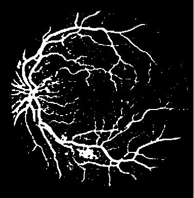
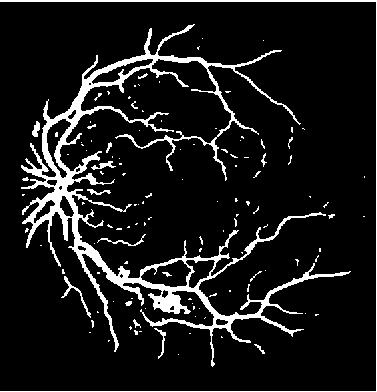
4 (e) (f) (l) (g) (h) (i) (j) (m) (k) (n) Fig.3 (a)original image (b)green channel (c)preprocessing operation (d)a RGB block image (e)normalized block image (f),(g),(h),(i) image filtering (j),(k)morphological operation (l)connected blocks (m)proposed method result (n)manual segment Page 4
5 True positive rate Jan. 31 Image block histograms are shown in Figure 4. This figure is used to obtain maximum and minimum values of block histograms and the maximum and minimum values are used to define suitable threshold. Performance of presented algorithm was also evaluated with receiver operating characteristic (ROC) curve. These trends were generated by measuring the true and the false rate on all DRIVE database images. The ROC curve for the DRIVE database shown in Fig 5. False positive rate Fig5. ROC curve for the DRIVE database Table I shows the comparison of performance of this work with the other approaches in terms of average accuracy, sensitivity, specificity and FPR. TABLE I COMPARISON OF PERFORMANCE BETWEEN THE RECENT STUDIESAND THIS WORK Method Average accuracy Sensitivity Specificit y FPR Fraz et al. [4] Zana et al. [5],[6] This work Fig4. Block histograms According to this method, the sensitivity, average accuracy and specificity in the diagnosis are and and respectively. IV. CONCLUSION A new algorithm for vessel extraction in retinal color fundus images was presented in this work. This approach is effective in medical and biomedical applications as automated retinal image analyses system. In this paper, the two methods of histogram maximum and minimum points and mathematical morphology were combined and used to Page 5
6 characterize the retinal blood vessels. The proposed method is applied for a database of 40 images and an accuracy of with and sensitivity and specificity, respectively on the DRIVE database obtained. The results of proposed method were compared to those obtained from existing methods and better performance has been achieved. In morphological operation needs two principal components, the maximum and minimum points of the histogram for these two principle component have been used. This method provides clear blood vessels of retinal images, suitable for detection retinal pathologies. For further study an efficient classification process can be considered for every image point in order to minimize or even eliminated some of misdetections of retina blood vessels. REFERENCES [1] J. Lowell, A. Hunter, D. Steel, A. Basu, R. Ryder, R.L. Kennedy, Measurement of retinal vessel widths from fundus images based on 2- D modeling, IEEE Transactions on Medical Imaging 23 (2004) [2] J.J. Kanski, Clinical Ophthalmology, 6th ed., Elsevier Health Sciences, London, UK, [3] M. Foracchia, E. Grisan, and A. Ruggeri, "Detection of Optic Disc in Retinal Images by Means of a Geometrical Model of Vessel Structure," IEEE Trans. Medical Imaging, vol. 23, pp , Oct [4] M.M. Fraz, S.A. Barman, P. Remagnino, A. Hoppe, A. Basit, B. Uyyanonvara, A.R. Rudnicka, C.G. Owen, An approach to localize the retinal blood vessels using bit planes and centerline detection, Computer Methods and Programs in Biomedicine, in press. [5] F. Zana, J.C. Klein, A multimodal registration algorithm of eye fundus images using vessels detection and Hough transform, IEEE Transactions on Medical Imaging 18 (1999) [6] F. Zana, J.C. Klein, Segmentation of vessel-like patterns using mathematical morphology and curvature evaluation, IEEE Transactions on Image Processing 10 (2001) [7] M. Niemeijer, J.J. Staal, B.v. Ginneken, M. Loog, M.D. Abramoff, DRIVE: digital retinal images for vessel extraction, [8] Fraz, M.M., et al., Blood vessel segmentation methodologies in retinal images A survey. Computer methods and programs in biomedicine, (1): p [9] R. Gonzalez and R. Woods 1992 Digital Image Processing. Adison Wesley. [10] W. K. Pratt, Digital Image Processing, 3rd ed. New York: Wiley, Page 6
Fovea and Optic Disc Detection in Retinal Images with Visible Lesions
 Fovea and Optic Disc Detection in Retinal Images with Visible Lesions José Pinão 1, Carlos Manta Oliveira 2 1 University of Coimbra, Palácio dos Grilos, Rua da Ilha, 3000-214 Coimbra, Portugal 2 Critical
CHAPTER 4 LOCATING THE CENTER OF THE OPTIC DISC AND MACULA
 90 CHAPTER 4 LOCATING THE CENTER OF THE OPTIC DISC AND MACULA The objective in this chapter is to locate the centre and boundary of OD and macula in retinal images. In Diabetic Retinopathy, location of
Retinal blood vessel extraction
 Retinal blood vessel extraction Surya G 1, Pratheesh M Vincent 2, Shanida K 3 M. Tech Scholar, ECE, College, Thalassery, India 1,3 Assistant Professor, ECE, College, Thalassery, India 2 Abstract: Image
Automatic Detection Of Optic Disc From Retinal Images. S.Sherly Renat et al.,
 International Journal of Technology and Engineering System (IJTES) Vol 7. No.3 2015 Pp. 203-207 gopalax Journals, Singapore available at : www.ijcns.com ISSN: 0976-1345 AUTOMATIC DETECTION OF OPTIC DISC
Comparison of two algorithms in the automatic segmentation of blood vessels in fundus images
 Comparison of two algorithms in the automatic segmentation of blood vessels in fundus images ABSTRACT Robert LeAnder, Myneni Sushma Chowdary, Swapnashri Mokkapati, and Scott E Umbaugh Effective timing
OPTIC DISC LOCATION IN DIGITAL FUNDUS IMAGES
 OPTIC DISC LOCATION IN DIGITAL FUNDUS IMAGES Miss. Tejaswini S. Mane 1,Prof. D. G. Chougule 2 1 Department of Electronics, Shivaji University Kolhapur, TKIET,Wrananagar (India) 2 Department of Electronics,
Image Database and Preprocessing
 Chapter 3 Image Database and Preprocessing 3.1 Introduction The digital colour retinal images required for the development of automatic system for maculopathy detection are provided by the Department of
Blood Vessel Segmentation of Retinal Images Based on Neural Network
 Blood Vessel Segmentation of Retinal Images Based on Neural Network Jingdan Zhang 1( ), Yingjie Cui 1, Wuhan Jiang 2, and Le Wang 1 1 Department of Electronics and Communication, Shenzhen Institute of
Gaussian and Fast Fourier Transform for Automatic Retinal Optic Disc Detection
 Gaussian and Fast Fourier Transform for Automatic Retinal Optic Disc Detection Arif Muntasa 1, Indah Agustien Siradjuddin 2, and Moch Kautsar Sophan 3 Informatics Department, University of Trunojoyo Madura,
ANALYZING THE EFFECT OF MULTI-CHANNEL MULTI-SCALE SEGMENTATION OF RETINAL BLOOD VESSELS
 ANALYZING THE EFFECT OF MULTI-CHANNEL MULTI-SCALE SEGMENTATION OF RETINAL BLOOD VESSELS Ain Nazari 1, Mohd Marzuki Mustafa 2 and Mohd Asyraf Zulkifley 3 Department of EESE, Faculty of Engineering and Built
Blood vessel segmentation in pathological retinal image
 2014 IEEE International Conference on Data Mining Workshop Blood vessel segmentation in pathological retinal image Zhe Han, Yilong Yin*, Xianjing Meng,Gongping Yang, and Xiaowei Yan School of Computer
INTERNATIONAL JOURNAL OF PURE AND APPLIED RESEARCH IN ENGINEERING AND TECHNOLOGY
 INTERNATIONAL JOURNAL OF PURE AND APPLIED RESEARCH IN ENGINEERING AND TECHNOLOGY A PATH FOR HORIZING YOUR INNOVATIVE WORK BLOOD VESSEL SEGMENTATION PROF. SAGAR P. MORE 1, PROF. S. M. AGRAWAL 2, PROF. M.
DETECTION OF OPTIC DISC BY USING THE PRINCIPLES OF IMAGE PROCESSING
 DETECTION OF OPTIC DISC BY USING THE PRINCIPLES OF IMAGE PROCESSING SUSHMA G 1, VENKATESHAPPA 2 ' 1 Asst professor, 2 HoD, Dept of ECE, MSEC Bangalore E-mail: sushmavasu11@gmail.com, venkat_harishith@rediffmail.com
Introduction. American Journal of Cancer Biomedical Imaging
 American Journal of Cancer Biomedical Imaging American Journal of Biomedical Imaging http://www.ivyunion.org/index.php/ajbi/index Vo1. 1, Article ID 20130133, 12 pages Kumar T. A. et al. American Journal
Optic Disc Boundary Approximation Using Elliptical Template Matching
 Research Article Optic Disc Boundary Approximation Using Elliptical Template Matching P. Nagarajan a *, S.S. Vinsley b a Research Scholar, Anna University, Chennai, Tamil Nadu, India. b Principal, Lourdes
A Retinal Image Enhancement Technique for Blood Vessel Segmentation Algorithm
 A Retinal Image Enhancement Technique for Blood Vessel Segmentation Algorithm A. M. R. R. Bandara University of Moratuwa, Katubedda, Moratuwa, Sri Lanka. ravimalb@uom.lk P. W. G. R. M. P. B. Giragama Base
An Efficient Pre-Processing Method to Extract Blood Vessel, Optic Disc and Exudates from Retinal Images
 An Efficient Pre-Processing Method to Extract Blood Vessel, Optic Disc and Exudates from Retinal Images 1 K. Priya, 2 Dr. N. Jayalakshmi 1 (Research Scholar, Research & Development Centre, Bharathiar University,
Pattern Recognition 46 (2013) Contents lists available at SciVerse ScienceDirect. Pattern Recognition
 Contents lists available at SciVerse ScienceDirect. Pattern Recognition") Pattern Recognition 46 (2013) 703 715 Contents lists available at SciVerse ScienceDirect Pattern Recognition journal homepage: www.elsevier.com/locate/pr An effective retinal blood vessel segmentation
Pattern Recognition 46 (2013) 703 715 Contents lists available at SciVerse ScienceDirect Pattern Recognition journal homepage: www.elsevier.com/locate/pr An effective retinal blood vessel segmentation
Segmentation of Blood Vessel in Retinal Images and Detection of Glaucoma using BWAREA and SVM
 Segmentation of Blood Vessel in Retinal Images and Detection of Glaucoma using BWAREA and SVM P.Dhivyabharathi 1, Mrs. V. Priya 2 1 P. Dhivyabharathi, Research Scholar & Vellalar College for Women, Erode-12,
Research Article. Detection of blood vessel Segmentation in retinal images using Adaptive filters
 Available online www.jocpr.com Journal of Chemical and Pharmaceutical Research, 2016, 8(4):290-298 Research Article ISSN : 0975-7384 CODEN(USA) : JCPRC5 Detection of blood vessel Segmentation in retinal
ABSTRACT I. INTRODUCTION II. REVIEW OF PREVIOUS METHODS. et al., the OD is usually the brightest component on
 National Conference on Engineering Innovations and Solutions (NCEIS 2018) International Journal of Scientific Research in Computer Science, Engineering and Information Technology 2018 IJSRCSEIT Volume
Abstract The change in morphology, diameter, branching pattern and/or tortuosity of retinal blood vessels is an important
 A Supervised Method for Retinal Blood Vessel Segmentation Using Line Strength, Multiscale Gabor and Morphological Features M.M. Fraz 1, P. Remagnino 1, A. Hoppe 1, Sergio Velastin 1, B. Uyyanonvara 2,
Optic Disc Approximation using an Ensemble of Processing Methods
 Optic Disc Approximation using an Ensemble of Processing Methods Anmol Sadanand Manipal, Karnataka. Anurag Datta Roy Manipal, Karnataka Pramodith Manipal, Karnataka Abstract - This paper proposes a simple
High Quality - Low Computational Cost Technique for Automated Principal Object Segmentation Applied in Solar and Medical Imaging
 Computer and Information Science; Vol. 9, No. 2; 2016 ISSN 1913-8989 E-ISSN 1913-8997 Published by Canadian Center of Science and Education High Quality - Low Computational Cost Technique for Automated
Blood Vessel Tree Reconstruction in Retinal OCT Data
 Blood Vessel Tree Reconstruction in Retinal OCT Data Gazárek J, Kolář R, Jan J, Odstrčilík J, Taševský P Department of Biomedical Engineering, FEEC, Brno University of Technology xgazar03@stud.feec.vutbr.cz
Segmentation of Blood Vessels and Optic Disc in Fundus Images
 RESEARCH ARTICLE Segmentation of Blood Vessels and Optic Disc in Fundus Images 1 M. Dhivya, 2 P. Jenifer, 3 D. C. Joy Winnie Wise, 4 N. Rajapriya, Department of CSE, Francis Xavier Engineering College,
Automatic Detection of Optic Disc and Optic Cup using Simple Linear Iterative Clustering
 Automatic Detection of Optic Disc and Optic Cup using Simple Linear Iterative Clustering Stephie Wini Wilson M. Tech Student, Signal Processing Marian Engineering College Kazhakutttam, Thiruvananthapuram
The New Method for Blood Vessel Segmentation and Optic Disc Detection
 Volume 119 No. 7 2018, 1053-1059 ISSN: 1311-8080 (printed version); ISSN: 1314-3395 (on-line version) url: http://www.ijpam.eu ijpam.eu The New Method for Blood Vessel Segmentation and Optic Disc Detection
Automatic No-Reference Quality Assessment for Retinal Fundus Images Using Vessel Segmentation
 Automatic No-Reference Quality Assessment for Retinal Fundus Images Using Vessel Segmentation Thomas Köhler 1,2, Attila Budai 1,2, Martin F. Kraus 1,2, Jan Odstrčilik 4,5, Georg Michelson 2,3, Joachim
Hybrid Method based Retinal Optic Disc Detection
 Hybrid Method based Retinal Optic Disc Detection Arif Muntasa 1, Indah Agustien Siradjuddin, and Moch Kautsar Sophan 3 Informatics Department, University of Trunojoyo Madura, Bangkalan Madura Island, Indonesia
Localization of Optic Disc and Macula using Multilevel 2-D Wavelet Decomposition Based on Haar Wavelet Transform
 Localization of Optic Disc and Macula using Multilevel 2-D Wavelet Decomposition Based on Haar Wavelet Transform Deepali D. Rathod MS Ramesh R. Manza MS ogesh M. Rajput MS Manjiri B. Patwari Institute
8.2 IMAGE PROCESSING VERSUS IMAGE ANALYSIS Image processing: The collection of routines and
 8.1 INTRODUCTION In this chapter, we will study and discuss some fundamental techniques for image processing and image analysis, with a few examples of routines developed for certain purposes. 8.2 IMAGE
SEGMENTATION OF BRIGHT REGION OF THE OPTIC DISC FOR EYE DISEASE PREDICTION
 RAHUL JADHAV AND MANISH NARNAWARE: SEGMENTATION OF BRIGHT REGION OF THE OPTIC DISC FOR EYE DISEASE PREDICTION DOI: 10.21917/ijivp.2018.0239 SEGMENTATION OF BRIGHT REGION OF THE OPTIC DISC FOR EYE DISEASE
Research Article Robust Retinal Blood Vessel Segmentation Based on Reinforcement Local Descriptions
 Hindawi BioMed Research International Volume 2017, Article ID 2028946, 9 pages https://doi.org/10.1155/2017/2028946 Research Article Robust Retinal Blood Vessel Segmentation Based on Reinforcement Local
COMPUTER-AIDED DETECTION OF CLUSTERED CALCIFICATION USING IMAGE MORPHOLOGY
 COMPUTER-AIDED DETECTION OF CLUSTERED CALCIFICATION USING IMAGE MORPHOLOGY Ariya Namvong Department of Information and Communication Technology, Rajamangala University of Technology Isan, Nakhon Ratchasima,
Segmentation approaches of optic cup from retinal images: A Survey
 I J C T A, 10(8), 2017, pp. 377-382 International Science Press ISSN: 0974-5572 Segmentation approaches of optic cup from retinal images: A Survey Niharika Thakur* and Mamta Juneja** ABSTRACT Eye is a
Digital Retinal Images: Background and Damaged Areas Segmentation
 Digital Retinal Images: Background and Damaged Areas Segmentation Eman A. Gani, Loay E. George, Faisel G. Mohammed, Kamal H. Sager Abstract Digital retinal images are more appropriate for automatic screening
License Plate Localisation based on Morphological Operations
 License Plate Localisation based on Morphological Operations Xiaojun Zhai, Faycal Benssali and Soodamani Ramalingam School of Engineering & Technology University of Hertfordshire, UH Hatfield, UK Abstract
An Efficacious Method of Cup to Disc Ratio Calculation for Glaucoma Diagnosis Using Super pixel
 An Efficacious Method of Cup to Disc Ratio Calculation for Glaucoma Diagnosis Using Super pixel Dr.G.P.Ramesh 1, M.Malini 2, Professor 1, PG Scholar 2, St.Peter s University, TN, India. Abstract: Glaucoma
Blood Vessel Tracking Technique for Optic Nerve Localisation for Field 1-3 Color Fundus Images
 Blood Tracing Technique for Optic Nerve Localisation for Field 1-3 Color Fundus Images Hwee Keong Lam, Opas Chutatape School of Electrical and Electronic Engineering Nanyang Technological University, Nanyang
AUTOMATED MALARIA PARASITE DETECTION BASED ON IMAGE PROCESSING PROJECT REFERENCE NO.: 38S1511
 AUTOMATED MALARIA PARASITE DETECTION BASED ON IMAGE PROCESSING PROJECT REFERENCE NO.: 38S1511 COLLEGE : BANGALORE INSTITUTE OF TECHNOLOGY, BENGALURU BRANCH : COMPUTER SCIENCE AND ENGINEERING GUIDE : DR.
Segmentation Of Optic Disc And Macula In Retinal Images
 Segmentation Of Optic Disc And Macula In Retinal Images Gogila Devi. K #1, Vasanthi. S *2 # PG Student, K.S.Rangasamy College of Technology Tiruchengode, Namakkal, Tamil Nadu, India. * Associate Professor,
Drusen Detection in a Retinal Image Using Multi-level Analysis
 Drusen Detection in a Retinal Image Using Multi-level Analysis Lee Brandon 1 and Adam Hoover 1 Electrical and Computer Engineering Department Clemson University {lbrando, ahoover}@clemson.edu http://www.parl.clemson.edu/stare/
VEHICLE LICENSE PLATE DETECTION ALGORITHM BASED ON STATISTICAL CHARACTERISTICS IN HSI COLOR MODEL
 VEHICLE LICENSE PLATE DETECTION ALGORITHM BASED ON STATISTICAL CHARACTERISTICS IN HSI COLOR MODEL Instructor : Dr. K. R. Rao Presented by: Prasanna Venkatesh Palani (1000660520) prasannaven.palani@mavs.uta.edu
Procedure to detect anatomical structures in optical fundus images
 Procedure to detect anatomical structures in optical fundus images L. Gagnon *a, M. Lalonde *a, M. Beaulieu *a, M.-C. Boucher **b a Computer Research Institute of Montreal; b Dept. Of Ophthalmology, Maisonneuve-Rosemont
Retinal Blood Vessel Extraction Method Based on Basic Filtering Schemes
 Retinal Blood Vessel Extraction Method Based on Basic Filtering Schemes Toufique A. Soomro Bathurst, Australia. tsoomro@csu.edu.au Manoranjan Paul Bathurst, Australia. mpaul@csu.edu.au Junbin Gao Discipline
NON UNIFORM BACKGROUND REMOVAL FOR PARTICLE ANALYSIS BASED ON MORPHOLOGICAL STRUCTURING ELEMENT:
 IJCE January-June 2012, Volume 4, Number 1 pp. 59 67 NON UNIFORM BACKGROUND REMOVAL FOR PARTICLE ANALYSIS BASED ON MORPHOLOGICAL STRUCTURING ELEMENT: A COMPARATIVE STUDY Prabhdeep Singh1 & A. K. Garg2
A framework for retinal vasculature segmentation based on matched filters
 DOI 10.1186/s12938-015-0089-2 RESEARCH Open Access A framework for retinal vasculature segmentation based on matched filters Xianjing Meng 1, Yilong Yin 1,2*, Gongping Yang 1, Zhe Han 1 and Xiaowei Yan
Preprocessing and Segregating Offline Gujarati Handwritten Datasheet for Character Recognition
 Preprocessing and Segregating Offline Gujarati Handwritten Datasheet for Character Recognition Hetal R. Thaker Atmiya Institute of Technology & science, Kalawad Road, Rajkot Gujarat, India C. K. Kumbharana,
A Method of Segmentation For Glaucoma Screening Using Superpixel Classification
 A Method of Segmentation For Glaucoma Screening Using Superpixel Classification Eleesa Jacob 1, R.Venkatesh 2 PG Scholar, Applied Electronics, SNS College of Engineering, Coimbatore, India 1 Assistant
A Study On Preprocessing A Mammogram Image Using Adaptive Median Filter
 A Study On Preprocessing A Mammogram Image Using Adaptive Median Filter Dr.K.Meenakshi Sundaram 1, D.Sasikala 2, P.Aarthi Rani 3 Associate Professor, Department of Computer Science, Erode Arts and Science
Implementing Morphological Operators for Edge Detection on 3D Biomedical Images
 Implementing Morphological Operators for Edge Detection on 3D Biomedical Images Sadhana Singh M.Tech(SE) ssadhana2008@gmail.com Ashish Agrawal M.Tech(SE) agarwal.ashish01@gmail.com Shiv Kumar Vaish Asst.
DIABETIC retinopathy (DR) is the leading ophthalmic
 is the leading ophthalmic") 146 IEEE TRANSACTIONS ON MEDICAL IMAGING, VOL. 30, NO. 1, JANUARY 2011 A New Supervised Method for Blood Vessel Segmentation in Retinal Images by Using Gray-Level and Moment Invariants-Based Features Diego
146 IEEE TRANSACTIONS ON MEDICAL IMAGING, VOL. 30, NO. 1, JANUARY 2011 A New Supervised Method for Blood Vessel Segmentation in Retinal Images by Using Gray-Level and Moment Invariants-Based Features Diego
CHAPTER 4 BACKGROUND
 48 CHAPTER 4 BACKGROUND 4.1 PREPROCESSING OPERATIONS Retinal image preprocessing consists of detection of poor image quality, correction of non-uniform luminosity, color normalization and contrast enhancement.
Locating Blood Vessels in Retinal Images by Piece-wise Threshold Probing of a Matched Filter Response
 Locating Blood Vessels in Retinal Images by Piece-wise Threshold Probing of a Matched Filter Response Adam Hoover, Ph.D. +, Valentina Kouznetsova, Ph.D. +, Michael Goldbaum, M.D. + Electrical and Computer
Available online at ScienceDirect. Ehsan Golkar*, Anton Satria Prabuwono
 Available online at www.sciencedirect.com ScienceDirect Procedia Technology 11 ( 2013 ) 771 777 The 4th International Conference on Electrical Engineering and Informatics (ICEEI 2013) Vision Based Length
COMPARATIVE STUDY ON OPTIC DISC SEGMENTATION TECHNIQUES
 COMPARATIVE STUDY ON OPTIC DISC SEGMENTATION TECHNIQUES A.Padma 1, Dr.M.Sivajothi 2, Dr.M.Mohamed Sathik 3 1 Department of Computer Science, Sri ParaSakthi College for Women, (India) 2 Department of Computer
Retinal Blood Vessel Segmentation and Optic Disc Detection Using Combination of Spatial Domain Techniques
 Retinal Blood Vessel Segmentation and Optic Disc Detection Using Combination of Spatial Domain Techniques Sukanya.R M.Tech., ISE Dept PESIT, Bangalore, VTU, Belgaum, India suku.3112@gmail.com Ganga Holi
Segmentation of Liver CT Images
 Segmentation of Liver CT Images M.A.Alagdar 1, M.E.Morsy 2, M.M.Elzalabany 3 1,2,3 Electronics And Communications Department-.Faculty Of Engineering Mansoura University, Egypt. Abstract In this paper we
Finger print Recognization. By M R Rahul Raj K Muralidhar A Papi Reddy
 Finger print Recognization By M R Rahul Raj K Muralidhar A Papi Reddy Introduction Finger print recognization system is under biometric application used to increase the user security. Generally the biometric
Automatic Morphological Segmentation and Region Growing Method of Diagnosing Medical Images
 International Journal of Information & Computation Technology. ISSN 0974-2239 Volume 2, Number 3 (2012), pp. 173-180 International Research Publications House http://www. irphouse.com Automatic Morphological
An Efficient Method for Vehicle License Plate Detection in Complex Scenes
 Circuits and Systems, 011,, 30-35 doi:10.436/cs.011.4044 Published Online October 011 (http://.scirp.org/journal/cs) An Efficient Method for Vehicle License Plate Detection in Complex Scenes Abstract Mahmood
Colour Retinal Image Enhancement based on Domain Knowledge
 Colour Retinal Image Enhancement based on Domain Knowledge by Gopal Dutt Joshi, Jayanthi Sivaswamy in Proc. of the IEEE Sixth Indian Conference on Computer Vision, Graphics and Image Processing (ICVGIP
COMPARATIVE PERFORMANCE ANALYSIS OF HAND GESTURE RECOGNITION TECHNIQUES
 International Journal of Advanced Research in Engineering and Technology (IJARET) Volume 9, Issue 3, May - June 2018, pp. 177 185, Article ID: IJARET_09_03_023 Available online at http://www.iaeme.com/ijaret/issues.asp?jtype=ijaret&vtype=9&itype=3
An Improved Bernsen Algorithm Approaches For License Plate Recognition
 IOSR Journal of Electronics and Communication Engineering (IOSR-JECE) ISSN: 78-834, ISBN: 78-8735. Volume 3, Issue 4 (Sep-Oct. 01), PP 01-05 An Improved Bernsen Algorithm Approaches For License Plate Recognition
An Efficient ELM Approach for Blood Vessel Segmentation in Retinal Images
 Bonfring International Journal of Man Machine Interface, Vol. 1, Special Issue, December 2011 15 An Efficient ELM Approach for Blood Vessel Segmentation in Retinal Images X. Merlin Sheeba and S. Vasanthi
PRACTICAL IMAGE AND VIDEO PROCESSING USING MATLAB
 PRACTICAL IMAGE AND VIDEO PROCESSING USING MATLAB OGE MARQUES Florida Atlantic University *IEEE IEEE PRESS WWILEY A JOHN WILEY & SONS, INC., PUBLICATION CONTENTS LIST OF FIGURES LIST OF TABLES FOREWORD
Automatic Optic Disc Localization in Color Retinal Fundus Images
 Advances in Computational Sciences and Technology ISSN 0973-6107 Volume 11, Number 1 (2018) pp. 1-13 Research India Publications http://www.ripublication.com Automatic Optic Disc Localization in Color
Automatic multiresolution age-related macular degeneration detection from fundus images
 Automatic multiresolution age-related macular degeneration detection from fundus images Mickaël Garnier, Thomas Hurtut, Houssem Ben Tahar, Farida Cheriet To cite this version: Mickaël Garnier, Thomas Hurtut,
Comparison of Segmentation Framework on Digital Microscope Images for Acute Lymphoblastic Leukemia Diagnosis using RGB and HSV Color Spaces
 ` VOLUME 2 ISSUE 2 Comparison of Segmentation Framework on Digital Microscope Images for Acute Lymphoblastic Leukemia Diagnosis using RGB and HSV Color Spaces 1 Kamal A. ElDahshan, 2 Mohammed I. Youssef,
Research Article An Approach to Evaluate Blurriness in Retinal Images with Vitreous Opacity for Cataract Diagnosis
 Hindawi Journal of Healthcare Engineering Volume 2017, Article ID 5645498, 16 pages https://doi.org/10.1155/2017/5645498 Research Article An Approach to Evaluate Blurriness in Retinal Images with Vitreous
Preparing Remote Sensing Data for Natural Resources Mapping (image enhancement, rectifications )
") Preparing Remote Sensing Data for Natural Resources Mapping (image enhancement, rectifications ) Why is this important What are the major approaches Examples of digital image enhancement Follow up exercises
Preparing Remote Sensing Data for Natural Resources Mapping (image enhancement, rectifications ) Why is this important What are the major approaches Examples of digital image enhancement Follow up exercises
Department of Ophthalmology, Perelman School of Medicine at the University of Pennsylvania
 Yuanjie Zheng 1, Dwight Stambolian 2, Joan O'Brien 2, James Gee 1 1 Penn Image Computing & Science Lab, Department of Radiology, 2 Department of Ophthalmology, Perelman School of Medicine at the University
Usefulness of Retina Codes in Biometrics
 Usefulness of Retina Codes in Biometrics Thomas Fuhrmann, Jutta Hämmerle-Uhl, and Andreas Uhl Department of Computer Sciences, Salzburg University, Austria uhl@cosy.sbg.ac.at Abstract. We discuss methods
Research Article Blood Vessel Extraction in Color Retinal Fundus Images with Enhancement Filtering and Unsupervised Classification
 Hindawi Journal of Healthcare Engineering Volume 2017, Article ID 4897258, 12 pages https://doi.org/10.1155/2017/4897258 Research Article Blood Vessel Extraction in Color Retinal Fundus Images with Enhancement
Automated Detection of Early Lung Cancer and Tuberculosis Based on X- Ray Image Analysis
 Proceedings of the 6th WSEAS International Conference on Signal, Speech and Image Processing, Lisbon, Portugal, September 22-24, 2006 110 Automated Detection of Early Lung Cancer and Tuberculosis Based
SEGMENTATION OF CUP AND DISC FOR GLAUCOMA DETECTION 1
 SEGMENTATION OF CUP AND DISC FOR GLAUCOMA DETECTION 1 Priyanka Verma 1 PG Scholar, Department Of Electronics and Communication Engineering, GSMCOE Savitri Bai Phule Pune University, Pune, India Email:
Research Article Vessel Extraction of Conjunctival Images Using LBPs and ANFIS
 International Scholarly Research Network ISRN Machine Vision Volume 22, Article ID 42467, 6 pages doi:.542/22/42467 Research Article Vessel Extraction of Conjunctival Images Using LBPs and ANFIS Seyed
Exudates Detection Methods in Retinal Images Using Image Processing Techniques
 International Journal of Scientific & Engineering Research, Volume 1, Issue 2, November-2010 1 Exudates Detection Methods in Retinal Images Using Image Processing Techniques V.Vijayakumari, N. Suriyanarayanan
Detection and Verification of Missing Components in SMD using AOI Techniques
 , pp.13-22 http://dx.doi.org/10.14257/ijcg.2016.7.2.02 Detection and Verification of Missing Components in SMD using AOI Techniques Sharat Chandra Bhardwaj Graphic Era University, India bhardwaj.sharat@gmail.com
Proposed Method for Off-line Signature Recognition and Verification using Neural Network
 e-issn: 2349-9745 p-issn: 2393-8161 Scientific Journal Impact Factor (SJIF): 1.711 International Journal of Modern Trends in Engineering and Research www.ijmter.com Proposed Method for Off-line Signature
Multichannel Blind Deconvolution in Eye Fundus Imaging
 Multichannel Blind Deconvolution in Eye Fundus Imaging Andrés G. Marrugo Dept. of Optics and Optometry Universitat Politècnica de Catalunya, Spain andres.marrugo@upc.edu Filip Šroubek UTIA Academy of Sciences
A Review of Optical Character Recognition System for Recognition of Printed Text
 IOSR Journal of Computer Engineering (IOSR-JCE) e-issn: 2278-0661,p-ISSN: 2278-8727, Volume 17, Issue 3, Ver. II (May Jun. 2015), PP 28-33 www.iosrjournals.org A Review of Optical Character Recognition
Image Modeling of the Human Eye
 Image Modeling of the Human Eye Rajendra Acharya U Eddie Y. K. Ng Jasjit S. Suri Editors ARTECH H O U S E BOSTON LONDON artechhouse.com Contents Preface xiiii CHAPTER1 The Human Eye 1.1 1.2 1. 1.4 1.5
An Image Processing Approach for Screening of Malaria
 An Image Processing Approach for Screening of Malaria Nagaraj R. Shet 1 and Dr.Niranjana Sampathila 2 1 M.Tech Student, Department of Biomedical Engineering, Manipal Institute of Technology, Manipal University,
Segmentation of Microscopic Bone Images
 International Journal of Electronics Engineering, 2(1), 2010, pp. 11-15 Segmentation of Microscopic Bone Images Anand Jatti Research Scholar, Vishveshvaraiah Technological University, Belgaum, Karnataka
Image Enhancement Analysis using Various Image Processing Techniques
 Image Enhancement Analysis using Various Image Processing Techniques Ilham Majid Rabbani 1 and Tito Waluyo Purboyo 2 1,2 Department of Computer Engineering, Faculty of Electrical Engineering, Telkom University,
Performance Evaluation of Edge Detection Techniques for Square Pixel and Hexagon Pixel images
 Performance Evaluation of Edge Detection Techniques for Square Pixel and Hexagon Pixel images Keshav Thakur 1, Er Pooja Gupta 2,Dr.Kuldip Pahwa 3, 1,M.Tech Final Year Student, Deptt. of ECE, MMU Ambala,
A fast approach to human retina optic disc segmentation using fuzzy c-means level set evolution
 Journal of Engineering Research and Applied Science Available at www.journaleras.com Volume 6 (1), June 017, pp 543-555 ISSN 147-3471 017 A fast approach to human retina optic disc segmentation using fuzzy
Digital Image Processing
 Digital Image Processing Part 2: Image Enhancement Digital Image Processing Course Introduction in the Spatial Domain Lecture AASS Learning Systems Lab, Teknik Room T26 achim.lilienthal@tech.oru.se Course
Automatic Licenses Plate Recognition System
 Automatic Licenses Plate Recognition System Garima R. Yadav Dept. of Electronics & Comm. Engineering Marathwada Institute of Technology, Aurangabad (Maharashtra), India yadavgarima08@gmail.com Prof. H.K.
Implementation of Barcode Localization Technique using Morphological Operations
 Implementation of Barcode Localization Technique using Morphological Operations Savreet Kaur Student, Master of Technology, Department of Computer Engineering, ABSTRACT Barcode Localization is an extremely
Detection of Retinal Blood Vessels from Ophthalmoscope Images Using Morphological Approach
 Electronic Letter on Computer Vision and Image Analysis 16(1):1-14; 2017 Detection of Retinal Blood Vessels from Ophthalmoscope Images Using Morphological Approach Jyotiprava Dash* and Nilamani Bhoi+ *Department
A Novel Morphological Method for Detection and Recognition of Vehicle License Plates
 American Journal of Applied Sciences 6 (12): 2066-2070, 2009 ISSN 1546-9239 2009 Science Publications A Novel Morphological Method for Detection and Recognition of Vehicle License Plates 1 S.H. Mohades
Segmentation of retinal blood vessels using normalized Gabor filters and automatic thresholding
 12 Research Article SACJ No. 55, December 2014 Segmentation of retinal blood vessels using normalized Gabor filters and automatic thresholding Mandlenkosi Victor Gwetu, Jules Raymond Tapamo, Serestina
Extraction and Recognition of Text From Digital English Comic Image Using Median Filter
 Extraction and Recognition of Text From Digital English Comic Image Using Median Filter S.Ranjini 1 Research Scholar,Department of Information technology Bharathiar University Coimbatore,India ranjinisengottaiyan@gmail.com
MATHEMATICAL MORPHOLOGY AN APPROACH TO IMAGE PROCESSING AND ANALYSIS
 MATHEMATICAL MORPHOLOGY AN APPROACH TO IMAGE PROCESSING AND ANALYSIS Divya Sobti M.Tech Student Guru Nanak Dev Engg College Ludhiana Gunjan Assistant Professor (CSE) Guru Nanak Dev Engg College Ludhiana
An Effective Method for Removing Scratches and Restoring Low -Quality QR Code Images
 An Effective Method for Removing Scratches and Restoring Low -Quality QR Code Images Ashna Thomas 1, Remya Paul 2 1 M.Tech Student (CSE), Mahatma Gandhi University Viswajyothi College of Engineering and
International Journal of Innovative Research in Engineering Science and Technology APRIL 2018 ISSN X
 HIGH DYNAMIC RANGE OF MULTISPECTRAL ACQUISITION USING SPATIAL IMAGES 1 M.Kavitha, M.Tech., 2 N.Kannan, M.E., and 3 S.Dharanya, M.E., 1 Assistant Professor/ CSE, Dhirajlal Gandhi College of Technology,
Image Processing Of Oct Glaucoma Images And Information Theory Analysis
 University of Denver Digital Commons @ DU Electronic Theses and Dissertations Graduate Studies 1-1-2009 Image Processing Of Oct Glaucoma Images And Information Theory Analysis Shuting Wang University of