Colour Retinal Image Enhancement based on Domain Knowledge
|
|
|
- Cori Nash
- 5 years ago
- Views:
Transcription
1 Colour Retinal Image Enhancement based on Domain Knowledge by Gopal Dutt Joshi, Jayanthi Sivaswamy in Proc. of the IEEE Sixth Indian Conference on Computer Vision, Graphics and Image Processing (ICVGIP 2008),pp ,Dec , Bhubaneswar, India. Report No: IIIT/TR/2009/9 Centre for Visual Information Technology International Institute of Information Technology Hyderabad , INDIA January 2009
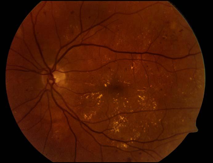
2 Sixth Indian Conference on Computer Vision, Graphics & Image Processing Colour Retinal Image Enhancement based on Domain Knowledge Gopal Datt Joshi, Jayanthi Sivaswamy Centre for Visual Information Technology IIIT-Hyderabad, India Abstract Retinal images are widely used to manually or automatically detect and diagnose many diseases. Due to the complex imaging setup, there is a large luminosity and contrast variability within and across images. Here, we use the knowledge of the imaging geometry and propose an enhancement method for colour retinal images, with a focus on contrast improvement with no introduction of artifacts. The method uses non-uniform sampling to estimate the degradation and derive a correction factor from a single plane. We also propose a scheme for applying the derived correction factor to enhance all the colour planes of a given image. The proposed enhancement method has been tested on a publicly available dataset [8]. Results show marked improvement over existing methods. 1. Introduction Among the many uses of retinal images are in the early detection and diagnosis of many eye diseases such as diabetic retinopathy (DR) and age-related macular degeneration (AMD). Automated analysis techniques for retinal images has been an important area of research of late for developing screening programmes [8]. In retinal images, vascular topography, dark and bright pathology (subtle or otherwise) are mainly of interest. A good quality of image is essential for a reliable diagnosis performed either manually or automatically. Therefore, improvement of image quality is a fundamental problem in retinal image analysis. Retinal images are acquired with a digital fundus camera, which captures the illumination reflected from the retinal surface. Despite the controlled conditions under which imaging takes place, there are many patient-dependent aspects which are difficult to control. Thus, most retinal images suffer from non-uniform illumination. Some of the contributing factors are: (a) The curved surface of the retina. Consequently, all retinal regions cannot be illuminated uniformly; (b) Imaging requires a dilated pupil. The degree of Figure 1. A retinal image with uneven illumination and contrast. dilation is highly variable across patients; (c) Unexpected movements of the patients eye. The bright flash-light makes the patient move his/her eye away from the view of the camera involuntarily; (d) Presence of other diseases such as cataract which can block the light reaching the retina. These factors result in images having a large luminosity and contrast variability within and across images. Hence, for a reliable diagnosis, whether manual or automated, an image normalization step is necessary. A sample of typical retinal image is shown in figure 1 affected by non-uniform illumination. In can be observed that luminosity and contrast distribution is not uniform across the image. Such variations affect the detection, for instance, of important objects such as microaneurysms (MA) which are of interest in early diagnosis of DR. These appear as a tiny red dots in a colour retinal images as highlighted in images shown in figure 2. The sample MA regions and the blood vessels (red lines/curves) also occur with varying local contrast across images. A normalisation step is hence /08 $ IEEE DOI /ICVGIP
![an important preprocessing step which has been shown to improve vessel segmentation [12]. Figure 2. Image regions containing microaneurysms.](/docs-images/87/95034926/images/3-0.jpg "Based on the above observations, few desired characteristics of retinal image enhancement technique can be inferred. image [7] but it smooths brighter structure present in the image.")
![Other methods estimate illumination function drift from segmented vessel pixels and use it for illumination correction [5][13].](/docs-images/87/95034926/images/3-1.jpg "These methods rely on vessel segmentation accuracy which is highly sensitive to the underlying luminosity and contrast of the image.")
![A contourlet transformbased enhancement has also been proposed [10]. This does not produce satisfactory results in poor contrast regions of the retinal image.](/docs-images/87/95034926/images/3-2.jpg "Enhancement in all of the above mentioned methods has been motivated by automatic analysis where, to this day, only one colour (green) plane of the colour image is processed since it contains maximal")
![information about structures of interest, compared to the red and blue planes [5][13].](/docs-images/87/95034926/images/3-3.jpg "Thus, enhancement performance is demonstrated by showing improvement in a specific task such as vessel/lesion segmentation from the green plane image.")


![A local normalisation of each pixel to zero mean and unit variance aims to compensate lighting variation and enhancing local contrast but also introduces artifacts due to amplification of noise [10].](/docs-images/87/95034926/images/3-7.jpg "Enhancement using matched filters [1][6][9] improves local contrast and aids in vessel segmentation but does not preserve the fidelity of the image.")
3 an important preprocessing step which has been shown to improve vessel segmentation [12]. Figure 2. Image regions containing microaneurysms. Based on the above observations, few desired characteristics of retinal image enhancement technique can be inferred. image [7] but it smooths brighter structure present in the image. Other methods estimate illumination function drift from segmented vessel pixels and use it for illumination correction [5][13]. These methods rely on vessel segmentation accuracy which is highly sensitive to the underlying luminosity and contrast of the image. A contourlet transformbased enhancement has also been proposed [10]. This does not produce satisfactory results in poor contrast regions of the retinal image. Enhancement in all of the above mentioned methods has been motivated by automatic analysis where, to this day, only one colour (green) plane of the colour image is processed since it contains maximal information about structures of interest, compared to the red and blue planes [5][13]. Thus, enhancement performance is demonstrated by showing improvement in a specific task such as vessel/lesion segmentation from the green plane image. These methods have not been extended for colour retinal image enhancement which is essential to aid manual diagnosis which is currently used heavily in practice due to immaturity of automatic diagnosis methods. We argue that focus on colour image enhancement can also benefit automated analysis as vector processing techniques can then be employed. It is a low-level technique. The technique should not depend on high-level information such as knowledge of the location of the sub-parts of the retina. This is necessary because it is a preprocessing step which can influence the detection of the sub-parts of the retina. It should be performed without manual intervention. It should not change the basic characteristics of any anatomical structure or bright/dark lesions present in the retinal image. Several techniques have been used to enhance retinal images. Histogram equalisation has been shown to be inappropriate for retinal images [10]. Unsharp masking-based enhancement is partly effective but is not capable of handling uneven illumination [10]. A local normalisation of each pixel to zero mean and unit variance aims to compensate lighting variation and enhancing local contrast but also introduces artifacts due to amplification of noise [10]. Enhancement using matched filters [1][6][9] improves local contrast and aids in vessel segmentation but does not preserve the fidelity of the image. It also affects other structures present in the image [10]. Histogram matching between the red and green planes has been used as a preprocessing step for vessel segmentation [12]. This improves the contrast of gross dark features like vessels but reduces the contrast of bright objects and tiny dark objects like MA. Slow variations of luminosity have been extracted via median (large) filtering and then subtracted from the observed (a) (b) Figure 3. (a) A region containing hemorrhage, (b) region after enhancement There have been attempts to extend histogram equalisation designed for grayscale images to restore colour images [11]. In [11], a method is proposed to generate an almost uniform colour histogram, which however, would change the ratios of the RGB components and thus producing hueshifting related artifacts [2] and introduces new colours to the objects in an image. This method was claimed to be more suitable for better visualisation of pseudo-colour scientific pictures than for ordinary image enhancement. In order to avoid hue-shifting related artifacts, colour image enhancement methods were formulated in HSI colour space [11] [4]. In this, the hue plane is kept intact and image correction is independently applied to the intensity (I) and saturation (S) components of the retinal image. The enhanced intensity and saturation components are combined 592

4 with hue component and colour image is recovered in RGB colour space. This shows promising results and it has been compared with the proposed scheme in the later section. A colour remapping scheme is suggested for colour retinal image enhancement[3]. It does luminosity and contrast enhancement on each colour plane of RGB colour space, independently. Later, they recover original chromatic distribution by identifying an overall image chromatic statistical distribution in the observed image. Figure 3 shows distortion introduced by this method [4] in a region containing large hemorrhage 1. In this paper, we propose a solution for colour retinal image enhancement which is based on the knowledge of the retina geometry and imaging conditions. Specifically, we present (i) a novel enhancement method which uses a nonuniform sampling-based estimation of the degradation components and report on results of applying it to a single colour plane; (ii) a solution for colour image enhancement which combines (i) and a linear color remapping technique. In the following sections, we will present enhancement methods and performed experiments in detail. 2 Image Enhancement on a Given Colour Plane Starting with a linear model of image formation, the relation between the true (ideal, uniform illumination) image U(x, y) and observed image I(x, y) can be written as: I(x, y) =U(x, y) S M (x, y)+s A (x, y) (1) where S M (x, y) is the multiplicative and S A (x, y) the additive component. Both S A and S M are generally assumed to be continuous and slowly varying functions. S M and S A represent the degradation components namely, the contrast and luminosity components of the image, respectively. This model does not incorporate blurring or additive noise. The recovery of true image U is based on the estimation of S A and S M and the correction of the observed image I as: U(x, y) = I(x, y) S A(x, y) (2) S M (x, y) In the retinal imaging scenario, the degradation functions have to be estimated from a given image. An understanding of the imaging mechanism is useful in the designing the estimation technique. The retina is a curved surface which is illuminated by a source of light located close to the pupil of the eye a few centimetres away. The camera, which is also locate close to the pupil, captures the reflected illumination from the retina. Due to this imaging geometry, the peripheral part of the retinal surface receives less illumination. Hence, the peripheral region appears darker than the central region of the retinal image. 1 The sample image regions are taken from [4]. Figure 4. Non-uniform sampling used for estimation. With the above in mind, we propose a non-uniform sampling scheme on a polar grid to estimate the degradation components for the acquired image. The sampling is coarse in the central region (well illuminated region) and dense in the periphery (poorly illuminated region). Thus, with the sampling points defined on a (r, θ) space, the sampling is non-uniform in both r and θ dimensions, as illustrated in fig Estimation of S A and S M functions In order to have an effective estimation, we follow the strategy of [3]. Here, the image (green channel) is separated into a set of background and foreground (made up of retinal structures of interest) pixels first. Next, the degradation components are estimated from the background image. This strategy is motivated by the fact that the retinal structures can bias the luminosity component. For instance, the optic disk (bright circular region on left in fig. 1) is a naturally high luminosity zone and the vessels (dark) are a low luminosity zone. The background pixels are extracted from I using the local mean and standard deviation as follows: 1. For every point on the sampling grid compute the local mean μ and σ within a window of size w w. 2. Interpolate between the sampling points to obtain μ(x, y) and σ(x, y) for all (x, y). 3. Computethe MahalanobisdistanceD(x, y) as follows. I(x, y) μ(x, y) D(x, y) = (3) σ(x, y) 593
 Given an image, a pixel is taken to belong to the background if D(x, y) t where t is a fixed threshold. Figure 5 shows a computed background image on the green plane of the image in fig. 1.")

 is obtained by applying the point transformation (equation 2) to each pixel of the image.")
5 Figure 5. A background image for the image in fig 1. Black pixels belong to foreground and white pixels to background. (a) Given an image, a pixel is taken to belong to the background if D(x, y) t where t is a fixed threshold. Figure 5 shows a computed background image on the green plane of the image in fig. 1. The degradation components are estimated from the background image by computing the local mean and the standard deviation values at every point (x, y), within a window of size (w 0 w 0 ). The desired contrast component S M is nothing but the standard deviation and the luminosity component is S A. In our experiments, bilinear interpolation was used in step 2 and w 0 was set to 50, t =1and a large enough window size (w = 125) was chosen to include retinal structures as well as the background. A sample estimated S A and S M functions are shown in fig. 6. Finally, the true image U(x, y) is obtained by applying the point transformation (equation 2) to each pixel of the image. A sample result of applying the proposed method is shown in fig. 7(f). The enhanced image has good luminosity and different retinal structures are contrasted well against the background. 2.2 Strengths of the proposed method The proposed technique differs from the method in [3] which results in some advantages. The approach in [3] uses a square sampling grid and determines local (over a w w neighbourhood) mean and variance. Since w is taken to be the sampling interval, the computation of background thus uses contiguous windows on the given image. The value of w impacts on the success of the background/foreground separation. A small w will lead to inability to discriminate between retinal structures and background, thus result in a (b) Figure 6. Estimated functions (a) S A and (b) S M for the image in fig 1. poor background image. A large w leads to fewer samples and hence an imprecise background determination. Our proposed non-uniform sampling technique addresses this problem very effectively. Firstly, it permits taking differential number of samples in different regions of the retina which is desirable, given the imaging geometry. Secondly, once a non-uniform sampling pattern is fixed, a window size w can also be fixed to result in computation in overlapping windows over the peripheral region and contiguous windows in the centre, which is not possible in a uniform grid. 594
 Input image; Image enhancement results obtained from (b) global histogram equalisation, (c) local histogram equalisation, (d) adaptive correction using red colour channel [12], (e) lowpass")
![subtraction: standard correction method used for retinal image [7] and (f) proposed method 3 Colour Retinal Image Enhancement We propose a colour remapping process for retinal colour enhancement](/docs-images/87/95034926/images/6-2.jpg "images which is based on the chromatic information of the original image.")
 is computed as : ˆr = g corr v r, ĝ = g corr v g, ˆb = g corr v b, (4) Since, v = max[r, g, b], it plays a normalisation role in the enhancement.")
6 (a) (b) (c) (d) (e) (f) Figure 7. (a) Input image; Image enhancement results obtained from (b) global histogram equalisation, (c) local histogram equalisation, (d) adaptive correction using red colour channel [12], (e) lowpass subtraction: standard correction method used for retinal image [7] and (f) proposed method 3 Colour Retinal Image Enhancement We propose a colour remapping process for retinal colour enhancement images which is based on the chromatic information of the original image. Given a colour image with colour components (r, g, b) or (h, s, v), the single plane correction described in section 2 is applied to the g plane and g corr is obtained. Next the enhanced colour image (ˆr, ĝ,ˆb) is computed as : ˆr = g corr v r, ĝ = g corr v g, ˆb = g corr v b, (4) Since, v = max[r, g, b], it plays a normalisation role in the enhancement. Thus, the ratio of the original r, g and b is maintained in the above linear color remapping and the chromatic content in the original image is preserved in the enhanced color image. Figure 8(d) shows the enhanced colour retinal image by the proposed remapping technique. It can be seen that a good contrast and uniform illumination is obtained and colour distribution is also well preserved. 4 Experimentation Results For all our experiments, we have used a public retinal image database aimed for benchmarking diabetic retinopathy detection from digital images [8]. Images were captured using the 50 degree field-of-view digital fundus camera with varying imaging settings. This dataset corresponds to a good practical situation and consists of total 89 digital retinal images. The single plane enhancement method presented in section 2 was applied to green plane images and compared against existing retinal image enhancement techniques. Figure 7 shows results obtained from the different techniques. It can be seen that proposed method gives better visual quality of the enhanced image while keeping good contrast of retinal structures. Other methods including local (adaptive) and global ones either fail in the enhancement or affect contrast of retinal structures. For instance, optic disk (bright circular region in left) is smoothed by method [7] (shown in fig. 7(e)) and by local histogram equalisation (shown in fig. 7(c)). Next, we present results of evaluating the proposed 595
 method proposed in [3], (c) method performing enhancement on saturation and intensity of HSI colour space, and (d) proposed method.")
 and saturation (S) components of the HSI space.")

7 (a) (b) (c) (d) Figure 8. Comparison of colour enhancement methods on (a) test input image. Results obtained from (b) method proposed in [3], (c) method performing enhancement on saturation and intensity of HSI colour space, and (d) proposed method. colour enhancement scheme (section3) against some existing schemes. Sample results are in Figure 8. In the method presented in [4] and [11], the hue plane is kept intact and image enhancement is performed independently on the intensity (I) and saturation (S) components of the HSI space. The enhanced intensity and saturation components are later combined with original hue component and colour image is recovered in RGB colour space. The obtained results are shown in fig.8(c). It can be seen that the resulting colour image maintains the visual appearance of retinal structures but compromised contrast of the retinal structures. This is mainly due to the inclusion of the I component in the correction process. Furthermore, the output image does not preserve the original colour distribution. Next, we have tested the colour enhancement method suggested in [3]. In our implementation, an identical enhancement technique is applied to all the three (r, g, b) channels separately. Given an image plane I x of image I, enhanced image Ix corr is obtained using method presented in section 2. To preserve the original chromatic distribution, a normalisation step is performed on Ix corr suggested in [3] as follows: Îcorr x = Ix corr σ x + μ x.where,μ x and σ x are the mean and standard deviation of the observed image I x, respectively. The colour output shown in fig. 8(b), is obtained by applying above procedure on each r, g, b plane. Though, the original colour distribution appears to preserved, the overall contrast in the colour image gets reduced due to colour normalisation. Our proposed colour enhancement scheme on the other hand, preserves the colour distribution and improves the 596
 (d)")


8 (a) (b) (c) (d) (e) (f) Figure 9. Results on a set of test images using proposed method. The odd rows show the test image and even rows show their corresponding enhanced colour image. 597

![It can be seen that proposed method is able to retain large dark lesion (fig. 10(a)) which was getting smoothed in [3]. No artifacts have also been introduced.](/docs-images/87/95034926/images/9-1.jpg "In fact, the proposed method significantly enhances the contrast of the lesion against the background. For instance, the tiny MA and small yellowish regions (fig.")
 (a-1) (b) (b-1) Figure 10.")
9 overall contrast in the output image shown in Figure 8(d). Additional results are shown in fig. 9. Next, we have evaluated the effect of enhancement on the dark and bright lesions present in a retinal image. Figure 10 shows the subimages containing the lesions and the processed results. It can be seen that proposed method is able to retain large dark lesion (fig. 10(a)) which was getting smoothed in [3]. No artifacts have also been introduced. In fact, the proposed method significantly enhances the contrast of the lesion against the background. For instance, the tiny MA and small yellowish regions (fig. 10(b)) are visible due to the improved contrast as compared to the original image. It can however be noted that the colour has shifted on this image. This effect was noted to occur only when the red content in the input image was dominant. (a) (a-1) (b) (b-1) Figure 10. First row shows sample region image regions and second row shows corresponding results obtained from the proposed method. 5 Conclusion In retinal images, vascular topography, dark and bright pathology (subtle or otherwise) are mainly of interest. In this paper, we presented a method for colour retinal image enhancement which is based on the knowledge of the retina geometry and imaging conditions. The method determines a correction factor using a single plane and then applies a normalised correction to all three (r, g, b) planes. The correction factor is found using a non-uniform samplingbased estimation of the degradation components. The results of testing the proposed colour enhancement method on 89 colour images show that it is able to improve the overall contrast and correct for non-uniform illumination successfully. There is a minimal shift in the colour content and no new artifacts are introduced. All of these features are attractive in applications which require manual as well as automatic examination of colour retinal images. References [1] S. Chaudhuri, S. Chatterjee, and N. Katz. Detection of blood vessels in retinal images using two-dimensional matched filters. IEEE Trans. Med. Imaging, 3(8): , [2] J. Duan and G. Qiu. Novel histogram processing for colour image enhancement. Proc. Int. Conf. Image and Graphics, pages 55 58, [3] M. Foracchia, E. Grisan, and A. Ruggeri. Luminosity and contrast normalization in retinal images. Medical Image Analysis, 3(9): , [4] E. Grisan, A. Giani, E. Ceseracciu, and A. Ruggeri. Modelbased illumination correction in retinal images. IEEE Int. Symp. Biomedical Imaging: Nano to Macro, pages , [5] A. Hoover. Equalizing illumination in a retinal image using blood vessels as a reference. Technical Report:URL [6] A. Hoover, V. Kouznetsova, and M. Goldbaum. Locating blood vessels in retinal images by piecewise threshold probing of a matched filter response. IEEE Trans. Med. Imaging, 3(19): , [7] G. ien and P. Osnes. Diabetic retinopathy: automatic detection of early symptoms from retinal images. Proc. Norwegian Signal Processing Society Symposium(NORSIG), [8] T. Kauppi, V. Kalesnykiene, J. Kmrinen, L. Lensu, I. Sorri, A. Raninen, R. Voutilainen, H. Uusitalo, H. Klviinen, and J. Pietil. Diaretdb1 diabetic retinopathy database and evaluation protocol. Proc. Medical Image Understanding and Analysis (MIUA), pages 61 65, [9] T. S. Lin, M. H. Du, and J. T. Xu. The preprocessing of subtraction and the enhancement for biomedical image of retinal blood vessels. Journal of Biomedical Engg., 1(20):56 59, [10] P.Feng, Y. Pan, B. Wei, W. Jin, and D. Mi. Enhancing retinal image by the contourlet transform. Pattern Recognition Letters, 4(28): , [11] E. Pichon, M. Niethammer, and G. Sapiro. Color histogram qualization though mesh deformation. Proc. Int. Conf. Image Processing, (2): , [12] N. M. Salem and A. K. Nandi. Novel and adaptive contribution of the red channel in pre-processing of colour fundus images. Journal of the Franklin Institute, (344): , [13] Y. Wang, W. Tsu, and S. Lee. llumination normalization of retinal images using sampling and interpolation. Proc. SPIE, Medical Imaging, pages ,
Image Database and Preprocessing
 Chapter 3 Image Database and Preprocessing 3.1 Introduction The digital colour retinal images required for the development of automatic system for maculopathy detection are provided by the Department of
Fovea and Optic Disc Detection in Retinal Images with Visible Lesions
 Fovea and Optic Disc Detection in Retinal Images with Visible Lesions José Pinão 1, Carlos Manta Oliveira 2 1 University of Coimbra, Palácio dos Grilos, Rua da Ilha, 3000-214 Coimbra, Portugal 2 Critical
CHAPTER 4 LOCATING THE CENTER OF THE OPTIC DISC AND MACULA
 90 CHAPTER 4 LOCATING THE CENTER OF THE OPTIC DISC AND MACULA The objective in this chapter is to locate the centre and boundary of OD and macula in retinal images. In Diabetic Retinopathy, location of
An Efficient Pre-Processing Method to Extract Blood Vessel, Optic Disc and Exudates from Retinal Images
 An Efficient Pre-Processing Method to Extract Blood Vessel, Optic Disc and Exudates from Retinal Images 1 K. Priya, 2 Dr. N. Jayalakshmi 1 (Research Scholar, Research & Development Centre, Bharathiar University,
Segmentation Of Optic Disc And Macula In Retinal Images
 Segmentation Of Optic Disc And Macula In Retinal Images Gogila Devi. K #1, Vasanthi. S *2 # PG Student, K.S.Rangasamy College of Technology Tiruchengode, Namakkal, Tamil Nadu, India. * Associate Professor,
CHAPTER 4 BACKGROUND
 48 CHAPTER 4 BACKGROUND 4.1 PREPROCESSING OPERATIONS Retinal image preprocessing consists of detection of poor image quality, correction of non-uniform luminosity, color normalization and contrast enhancement.
Digital Retinal Images: Background and Damaged Areas Segmentation
 Digital Retinal Images: Background and Damaged Areas Segmentation Eman A. Gani, Loay E. George, Faisel G. Mohammed, Kamal H. Sager Abstract Digital retinal images are more appropriate for automatic screening
Automatic Detection Of Optic Disc From Retinal Images. S.Sherly Renat et al.,
 International Journal of Technology and Engineering System (IJTES) Vol 7. No.3 2015 Pp. 203-207 gopalax Journals, Singapore available at : www.ijcns.com ISSN: 0976-1345 AUTOMATIC DETECTION OF OPTIC DISC
OPTIC DISC LOCATION IN DIGITAL FUNDUS IMAGES
 OPTIC DISC LOCATION IN DIGITAL FUNDUS IMAGES Miss. Tejaswini S. Mane 1,Prof. D. G. Chougule 2 1 Department of Electronics, Shivaji University Kolhapur, TKIET,Wrananagar (India) 2 Department of Electronics,
FOG REMOVAL ALGORITHM USING ANISOTROPIC DIFFUSION AND HISTOGRAM STRETCHING
 FOG REMOVAL ALGORITHM USING DIFFUSION AND HISTOGRAM STRETCHING 1 G SAILAJA, 2 M SREEDHAR 1 PG STUDENT, 2 LECTURER 1 DEPARTMENT OF ECE 1 JNTU COLLEGE OF ENGINEERING (Autonomous), ANANTHAPURAMU-5152, ANDRAPRADESH,
Novel Histogram Processing for Colour Image Enhancement
 Novel Histogram Processing for Colour Image Enhancement Jiang Duan and Guoping Qiu School of Computer Science, The University of Nottingham, United Kingdom Abstract: Histogram equalization is a well-known
Retinal image analysis: preprocessing and feature extraction
 Journal of Physics: Conference Series Retinal image analysis: preprocessing and feature extraction To cite this article: Andrés G Marrugo and María S Millán 2011 J. Phys.: Conf. Ser. 274 012039 View the
CoE4TN4 Image Processing. Chapter 3: Intensity Transformation and Spatial Filtering
 CoE4TN4 Image Processing Chapter 3: Intensity Transformation and Spatial Filtering Image Enhancement Enhancement techniques: to process an image so that the result is more suitable than the original image
Exudates Detection Methods in Retinal Images Using Image Processing Techniques
 International Journal of Scientific & Engineering Research, Volume 1, Issue 2, November-2010 1 Exudates Detection Methods in Retinal Images Using Image Processing Techniques V.Vijayakumari, N. Suriyanarayanan
Gaussian and Fast Fourier Transform for Automatic Retinal Optic Disc Detection
 Gaussian and Fast Fourier Transform for Automatic Retinal Optic Disc Detection Arif Muntasa 1, Indah Agustien Siradjuddin 2, and Moch Kautsar Sophan 3 Informatics Department, University of Trunojoyo Madura,
AN AUTOMATIC SCREENING METHOD TO DETECT OPTIC DISC IN THE RETINA
 AN AUTOMATIC SCREENING METHOD TO DETECT OPTIC DISC IN THE RETINA Murugan.R 1, Dr.Reeba Korah 2 1 Research Scholar, Centre for Research, Anna University of Technology Chennai murugan.rmn@gmail.com 2 Professor,
DETECTION OF OPTIC DISC BY USING THE PRINCIPLES OF IMAGE PROCESSING
 DETECTION OF OPTIC DISC BY USING THE PRINCIPLES OF IMAGE PROCESSING SUSHMA G 1, VENKATESHAPPA 2 ' 1 Asst professor, 2 HoD, Dept of ECE, MSEC Bangalore E-mail: sushmavasu11@gmail.com, venkat_harishith@rediffmail.com
Segmentation approaches of optic cup from retinal images: A Survey
 I J C T A, 10(8), 2017, pp. 377-382 International Science Press ISSN: 0974-5572 Segmentation approaches of optic cup from retinal images: A Survey Niharika Thakur* and Mamta Juneja** ABSTRACT Eye is a
NON UNIFORM BACKGROUND REMOVAL FOR PARTICLE ANALYSIS BASED ON MORPHOLOGICAL STRUCTURING ELEMENT:
 IJCE January-June 2012, Volume 4, Number 1 pp. 59 67 NON UNIFORM BACKGROUND REMOVAL FOR PARTICLE ANALYSIS BASED ON MORPHOLOGICAL STRUCTURING ELEMENT: A COMPARATIVE STUDY Prabhdeep Singh1 & A. K. Garg2
A new method for segmentation of retinal blood vessels using morphological image processing technique
 A new method for segmentation of retinal blood vessels using morphological image processing technique Roya Aramesh Faculty of Computer and Information Technology Engineering,Qazvin Branch,Islamic Azad
ABSTRACT I. INTRODUCTION II. REVIEW OF PREVIOUS METHODS. et al., the OD is usually the brightest component on
 National Conference on Engineering Innovations and Solutions (NCEIS 2018) International Journal of Scientific Research in Computer Science, Engineering and Information Technology 2018 IJSRCSEIT Volume
A Global-Local Contrast based Image Enhancement Technique based on Local Standard Deviation
 A Global-Local Contrast based Image Enhancement Technique based on Local Standard Deviation Archana Singh Ch. Beeri Singh College of Engg & Management Agra, India Neeraj Kumar Hindustan College of Science
SEGMENTATION OF BRIGHT REGION OF THE OPTIC DISC FOR EYE DISEASE PREDICTION
 RAHUL JADHAV AND MANISH NARNAWARE: SEGMENTATION OF BRIGHT REGION OF THE OPTIC DISC FOR EYE DISEASE PREDICTION DOI: 10.21917/ijivp.2018.0239 SEGMENTATION OF BRIGHT REGION OF THE OPTIC DISC FOR EYE DISEASE
Drusen Detection in a Retinal Image Using Multi-level Analysis
 Drusen Detection in a Retinal Image Using Multi-level Analysis Lee Brandon 1 and Adam Hoover 1 Electrical and Computer Engineering Department Clemson University {lbrando, ahoover}@clemson.edu http://www.parl.clemson.edu/stare/
Procedure to detect anatomical structures in optical fundus images
 Procedure to detect anatomical structures in optical fundus images L. Gagnon *a, M. Lalonde *a, M. Beaulieu *a, M.-C. Boucher **b a Computer Research Institute of Montreal; b Dept. Of Ophthalmology, Maisonneuve-Rosemont
Computer Vision. Howie Choset Introduction to Robotics
 Computer Vision Howie Choset http://www.cs.cmu.edu.edu/~choset Introduction to Robotics http://generalrobotics.org What is vision? What is computer vision? Edge Detection Edge Detection Interest points
The First True Color Confocal Scanner on the Market
 The First True Color Confocal Scanner on the Market White color and infrared confocal images: the advantages of white color and confocality together for better fundus images. The infrared to see what our
Introduction. American Journal of Cancer Biomedical Imaging
 American Journal of Cancer Biomedical Imaging American Journal of Biomedical Imaging http://www.ivyunion.org/index.php/ajbi/index Vo1. 1, Article ID 20130133, 12 pages Kumar T. A. et al. American Journal
Chapter 6. [6]Preprocessing
![Chapter 6. [6]Preprocessing](/thumbs/88/115179409.jpg "Chapter 6. [6]Preprocessing") Chapter 6 [6]Preprocessing As mentioned in chapter 4, the first stage in the HCR pipeline is preprocessing of the image. We have seen in earlier chapters why this is very important and at the same time
Chapter 6 [6]Preprocessing As mentioned in chapter 4, the first stage in the HCR pipeline is preprocessing of the image. We have seen in earlier chapters why this is very important and at the same time
An Enhanced Biometric System for Personal Authentication
 IOSR Journal of Electronics and Communication Engineering (IOSR-JECE) e-issn: 2278-2834,p- ISSN: 2278-8735. Volume 6, Issue 3 (May. - Jun. 2013), PP 63-69 An Enhanced Biometric System for Personal Authentication
INTERNATIONAL JOURNAL OF PURE AND APPLIED RESEARCH IN ENGINEERING AND TECHNOLOGY
 INTERNATIONAL JOURNAL OF PURE AND APPLIED RESEARCH IN ENGINEERING AND TECHNOLOGY A PATH FOR HORIZING YOUR INNOVATIVE WORK BLOOD VESSEL SEGMENTATION PROF. SAGAR P. MORE 1, PROF. S. M. AGRAWAL 2, PROF. M.
A Review on Image Enhancement Technique for Biomedical Images
 A Review on Image Enhancement Technique for Biomedical Images Pankaj V.Gosavi 1, Prof. V. T. Gaikwad 2 M.E (Pursuing) 1, Associate Professor 2 Dept. Information Technology 1, 2 Sipna COET, Amravati, India
Blood Vessel Segmentation of Retinal Images Based on Neural Network
 Blood Vessel Segmentation of Retinal Images Based on Neural Network Jingdan Zhang 1( ), Yingjie Cui 1, Wuhan Jiang 2, and Le Wang 1 1 Department of Electronics and Communication, Shenzhen Institute of
Macula centred, giving coverage of the temporal retinal. Disc centred. Giving coverage of the nasal retina.
 3. Field positions, clarity and overall quality For retinopathy screening purposes in England two images are taken of each eye. These have overlapping fields of view and between them cover the main area
Segmentation using Saturation Thresholding and its Application in Content-Based Retrieval of Images
 Segmentation using Saturation Thresholding and its Application in Content-Based Retrieval of Images A. Vadivel 1, M. Mohan 1, Shamik Sural 2 and A.K.Majumdar 1 1 Department of Computer Science and Engineering,
The TRC-NW8F Plus: As a multi-function retinal camera, the TRC- NW8F Plus captures color, red free, fluorescein
 The TRC-NW8F Plus: By Dr. Beth Carlock, OD Medical Writer Color Retinal Imaging, Fundus Auto-Fluorescence with exclusive Spaide* Filters and Optional Fluorescein Angiography in One Single Instrument W
Multichannel Blind Deconvolution in Eye Fundus Imaging
 Multichannel Blind Deconvolution in Eye Fundus Imaging Andrés G. Marrugo Dept. of Optics and Optometry Universitat Politècnica de Catalunya, Spain andres.marrugo@upc.edu Filip Šroubek UTIA Academy of Sciences
Blood Vessel Tree Reconstruction in Retinal OCT Data
 Blood Vessel Tree Reconstruction in Retinal OCT Data Gazárek J, Kolář R, Jan J, Odstrčilík J, Taševský P Department of Biomedical Engineering, FEEC, Brno University of Technology xgazar03@stud.feec.vutbr.cz
A diabetic retinopathy detection method using an improved pillar K-means algorithm
 www.bioinformation.net Hypothesis Volume 10(1) A diabetic retinopathy detection method using an improved pillar K-means algorithm Susmitha valli Gogula 1 *, CH Divakar 2, CH Satyanarayana 3 & Allam Appa
Retinal blood vessel extraction
 Retinal blood vessel extraction Surya G 1, Pratheesh M Vincent 2, Shanida K 3 M. Tech Scholar, ECE, College, Thalassery, India 1,3 Assistant Professor, ECE, College, Thalassery, India 2 Abstract: Image
Impressive Wide Field Image Quality with Small Pupil Size
 Impressive Wide Field Image Quality with Small Pupil Size White color and infrared confocal images: the advantages of white color and confocality together for better fundus images. The infrared to see
The First True-Color Wide-Field Confocal Scanner
 The First True-Color Wide-Field Confocal Scanner 2 Company Profile CenterVue designs and manufactures highly automated medical devices for the diagnosis and management of ocular pathologies, including
Optic Disc Boundary Approximation Using Elliptical Template Matching
 Research Article Optic Disc Boundary Approximation Using Elliptical Template Matching P. Nagarajan a *, S.S. Vinsley b a Research Scholar, Anna University, Chennai, Tamil Nadu, India. b Principal, Lourdes
PERCEPTUALLY-ADAPTIVE COLOR ENHANCEMENT OF STILL IMAGES FOR INDIVIDUALS WITH DICHROMACY. Alexander Wong and William Bishop
 PERCEPTUALLY-ADAPTIVE COLOR ENHANCEMENT OF STILL IMAGES FOR INDIVIDUALS WITH DICHROMACY Alexander Wong and William Bishop University of Waterloo Waterloo, Ontario, Canada ABSTRACT Dichromacy is a medical
Single Image Haze Removal with Improved Atmospheric Light Estimation
 Journal of Physics: Conference Series PAPER OPEN ACCESS Single Image Haze Removal with Improved Atmospheric Light Estimation To cite this article: Yincui Xu and Shouyi Yang 218 J. Phys.: Conf. Ser. 198
USE OF HISTOGRAM EQUALIZATION IN IMAGE PROCESSING FOR IMAGE ENHANCEMENT
 USE OF HISTOGRAM EQUALIZATION IN IMAGE PROCESSING FOR IMAGE ENHANCEMENT Sapana S. Bagade M.E,Computer Engineering, Sipna s C.O.E.T,Amravati, Amravati,India sapana.bagade@gmail.com Vijaya K. Shandilya Assistant
INSTITUTIONEN FÖR SYSTEMTEKNIK LULEÅ TEKNISKA UNIVERSITET
 INSTITUTIONEN FÖR SYSTEMTEKNIK LULEÅ TEKNISKA UNIVERSITET Some color images on this slide Last Lecture 2D filtering frequency domain The magnitude of the 2D DFT gives the amplitudes of the sinusoids and
Local Contrast Enhancement using Local Standard Deviation
 Local ontrast Enhancement using Local Standard Deviation S. Somoreet Singh Th. Tangkeshwar Singh Department of omputer Science Asst. Prof. (Sr. Scale), Dept. of omputer Science Manipur University, anchipur
Automatic Morphological Segmentation and Region Growing Method of Diagnosing Medical Images
 International Journal of Information & Computation Technology. ISSN 0974-2239 Volume 2, Number 3 (2012), pp. 173-180 International Research Publications House http://www. irphouse.com Automatic Morphological
INDIAN VEHICLE LICENSE PLATE EXTRACTION AND SEGMENTATION
 International Journal of Computer Science and Communication Vol. 2, No. 2, July-December 2011, pp. 593-599 INDIAN VEHICLE LICENSE PLATE EXTRACTION AND SEGMENTATION Chetan Sharma 1 and Amandeep Kaur 2 1
Automatic Licenses Plate Recognition System
 Automatic Licenses Plate Recognition System Garima R. Yadav Dept. of Electronics & Comm. Engineering Marathwada Institute of Technology, Aurangabad (Maharashtra), India yadavgarima08@gmail.com Prof. H.K.
1200 "h278" 2500 "h563"
 Automatic visual quality assessment in optical fundus images Marc Lalondey, Langis Gagnony and Marie-Carole Boucherz ycentre de recherche informatique de Montréal 550 Sherbrooke W., Suite 100, Montréal,
International Conference on Computer, Communication, Control and Information Technology (C 3 IT 2009) Paper Code: DSIP-024
 Paper Code: DSIP-024") Paper Code: DSIP-024 Oral 270 A NOVEL SCHEME FOR BINARIZATION OF VEHICLE IMAGES USING HIERARCHICAL HISTOGRAM EQUALIZATION TECHNIQUE Satadal Saha 1, Subhadip Basu 2 *, Mita Nasipuri 2, Dipak Kumar Basu
Paper Code: DSIP-024 Oral 270 A NOVEL SCHEME FOR BINARIZATION OF VEHICLE IMAGES USING HIERARCHICAL HISTOGRAM EQUALIZATION TECHNIQUE Satadal Saha 1, Subhadip Basu 2 *, Mita Nasipuri 2, Dipak Kumar Basu
Image binarization techniques for degraded document images: A review
 Image binarization techniques for degraded document images: A review Binarization techniques 1 Amoli Panchal, 2 Chintan Panchal, 3 Bhargav Shah 1 Student, 2 Assistant Professor, 3 Assistant Professor 1
Preprocessing on Digital Image using Histogram Equalization: An Experiment Study on MRI Brain Image
 Preprocessing on Digital Image using Histogram Equalization: An Experiment Study on MRI Brain Image Musthofa Sunaryo 1, Mochammad Hariadi 2 Electrical Engineering, Institut Teknologi Sepuluh November Surabaya,
A Novel Method for Enhancing Satellite & Land Survey Images Using Color Filter Array Interpolation Technique (CFA)
") A Novel Method for Enhancing Satellite & Land Survey Images Using Color Filter Array Interpolation Technique (CFA) Suma Chappidi 1, Sandeep Kumar Mekapothula 2 1 PG Scholar, Department of ECE, RISE Krishna
A Novel Method for Enhancing Satellite & Land Survey Images Using Color Filter Array Interpolation Technique (CFA) Suma Chappidi 1, Sandeep Kumar Mekapothula 2 1 PG Scholar, Department of ECE, RISE Krishna
An Evaluation of Automatic License Plate Recognition Vikas Kotagyale, Prof.S.D.Joshi
 An Evaluation of Automatic License Plate Recognition Vikas Kotagyale, Prof.S.D.Joshi Department of E&TC Engineering,PVPIT,Bavdhan,Pune ABSTRACT: In the last decades vehicle license plate recognition systems
Image Filtering. Median Filtering
 Image Filtering Image filtering is used to: Remove noise Sharpen contrast Highlight contours Detect edges Other uses? Image filters can be classified as linear or nonlinear. Linear filters are also know
Analysis of various Fuzzy Based image enhancement techniques
 Analysis of various Fuzzy Based image enhancement techniques SONALI TALWAR Research Scholar Deptt.of Computer Science DAVIET, Jalandhar(Pb.), India sonalitalwar91@gmail.com RAJESH KOCHHER Assistant Professor
Background Subtraction Fusing Colour, Intensity and Edge Cues
 Background Subtraction Fusing Colour, Intensity and Edge Cues I. Huerta and D. Rowe and M. Viñas and M. Mozerov and J. Gonzàlez + Dept. d Informàtica, Computer Vision Centre, Edifici O. Campus UAB, 08193,
Implementation of global and local thresholding algorithms in image segmentation of coloured prints
 Implementation of global and local thresholding algorithms in image segmentation of coloured prints Miha Lazar, Aleš Hladnik Chair of Information and Graphic Arts Technology, Department of Textiles, Faculty
CX-1 digital retinal camera mydriatic & non-mydriatic. Redefining true versatility.
 CX-1 digital retinal camera mydriatic & non-mydriatic Redefining true versatility. Redefining True versatility The multifaceted CX-1 The CX-1 is a Mydriatic Retinal Camera with full Non-Mydriatic functionality.
A Retinal Image Enhancement Technique for Blood Vessel Segmentation Algorithm
 A Retinal Image Enhancement Technique for Blood Vessel Segmentation Algorithm A. M. R. R. Bandara University of Moratuwa, Katubedda, Moratuwa, Sri Lanka. ravimalb@uom.lk P. W. G. R. M. P. B. Giragama Base
Reference Free Image Quality Evaluation
 Reference Free Image Quality Evaluation for Photos and Digital Film Restoration Majed CHAMBAH Université de Reims Champagne-Ardenne, France 1 Overview Introduction Defects affecting films and Digital film
Blood Vessel Tracking Technique for Optic Nerve Localisation for Field 1-3 Color Fundus Images
 Blood Tracing Technique for Optic Nerve Localisation for Field 1-3 Color Fundus Images Hwee Keong Lam, Opas Chutatape School of Electrical and Electronic Engineering Nanyang Technological University, Nanyang
Hybrid Method based Retinal Optic Disc Detection
 Hybrid Method based Retinal Optic Disc Detection Arif Muntasa 1, Indah Agustien Siradjuddin, and Moch Kautsar Sophan 3 Informatics Department, University of Trunojoyo Madura, Bangkalan Madura Island, Indonesia
Keywords Fuzzy Logic, ANN, Histogram Equalization, Spatial Averaging, High Boost filtering, MSE, RMSE, SNR, PSNR.
 Volume 4, Issue 1, January 2014 ISSN: 2277 128X International Journal of Advanced Research in Computer Science and Software Engineering Research Paper Available online at: www.ijarcsse.com An Image Enhancement
Preprocessing and Segregating Offline Gujarati Handwritten Datasheet for Character Recognition
 Preprocessing and Segregating Offline Gujarati Handwritten Datasheet for Character Recognition Hetal R. Thaker Atmiya Institute of Technology & science, Kalawad Road, Rajkot Gujarat, India C. K. Kumbharana,
A Novel Algorithm for Hand Vein Recognition Based on Wavelet Decomposition and Mean Absolute Deviation
 Sensors & Transducers, Vol. 6, Issue 2, December 203, pp. 53-58 Sensors & Transducers 203 by IFSA http://www.sensorsportal.com A Novel Algorithm for Hand Vein Recognition Based on Wavelet Decomposition
Improved Region of Interest for Infrared Images Using. Rayleigh Contrast-Limited Adaptive Histogram Equalization
 Improved Region of Interest for Infrared Images Using Rayleigh Contrast-Limited Adaptive Histogram Equalization S. Erturk Kocaeli University Laboratory of Image and Signal processing (KULIS) 41380 Kocaeli,
Optic Disc Approximation using an Ensemble of Processing Methods
 Optic Disc Approximation using an Ensemble of Processing Methods Anmol Sadanand Manipal, Karnataka. Anurag Datta Roy Manipal, Karnataka Pramodith Manipal, Karnataka Abstract - This paper proposes a simple
Exercise questions for Machine vision
 Exercise questions for Machine vision This is a collection of exercise questions. These questions are all examination alike which means that similar questions may appear at the written exam. I ve divided
Coded Aperture for Projector and Camera for Robust 3D measurement
 Coded Aperture for Projector and Camera for Robust 3D measurement Yuuki Horita Yuuki Matugano Hiroki Morinaga Hiroshi Kawasaki Satoshi Ono Makoto Kimura Yasuo Takane Abstract General active 3D measurement
A Method of Using Digital Image Processing for Edge Detection of Red Blood Cells
 Sensors & Transducers 013 by IFSA http://www.sensorsportal.com A Method of Using Digital Image Processing for Edge Detection of Red Blood Cells 1 Jinping LI, Hongshan MU, Wei XU 1 Software School, East
A Study On Preprocessing A Mammogram Image Using Adaptive Median Filter
 A Study On Preprocessing A Mammogram Image Using Adaptive Median Filter Dr.K.Meenakshi Sundaram 1, D.Sasikala 2, P.Aarthi Rani 3 Associate Professor, Department of Computer Science, Erode Arts and Science
Linear Gaussian Method to Detect Blurry Digital Images using SIFT
 IJCAES ISSN: 2231-4946 Volume III, Special Issue, November 2013 International Journal of Computer Applications in Engineering Sciences Special Issue on Emerging Research Areas in Computing(ERAC) www.caesjournals.org
Lossless Image Watermarking for HDR Images Using Tone Mapping
 IJCSNS International Journal of Computer Science and Network Security, VOL.13 No.5, May 2013 113 Lossless Image Watermarking for HDR Images Using Tone Mapping A.Nagurammal 1, T.Meyyappan 2 1 M. Phil Scholar
Keyword: Morphological operation, template matching, license plate localization, character recognition.
 Volume 4, Issue 11, November 2014 ISSN: 2277 128X International Journal of Advanced Research in Computer Science and Software Engineering Research Paper Available online at: www.ijarcsse.com Automatic
Segmentation of Blood Vessels and Optic Disc in Fundus Images
 RESEARCH ARTICLE Segmentation of Blood Vessels and Optic Disc in Fundus Images 1 M. Dhivya, 2 P. Jenifer, 3 D. C. Joy Winnie Wise, 4 N. Rajapriya, Department of CSE, Francis Xavier Engineering College,
AUTOMATED DRUSEN DETECTION IN A RETINAL IMAGE USING MULTI-LEVEL ANALYSIS
 AUTOMATED DRUSEN DETECTION IN A RETINAL IMAGE USING MULTI-LEVEL ANALYSIS A Thesis Presented to the Graduate School of Clemson University In Partial Fulfillment of the Requirements for the Degree Master
A Review Paper on Image Processing based Algorithms for De-noising and Enhancement of Underwater Images
 IJSTE - International Journal of Science Technology & Engineering Volume 2 Issue 10 April 2016 ISSN (online): 2349-784X A Review Paper on Image Processing based Algorithms for De-noising and Enhancement
Colored Rubber Stamp Removal from Document Images
 Colored Rubber Stamp Removal from Document Images Soumyadeep Dey, Jayanta Mukherjee, Shamik Sural, and Partha Bhowmick Indian Institute of Technology, Kharagpur {soumyadeepdey@sit,jay@cse,shamik@sit,pb@cse}.iitkgp.ernet.in
Target detection in side-scan sonar images: expert fusion reduces false alarms
 Target detection in side-scan sonar images: expert fusion reduces false alarms Nicola Neretti, Nathan Intrator and Quyen Huynh Abstract We integrate several key components of a pattern recognition system
Automatic Detection of Optic Disc and Optic Cup using Simple Linear Iterative Clustering
 Automatic Detection of Optic Disc and Optic Cup using Simple Linear Iterative Clustering Stephie Wini Wilson M. Tech Student, Signal Processing Marian Engineering College Kazhakutttam, Thiruvananthapuram
Demosaicing Algorithm for Color Filter Arrays Based on SVMs
 www.ijcsi.org 212 Demosaicing Algorithm for Color Filter Arrays Based on SVMs Xiao-fen JIA, Bai-ting Zhao School of Electrical and Information Engineering, Anhui University of Science & Technology Huainan
Color Transformations
 Color Transformations It is useful to think of a color image as a vector valued image, where each pixel has associated with it, as vector of three values. Each components of this vector corresponds to
VLSI Implementation of Impulse Noise Suppression in Images
 VLSI Implementation of Impulse Noise Suppression in Images T. Satyanarayana 1, A. Ravi Chandra 2 1 PG Student, VRS & YRN College of Engg. & Tech.(affiliated to JNTUK), Chirala 2 Assistant Professor, Department
Segmentation of Blood Vessel in Retinal Images and Detection of Glaucoma using BWAREA and SVM
 Segmentation of Blood Vessel in Retinal Images and Detection of Glaucoma using BWAREA and SVM P.Dhivyabharathi 1, Mrs. V. Priya 2 1 P. Dhivyabharathi, Research Scholar & Vellalar College for Women, Erode-12,
An Efficacious Method of Cup to Disc Ratio Calculation for Glaucoma Diagnosis Using Super pixel
 An Efficacious Method of Cup to Disc Ratio Calculation for Glaucoma Diagnosis Using Super pixel Dr.G.P.Ramesh 1, M.Malini 2, Professor 1, PG Scholar 2, St.Peter s University, TN, India. Abstract: Glaucoma
DIABETIC retinopathy (DR) is the leading ophthalmic
 is the leading ophthalmic") 146 IEEE TRANSACTIONS ON MEDICAL IMAGING, VOL. 30, NO. 1, JANUARY 2011 A New Supervised Method for Blood Vessel Segmentation in Retinal Images by Using Gray-Level and Moment Invariants-Based Features Diego
146 IEEE TRANSACTIONS ON MEDICAL IMAGING, VOL. 30, NO. 1, JANUARY 2011 A New Supervised Method for Blood Vessel Segmentation in Retinal Images by Using Gray-Level and Moment Invariants-Based Features Diego
Wide-Band Enhancement of TV Images for the Visually Impaired
 Wide-Band Enhancement of TV Images for the Visually Impaired E. Peli, R.B. Goldstein, R.L. Woods, J.H. Kim, Y.Yitzhaky Schepens Eye Research Institute, Harvard Medical School, Boston, MA Association for
CSC 320 H1S CSC320 Exam Study Guide (Last updated: April 2, 2015) Winter 2015
 Winter 2015") Question 1. Suppose you have an image I that contains an image of a left eye (the image is detailed enough that it makes a difference that it s the left eye). Write pseudocode to find other left eyes in
Question 1. Suppose you have an image I that contains an image of a left eye (the image is detailed enough that it makes a difference that it s the left eye). Write pseudocode to find other left eyes in
Research Article Vessel Extraction of Conjunctival Images Using LBPs and ANFIS
 International Scholarly Research Network ISRN Machine Vision Volume 22, Article ID 42467, 6 pages doi:.542/22/42467 Research Article Vessel Extraction of Conjunctival Images Using LBPs and ANFIS Seyed
Region Adaptive Unsharp Masking Based Lanczos-3 Interpolation for video Intra Frame Up-sampling
 Region Adaptive Unsharp Masking Based Lanczos-3 Interpolation for video Intra Frame Up-sampling Aditya Acharya Dept. of Electronics and Communication Engg. National Institute of Technology Rourkela-769008,
AN EFFECTIVE APPROACH FOR IMAGE RECONSTRUCTION AND REFINING USING DEMOSAICING
 Research Article AN EFFECTIVE APPROACH FOR IMAGE RECONSTRUCTION AND REFINING USING DEMOSAICING 1 M.Jayasudha, 1 S.Alagu Address for Correspondence 1 Lecturer, Department of Information Technology, Sri
Achim J. Lilienthal Mobile Robotics and Olfaction Lab, AASS, Örebro University
 Achim J. Lilienthal Mobile Robotics and Olfaction Lab, Room T1227, Mo, 11-12 o'clock AASS, Örebro University (please drop me an email in advance) achim.lilienthal@oru.se 1 2. General Introduction Schedule
An Effective Method for Removing Scratches and Restoring Low -Quality QR Code Images
 An Effective Method for Removing Scratches and Restoring Low -Quality QR Code Images Ashna Thomas 1, Remya Paul 2 1 M.Tech Student (CSE), Mahatma Gandhi University Viswajyothi College of Engineering and
SCIENCE & TECHNOLOGY
 Pertanika J. Sci. & Technol. 25 (S): 163-172 (2017) SCIENCE & TECHNOLOGY Journal homepage: http://www.pertanika.upm.edu.my/ Performance Comparison of Min-Max Normalisation on Frontal Face Detection Using
Examples of image processing
 Examples of image processing Example 1: We would like to automatically detect and count rings in the image 3 Detection by correlation Correlation = degree of similarity Correlation between f(x, y) and
Applications of Flash and No-Flash Image Pairs in Mobile Phone Photography
 Applications of Flash and No-Flash Image Pairs in Mobile Phone Photography Xi Luo Stanford University 450 Serra Mall, Stanford, CA 94305 xluo2@stanford.edu Abstract The project explores various application