An Introduction: Radon Transform, X-ray Transform, Inverse Problems
|
|
|
- Sheila Whitehead
- 5 years ago
- Views:
Transcription
1 Other applications: SPECT and Attenuated An Introduction:, X-ray Transform, TING ZHOU Northeastern University January 9, 2018
2 Other applications: SPECT and Attenuated Outline 1 Course Information (syllabus, course webpage) Other applications: SPECT and Attenuated
3 Other applications: SPECT and Attenuated Outline 1 Course Information (syllabus, course webpage) Other applications: SPECT and Attenuated
4 Other applications: SPECT and Attenuated References The Mathematics of by Frank Natterer
5 Other applications: SPECT and Attenuated Outline 1 Course Information (syllabus, course webpage) Other applications: SPECT and Attenuated
6 Other applications: SPECT and Attenuated Tomography and Imaging Derived from Greek words "τ oµoσ" (tomo)= slice and graphein = to write. Diagnostic radiology: the imaging of cross sections of the human body. Applied in medicine (diagnostics and treatment), biology, geophysics, archeology, astronomy, material science, industry, oceanography, atmospheric sciences, homeland security... (Non-intrusive, non-destructive)
")

")

7 Course Information (syllabus, course webpage) Other applications: SPECT and Attenuated Medical Imaging (CT scan; MRI; Ultrasound; Photoacoustics) Why do we need so many imaging methods? T ING Z HOU Northeastern University
 The surface of Venus, as imaged by the Magellan probe using")
8 Other applications: SPECT and Attenuated Synthetic-Aperture Radar (SAR) The surface of Venus, as imaged by the Magellan probe using SAR.

9 Other applications: SPECT and Attenuated Geophysics and Seismology "The new studies rely on a computer-intensive technique called whole waveform tomography, which yields the highest resolution images ever of Earth s interior. Whereas previous studies only used the initial bursts of energy from an earthquake, the new ones now incorporate information from all the squiggles in a seismogram." - Science Journal
 Tomography CAT Scan")

10 Other applications: SPECT and Attenuated Computerized (Assisted) Tomography CAT Scan inventors Cormack and Hounsfield built the first CT scanners in 1960s, won 1979 Nobel Prize in medicine.


11 Other applications: SPECT and Attenuated CT scanners Cormack s scanner Modern scanners Cost: $300 Cost: $1,500,000

12 Other applications: SPECT and Attenuated CT = X-ray Tomography A cross-section (2D) of the human body is scanned by X-ray beams. The X-ray beam is attenuated when passing through the body.
13 Other applications: SPECT and Attenuated Beer s law The relative intensity loss of X-ray is proportional to the distance it travels. I(x) intensity of X-ray along the beam line L at point x. L Beer s law: f (x) dx for "all" L di I = f (x) }{{} attenuation coefficient / density at x f (x) dx = ln I(x) receiver:x 1 = ln(i source:x 1 /I 0 ) 0 L }{{} known! Processor = f (x) Plot = tomogram dx
14 Other applications: SPECT and Attenuated Beer s law The relative intensity loss of X-ray is proportional to the distance it travels. I(x) intensity of X-ray along the beam line L at point x. L Beer s law: f (x) dx for "all" L di I = f (x) }{{} attenuation coefficient / density at x f (x) dx = ln I(x) receiver:x 1 = ln(i source:x 1 /I 0 ) 0 L }{{} known! Processor = f (x) Plot = tomogram dx
15 Other applications: SPECT and Attenuated Transport of photons/particles (A general model) u(x, ω) density of particles at x moving in direction ω S 1. ω x u(x, ω) + f (x)u(x, ω) = σ(s, ω, ω)u(x, ω ) dω + s(x). S 1 f (x) - attenuation coefficient (density of the tissue); σ - scattering coefficient; s(x) - source density. In absence of scattering and source, one get s Beer s law. Optical tomography: fluorescence tomography etc. Highly scattering medium: diffusive optical tomography.

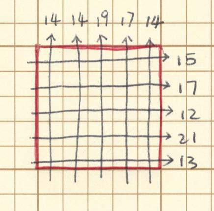


16 Other applications: SPECT and Attenuated CT (square body) Line integrals of f reconstruction of f
17 Other applications: SPECT and Attenuated Outline 1 Course Information (syllabus, course webpage) Other applications: SPECT and Attenuated
18 Other applications: SPECT and Attenuated in Imaging (inverse v.s. direct) Direct problem: Input signal = A = Output signal =? Inverse problem: Input signal = A =? = Output signal e.g. X-ray beam L = f =? = L f (x) dx
19 Other applications: SPECT and Attenuated Interesting questions for Uniqueness Inversion/Reconstruction (Optimization v.s. direct inversion) Stability Range of the direct operator Incomplete data Contrast* Resolution*
20 Other applications: SPECT and Attenuated Examples of Other Electrical Impedance Tomography (EIT) Inverse Scattering
21 Other applications: SPECT and Attenuated Outline 1 Course Information (syllabus, course webpage) Other applications: SPECT and Attenuated
22 Other applications: SPECT and Attenuated The R of f (x) in R 2 : Rf (L) := f (x) dx for all lines L R 2 L Can we reconstruct f (x) from Rf?
23 Other applications: SPECT and Attenuated Outline 1 Course Information (syllabus, course webpage) Other applications: SPECT and Attenuated
24 Other applications: SPECT and Attenuated 3D object and X-ray transform To image a medium in R 3 using CT scan, Layer by layer of 2D CT scan; X-ray transform X of f (x) along line L in R 3 : Xf (L) := f (x) dx. ( and X-ray transform differ in dimension n 3.) L
25 Other applications: SPECT and Attenuated Outline 1 Course Information (syllabus, course webpage) Other applications: SPECT and Attenuated
26 Other applications: SPECT and Attenuated SPECT: Single Photon Emission Computed Tomography A nuclear medical tomographic imaging technique using gamma rays, with a true 3D reconstruction (v.s. cross-sectional slices 2D reconstruction). The γ-ray density solves the equation ω x u(x, ω) + a(x)u(x, ω) = f (x). Here a(x) is the known attenuation coefficient and f (x) is the unknown source. The inverse source problem consists of reconstructing f (x) from u out (x, ω).
27 Other applications: SPECT and Attenuated Attenuated Knowing u out (x, ω), one can compute the AtRT R a of the source f (x) given by R a f (L) := f (x)e L(x) a(y) dy dx where L(x) is the line section from x to the receiver on L. L
28 Other applications: SPECT and Attenuated of Attenuated The inverse problem consists then in answering the following questions: 1 Knowing the AtRT R a f (L) for all L and a(x), what can we reconstruct in f (x)? What if f also depends on ω? 2 Can we reconstruct f (x) from half of the measurements or do we at least have uniqueness of the reconstruction? 3 Do we have a reliable numerical technique to obtain fast reconstructions?
Medical Imaging. X-rays, CT/CAT scans, Ultrasound, Magnetic Resonance Imaging
 Medical Imaging X-rays, CT/CAT scans, Ultrasound, Magnetic Resonance Imaging From: Physics for the IB Diploma Coursebook 6th Edition by Tsokos, Hoeben and Headlee And Higher Level Physics 2 nd Edition
Introduction. Chapter 16 Diagnostic Radiology. Primary radiological image. Primary radiological image
 Introduction Chapter 16 Diagnostic Radiology Radiation Dosimetry I Text: H.E Johns and J.R. Cunningham, The physics of radiology, 4 th ed. http://www.utoledo.edu/med/depts/radther In diagnostic radiology
Introduction to image processing
 Part I Introduction to image processing 1 Introduction Overview Imaging systems construct an (output) image in response to (input) signals from diverse types of objects. They can be classified in a number
Introduction, Review of Signals & Systems, Image Quality Metrics
 Introduction, Review of Signals & Systems, Image Quality Metrics Yao Wang Polytechnic University, Brooklyn, NY 11201 Based on Prince and Links, Medical Imaging Signals and Systems and Lecture Notes by
PD233: Design of Biomedical Devices and Systems
 PD233: Design of Biomedical Devices and Systems (Lecture-8 Medical Imaging Systems) (Imaging Systems Basics, X-ray and CT) Dr. Manish Arora CPDM, IISc Course Website: http://cpdm.iisc.ac.in/utsaah/courses/
Radionuclide Imaging MII Single Photon Emission Computed Tomography (SPECT)
") Radionuclide Imaging MII 3073 Single Photon Emission Computed Tomography (SPECT) Single Photon Emission Computed Tomography (SPECT) The successful application of computer algorithms to x-ray imaging in
Radionuclide Imaging MII 3073 Single Photon Emission Computed Tomography (SPECT) Single Photon Emission Computed Tomography (SPECT) The successful application of computer algorithms to x-ray imaging in
HIGH RESOLUTION COMPUTERIZED TOMOGRAPHY SYSTEM USING AN IMAGING PLATE
 HIGH RESOLUTION COMPUTERIZED TOMOGRAPHY SYSTEM USING AN IMAGING PLATE Takeyuki Hashimoto 1), Morio Onoe 2), Hiroshi Nakamura 3), Tamon Inouye 4), Hiromichi Jumonji 5), Iwao Takahashi 6); 1)Yokohama Soei
X-rays in medical diagnostics
 X-rays in medical diagnostics S.Dolanski Babić 2017/18. History W.C.Röntgen (1845-1923) discovered a new type of radiation Nature, Jan. 23. 1896.; Science, Feb.14. 1896. X- rays: Induced the ionization
Digitization and fundamental techniques
 Digitization and fundamental techniques Chapter 2.2-2.6 Robin Strand Centre for Image analysis Swedish University of Agricultural Sciences Uppsala University Outline Imaging Digitization Sampling Labeling
Explain what is meant by a photon and state one of its main properties [2]
![Explain what is meant by a photon and state one of its main properties [2]](/thumbs/80/82516318.jpg "Explain what is meant by a photon and state one of its main properties [2]") 1 (a) A patient has an X-ray scan taken in hospital. The high-energy X-ray photons interact with the atoms inside the body of the patient. Explain what is meant by a photon and state one of its main properties....
1 (a) A patient has an X-ray scan taken in hospital. The high-energy X-ray photons interact with the atoms inside the body of the patient. Explain what is meant by a photon and state one of its main properties....
Photomultiplier Tube
 Nuclear Medicine Uses a device known as a Gamma Camera. Also known as a Scintillation or Anger Camera. Detects the release of gamma rays from Radionuclide. The radionuclide can be injected, inhaled or
Parameter Estimation Techniques for Ultrasound Phase Reconstruction. Fatemeh Vakhshiteh Sept. 16, 2010
 Parameter Estimation Techniques for Ultrasound Phase Reconstruction Fatemeh Vakhshiteh Sept. 16, 2010 Presentation Outline Motivation Thesis Objectives Background Simulation Quadrature Phase Measurement
HISTORY. CT Physics with an Emphasis on Application in Thoracic and Cardiac Imaging SUNDAY. Shawn D. Teague, MD
 CT Physics with an Emphasis on Application in Thoracic and Cardiac Imaging Shawn D. Teague, MD DISCLOSURES 3DR- advisory committee CT PHYSICS WITH AN EMPHASIS ON APPLICATION IN THORACIC AND CARDIAC IMAGING
SYLLABUS. 1. Identification of Subject:
 SYLLABUS Date/ Revision : 30 January 2017/1 Faculty : Life Sciences Approval : Dean, Faculty of Life Sciences SUBJECT : Biophysics 1. Identification of Subject: Name of Subject : Biophysics Code of Subject
Reconstruction Filtering in Industrial gamma-ray CT Application
 Reconstruction Filtering in Industrial gamma-ray CT Application Lakshminarayana Yenumula *, Rajesh V Acharya, Umesh Kumar, and Ashutosh Dash Industrial Tomography and Instrumentation Section, Isotope Production
Ultrasonic Linear Array Medical Imaging System
 Ultrasonic Linear Array Medical Imaging System R. K. Saha, S. Karmakar, S. Saha, M. Roy, S. Sarkar and S.K. Sen Microelectronics Division, Saha Institute of Nuclear Physics, 1/AF Bidhannagar, Kolkata-700064.
COMPUTATIONAL IMAGING. Berthold K.P. Horn
 COMPUTATIONAL IMAGING Berthold K.P. Horn What is Computational Imaging? Computation inherent in image formation What is Computational Imaging? Computation inherent in image formation (1) Computing is getting
Medical Images Analysis and Processing
 Medical Images Analysis and Processing - 25642 Emad Course Introduction Course Information: Type: Graduated Credits: 3 Prerequisites: Digital Image Processing Course Introduction Reference(s): Insight
Introduction. MIA1 5/14/03 4:37 PM Page 1
 MIA1 5/14/03 4:37 PM Page 1 1 Introduction The last two decades have witnessed significant advances in medical imaging and computerized medical image processing. These advances have led to new two-, three-
Fig. 1
 PhysicsAndMathsTutor.com 1 1. Fig. 1 shows data for the intensity of a parallel beam of X-rays after penetration through varying thicknesses of a material. intensity / MW m 2 thickness / mm 0.91 0.40 0.69
SECTION I - CHAPTER 2 DIGITAL IMAGING PROCESSING CONCEPTS
 RADT 3463 - COMPUTERIZED IMAGING Section I: Chapter 2 RADT 3463 Computerized Imaging 1 SECTION I - CHAPTER 2 DIGITAL IMAGING PROCESSING CONCEPTS RADT 3463 COMPUTERIZED IMAGING Section I: Chapter 2 RADT
Gamma emission tomography of nuclear fuel; Objectives and status of the IAEA UGET project.
 Gamma emission tomography of nuclear fuel; Objectives and status of the IAEA UGET project. Peter Jansson Staffan Jacobsson Svärd Sophie Grape Div. of Applied Nuclear Physics Dept. of Physics and Astronomy
Introduction, Review of Signals & Systems, Image Quality Metrics
 EL-GY 5823 / BE-GY 6203 / G16.4426 Medical Imaging Introduction, Review of Signals & Systems, Image Quality Metrics Jonathan Mamou & Yao Wang Tandon School of Engineering New York University, Brooklyn,
Computerized transverse axial scanning (tomography): Part I. Description of system
: Part I. Description of system") 1973, British Journal of Radiology, 46, 1016-1022 Computerized transverse axial scanning (tomography): Part I. Description of system Central Research Laboratories of EMI Limited, Hayes, Middlesex (Received
1973, British Journal of Radiology, 46, 1016-1022 Computerized transverse axial scanning (tomography): Part I. Description of system Central Research Laboratories of EMI Limited, Hayes, Middlesex (Received
Digital Image Processing. Lecture 1 (Introduction) Bu-Ali Sina University Computer Engineering Dep. Fall 2011
 Bu-Ali Sina University Computer Engineering Dep. Fall 2011") Digital Processing Lecture 1 (Introduction) Bu-Ali Sina University Computer Engineering Dep. Fall 2011 Introduction One picture is worth more than ten thousand p words Outline Syllabus References Course
Digital Processing Lecture 1 (Introduction) Bu-Ali Sina University Computer Engineering Dep. Fall 2011 Introduction One picture is worth more than ten thousand p words Outline Syllabus References Course
Tomostatic Waveform Tomography on Near-surface Refraction Data
 Tomostatic Waveform Tomography on Near-surface Refraction Data Jianming Sheng, Alan Leeds, and Konstantin Osypov ChevronTexas WesternGeco February 18, 23 ABSTRACT The velocity variations and static shifts
LaBr 3 :Ce, the latest crystal for nuclear medicine
 10th Topical Seminar on Innovative Particle and Radiation Detectors 1-5 October 2006 Siena, Italy LaBr 3 :Ce, the latest crystal for nuclear medicine Roberto Pani On behalf of SCINTIRAD Collaboration INFN
PRODUCT 4.06 IMAGE MANAGEMENT
 IT EDUCTRA TELEMATICS APPLICATIONS PROGRAMME Sector: Healthcare PRODUCT 4.06 IMAGE MANAGEMENT Arie HASMAN This Product Section outlines the different methods of generating and analysing images, where each
SPARSE ARRAY TOMOGRAPHY SYSTEM FOR CORROSION EXTENT MONITORING H. Bian, H. Gao, J. Rose Pennsylvania State University, University Park, PA, USA
 SPARSE ARRAY TOMOGRAPHY SYSTEM FOR CORROSION EXTENT MONITORING H. Bian, H. Gao, J. Rose Pennsylvania State University, University Park, PA, USA Abstract: A sparse array guided wave tomography system is
FROM PHOTOGRAPHY TO *.GRAPHIES: UNCONVENTIONAL IMAGING TECHNIQUES
 L. Yaroslavsky FROM PHOTOGRAPHY TO *.GRAPHIES: UNCONVENTIONAL IMAGING TECHNIQUES A short course at Tampere University of Technology, Tampere, Finland, Sept. 3 Sept. 14, 2001 Lecture 2. EVOLUTION OF IMAGING:
Breast Tomosynthesis. Bob Liu, Ph.D. Department of Radiology Massachusetts General Hospital And Harvard Medical School
 Breast Tomosynthesis Bob Liu, Ph.D. Department of Radiology Massachusetts General Hospital And Harvard Medical School Outline Physics aspects of breast tomosynthesis Quality control of breast tomosynthesis
JEFFERSON COLLEGE COURSE SYLLABUS BET220 DIAGNOSTIC IMAGING. 3 Credit Hours. Prepared by: Scott Sebaugh Date: 2/20/2012
 JEFFERSON COLLEGE COURSE SYLLABUS BET220 DIAGNOSTIC IMAGING 3 Credit Hours Prepared by: Scott Sebaugh Date: 2/20/2012 Mary Beth Ottinger, Division Chair Elizabeth Check, Dean, Career & Technical Education
Digital Image Processing
 What is an image? Digital Image Processing Picture, Photograph Visual data Usually two- or three-dimensional What is a digital image? An image which is discretized, i.e., defined on a discrete grid (ex.
Nuclear Associates , , CT Head and Body Dose Phantom
 Nuclear Associates 76-414,76-414-4150,76-415 CT Head and Body Dose Phantom Users Manual March 2005 Manual No. 76-414-1 Rev. 2 2004, 2005 Fluke Corporation, All rights reserved. Printed in U.S.A. All product
Imaging with Electricity, Andy Adler, 2016/11/02 1 / 39
 Imaging with Electricity, Andy Adler, 2016/11/02 1 / 39 Tomography imaging by sections τόμος tomos, "slice, section" γράφω graphō, "to write" Imaging with Electricity, Andy Adler, 2016/11/02 2 / 39 Image:
MC SIMULATION OF SCATTER INTENSITIES IN A CONE-BEAM CT SYSTEM EMPLOYING A 450 kv X-RAY TUBE
 MC SIMULATION OF SCATTER INTENSITIES IN A CONE-BEAM CT SYSTEM EMPLOYING A 450 kv X-RAY TUBE A. Miceli ab, R. Thierry a, A. Flisch a, U. Sennhauser a, F. Casali b a Empa - Swiss Federal Laboratories for
Radiation Imaging. By: Engr. Joseph Ronald Cañedo
 Radiation Imaging By: Engr. Joseph Ronald Cañedo Objectives Tounderstand the fundamental principles of radioactivity. To understand that ionizing radiation, as generally employed for medical imaging, is
A NOVEL CONCEPT FOR A POSITRON EMISSION TOMOGRAPHY SCANNER
 A NOVEL CONCEPT FOR A POSITRON EMISSION TOMOGRAPHY SCANNER An Undergraduate Research Scholars Thesis by BRIAN KELLY, MATTHEW LEE ELLIOT LEVIN and JEENA KHATRI Submitted to Honors and Undergraduate Research
Local ROI Reconstruction via Generalized FBP and BPF Algorithms along More Flexible Curves
 Hindawi Publishing Corporation International Journal of Biomedical Imaging Volume 2, Article ID 48, Pages 7 DOI.55/IJBI/2/48 Local ROI Reconstruction via Generalized FBP and BPF Algorithms along More Flexible
4.3 Ultrasonic Computed Tomography
 reconstruction in this study) to record total transmission loss for each ray in each projection. Then, in the positron emission study, the data for each ray can simply be attenuation compensated when corrected
3-D tomographic Q inversion for compensating frequency dependent attenuation and dispersion. Kefeng Xin* and Barry Hung, CGGVeritas
 P-75 Summary 3-D tomographic Q inversion for compensating frequency dependent attenuation and dispersion Kefeng Xin* and Barry Hung, CGGVeritas Following our previous work on Amplitude Tomography that
How Maths Can Save Your Life
 17 October 2017 How Maths Can Save Your Life PROFESSOR CHRIS BUDD OBE Introduction Can Maths really save your life? Of course it can! Maths has applications to many problems that are vital to human health
Digital Image Processing
 Digital Processing Introduction Christophoros Nikou cnikou@cs.uoi.gr s taken from: R. Gonzalez and R. Woods. Digital Processing, Prentice Hall, 2008. Digital Processing course by Brian Mac Namee, Dublin
An Activity in Computed Tomography
 Pre-lab Discussion An Activity in Computed Tomography X-rays X-rays are high energy electromagnetic radiation with wavelengths smaller than those in the visible spectrum (0.01-10nm and 4000-800nm respectively).
QC Testing for Computed Tomography (CT) Scanner
 Scanner") QC Testing for Computed Tomography (CT) Scanner QA - Quality Assurance All planned and systematic actions needed to provide confidence on a structure, system or component. all-encompassing program, including
QC Testing for Computed Tomography (CT) Scanner QA - Quality Assurance All planned and systematic actions needed to provide confidence on a structure, system or component. all-encompassing program, including
Near-Field Scanning. Searching for Root Causes
 Near-Field Scanning Searching for Root Causes Feb. 06, 2018 Outline Susceptibility Scanning Conducted susceptibility: where does ESD current go? Near-field effects of electrostatic discharge events Emission
Medical Imaging (EL582/BE620/GA4426)
") Medical Imaging (EL582/BE620/GA4426) Jonathan Mamou, PhD Riverside Research Lizzi Center for Biomedical Engineering New York, NY jmamou@riversideresearch.org On behalf of Prof. Daniel Turnbull Outline
Medical Imaging (EL582/BE620/GA4426) Jonathan Mamou, PhD Riverside Research Lizzi Center for Biomedical Engineering New York, NY jmamou@riversideresearch.org On behalf of Prof. Daniel Turnbull Outline
imaging of the ionosphere and its applications to radio propagation Fundamentals of tomographic Ionospheric Tomography I: Ionospheric Tomography I:
 Ionospheric Tomography I: Ionospheric Tomography I: Fundamentals of tomographic imaging of the ionosphere and its applications to radio propagation Summary Introduction to tomography Introduction to tomography
H 2 O and fat imaging
 H 2 O and fat imaging Xu Feng Outline Introduction benefit from the separation of water and fat imaging Chemical Shift definition of chemical shift origin of chemical shift equations of chemical shift
EE 529 Remote Sensing Techniques. Radar
 EE 59 Remote Sensing Techniques Radar Outline Radar Resolution Radar Range Equation Signal-to-Noise Ratio Doppler Frequency Basic function of an active radar Radar RADAR: Radio Detection and Ranging Detection
Lecture 1 Introduction. Lin ZHANG, PhD School of Software Engineering Tongji University Fall 2016
 Lecture 1 Introduction Lin ZHANG, PhD School of Software Engineering Tongji University Fall 2016 Self Introduction B.Sc., Computer Science and Engineering, Shanghai JiaoTong University, 2003 M.Sc., Computer
Multi-Element Synthetic Transmit Aperture Method in Medical Ultrasound Imaging Ihor Trots, Yuriy Tasinkevych, Andrzej Nowicki and Marcin Lewandowski
 Multi-Element Synthetic Transmit Aperture Method in Medical Ultrasound Imaging Ihor Trots, Yuriy Tasinkevych, Andrzej Nowicki and Marcin Lewandowski Abstract The paper presents the multi-element synthetic
Design of a Tapered Stripline Fast Faraday Cup for Measurements on Heavy Ion Beams: Problems and Solutions
 Design of a Tapered Stripline Fast Faraday Cup for Measurements on Heavy Ion Beams: Problems and Solutions F. Marcellini* and M. Poggi** * INFN, Laboratori Nazionali di Frascati, Frascati (Italy) and **INFN,
12/26/2017. Alberto Ardon M.D.
 Alberto Ardon M.D. 1 Preparatory Work Ultrasound Physics http://www.nysora.com/mobile/regionalanesthesia/foundations-of-us-guided-nerve-blockstechniques/index.1.html Basic Ultrasound Handling https://www.youtube.com/watch?v=q2otukhrruc
Radiology Physics Lectures: Digital Radiography. Digital Radiography. D. J. Hall, Ph.D. x20893
 Digital Radiography D. J. Hall, Ph.D. x20893 djhall@ucsd.edu Background Common Digital Modalities Digital Chest Radiograph - 4096 x 4096 x 12 bit CT - 512 x 512 x 12 bit SPECT - 128 x 128 x 8 bit MRI -
Introduction. Parametric Imaging. The Ultrasound Research Interface: A New Tool for Biomedical Investigations
 The Ultrasound Research Interface: A New Tool for Biomedical Investigations Shelby Brunke, Laurent Pelissier, Kris Dickie, Jim Zagzebski, Tim Hall, Thaddeus Wilson Siemens Medical Systems, Issaquah WA
PET/CT Instrumentation Basics
 / Instrumentation Basics 1. Motivations for / imaging 2. What is a / Scanner 3. Typical Protocols 4. Attenuation Correction 5. Problems and Challenges with / 6. Examples Motivations for / Imaging Desire
Augmentation of X-Rays Images using Pixel Intensity Values Adjustments
 Augmentation of X-Rays Images using Pixel Intensity Values Adjustments Yousif Mohamed Y. Abdallah 1, 2, Rajab M. Ben Yousef 3 1 College of Medical Radiological Science, Sudan University of Science and
CHAPTER 8 GENERIC PERFORMANCE MEASURES
 GENERIC PERFORMANCE MEASURES M.E. DAUBE-WITHERSPOON Department of Radiology, University of Pennsylvania, Philadelphia, Pennsylvania, United States of America 8.1. INTRINSIC AND EXTRINSIC MEASURES 8.1.1.
M R I Physics Course. Jerry Allison Ph.D., Chris Wright B.S., Tom Lavin B.S., Nathan Yanasak Ph.D. Department of Radiology Medical College of Georgia
 M R I Physics Course Jerry Allison Ph.D., Chris Wright B.S., Tom Lavin B.S., Nathan Yanasak Ph.D. Department of Radiology Medical College of Georgia M R I Physics Course Magnetic Resonance Imaging Spatial
Modern X-ray vision: 3D imaging with sparse data
 Modern X-ray vision: 3D imaging with sparse data Samuli Siltanen Department of Mathematics and Statistics University of Helsinki, Finland samuli.siltanen@helsinki.fi www.siltanen-research.net Finland at
ELE 882: Introduction to Digital Image Processing (DIP)
") ELE882 Introduction to Digital Image Processing Course Instructor: Prof. Ling Guan Department of Electrical & Computer Engineering Room 315, ENG Building Tel: (416)979-5000 ext 6072 Email: lguan@ee.ryerson.ca
ELE882 Introduction to Digital Image Processing Course Instructor: Prof. Ling Guan Department of Electrical & Computer Engineering Room 315, ENG Building Tel: (416)979-5000 ext 6072 Email: lguan@ee.ryerson.ca
CHAPTER 1 INTRODUCTION
 CHAPTER 1 INTRODUCTION Spatial resolution in ultrasonic imaging is one of many parameters that impact image quality. Therefore, mechanisms to improve system spatial resolution could result in improved
Chapter 4. Pulse Echo Imaging. where: d = distance v = velocity t = time
 Chapter 4 Pulse Echo Imaging Ultrasound imaging systems are based on the principle of pulse echo imaging. These systems require the use of short pulses of ultrasound to create two-dimensional, sectional
Waves, Sound and Light. Grade 10 physics Robyn Basson
 Waves, Sound and Light Grade 10 physics Robyn Basson Heartbeat Flick in hose pipe What is a pulse? A single disturbance that moves through a medium. Stone in water Other? moving Transverse pulse: A pulse
Introduction. Stefano Ferrari. Università degli Studi di Milano Methods for Image Processing. academic year
 Introduction Stefano Ferrari Università degli Studi di Milano stefano.ferrari@unimi.it Methods for Image Processing academic year 2015 2016 Image processing Computer science concerns the representation,
DICOM Conformance. DICOM Detailed Specification for Diagnostic Labs and Radiology Center Connectivity
 DICOM Detailed Specification for Diagnostic Labs and Radiology Center Connectivity Authored by Global Engineering Team, Health Gorilla April 10, 2014 Table of Contents About Health Gorilla s Online Healthcare
IMPROVEMENTS IN X-RAY CT
 The First International Proficiency Testing Conference Sinaia, România 11 th 13 th October, 2007 IMPROVEMENTS IN X-RAY CT Emilia Dana Seleţchi Faculty of Physics, University of Bucharest, Măgurele, CP
Microwave and optical systems Introduction p. 1 Characteristics of waves p. 1 The electromagnetic spectrum p. 3 History and uses of microwaves and
 Microwave and optical systems Introduction p. 1 Characteristics of waves p. 1 The electromagnetic spectrum p. 3 History and uses of microwaves and optics p. 4 Communication systems p. 6 Radar systems p.
RADIATIONS. ELECTROMAGNETIC WAVES. Talián Csaba Gábor Dept. Biophysics Apr
 RADIATIONS. ELECTROMAGNETIC WAVES. Talián Csaba Gábor Dept. Biophysics Apr 16. 2012. WHAT IS RADIATION? PROPAGATION OF ENERGY IN SPACE THROUGH TRAVELLING OF PARTICLES OR WAVES Particle: alfa-, beta-radiantion
2008 Monitoring Research Review: Ground-Based Nuclear Explosion Monitoring Technologies
 ABSTRACT SEMI-EMPIRICAL YIELD ESTIMATES FOR THE 2006 NORTH KOREAN EXPLOSION David H. Salzberg Science Applications International Corporation Sponsored by Air Force Research Laboratory Contract number FA8718-08-C-0011
An overview of our lab and our activities. Giuseppe Vecchi March 2015
 An overview of our lab and our activities Giuseppe Vecchi March 2015 ISMB Research Areas Navigation Technologies Pervasive Technologies Multi-Layer Wireless Solutions LACE @ ISMB ISMB is organized in Research
Aperture antennas. Ahmed FACHAR, Universidad Politécnica de Madrid (Technical University of Madrid, UPM)
") Aperture antennas Ahmed FACHAR, ahmedfach@gr.ssr.upm.es Universidad Politécnica de Madrid (Technical University of Madrid, UPM) Outline Introduction Horn antennas Introduction Rectangular horns Conical
Aperture antennas Ahmed FACHAR, ahmedfach@gr.ssr.upm.es Universidad Politécnica de Madrid (Technical University of Madrid, UPM) Outline Introduction Horn antennas Introduction Rectangular horns Conical
Imaging Techniques. Introduction. Patrícia Figueiredo IST
 Imaging Techniques Introduction Patrícia Figueiredo IST 2012-2013 Faculty: Patrícia Figueiredo, IST (patricia.figueiredo@ist.utl.pt, IST North Tower, 6th floor, Tel: 218418277 ext 2277) Jorge Campos, FMUL
X-RAY COMPUTED TOMOGRAPHY
 X-RAY COMPUTED TOMOGRAPHY Bc. Jan Kratochvíla Czech Technical University in Prague Faculty of Nuclear Sciences and Physical Engineering Abstract Computed tomography is a powerful tool for imaging the inner
ESA Radar Remote Sensing Course ESA Radar Remote Sensing Course Radar, SAR, InSAR; a first introduction
 Radar, SAR, InSAR; a first introduction Ramon Hanssen Delft University of Technology The Netherlands r.f.hanssen@tudelft.nl Charles University in Prague Contents Radar background and fundamentals Imaging
3D light microscopy techniques
 3D light microscopy techniques The image of a point is a 3D feature In-focus image Out-of-focus image The image of a point is not a point Point Spread Function (PSF) 1D imaging 2D imaging 3D imaging Resolution
IBEX MATERIALS DETECTION TECHNOLOGY
 WHITE PAPER: IBEX MATERIALS DETECTION TECHNOLOGY IBEX Innovations Ltd. Registered in England and Wales: 07208355 Address: Discovery 2, NETPark, William Armstrong Way, Sedgefield, TS21 3FH, UK Patents held
COMPUTED TOMOGRAPHY 1
 COMPUTED TOMOGRAPHY 1 Why CT? Conventional X ray picture of a chest 2 Introduction Why CT? In a normal X-ray picture, most soft tissue doesn't show up clearly. To focus in on organs, or to examine the
Digital Image Processing COSC 6380/4393
 Digital Image Processing COSC 6380/4393 Lecture 2 Aug 24 th, 2017 Slides from Dr. Shishir K Shah, Rajesh Rao and Frank (Qingzhong) Liu 1 Instructor TA Digital Image Processing COSC 6380/4393 Pranav Mantini
INTRODUCTION. Have applications for imaging, detection and navigation.
 ULTRASONICS INTRODUCTION The word ultrasonic combines the Latin roots ultra - beyond sonic - sound. Having frequencies above the audible range i.e. above 20000Hz Have applications for imaging, detection
Moving from biomedical to industrial applications: OCT Enables Hi-Res ND Depth Analysis
 Moving from biomedical to industrial applications: OCT Enables Hi-Res ND Depth Analysis Patrick Merken a,c, Hervé Copin a, Gunay Yurtsever b, Bob Grietens a a Xenics NV, Leuven, Belgium b UGENT, Ghent,
Towards accurate measurements with synchrotron tomography Problems and pitfalls. Robert C. Atwood. Nghia T. Vo, Michael Drakopoulos, Thomas Connolley
 Towards accurate measurements with synchrotron tomography Problems and pitfalls Robert C. Atwood Nghia T. Vo, Michael Drakopoulos, Thomas Connolley Artefacts in Synchrotron X-ray Tomography Rings Rings
LECTURE 20 ELECTROMAGNETIC WAVES. Instructor: Kazumi Tolich
 LECTURE 20 ELECTROMAGNETIC WAVES Instructor: Kazumi Tolich Lecture 20 2 25.6 The photon model of electromagnetic waves 25.7 The electromagnetic spectrum Radio waves and microwaves Infrared, visible light,
Development of an innovative LSO-SiPM detector module for high-performance Positron Emission Tomography
 Development of an innovative LSO-SiPM detector module for high-performance Positron Emission Tomography Maria Leonor Trigo Franco Frazão leonorfrazao@ist.utl.pt Instituto Superior Técnico, Lisboa, Portugal
Methods of processing and image compression in an X-ray micro tomographic scanner. Syryamkin V.I. Osipov A.V., Kutsov M.S.
 11th European Conference on Non-Destructive Testing (ECNDT 2014), October 6-10, 2014, Prague, Czech Republic Methods of processing and image compression in an X-ray micro tomographic scanner Syryamkin
Digital Images & Image Quality
 Introduction to Medical Engineering (Medical Imaging) Suetens 1 Digital Images & Image Quality Ho Kyung Kim Pusan National University Radiation imaging DR & CT: x-ray Nuclear medicine: gamma-ray Ultrasound
Optical coherence tomography
 Optical coherence tomography Peter E. Andersen Optics and Plasma Research Department Risø National Laboratory E-mail peter.andersen@risoe.dk Outline Part I: Introduction to optical coherence tomography
Ground Penetrating Radar
 Ground Penetrating Radar Begin a new section: Electromagnetics First EM survey: GPR (Ground Penetrating Radar) Physical Property: Dielectric constant Electrical Permittivity EOSC 350 06 Slide Di-electric
Image Analysis in X-ray Computed Tomography
 Image Analysis in X-ray Computed Tomography Emilia Dana Seleţchi 1, Victor Şutac 2 1 University of Bucharest, Faculty of Physics, Bucharest, ROMANIA E-mail: seletchi@gmail.com 2 Cygnus Scientific Society,
Ultrasound Beamforming and Image Formation. Jeremy J. Dahl
 Ultrasound Beamforming and Image Formation Jeremy J. Dahl Overview Ultrasound Concepts Beamforming Image Formation Absorption and TGC Advanced Beamforming Techniques Synthetic Receive Aperture Parallel
Synchrotron X-ray tomographic microscopy Theory vs. practice
 Synchrotron X-ray tomographic microscopy Theory vs. practice Federica Marone Swiss Light Source, Paul Scherrer Institut, Villigen, Switzerland Theory Radon transform Rf x = Beer-Lambert law I E = I 0 (E)e
Postprocessing of nonuniform MRI
 Postprocessing of nonuniform MRI Wolfgang Stefan, Anne Gelb and Rosemary Renaut Arizona State University Oct 11, 2007 Stefan, Gelb, Renaut (ASU) Postprocessing October 2007 1 / 24 Outline 1 Introduction
Medical Imaging and its Associated Analysis
 Medical Imaging and its Associated Analysis Saurabh Singh 1, Anurag Singh 2,Pranay Surana3, Priyen Dang 4, Anand Ranka 5, Saurabh Burange 6 1 Department of Electronics and Communication Engineering 2,3,4,5,6
Simulations of the J-PET detector response with the GATE package
 Simulations of the J-PET detector response with the GATE package Author: pawel.kowalski@ncbj.gov.pl 22nd to 24th September 2014 II Symposium on Positron Emission Tomography Outline 1. Introduction 2. Simulation
diagnostic examination
 RADIOLOGICAL PHYSICS 2011 Raphex diagnostic examination Adel A. Mustafa, Ph.D., Editor PUBLISHED FOR: RAMPS (Radiological and Medical Physics Society of New York) preface The RAPHEX Diagnostic exam 2011
COMPUTED IMAGE BACKSCATTER RADIOGRAPHY: PROOF OF PRINCIPLE AND INITIAL DEVELOPMENT
 COMPUTED IMAGE BACKSCATTER RADIOGRAPHY: PROOF OF PRINCIPLE AND INITIAL DEVELOPMENT By CHRISTOPHER LLOYD MENG A DISSERTATION PRESENTED TO THE GRADUATE SCHOOL OF THE UNIVERSITY OF FLORIDA IN PARTIAL FULFILLMENT
Measurement, Sensors, and Data Acquisition in the Two-Can System
 Measurement, Sensors, and Data Acquisition in the Two-Can System Prof. R.G. Longoria Updated Fall 2010 Goal of this week s lab Gain familiarity with using sensors Gain familiarity with using DAQ hardware
An Activity in Computed Tomography
 Pre-lab Discussion An Activity in Computed Tomography X-rays X-rays are high energy electromagnetic radiation with wavelengths smaller than those in the visible spectrum (0.01-10nm and 4000-800nm respectively).
Acknowledgment. Process of Atmospheric Radiation. Atmospheric Transmittance. Microwaves used by Radar GMAT Principles of Remote Sensing
 GMAT 9600 Principles of Remote Sensing Week 4 Radar Background & Surface Interactions Acknowledgment Mike Chang Natural Resources Canada Process of Atmospheric Radiation Dr. Linlin Ge and Prof Bruce Forster