2/4/15. Brightfield Microscopy! It s all about Magnification..! or is it?!
|
|
|
- Roland Pearson
- 5 years ago
- Views:
Transcription


1 Brightfield Microscopy It s all about Magnification.. or is it? 1

2 What actually does go into chosing a microscope Choice depends on what you need the microscope to do. Do you want to magnify stained specimens? Do you want high magnification? What are the resolution requirements? What does that mean magnification and resolution? Magnification versus Resolution Magnification Think of it as experimenting on a computer by typing the letter A and magnifying it. What happens? Resolution Accepting each pixel as a part of the puzzle, take some away. What happens? 2


3 Magnification versus Resolution - magnification in a good dissecting scope can be similar - however, as magnification increases, resolution does not => empty magnification Dissecting Microscope Compound Microscope If magnification is the same, why does this occur? Solving Resolution differences in lens systems in the dissecting (stereo-) microscope compared to the compound microscope SIMPLE MICROSCOPE 3


4 The Compound Microscope Take the time and see whether you can still identify the different parts of the microscope. I know you have already done that before, but during this class you will really have to know the microscope in the dark. The Optical Path in the Brightfield Microscope = that s why the NA has to match between objective and condenser 4

5 The Numerical Aperture = dimensionless number of an optical system characterizing the angles over which the system can transmit light - How do you compensate for the change in numerical aperture when you change objective magnifications? Answers: How to address and compensate for changes in the NA between different objective magnifications? To adjust for the NA adjust the settings above the condenser (numbered lines) to the same numerical value as the NA Thickness and the texture of the condenser lens. The Iris Diaphragm can be adjusted to the correct measurement to match the aperture of the objective lens. The iris diaphragm knob would have to be adjusted to the specific numerical aperture that would be on the objective lens in use. What you have to do is to open and close the diaphragm of the iris to condense light through the condenser. 5


 Plan =")
6 Correct Answer The NA on the condenser should be adjusted so that it equals the NA on objective lens used to view the specimen. This should allow the appropriate cone of light light to be gathered from the light source to focus on the image. - objective and condenser NA match = increased resolution - objective NA increases with increased magnification, therefore condenser NA needs to be matched = match condenser setting to NA on objective The Objectives Achromatic = chromatic aberration, generally doublet Apochromatic = best correction, spherical/chromatic aberration, generally triplet Semi-plan = flatter lenses (80% flat field) Plan = flatter lenses (95% flat field, generally triplet) 6


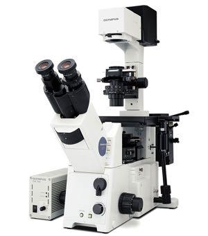
7 When to use Brightfield Microscopy Brightfield = stained specimens amplitude specimens Unstained specimen Amplitude specimen Two Compound Microscope Options - upright versus inverted How to chose and why 7


8 Again.. what do you want to look at? If you have regular specimens, i.e. a stained specimen mounted on a slide. If you are looking at 96-well plates, petri dishes or culture dishes. Does that mean you cannot use regular slides on an inverted microscope? of course you can You just have to adjust your settings: - have slide upside down - buy specific cover slips - buy specific objective lenses 8


 Focus edges of")
 Open the")
9 Koehler Illumination - set up and adjust microscope => combination of contrast & resolution - condenser diaphragm controls light => condenser+objective aperture = realized NA - higher magnification = closer to specimen = large NA = smaller lens opening Koehler Illumination: Getting started Focus sample in brightfield Close field diaphragm (light source) If image view not centered, align using condenser screws (silver screws) Focus edges of the internal, centered image view by moving the condenser (black condenser screws) Open the field diaphragm (light source) 9
10 Exercises today: (work in pairs) 1. Find/identify the important components of the different microscopes (i.e. Inverted versus upright microscope). Are you able to name the parts for the purpose of an exam? 2. Understand where and how to set Koehler illumination in the different microscopes (i.e. inverted, upright, polarizing, DIC). 3. Are you capable of adjusting the NA on the associated parts of the different microscopes? Are all microscopes capable to compensate for objective changes in NA? 4. Observe differences when you open up and or stop down the condenser entirely (disregarding the NA compensation) = make slides of material you brought for exercises 3&4 10
Figure 3.4 Approximate size of various types of cells. ~10 um. Red Blood Cells = mm 1500 um. Width of penny Pearson Education, Inc.
 Figure 3.4 Approximate size of various types of cells. ~10 um Red Blood Cells 1.5mm 1500 um Width of penny = 1500 Figure 4.3 The limits of resolution (and some representative objects within those ranges)
Imaging Introduction. September 24, 2010
 Imaging Introduction September 24, 2010 What is a microscope? Merriam-Webster: an optical instrument consisting of a lens or combination of lenses for making enlarged images of minute objects; especially:
Biology 29 Cell Structure and Function Spring, 2009 Springer LABORATORY 1: THE LIGHT MICROSCOPE
 Biology 29 Cell Structure and Function Spring, 2009 Springer LABORATORY 1: THE LIGHT MICROSCOPE Prior to lab: 1) Read these instructions (p 1-6) 2) Go through the online tutorial, the microscopy pre-lab
Reflection! Reflection and Virtual Image!
 1/30/14 Reflection - wave hits non-absorptive surface surface of a smooth water pool - incident vs. reflected wave law of reflection - concept for all electromagnetic waves - wave theory: reflected back
Instruction Manual T Binocular Acromat Research Scope T Trinocular Acromat Research Scope
 Research Scope Instruction Manual T-29031 Binocular Acromat Research Scope T-29041 Trinocular Acromat Research Scope T-29032 Binocular Semi-Plan Research Scope T-29042 Trinocular Semi-Plan Research Scope
Microscopy. Danil Hammoudi.MD
 Microscopy Danil Hammoudi.MD Care and Handling of the Microscope: A microscope is a delicate piece of equipment and should be treated with care. Use two hands when carrying the microscope. Place one hand
OPELCO OPtical ELements COrporation LB Objective Series for Biological Use
 LB Objective Series for Biological Use 105 Executive Drive Suite 100 Dulles, VA 20166-9558 Tel: (703) 471-0080 S PLAN APOCHROMAT OBJECTIVES These objectives compensate for three wavelength of chromatic
The Compound Microscope. Brightfield: Köhler Illumination
 Outline History of Microscopy The Magnifying Glass The Compound Microscope Brightfield: Köhler Illumination Microscopy µικροσ (mikros): small σκοπειν (skopein): to observe History of Microscopy Well :
BASICS IN BIOIMAGING AND OPTICS PLATFORM EPFL SV PTBIOP LIGHT MICROSCOPY
 BASICS IN LIGHT MICROSCOPY OVERVIEW 1. Motivation 2. Basic in optics 3. How microscope works 4. Illumination and resolution 5. Microscope optics 6. Contrasting methods -2- MOTIVATION Why do we need microscopy?
 Microscopy http://www.microscopyu.com/articles/phasecontrast/phasemicroscopy.html http://micro.magnet.fsu.edu/primer/anatomy/anatomy.html 2005, Dr. Jack Ikeda & Dr. Gail Grabner 9 Nikon Labophot (Question
Basics of Light Microscopy and Metallography
 ENGR45: Introduction to Materials Spring 2012 Laboratory 8 Basics of Light Microscopy and Metallography In this exercise you will: gain familiarity with the proper use of a research-grade light microscope
A BRIEF INTRODUCTION TO MICROSCOPY The two key properties of a microscope that allow you to see microbes are resolution and magnification.
 A BRIEF INTRODUCTION TO MICROSCOPY The two key properties of a microscope that allow you to see microbes are resolution and magnification. Magnification refers to the enlargement of the specimen when seen
Microscopy: Fundamental Principles and Practical Approaches
 Microscopy: Fundamental Principles and Practical Approaches Simon Atkinson Online Resource: http://micro.magnet.fsu.edu/primer/index.html Book: Murphy, D.B. Fundamentals of Light Microscopy and Electronic
Introduction. Laboratory Equipment & Supplies. Model 1333PHi Shown (Phase Contrast) (2) Eyepieces (Eyecups installed) Diopter Adjustment Mechanism
 (2) Eyepieces (Eyecups installed) Diopter Adjustment Mechanism") Introduction With the invention of the microscope in the early 17th century, it was made possible to view objects which were too small for the human eye to see. As the microscope evolved, the structure
Introduction With the invention of the microscope in the early 17th century, it was made possible to view objects which were too small for the human eye to see. As the microscope evolved, the structure
Match the microscope structures given in the left column with the statements in the right column that identify or describe them.
 49 Prelab for Name Match the microscope structures given in the left column with the statements in the right column that identify or describe them. Key: a. coarse adjustment knob f. turret or nosepiece
Basic Microscopy. OBJECTIVES After completing this exercise, you should be able to do the following:
 Page 1 of 10 Basic Microscopy OBJECTIVES After completing this exercise, you should be able to do the following: a. Name the parts of the compound microscope and the functions of each. b. Describe how
EPIFLUORESCENCE &/OR BRIGHTFIELD MICROSCOPY
 EPIFLUORESCENCE &/OR BRIGHTFIELD MICROSCOPY TURN ON THE FOLLOWING EQUIPMENT The fluorescent light (if needed) The power strip for the microscope and accessories The CoolSNAP HQ camera on the right (Turn
Microscope anatomy, image formation and resolution
 Microscope anatomy, image formation and resolution Ian Dobbie Buy this book for your lab: D.B. Murphy, "Fundamentals of light microscopy and electronic imaging", ISBN 0-471-25391-X Visit these websites:
Introduction to Light Microscopy. (Image: T. Wittman, Scripps)
") Introduction to Light Microscopy (Image: T. Wittman, Scripps) The Light Microscope Four centuries of history Vibrant current development One of the most widely used research tools A. Khodjakov et al. Major
Introduction to Light Microscopy (Image: T. Wittman, Scripps) The Light Microscope Four centuries of history Vibrant current development One of the most widely used research tools A. Khodjakov et al. Major
Education in Microscopy and Digital Imaging
 Contact Us Carl Zeiss Education in Microscopy and Digital Imaging ZEISS Home Products Solutions Support Online Shop ZEISS International ZEISS Campus Home Interactive Tutorials Basic Microscopy Spectral
VISUAL PHYSICS ONLINE DEPTH STUDY: ELECTRON MICROSCOPES
 VISUAL PHYSICS ONLINE DEPTH STUDY: ELECTRON MICROSCOPES Shortly after the experimental confirmation of the wave properties of the electron, it was suggested that the electron could be used to examine objects
Microscopy Training & Overview
 Microscopy Training & Overview Product Marketing October 2011 Stephan Briggs - PLE OVERVIEW AND PRESENTATION FLOW Glossary and Important Terms Introduction Timeline Innovation and Advancement Primary Components
Visual Anatomy ansd Physiology Lab Manual Pig Version 2nd Edition Sarikas TEST BANK
 Visual Anatomy ansd Physiology Lab Manual Pig Version 2nd Edition Sarikas TEST BANK https://testbankreal.com/download/visual-anatomy-ansd-physiology-labmanual-pig-version-2nd-edition-sarikas-test-bank/
Basic Microscopy for Plant Biology
 Page 1 of 8 Basic Microscopy for Plant Biology OBJECTIVES After completing this exercise, you should be able to do the following: a. Name the parts of the compound microscope and the functions of each.
Microscopy, Staining, and Classification
 PowerPoint Lecture Presentations prepared by Mindy Miller-Kittrell, North Carolina State University C H A P T E R 4 Microscopy, Staining, and Classification Figure 3.4 Approximate size of various types
Optics Day 3 Kohler Illumination (Philbert Tsai July 2004) Goal : To build an bright-field microscope with a Kohler illumination pathway
 Goal : To build an bright-field microscope with a Kohler illumination pathway") Optics Day 3 Kohler Illumination (Philbert Tsai July 2004) Goal : To build an bright-field microscope with a Kohler illumination pathway Prepare the Light source and Lenses Set up Light source Use 3 rail
Optics Day 3 Kohler Illumination (Philbert Tsai July 2004) Goal : To build an bright-field microscope with a Kohler illumination pathway Prepare the Light source and Lenses Set up Light source Use 3 rail
SWIFT SERIES M2252DGL MICROSCOPE
 SWIFT SERIES M2252DGL MICROSCOPE The M2252DGL Series is ideal for elementary to high school classrooms. Built to withstand student use, this series has locked-on eyepieces, objectives, illuminator housing
MICROSCOPY MICROSCOPE TERMINOLOGY
 1 MICROSCOPY Most of the microorganisms that we talk about in this class are too small to be seen with the naked eye. The instruments we will use to visualize these microbes are microscopes. The laboratory
STEINDORFF NYMC Polarizing Microscope
 NYMC38000 Polarizing Microscope In order to exert performance of this microscope and to ensure the safety, please read the operating instruction carefully before use. 1 I. APPLICATION: NYMC38000 series
Very short introduction to light microscopy and digital imaging
 Very short introduction to light microscopy and digital imaging Hernan G. Garcia August 1, 2005 1 Light Microscopy Basics In this section we will briefly describe the basic principles of operation and
Laboratory Introduction
 Laboratory Introduction There are two basic categories of microscopes: light microscopes and electron microscopes. Light, or optical, microscopes require light waves to provide the illumination while electron
10. Power Cord 8. Base
 1. Eyepiece 2. Eyepiece Tube 3. Coarse Focus Knob 10. Arm 11. Fine Focus Knob 12. Nosepiece 4. Objective Lens 9. Safety Rack Stop 8. Stage Holder Clamp 5. Specimen Stage 6. Illumination Mirror 7. Base
DIC Imaging using Laser Scanning Microscopes (LSM) on Inverted Stands
 on Inverted Stands") DIC Imaging using Laser Scanning Microscopes (LSM) on Inverted Stands Differential Interference Contrast (DIC) imaging is a technique used to increase contrast in brightfield images. In confocal systems,
DIC Imaging using Laser Scanning Microscopes (LSM) on Inverted Stands Differential Interference Contrast (DIC) imaging is a technique used to increase contrast in brightfield images. In confocal systems,
MICROSCOPE LAB. Resolving Power How well specimen detail is preserved during the magnifying process.
 AP BIOLOGY Cells ACTIVITY #2 MICROSCOPE LAB OBJECTIVES 1. Demonstrate proper care and use of a compound microscope. 2. Identify the parts of the microscope and describe the function of each part. 3. Compare
Ocular Lenses. Head. Arm. Objective Lenses. Slide Holder Stage. On / Off Switch. Condenser with Iris Diaphragm. Light Intensity Control
 BIOLOGY 211: HUMAN ANATOMY & PHYSIOLOGY ********************************************************************************************************* USE OF THE LIGHT MICROSCOPE **********************************************************************************************************
THE COMPOUND BRIGHTFIELD MICROSCOPE
 THE COMPOUND BRIGHTFIELD MICROSCOPE Microbiology is the study of microscopic organisms that are so small that they are below the limit of vision of the human eye. Bacteria are the smallest of microorganisms
Chapter 1 Parts. Figure 1.1. Parts of a Compound Light Microscope
 Chapter 1 Parts Chapter 1 Parts Figure 1.1 illustrates the parts of an upright compound microscope and indicates the terminology that I use in these notes. Figure 1.1. Parts of a Compound Light Microscope
Motorized Axio Observer Start-up instructions
 Start-up instructions 1. If using fluorescence turn on Fluorescent light source. TL light Source (Hal 100) 2. Turn on microscope using switch on lower left side of the microscope. 3. If imaging, turn on
Characterization Microscope Nikon LV150
 Characterization Microscope Nikon LV150 Figure 1: Microscope Nikon LV150 Introduction This upright optical microscope is designed for investigating up to 150 mm (6 inch) semiconductor wafers but can also
Components of the Microscope
 Swift M3 Microscope The Swift M3 is a versatile microscope designed for both microscopic (high magnification, small field of view) and macroscopic (low magnification, large field of view) applications.
Manual for BMS E1 eplan series, compound microscope
 Manual for BMS E1 eplan series, compound microscope The compound microscope allows it to study, at cell level, structures of textures of botanical and zoological nature. (e.g. slides of roots, leaves and
CCAM Microscope Objectives
 CCAM Microscope Objectives Things to consider when selecting an objective Magnification Numerical Aperture (NA) resolving power and light intensity of the objective Working Distance distance between the
DIC Imaging using Laser Scanning Microscopes (LSMs) on Axio Imager Stands
 on Axio Imager Stands") DIC Imaging using Laser Scanning Microscopes (LSMs) on Axio Imager Stands Differential Interference Contrast (DIC) imaging is a technique used to increase contrast in brightfield images. In confocal systems,
DIC Imaging using Laser Scanning Microscopes (LSMs) on Axio Imager Stands Differential Interference Contrast (DIC) imaging is a technique used to increase contrast in brightfield images. In confocal systems,
Marine Invertebrate Zoology Microscope Introduction
 Marine Invertebrate Zoology Microscope Introduction Introduction A laboratory tool that has become almost synonymous with biology is the microscope. As an extension of your eyes, the microscope is one
The Care and Use of the Microscope. Lab Exercise #4
 Lab Safety No eating or drinking!!! Long hair must be tied back Clean up your workstation before you leave! Return all materials to the storage sites Clean glassware and wipe down countertops Follow directions
The Nature of Light. Light and Energy
 The Nature of Light Light and Energy - dependent on energy from the sun, directly and indirectly - solar energy intimately associated with existence of life -light absorption: dissipate as heat emitted
Microscopy Techniques that make it easy to see things this small.
 Microscopy Techniques that make it easy to see things this small. What is a Microscope? An instrument for viewing objects that are too small to be seen easily by the naked eye. Dutch spectacle-makers Hans
Chapter 2 Alignment C. Robert Bagnell, Jr., Ph.D., 2012
 Chapter 2 Alignment C. Robert Bagnell, Jr., Ph.D., 2012 Figure 2.1 is an image of striated muscle taken with a misaligned microscope and figure 2.2 is with a properly aligned microscope. To the untrained
Tissue Preparation ORGANISM IMAGE TISSUE PREPARATION. 1) Fixation: halts cell metabolism, preserves cell/tissue structure
 Fixation: halts cell metabolism, preserves cell/tissue structure") Lab starts this week! ANNOUNCEMENTS - Tuesday or Wednesday 1:25 ISB 264 - Read Lab 1: Microscopy and Imaging (see Web Page) - Getting started on Lab Group project - Organ for investigation - Lab project
Lab starts this week! ANNOUNCEMENTS - Tuesday or Wednesday 1:25 ISB 264 - Read Lab 1: Microscopy and Imaging (see Web Page) - Getting started on Lab Group project - Organ for investigation - Lab project
used for low power magnification of a sample image is 3 dimensional
 MICROSCOPES One of the most important inventions in the advancement of Biology 1. Simple Microscopes ie. magnifying glass, stereoscope (dissecting scope) have a single lens or a pair of lenses combined
MICROSCOPY FOR THE DEVELOPMENTAL BIOLOGY STUDENT...
 MICROSCOPY FOR THE DEVELOPMENTAL BIOLOGY STUDENT... You will be using two configurations of microscope during the course of the semester to observe specimens and record your results: compound microscopes
Nikon E800 Operating Instructions.
 Nikon E800 Operating Instructions. You can request electronic copies of this manual by contacting lshats@jhsph.edu Copies are also available on the JHU MMI Department web site. Please send your comments
Name: Date Completed: Class: Lab Minutes: Teacher:
 Name: Date Completed: _ Class: Lab Minutes: _ Teacher: Introduction to the Microscope Lab Activity This lab was created by Mr. Buckley from Edward Knox High School. Credit is given for this original activity
OMM300. Inverted Metallurgical Microscope
 OMM300 Inverted Metallurgical Microscope Instruction Manual Please read the instructions carefully before operating CONTENTS Safety 2 Parts List 2 Features 3 Assembly 5 Operation 7 Maintenance 9 Specifications
University of Rochester Department of Physics and Astronomy Physics123, Spring Homework 5 - Solutions
 Problem 5. University of Rochester Department of Physics and Astronomy Physics23, Spring 202 Homework 5 - Solutions An optometrist finds that a farsighted person has a near point at 25 cm. a) If the eye
Lab 1, 2 and 3: Stain, Observe and Identify the Microbes. BIOHAZARD Rules. VIOLATORS will lose points. A) Lab Safety Rules Lab Safety Form Signup
 Lab Safety Rules Lab Safety Form Signup") MICROLAB PREPARATIONS A) Lab Safety Rules Lab Safety Form Signup B) Lab Participation Instructor Review Peer Review Form C) Biohazard Rules How to dispose Trash REQUIRED Items: 1) LAB Manual/Journal 2)
MICROLAB PREPARATIONS A) Lab Safety Rules Lab Safety Form Signup B) Lab Participation Instructor Review Peer Review Form C) Biohazard Rules How to dispose Trash REQUIRED Items: 1) LAB Manual/Journal 2)
Observing Microorganisms through a Microscope
 2016/2/19 PowerPoint Lecture Presentations prepared by Bradley W. Christian, McLennan Community College CHAPTER 3 Observing Microorganisms through a Microscope 1 Figure 3.2 Microscopes and Magnification.
Light microscopy BMB 173, Lecture 14, Feb. 21, 2018
 Light microscopy The Structural Biology Continuum Next two lectures: Light microscopy Many slides taken from Scott Fraser, Murphy s Fundamentals of light microscopy, Alberts Molecular Biology of the Cell,
INSTRUCTIONS FOR COURSE WORK 4 (AxioVert) Instructor: Anne Vaahtokari (MIU) 1. Purpose of the work
 Instructor: Anne Vaahtokari (MIU) 1. Purpose of the work") INSTRUCTIONS FOR COURSE WORK 4 (AxioVert) Instructor: Anne Vaahtokari (MIU) 1. Purpose of the work In this work, you will get familiar with an inverted epifluorescence microscope. Also, you will learn
INSTRUCTIONS FOR COURSE WORK 4 (AxioVert) Instructor: Anne Vaahtokari (MIU) 1. Purpose of the work In this work, you will get familiar with an inverted epifluorescence microscope. Also, you will learn
CCAM s Selection of. Zeiss Microscope Objectives
 CCAM s Selection of Zeiss Microscope Objectives 1. Magnification Image scale 2. Resolution The minimum separation distance between two points that are clearly resolved. The resolution of an objective is
Microscopy Primer. Fig A compound light microscope with important parts labeled.
 BIOL 221 Concepts of Botany Fall 2010 Microscopy Primer A. Introduction: The microscope is a vital scientific tool that will be used often to study plants. We shall begin our studies of plants with a brief
Chapter 25 Optical Instruments
 Chapter 25 Optical Instruments Units of Chapter 25 Cameras, Film, and Digital The Human Eye; Corrective Lenses Magnifying Glass Telescopes Compound Microscope Aberrations of Lenses and Mirrors Limits of
Easy Kohler Illumination Method
 Easy Kohler Illumination Method ACADEMIC SKILLS CENTRE (ASC) A. Silverberg Completion of a Kohler illumination method is required before a microscope can be used efficiently. The Kohler method is designed
BASICS IN LIGHT MICROSCOPY
 BASICS IN LIGHT MICROSCOPY INTERNAL COURSE 2015 26 TH JANUARY OVERVIEW Light microscopy Why do we need it? How does it work? What are its limitations? What do we need to consider? - 2 - HUMAN EYE Normal
Care and Use of the Compound Light Microscope
 EXERCISE 2 Care and Use of the Compound Light Microscope Time Estimates for Completing This Lab The activities in this laboratory exercise can be completed in 2 to 2.5 hours. Extra time will be required
Lecture Outline Chapter 27. Physics, 4 th Edition James S. Walker. Copyright 2010 Pearson Education, Inc.
 Lecture Outline Chapter 27 Physics, 4 th Edition James S. Walker Chapter 27 Optical Instruments Units of Chapter 27 The Human Eye and the Camera Lenses in Combination and Corrective Optics The Magnifying
2018 MICROSCOPE REVIEW by Karen L. Lancour RELATIVE SIZE OF MICROBES
 2018 MICROSCOPE REVIEW by Karen L. Lancour RELATIVE SIZE OF MICROBES 1000 millimeters (mm) = 1 meter (m) 1000 micrometers (µm or mcm) = 1 millimeter (mm) 1000 nanometers (nm) = 1 micrometer (mcm) Size
What you should have learned from the microscope labs.
 What you should have learned from the microscope labs. Microscope Lab 1 Directionality Items appear backwards and inverted On Stage In Microscope NOT!!!! Microscope Lab 1 More Directionality Items move
2017 MICROSCOPE REVIEW by Karen L. Lancour RELATIVE SIZE OF MICROBES
 2017 MICROSCOPE REVIEW by Karen L. Lancour RELATIVE SIZE OF MICROBES 1000 millimeters (mm) = 1 meter (m) 1000 micrometers (µm or mcm) = 1 millimeter (mm) 1000 nanometers (nm) = 1 micrometer (mcm) Size
Nikon Ti-E Microscope Manual. Rightmire Hall Ohio State University. Director: Tony Brown Rightmire
 Nikon Ti-E Microscope Manual Rightmire Hall Ohio State University Director: Tony Brown Rightmire 060 292-1205 brown.2302@osu.edu Facility Manager: Paula Monsma Rightmire 062 293-0939 292-1367 monsma.1@osu.edu
Observing Microorganisms through a Microscope LIGHT MICROSCOPY: This type of microscope uses visible light to observe specimens. Compound Light Micros
 PHARMACEUTICAL MICROBIOLOGY JIGAR SHAH INSTITUTE OF PHARMACY NIRMA UNIVERSITY Observing Microorganisms through a Microscope LIGHT MICROSCOPY: This type of microscope uses visible light to observe specimens.
LAB 1 Introduction to Microscopy
 I. Ubiquity of Microorganisms II. Microscopy LAB 1 Introduction to Microscopy I. UBIQUITY OF MICROORGANISMS Microorganisms are ubiquitous; that is, they are present nearly everywhere. In this lab you will
Name: Period: Week of: January 21st-25th Root Words In-Class Homework. Picture: -Microscope Notes -Lesson on Focusing the Microscope
 Day 1/21: Monday Biology Week #21 Week of: January 21st-25th Root Words In-Class Homework Word: Definition: As in: - Picture: NO SCHOOL: MLK Day 1/22: Tuesday Word: Definition: As in: - Picture: -Microscope
Microscope Review. 1. A compound light microscope is represented in the diagram below.
 Name Microscope Review Date 1. A compound light microscope is represented in the diagram below. 5. The diagram below represents a hydra as viewed with a compound light microscope. If the hydra moves toward
Zeiss Axioskop II. The AIF's "routine" light microscope. (Installed 8/24/04)AxioCam installed July 11th 2005
AxioCam installed July 11th 2005") Zeiss Axioskop II The AIF's "routine" light microscope. (Installed 8/24/04)AxioCam installed July 11th 2005 Featuring: Phase Contrast Darkfield DIC/Nomarski Brightfield Fluorescent filters for Dapi, FITC,Rhodamine
Zeiss Axioskop II The AIF's "routine" light microscope. (Installed 8/24/04)AxioCam installed July 11th 2005 Featuring: Phase Contrast Darkfield DIC/Nomarski Brightfield Fluorescent filters for Dapi, FITC,Rhodamine
OPTICAL PRINCIPLES OF MICROSCOPY. Interuniversity Course 28 December 2003 Aryeh M. Weiss Bar Ilan University
 OPTICAL PRINCIPLES OF MICROSCOPY Interuniversity Course 28 December 2003 Aryeh M. Weiss Bar Ilan University FOREWORD This slide set was originally presented at the ISM Workshop on Theoretical and Experimental
CALIBRATION OF MICROSCOPE EYEPIECE GRATICULE
 CALIBRATION OF MICROSCOPE EYEPIECE GRATICULE A typical eyepiece graticule looks like this: It is 10mm in length and each mm is divided into 10 parts So each small division = 0.1mm = 100µm The eyepiece
Anatomy: Introduction to the Light Microscope
 Anatomy: Introduction to the Light Microscope Background: Microscopes are very important tools in biology. The term microscope can be translated as to view the tiny, because microscopes are used to study
a) How big will that physical image of the cells be your camera sensor?
 How big will that physical image of the cells be your camera sensor?") 1. Consider a regular wide-field microscope set up with a 60x, NA = 1.4 objective and a monochromatic digital camera with 8 um pixels, properly positioned in the primary image plane. This microscope is
1. Consider a regular wide-field microscope set up with a 60x, NA = 1.4 objective and a monochromatic digital camera with 8 um pixels, properly positioned in the primary image plane. This microscope is
Light microscopy. Part II
 Light microscopy Part II What is numerical aperature (NA)? Usually, higher magnifica>on objec>ves have greater NAs Sample specifica>ons objective magnification NA working distance (mm) Achromat 10x 0.25
Practical Light Microscopy
 Biomedical & X-ray Physics Kjell Carlsson Important: Study the preparatory exercises carefully before the lab session starts! Practical Light Microscopy Laboratory instructions for course SK2500/01, Physics
Microscopy. Matti Hotokka Department of Physical Chemistry Åbo Akademi University
 Microscopy Matti Hotokka Department of Physical Chemistry Åbo Akademi University What s coming Anatomy of a microscope Modes of illumination Practicalities Special applications Basic microscope Ocular
Microscope. Dr. Leena Barhate Department of Microbiology M.J.College, Jalgaon
 Microscope Dr. Leena Barhate Department of Microbiology M.J.College, Jalgaon Acknowledgement http://www.cerebromente.org.br/n17/histor y/neurons1_i.htm Google Images http://science.howstuffworks.com/lightmicroscope1.htm
Light Microscopy. Upon completion of this lecture, the student should be able to:
 Light Light microscopy is based on the interaction of light and tissue components and can be used to study tissue features. Upon completion of this lecture, the student should be able to: 1- Explain the
Family of Stereo Microscopes Quality microscopes for industry and life sciences
 SX2 Family of Stereo Microscopes Quality microscopes for industry and life sciences Competitive family of microscopes with first-class performance Precision optics deliver high resolution, flat field and
Reflected Light COL (Circular Oblique Illumination), an Almost Forgotten Technique 1 Theodore M. Clarke Retired Materials Engineer*
, an Almost Forgotten Technique 1 Theodore M. Clarke Retired Materials Engineer*") MICROSCOPE Vol 56:2 p.53-60 (2008) Reflected Light COL (Circular Oblique Illumination), an Almost Forgotten Technique 1 Theodore M. Clarke Retired Materials Engineer* ABSTRACT I recently designed and made
MICROSCOPE Vol 56:2 p.53-60 (2008) Reflected Light COL (Circular Oblique Illumination), an Almost Forgotten Technique 1 Theodore M. Clarke Retired Materials Engineer* ABSTRACT I recently designed and made
Chapter 29/30. Wave Fronts and Rays. Refraction of Sound. Dispersion in a Prism. Index of Refraction. Refraction and Lenses
 Chapter 29/30 Refraction and Lenses Refraction Refraction the bending of waves as they pass from one medium into another. Caused by a change in the average speed of light. Analogy A car that drives off
BIOLOGY 1101 LAB 2: MICROSCOPES AND CELLS
 BIOLOGY 1101 LAB 2: MICROSCOPES AND CELLS READING: Please read Chapter 4 in your text book to learn about the history of microscopy and basic cell structure. INTRODUCTION: The microscope is an important
The microscope is useful in making observations and collecting data in scientific experiments. Microscopy involves three basic concepts:
 AP BIOLOGY Chapter 6 NAME DATE Block MICROSCOPE LAB PART I: COMPOUND MICROSCOPE OBJECTIVES: After completing this exercise you should be able to: Demonstrate proper care and use of a compound microscope.
Compound Light Microscopy. Microscopy. Things to remember... 1/22/2017. This is what we use in the laboratory
 Compound Light Microscopy This is what we use in the laboratory Microscopy Chapter 3 BIO 440 A series of finely ground lenses is used to form a magnified image Specimen is illuminated with visible light
Indian Institute of technology Madras Presents NPTEL NATIONAL PROGRAMME ON TECHNOLOGY ENHANCED LEARNING
 Indian Institute of technology Madras Presents NPTEL NATIONAL PROGRAMME ON TECHNOLOGY ENHANCED LEARNING Lecture - 5 Materials Characterization Fundamentals of Optical microscopy Dr. S. Sankaran Associate
Lecture PowerPoint. Chapter 25 Physics: Principles with Applications, 6 th edition Giancoli
 Lecture PowerPoint Chapter 25 Physics: Principles with Applications, 6 th edition Giancoli 2005 Pearson Prentice Hall This work is protected by United States copyright laws and is provided solely for the
BIOIMAGING AND OPTICS PLATFORM EPFL SV PTBIOP BASICS IN LIGHT MICROSCOPY
 BASICS IN LIGHT MICROSCOPY INTERNAL COURSE 2014 13 TH JANUARY OVERVIEW 1. Motivation 2. Basic in optics 3. How microscope works 4. Illumination and resolution 5. Microscope optics 6. Contrasting methods
Instructions for Making On-Line Reservations for Microscopes in NB11-204
 Instructions for Making On-Line Reservations for Microscopes in NB11-204 1. Log into Mail using Mail.swmed.edu 2. Log in using your university id and password. 3. Click the Calendar Tab at the top right
Test Review # 8. Physics R: Form TR8.17A. Primary colors of light
 Physics R: Form TR8.17A TEST 8 REVIEW Name Date Period Test Review # 8 Light and Color. Color comes from light, an electromagnetic wave that travels in straight lines in all directions from a light source
Swift M10 Series Microscope Use and Care Manual
 Swift M10 Series Microscope Use and Care Manual SWIFT OPTICAL Enduring Quality and Technical Excellence SWIFT M10 SERIES (Non-digital) Your Swift M10 microscope is an instrument of precision, both optically
There is a range of distances over which objects will be in focus; this is called the depth of field of the lens. Objects closer or farther are
 Chapter 25 Optical Instruments Some Topics in Chapter 25 Cameras The Human Eye; Corrective Lenses Magnifying Glass Telescopes Compound Microscope Aberrations of Lenses and Mirrors Limits of Resolution
FLUORESCENCE MICROSCOPY. Matyas Molnar and Dirk Pacholsky
 FLUORESCENCE MICROSCOPY Matyas Molnar and Dirk Pacholsky 1 The human eye perceives app. 400-700 nm; best at around 500 nm (green) Has a general resolution down to150-300 μm (human hair: 40-250 μm) We need
The light microscope
 What is a microscope? The microscope is an essential tool in modern biology. It allows us to view structural details of organs, tissue, and cells not visible to the naked eye. The microscope should always
IX2-MLWCD INSTRUCTIONS MID-LONG WORKING DISTANCE CONDENSER
 INSTRUCTIONS IX2-MLWCD MID-LONG WORKING DISTANCE CONDENSER This instruction manual is for the Olympus Mid-Long Working Distance Condenser Model IX2-MLWCD. To ensure the safety, obtain optimum performance