Simplified Instructions: Zeiss Brightfield Microscope S1000
|
|
|
- Edith Walsh
- 6 years ago
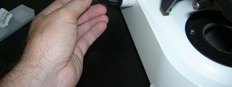
- Views:
Transcription
1 Contents General Microscope Set-Up Adjust Illumination Focus Condenser Open Software Image Capture Settings Shading & Color Corrections Image Capture & Viewing Scaling and Measurements Synopsis of Other Functions Pages Simplified Instructions: Zeiss Brightfield Microscope S1000 This is an Overview Only intended to refresh specific items learned in the Hands-On Training Use only in conjunction with your own much more detailed notes taken during your training
2 1, Reserve Time on Scheduler 2, Remove Eye-Piece Covers
3 3, Power-Up Computer is Usually On (if not, turn on)


4 3, Power-Up Turn on Lamp (on Left of Stand)


5 3, Power-Up Adjust Ocular/Camera Light Path To 50:50 (middle position)


6 3, Power-Up Manually Move Desired Objective Into Position


7 3, Power-Up Move Condenser Magnifier INTO LIGHT PATH If Using 10x or Higher Objective


8 3, Power-Up Move Condenser Magnifier OUT OF LIGHT PATH If Using 5x or Lower Objective
9 4, Adjust Illumination Adjust Light Filters (recommend none, e.g. II, H) (i.e. anything where light is brightest)


10 4, Adjust Illumination Adjust Lamp Power (on Right of Stand) Recommend 3200K


11 4, Adjust Illumination Set to Frosted or Clear Glass (Frosted Recommended, which is as shown below)

12 4, Adjust Illumination Focus on your slide


13 Note: Altering Brightness Through Neutral Density Adjustment is Preferred Over Changing Lamp Power 4, Adjust Illumination Adjust Neutral Density Filters To Alter Brightness to Good Level While Viewing Sample via Oculars


14 RECOMMENDATION Each User Should Define A Standard Frosted Glass/ Neutral Density/Condenser Setting. Make note of that setting and use each time for consistent imaging. 4, Adjust Illumination Can also Adjust Illumination Level by Changing Light Path Condenser
15 5, Focus Condenser (must do for Every Objective) A, Focus Sample, use Eyepieces B, Close Aperture F (right of stand) to Create Ring On Image (see pic)


16 5, Focus Condenser (must do for Every Objective) C, Move Condenser Knob Until Margins of Ring are Sharp (see pic)
 D,")

17 5, Focus Condenser (must do for Every Objective) D, Center Ring in Middle of Field Using Adjusting Screws
18 IMPORTANT You have focused the illumination light, which is different for each objective. If you change the objective, you must re-focus the condenser for the new objective. 5, Focus Condenser (must do for Every Objective) E, Once Centered and Sharp, Expand Ring (with Aperture, p 15) to Slightly Bigger Than Viewing Field


19 6, Open Software Use Windows Log-In/Password provided to you at training Click on AxioVision Rel. 4.6 icon
20 6, OpenSoftware Workarea icon open the window on the left. Each laboratory may have modified the workarea. Note: right-click on upper area permits you to set toolbars available.

21 7, Image Capture A, Click Live to view sample on screen.
22 7, Image Capture B, Use Magnification drop-down to tell software which objective is used.
23 7, Image Capture C, Select speed of Live Image update to Slow D, Focus Image on screen
24 RECOMMENDATION Do not use any Display other than Linear. Under NO circumstances do you use G Best Fit and Min/Max Settings are more for fluorescence microscopy and are not recommended here. 7, Image Capture E, Make sure Linear Image Display is highlighted.
25 7, Image Capture F, Click on Properties to open the main capture settings menu. G, Make NO adjustments under Display tab. Using Linear (already set) and next pages adjustments ensures consistency.

26 7, Image Capture H, Under Adjust tab, Click Measure to have the computer adjust exposure time for you. You also can manually adjust by typing in the desired number of msecs to be exposed.

27 7, Image Capture I, Shading Correction Open General Tab Move Slide to a field of no sample and no debris Moving to slightly out of focus can minimize presence of any debris
 are now even across the")
28 7, Image Capture I, Shading Correction Click Shading Correction Note: the slight variations in darkness (see last slide) are now even across the image.
29 7, Image Capture J, Set Color Balance Return to partial sample in field Return to Adjust Tab Focus Sample Click Interactive (changes to picking )
 7, Image Capture J, Set Color Balance Move mouse to empty portion of field and click Note:")
30 Note, after adjustment display shows equal blue, green and red. (compare to prior page) 7, Image Capture J, Set Color Balance Move mouse to empty portion of field and click Note: background color now has similar blue, green and red components.
31 7, Image Capture K, Capture Image Move to desired image field. Click Snap to capture image
32 8, Save Image Under File

33 8, Save Image Save Images only to a mapped server or to a temporary storage device. If you need assistance with server mapping, contact your IT support. Under NO circumstance do you save to the microscope computer.
34 9, Viewing Image Image captured is larger than Display. Use sliders to display area of image you are interested in. Note: Despite display, whole image will be saved, not just the visible part.

35 9, Viewing Image Can use Maximize to fill image window (see next page)
36 RECOMMENDATION Follow all the above steps prior to image collection and change nothing post-capture. If you make changes here, this image will be displayed different from your others. 9, Viewing Image Can open Properties pop-up, which Allows changes similar to those Described under live properties (p. 24)
37 10, Add Scale Bar Under Annotation, select Scale Bar
38 10, Add Scale Bar Scale Bar Appears Click on Scale Bar to move it. Note: Important to select correct magnification at collection (slide 22)
39 10, Add Scale Bar If incorrect magnification selected: A, Open Scalings under Measure

40 10, Add Scale Bar If incorrect magnification selected: B, Highlight correct magnification C, Click Apply selection to image NEVER touch anything else here as you may incorrectly alter the scalings. RECOMMENDATION Ideally you will always set the Magnification during set-up. It is unlikely you will remember this settings weeks later. i.e. use for your record-keeping.
41 11, Measurements Under Measure, select type of measurement to be conducted
42 11, Measurements Create Region of Interest (ROI) by holding down left-click of mouse Right-click mouse to close the ROI after defining the circle. Create as many ROIs as you would like
43 11, Measurements After creating all ROIs, click on Create Data Table

44 11, Measurements Apply Data to Table


45 11, Measurements Go to File Save As to store data table.


46 11, Measurements Save your data to the mapped server or your external storage device. Recommend to save as a.csv file (can be opened in Excel)

47 11, Measurements Use Window to toggle between open images or tables, or Simply click the desired Tab
48 12, Synopsis of Other Functions in Software Most Important Tab: Help Tab This is your Go-To Tab for all of your further questions!
49 12, Synopsis of Other Functions in Software Highlight each Tab to view functions not discussed in this handout. 1. File Tab
50 12, Synopsis of Other Functions in Software Highlight each Tab to view functions not discussed in this handout. 2. Edit Tab
51 12, Synopsis of Other Functions in Software Highlight each Tab to view functions not discussed in this handout. 3. View Tab
52 12, Synopsis of Other Functions in Software Highlight each Tab to view functions not discussed in this handout. 4. Acquisition Tab
53 12, Synopsis of Other Functions in Software Highlight each Tab to view functions not discussed in this handout. 5. Functions Tab
54 12, Synopsis of Other Functions in Software Highlight each Tab to view functions not discussed in this handout. 6. Annotation Tab
55 12, Synopsis of Other Functions in Software Highlight each Tab to view functions not discussed in this handout. 7. Measure Tab
56 12, Synopsis of Other Functions in Software Highlight each Tab to view functions not discussed in this handout. 8. Tools Tab Options dialog can be helpful to the User
57 12, Synopsis of Other Functions in Software Highlight each Tab to view functions not discussed in this handout. 8. Tools Tab A, Options: General
58 12, Synopsis of Other Functions in Software Highlight each Tab to view functions not discussed in this handout. 8. Tools Tab B, Options: Layout
59 12, Synopsis of Other Functions in Software Highlight each Tab to view functions not discussed in this handout. 8. Tools Tab C, Options: Folder


60 12, Synopsis of Other Functions in Software Highlight each Tab to view functions not discussed in this handout. 8. Tools Tab D, Options: Storage
61 12, Synopsis of Other Functions in Software Highlight each Tab to view functions not discussed in this handout. 8. Tools Tab E, Options: Acquisition

62 12, Synopsis of Other Functions in Software Highlight each Tab to view functions not discussed in this handout. 8. Tools Tab F, Options: Naming
63 12, Synopsis of Other Functions in Software Highlight each Tab to view functions not discussed in this handout. 8. Tools Tab G, Options: Display

64 12, Synopsis of Other Functions in Software Highlight each Tab to view functions not discussed in this handout. 8. Tools Tab H, Options: ID
Simplified Instructions: Olympus Widefield Microscope S1230
 Contents General Microscope Operation Simple Image Capture Multi-Wavelength Capture Z-Series Timelapse Combining Capture Modes Synopsis of Other Functions Pages 2-23 24-40 41-47 48-56 57-59 60-68 69-83
AxioVision 4.5 Brightfield Image Capture Procedure
 AxioVision 4.5 Brightfield Image Capture Procedure 1. STARTING-UP PROCEDURE: Remove blue dust cover and place on shelf under microscope. Turn on the halogen lamp by pushing the switch at the back right
BX-61: Brightfield Instruction /Continue to scroll for Fluorescent Instuctions
 BX-61: Brightfield Instruction /Continue to scroll for Fluorescent Instuctions Starting up: Schematic of Olympus BX-61. 1. Turn on Olympus microscope power box (left of microscope) with toggle switch on
Contents STARTUP MICROSCOPE CONTROLS CAMERA CONTROLS SOFTWARE CONTROLS EXPOSURE AND CONTRAST MONOCHROME IMAGE HANDLING
 Operations Guide Contents STARTUP MICROSCOPE CONTROLS CAMERA CONTROLS SOFTWARE CONTROLS EXPOSURE AND CONTRAST MONOCHROME IMAGE HANDLING Nikon Eclipse 90i Operations Guide STARTUP Startup Powering Up Fluorescence
Zeiss AxioImager.Z2 Brightfield Protocol
 Zeiss AxioImager.Z2 Brightfield Protocol 1) System Startup Please note put sign-up policy. You must inform the facility at least 24 hours beforehand if you can t come; otherwise, you will receive a charge
Zeiss Axio Imager.A1 manual
 Zeiss Axio Imager.A1 manual Power-up protocol 1. Mercury lamp 2. Power strip on shelf 3. Computer The Mercury lamp should always be first-on and last-off. This prevents any electrical surges caused by
Zeiss Deconvolution Microscope: A Quick Guide
 Zeiss Deconvolution Microscope: A Quick Guide Start-up Uncover microscope. Do not put dust cover on the floor. Plug in both cameras. The default camera is the AxioCam HRm (monochrome camera) for fluorescence
CAPTURING IMAGES ON THE HIGH-MAGNIFICATION MICROSCOPE
 University of Virginia ITC Academic Computing Health Sciences CAPTURING IMAGES ON THE HIGH-MAGNIFICATION MICROSCOPE Introduction The Olympus BH-2 microscope in ACHS s microscope lab has objectives from
Zeiss Axioskop II. The AIF's "routine" light microscope. (Installed 8/24/04)AxioCam installed July 11th 2005
AxioCam installed July 11th 2005") Zeiss Axioskop II The AIF's "routine" light microscope. (Installed 8/24/04)AxioCam installed July 11th 2005 Featuring: Phase Contrast Darkfield DIC/Nomarski Brightfield Fluorescent filters for Dapi, FITC,Rhodamine
Zeiss Axioskop II The AIF's "routine" light microscope. (Installed 8/24/04)AxioCam installed July 11th 2005 Featuring: Phase Contrast Darkfield DIC/Nomarski Brightfield Fluorescent filters for Dapi, FITC,Rhodamine
Zeiss Axiovert 135 Fluorescence Microscope Quick Guide / Operations Manual (v. 1.0 February 09)
") University of Chicago Integrated Light Microscopy Core Dr. Vytas Bindokas, Director http://digital.bsd.uchicago.edu By: Christine Labno, Assistant Director Room: AB-129 Phone: 4-9040 Zeiss Axiovert 135
University of Chicago Integrated Light Microscopy Core Dr. Vytas Bindokas, Director http://digital.bsd.uchicago.edu By: Christine Labno, Assistant Director Room: AB-129 Phone: 4-9040 Zeiss Axiovert 135
Brightfield Microscopy and Image Acquisition on Spotcam1. by Ryan Taylor/Nancy Kleene Last modified 10/02/05 by Birgit Ehmer
 Brightfield Microscopy and Image Acquisition on Spotcam1 by Ryan Taylor/Nancy Kleene Last modified 10/02/05 by Birgit Ehmer Log onto the computer. Enter your username and password to log onto the server.
Simplified Instructions: IXMicro High Throughput Microscope S1225
 Simplified Instructions: IXMicro High Throughput Microscope S1225 This is an Overview Only intended to refresh specific items learned in the Hands-On Training Use only in conjunction with your own much
Easy Kohler Illumination Method
 Easy Kohler Illumination Method ACADEMIC SKILLS CENTRE (ASC) A. Silverberg Completion of a Kohler illumination method is required before a microscope can be used efficiently. The Kohler method is designed
SOP: Polyvar Met Light Microscope
 SOP Polyvar Met Light Microscope Page 1 of 8 SOP: Polyvar Met Light Microscope 1. Scope 1.1 This document describes the standard operating procedure (SOP) for the Polyvar Met Light Microscope. 2. Table
Motorized Axio Observer Start-up instructions
 Start-up instructions 1. If using fluorescence turn on Fluorescent light source. TL light Source (Hal 100) 2. Turn on microscope using switch on lower left side of the microscope. 3. If imaging, turn on
Biology 29 Cell Structure and Function Spring, 2009 Springer LABORATORY 1: THE LIGHT MICROSCOPE
 Biology 29 Cell Structure and Function Spring, 2009 Springer LABORATORY 1: THE LIGHT MICROSCOPE Prior to lab: 1) Read these instructions (p 1-6) 2) Go through the online tutorial, the microscopy pre-lab
Characterization Microscope Nikon LV150
 Characterization Microscope Nikon LV150 Figure 1: Microscope Nikon LV150 Introduction This upright optical microscope is designed for investigating up to 150 mm (6 inch) semiconductor wafers but can also
EPIFLUORESCENCE &/OR BRIGHTFIELD MICROSCOPY
 EPIFLUORESCENCE &/OR BRIGHTFIELD MICROSCOPY TURN ON THE FOLLOWING EQUIPMENT The fluorescent light (if needed) The power strip for the microscope and accessories The CoolSNAP HQ camera on the right (Turn
Using the Nikon TE2000 Inverted Microscope
 Wellcome Trust Centre for Human Genetics Molecular Cytogenetics and Microscopy Core Using the Nikon TE2000 Inverted Microscope Fluorescence image acquisition using Scanalytic s IPLab software and the B&W
Standard Operating Procedure
 CENTER FOR NANOSCALE SCIENCE AND ENGINEERING Standard Operating Procedure Microscope Software Brian Wajdyk Page 1 of 6 Important Images are not to be saved to the computer. They will be deleted without
DIC Imaging using Laser Scanning Microscopes (LSM) on Inverted Stands
 on Inverted Stands") DIC Imaging using Laser Scanning Microscopes (LSM) on Inverted Stands Differential Interference Contrast (DIC) imaging is a technique used to increase contrast in brightfield images. In confocal systems,
DIC Imaging using Laser Scanning Microscopes (LSM) on Inverted Stands Differential Interference Contrast (DIC) imaging is a technique used to increase contrast in brightfield images. In confocal systems,
Leica DB LB Research microscope and Studo Lite Imaging software
 Leica DB LB Research microscope and Studo Lite Imaging software Room B523 User Guide Molecular Imaging Unit University of Helsinki www.miu.helsinki.fi 9.4.2008 1 GENERAL USER INFORMATION... 1 2 SETTINGS
Training Guide for Carl Zeiss AxioZoom V16 Stereo Microscope
 Training Guide for Carl Zeiss AxioZoom V16 Stereo Microscope ZEN 2012 Optical Imaging & Vital Microscopy Core Baylor College of Medicine (2017) Power ON Routine 1 2 If you require fluorescence imaging,
Nikon E800 Operating Instructions.
 Nikon E800 Operating Instructions. You can request electronic copies of this manual by contacting lshats@jhsph.edu Copies are also available on the JHU MMI Department web site. Please send your comments
INSTRUCTIONS FOR COURSE WORK 4 (AxioVert) Instructor: Anne Vaahtokari (MIU) 1. Purpose of the work
 Instructor: Anne Vaahtokari (MIU) 1. Purpose of the work") INSTRUCTIONS FOR COURSE WORK 4 (AxioVert) Instructor: Anne Vaahtokari (MIU) 1. Purpose of the work In this work, you will get familiar with an inverted epifluorescence microscope. Also, you will learn
INSTRUCTIONS FOR COURSE WORK 4 (AxioVert) Instructor: Anne Vaahtokari (MIU) 1. Purpose of the work In this work, you will get familiar with an inverted epifluorescence microscope. Also, you will learn
User Manual. Copyright 2010 Lumos. All rights reserved
 User Manual The contents of this document may not be copied nor duplicated in any form, in whole or in part, without prior written consent from Lumos. Lumos makes no warranties as to the accuracy of the
DIC Imaging using Laser Scanning Microscopes (LSMs) on Axio Imager Stands
 on Axio Imager Stands") DIC Imaging using Laser Scanning Microscopes (LSMs) on Axio Imager Stands Differential Interference Contrast (DIC) imaging is a technique used to increase contrast in brightfield images. In confocal systems,
DIC Imaging using Laser Scanning Microscopes (LSMs) on Axio Imager Stands Differential Interference Contrast (DIC) imaging is a technique used to increase contrast in brightfield images. In confocal systems,
Zeiss LSM 880 Protocol
 Zeiss LSM 880 Protocol 1) System Startup Please note put sign-up policy. You must inform the facility at least 24 hours beforehand if you can t come; otherwise, you will receive a charge for unused time.
Zeiss AxioObserver with ApoTome
 Zeiss AxioObserver with ApoTome Quick Start User Guide LSU Health Sciences Center-Shreveport Research Core Facility (RCF) Microscopy Table of Contents 1 Start up the system.. Page 3 2 Touch screen controller
Nikon E800 Microscope. Operating Instructions
 Nikon E800 Microscope Operating Instructions B Watson 12/2005 Table of contents: 1. The Nikon E800 Microscope 2. Turning the system ON and OFF 3. Selecting the light path 4. Operating in transmitted light
PIXERA InStudio Software Package
 PIXERA InStudio Software Package Background InStudio is a comprehensive imaging capture and manipulation software program from Pixera. It combines the features of the original ViewfinderPro and StudioPro
User Guide for TWAIN / DirectX interface for GRYPHAX USB 3.0 cameras
 User Guide for TWAIN / DirectX interface for GRYPHAX USB 3.0 cameras The TWAIN & DirectX driver for PROGRES GRYPHAX USB 3.0 cameras enables user to operate with TWAIN and DirectX supported 3 rd party software
QUICKSTART GUIDE: WIDEFIELD WF3 Zeiss Cell Observer Live Cell Imaging System (SAF, ROOM 409) Imperial College London
 Imperial College London") Imperial College London Facility for Imaging by Light Microscopy QUICKSTART GUIDE: WIDEFIELD WF3 Zeiss Cell Observer Live Cell Imaging System (SAF, ROOM 409) Observing Life As It Happens Startup procedure...
Imperial College London Facility for Imaging by Light Microscopy QUICKSTART GUIDE: WIDEFIELD WF3 Zeiss Cell Observer Live Cell Imaging System (SAF, ROOM 409) Observing Life As It Happens Startup procedure...
Zeiss Axioplan 2 imaging microscope and Axiovision software
 Zeiss Axioplan 2 imaging microscope and Axiovision software Microscopes 1 and 2 in room B501b User Guide Molecular Imaging Unit University of Helsinki www.miu.helsinki.fi 20.5.2010 1 GENERAL... 1 1.1...
MIF ZEISS VIOLET CONFOCAL ZEN 2009 PROTOCOL
 MIF ZEISS VIOLET CONFOCAL ZEN 2009 PROTOCOL START-UP On the Switchbox, turn both black switches to the ON position. Wait for the microscope to boot up completely (watch the screen on the side of the microscope).
Olympus IX71 Microscope and DP71 Camera Instructions
 Olympus IX71 Microscope and DP71 Camera Instructions Microscopy in Medicine (MiM) Core Emory University Department of Medicine 1 Olympus IX71 Image Capture Procedure 2 3 1. STARTING-UP PROCEDURE: Remove
AxioVision User's Guide. Release 4.1
 AxioVision User's Guide Release 4.1 Number of this manual: B 48-0038 e 10.2003 Date of issue: 10.2003 Carl Zeiss Vision draws the User's attention to the fact that the information and references contained
Nikon E800 Operating Instructions.
 Nikon E800 Operating Instructions. You can request electronic copies of this manual by contacting imaging@fhcrc.org. Copies are also available on the Scientific Imaging web site. Please send your comments
Leica DMi8A Quick Guide
 Leica DMi8A Quick Guide 1 Optical Microscope Quick Start Guide The following instructions are provided as a Quick Start Guide for powering up, running measurements, and shutting down Leica s DMi8A Inverted
Leica SP8 TCS Users Manual
 Version : 07/08/0 Leica SP8 TCS Users Manual Start up:. Turn the PC Microscope, Scanner Power, Laser Power, and the Laser Emission key to on (bottom right of desk).. Turn on the fluorescent lamp (top left
ZEISS LSM 710 NLO Multiphoton microscope Manual/Quick guide
 ZEISS LSM 710 NLO Multiphoton microscope Manual/Quick guide Matyas Molnar, Biovis 2016 Starting the microscpe 1. Check the microscope if everything looks clean and normal. If not, report it in the logbook.
Using the Microscope for a NANSLO Remote Web-based Science Lab Activity
 Using the Microscope for a NANSLO Remote Web-based Science Lab Activity MICROSCOPE RWSL LAB INTERFACE INSTRUCTIONS The Remote Web-based Science Lab (RWSL) microscope is a high quality digital microscope
Using the AmScope Microscope Cameras
 Using the AmScope Microscope Cameras Part 1 Setup. In order to use the camera, you will need: a) the camera system; b) a computer running the camera software. The camera system is contained in a Pelican
QUICKSTART GUIDE: WIDEFIELD HWF1 Zeiss Cell Observer Live Cell Imaging System (HAMMERSMITH, L BLOCK, ROOM 314) Imperial College London
 Imperial College London") Imperial College London Facility for Imaging by Light Microscopy QUICKSTART GUIDE: WIDEFIELD HWF1 Zeiss Cell Observer Live Cell Imaging System (HAMMERSMITH, L BLOCK, ROOM 314) Observing Life As It Happens
Imperial College London Facility for Imaging by Light Microscopy QUICKSTART GUIDE: WIDEFIELD HWF1 Zeiss Cell Observer Live Cell Imaging System (HAMMERSMITH, L BLOCK, ROOM 314) Observing Life As It Happens
Guide to Confocal 5. Starting session
 Guide to Confocal 5 Remember that when booking and before starting session you can check for any problems at https://www.bris.ac.uk/biochemistry/uobonly/cif/index.html Starting session Switch on microscope
Instruction Manual T Binocular Acromat Research Scope T Trinocular Acromat Research Scope
 Research Scope Instruction Manual T-29031 Binocular Acromat Research Scope T-29041 Trinocular Acromat Research Scope T-29032 Binocular Semi-Plan Research Scope T-29042 Trinocular Semi-Plan Research Scope
Horiba LabRAM ARAMIS Raman Spectrometer Revision /28/2016 Page 1 of 11. Horiba Jobin-Yvon LabRAM Aramis - Raman Spectrometer
 Page 1 of 11 Horiba Jobin-Yvon LabRAM Aramis - Raman Spectrometer The Aramis Raman system is a software selectable multi-wavelength Raman system with mapping capabilities with a 400mm monochromator and
Leica SP8 TCS Users Manual
 Leica SP8 TCS Users Manual Follow the procedure for start up and log on as posted in the lab. Please log on with your account only and do not share your password with anyone. We track and confirm usage
MIF ZEISS LSM510 CONFOCAL USER PROTOCOL
 MIF ZEISS LSM510 CONFOCAL USER PROTOCOL START-UP Turn on the Mercury Bulb Power Supply (if needed). Power-on the Control Box. Turn on the computer. Open the LSM 510 software. Choose Scan New Images and
Using a Compound Light Microscope
 Name Class Date Laboratory Skills 5 Using a Compound Light Microscope Introduction Many objects are too small to be seen by the eye alone. They can be seen, however, with the use of an instrument that
USING LEICA AS LASER MICRODISSECTION (LMD6000) MICROSCOPE Written By Jungim Hur
 MICROSCOPE Written By Jungim Hur") USING LEICA AS LASER MICRODISSECTION (LMD6000) MICROSCOPE Written By Jungim Hur Digital Video Camera Eyepieces Laser module Laser safety UV shield Specimen holder Smart move control LEICA CTR6500 electronics
USING LEICA AS LASER MICRODISSECTION (LMD6000) MICROSCOPE Written By Jungim Hur Digital Video Camera Eyepieces Laser module Laser safety UV shield Specimen holder Smart move control LEICA CTR6500 electronics
Olympus xcellence Software - basic user guide
 Olympus xcellence Software - basic user guide This is a basic overview of setting up time lapse experiments using Olympus's xcellence software on BIU's IX81 inverted phase contrast system - the software
1.3. Before loading the holder into the TEM, make sure the X tilt is set to zero and the goniometer locked in place (this will make loading easier).
.") JEOL 200CX operating procedure Nicholas G. Rudawski ngr@ufl.edu (805) 252-4916 1. Specimen loading 1.1. Unlock the TUMI system. 1.2. Load specimen(s) into the holder. If using the double tilt holder, ensure
JEOL 200CX operating procedure Nicholas G. Rudawski ngr@ufl.edu (805) 252-4916 1. Specimen loading 1.1. Unlock the TUMI system. 1.2. Load specimen(s) into the holder. If using the double tilt holder, ensure
Quick Guide for Zeiss 710 Laser Scanning Confocal MGH Cancer Center
 Quick Guide for Zeiss 710 Laser Scanning Confocal MGH Cancer Center For any questions or concerns, please contact: Linda Nieman lnieman@mgh.harvard.edu Office: (617) 643-9684 Cell: (512) 565-8076 Chenyue
Nikon C1si Spectral Laser Scanning Confocal Microscope. User Guide
 Nikon C1si Spectral Laser Scanning Confocal Microscope User Guide Contents: C1Si Turn-On/ShutDown Procedures... 2 Overview... 4 Setup for epi-illumination to view through the eyepieces:... 5 Setup for
Microscopy. Matti Hotokka Department of Physical Chemistry Åbo Akademi University
 Microscopy Matti Hotokka Department of Physical Chemistry Åbo Akademi University What s coming Anatomy of a microscope Modes of illumination Practicalities Special applications Basic microscope Ocular
Nikon Eclipse Ti A1-A Confocal Operating Manual. Start-up. Microscope
 Nikon Eclipse Ti A1-A Confocal Operating Manual Start-up 1. Turn on Excite Fluorescent light power supply- metal halide. a. Cool down as for mercury bulb b. Wheel closed liquid light guide 2. Turn on power
ZEISS LSM 710 CONFOCAL MICROSCOPE USER MANUAL
 ZEISS LSM 710 CONFOCAL MICROSCOPE USER MANUAL START THE SYSTEM... 2 START ZEN SOFTWARE... 3 SET THE TEMPERATURE AND THE CO2 CONTROLLERS... OBSERVATION AT OCULARS... 5 STATIF PRESENTATION... 6 ACQUIRE ONE
used for low power magnification of a sample image is 3 dimensional
 MICROSCOPES One of the most important inventions in the advancement of Biology 1. Simple Microscopes ie. magnifying glass, stereoscope (dissecting scope) have a single lens or a pair of lenses combined
Instructions for Making On-Line Reservations for Microscopes in NB11-204
 Instructions for Making On-Line Reservations for Microscopes in NB11-204 1. Log into Mail using Mail.swmed.edu 2. Log in using your university id and password. 3. Click the Calendar Tab at the top right
Operating Instructions for Zeiss LSM 510
 Operating Instructions for Zeiss LSM 510 Location: GNL 6.312q (BSL3) Questions? Contact: Maxim Ivannikov, maivanni@utmb.edu 1 Attend A Complementary Training Before Using The Microscope All future users
LSM 510 Training Notes
 LSM 510 Training Notes Turning on the system Turn on the arc lamp, found on the bench top left of the microscope. This supplies light for epifluorescence for viewing your samples through the microscope.
Nikon Eclipse Ti2-E Widefield/Spinning Disk Confocal Microscope Standard Operation Protocol
 Nikon Eclipse Ti-E Widefield/Spinning Disk Confocal Microscope Standard Operation Protocol Please sign on the log sheet before switching on system. Turn on system Turn on A only if confocal mode or laser
Basic Microscopy. OBJECTIVES After completing this exercise, you should be able to do the following:
 Page 1 of 10 Basic Microscopy OBJECTIVES After completing this exercise, you should be able to do the following: a. Name the parts of the compound microscope and the functions of each. b. Describe how
User Operation of JEOL 1200 EX II
 **Log onto Computer** Open item program Start Up Procedure User Operation of JEOL 1200 EX II 1. If scope is not running, locate an electron microscopy technician (EMT) to find out why not. 2. Turn up brightness
Olympus Digital Microscope Camera (DP70) checklist
 checklist") Smith College - July 2005 Olympus Digital Microscope Camera (DP70) checklist CONTENT, page no. Camera Information, 1 Startup, 1 Retrieve an Image, 2 Microscope Setup, 2 Capture, 3 Preview. 3 Color Balans,
Smith College - July 2005 Olympus Digital Microscope Camera (DP70) checklist CONTENT, page no. Camera Information, 1 Startup, 1 Retrieve an Image, 2 Microscope Setup, 2 Capture, 3 Preview. 3 Color Balans,
Training Guide for Carl Zeiss LSM 880 with AiryScan FAST
 Training Guide for Carl Zeiss LSM 880 with AiryScan FAST ZEN 2.3 Optical Imaging & Vital Microscopy Core Baylor College of Medicine (2018) Power ON Routine 1 2 Turn ON Main Switch from the remote control
Training Guide for Carl Zeiss LSM 7 MP Multiphoton Microscope
 Training Guide for Carl Zeiss LSM 7 MP Multiphoton Microscope ZEN 2009 Optical Imaging & Vital Microscopy Core Baylor College of Medicine (2017) Power ON Routine 1 2 Turn Chameleon TiS laser key from Standby
The invention of the microscope made it possible for scientists to view and study cells. Cells the basic units of all living organisms.
 The Discovery of Cells The invention of the microscope made it possible for scientists to view and study cells. Cells the basic units of all living organisms. The Cell Theory All living things are made
Operating Checklist for using the Laser Scanning Confocal Microscope. Leica TCS SP5.
 Smith College August 2010 Operating Checklist for using the Laser Scanning Confocal Microscope Leica TCS SP5. CONTENT, page no. Startup, 1 Initial set-up, 1 Software, 2 Microscope Specimen observation
The ideal K-12 science microscope solution. User Guide. for use with the Nova5000
 The ideal K-12 science microscope solution User Guide for use with the Nova5000 NovaScope User Guide Information in this document is subject to change without notice. 2009 Fourier Systems Ltd. All rights
Things to check before start-up.
 Byeong Cha Page 1 11/24/2009 Manual for Leica SP2 Confocal Microscope Enter you name, the date, the time, and the account number in the user log book. Things to check before start-up. Make sure that your
Therefore, all descriptions and illustrations in this instruction manual, including all specifications are subject to change without notice.
 We are constantly endeavouring to improve our instruments and to adapt them to the requirements of modern research techniques and testing methods. This involves modification to the mechanical structure
SHORT INSTRUCTIONS FOR OPERATING LSM1/2 (Zeiss LSM510) AT CIAN Version 1.4, September 2014
 AT CIAN Version 1.4, September 2014") CIAN LSM1 or LSM2 short instructions, version 1.4, September 2014 page 1 of 6 SHORT INSTRUCTIONS FOR OPERATING LSM1/2 (Zeiss LSM510) AT CIAN Version 1.4, September 2014 Before starting To work with LSM1
CIAN LSM1 or LSM2 short instructions, version 1.4, September 2014 page 1 of 6 SHORT INSTRUCTIONS FOR OPERATING LSM1/2 (Zeiss LSM510) AT CIAN Version 1.4, September 2014 Before starting To work with LSM1
A quick overview of the basics of my workflow in. Those gaps in Photoshop s Histogram indicate missing information.
 Another Photoshop tutorial by Bruce Philpott Copyright 2007 Bruce Philpott A quick overview of the basics of my workflow in Adobe Camera Raw This short tutorial certainly won t cover everything about Adobe
Zeiss LSM 780 Protocol
 Zeiss LSM 780 Protocol 1) System Startup F Please note the sign-up policy. You must inform the facility at least 24 hours beforehand if you can t come; otherwise, you will receive a charge for unused time.
Ch. 1 - Installation Guidelines
 Ch. 1 - Installation Guidelines Table of Contents Ch. 1 - Installation Guidelines Introduction... 8 The Image-Pro Driver Interface... 8 Installing the SPOT Image-Pro Driver... 8 Image-Pro Driver Supplement
Operating Checklist for using the Scanning Electron. Microscope, JEOL JSM 6400.
 Smith College August 2009 Operating Checklist for using the Scanning Electron Microscope, JEOL JSM 6400. CONTENT, page no. Pre-Check 1 Startup 1 Specimen Insertion 2 Filament Saturation 2 Beam Alignment
1 Placing particles on the slide
 Aerosols Transport Particle Removal Experiment E.S. Kenney, J.A. Taylor, and G. Ahmadi 1 Placing particles on the slide 1.1 Swing the light beneath the test section down and to the left. Figure 1: Light
Everest System / Slidebook Operating Procedures
 Everest System / Slidebook Operating Procedures NOTICE: This guide is meant to supplement training, not replace it. All users must be trained first hand by a core employee. Training of others in your lab
Figure 3.4 Approximate size of various types of cells. ~10 um. Red Blood Cells = mm 1500 um. Width of penny Pearson Education, Inc.
 Figure 3.4 Approximate size of various types of cells. ~10 um Red Blood Cells 1.5mm 1500 um Width of penny = 1500 Figure 4.3 The limits of resolution (and some representative objects within those ranges)
Takeoff Guide Tutorial to get started with the AxioVision Imaging System (Based on Release 4.8 June 2009)
") Takeoff Guide Tutorial to get started with the AxioVision Imaging System (Based on Release 4.8 June 2009) This Takeoff Guide has been prepared for Carl Zeiss by Imaging Associates Ltd. www.imas.co.uk Copyright
Takeoff Guide Tutorial to get started with the AxioVision Imaging System (Based on Release 4.8 June 2009) This Takeoff Guide has been prepared for Carl Zeiss by Imaging Associates Ltd. www.imas.co.uk Copyright
Microscopy from Carl Zeiss
 Microscopy from Carl Zeiss Contents Page Contents... 1 Introduction... 1 Starting the System... 2 Introduction to ZEN Efficient Navigation... 5 Setting up the microscope... 10 Configuring the beam path
Center for Microscopy and Image Analysis Axio Scan.Z1 Operating Manual
 No index entries found. Center for Microscopy and Image Analysis Axio Scan.Z1 Operating Manual Table of contents 1. Starting procedure... 3 1.1. Turn on hardware... 3 1.2. Starting ZEN blue... 4 2. Load
Ocular Lenses. Head. Arm. Objective Lenses. Slide Holder Stage. On / Off Switch. Condenser with Iris Diaphragm. Light Intensity Control
 BIOLOGY 211: HUMAN ANATOMY & PHYSIOLOGY ********************************************************************************************************* USE OF THE LIGHT MICROSCOPE **********************************************************************************************************
Indian Institute of technology Madras Presents NPTEL NATIONAL PROGRAMME ON TECHNOLOGY ENHANCED LEARNING
 Indian Institute of technology Madras Presents NPTEL NATIONAL PROGRAMME ON TECHNOLOGY ENHANCED LEARNING Lecture - 5 Materials Characterization Fundamentals of Optical microscopy Dr. S. Sankaran Associate
LSM 780 Confocal Microscope Standard Operation Protocol
 LSM 780 Confocal Microscope Standard Operation Protocol Basic Operation Turning on the system 1. Sign on log sheet according to Actual start time 2. Check Compressed Air supply for the air table 3. Switch
Jeol JEM Responsible personell: Endy ( ) Online booking is compulsory!
 Online booking is compulsory!") Jeol JEM 1230 Responsible personell: Endy (45279377) Online booking is compulsory! After training you will have access to working alone on the instrument. All insertion of samples is done by responsible
Jeol JEM 1230 Responsible personell: Endy (45279377) Online booking is compulsory! After training you will have access to working alone on the instrument. All insertion of samples is done by responsible
User instructions Compound laboratory microscope
 KERN & Sohn GmbH Ziegelei 1 D-72336 Balingen E-mail: info@kern-sohn.com User instructions Compound laboratory microscope Tel: +49-[0]7433-9933-0 Fax: +49-[0]7433-9933-149 Internet: www.kern-sohn.com KERN
Basic Microscopy for Plant Biology
 Page 1 of 8 Basic Microscopy for Plant Biology OBJECTIVES After completing this exercise, you should be able to do the following: a. Name the parts of the compound microscope and the functions of each.
Quick Guide for Zeiss 710 Laser Scanning Confocal MGH Cancer Center
 Quick Guide for Zeiss 710 Laser Scanning Confocal MGH Cancer Center For any questions or concerns, please contact: Linda Nieman lnieman@mgh.harvard.edu Office: (617) 643-9684 Cell: (512) 565-8076 Chenyue
Nikon TE300 Eclipse Wide-Field Microscope
 Nikon TE300 Eclipse Wide-Field Microscope User Guide LSU Health Science Center-Shreveport Research Core Facility 1 User manual for Nikon Elements software Equipment: Nikon TE300 Eclipse microscope Photometrics
 Microscopy http://www.microscopyu.com/articles/phasecontrast/phasemicroscopy.html http://micro.magnet.fsu.edu/primer/anatomy/anatomy.html 2005, Dr. Jack Ikeda & Dr. Gail Grabner 9 Nikon Labophot (Question
Training Guide for Carl Zeiss LSM 5 LIVE Confocal Microscope
 Training Guide for Carl Zeiss LSM 5 LIVE Confocal Microscope AIM 4.2 Optical Imaging & Vital Microscopy Core Baylor College of Medicine (2017) Power ON Routine 1 2 Verify that main power switches on the
Operating Procedures for MICROCT1 Nikon XTH 225 ST
 Operating Procedures for MICROCT1 Nikon XTH 225 ST Ensuring System is Ready (go through to ensure all windows and tasks below have been completed either by you or someone else prior to mounting and scanning
Using a Compound Light Microscope Lab Pre-Lab Assignment
 Name: Block: Due Date: Using a Compound Light Microscope Lab Pre-Lab Assignment Pre-Lab Assignment This assignment must be completed by the next class period in order to be allowed to participate in the
LSM 710 Confocal Microscope Standard Operation Protocol
 LSM 710 Confocal Microscope Standard Operation Protocol Basic Operation Turning on the system 1. Switch on Main power switch 2. Switch on System / PC power button 3. Switch on Components power button 4.
Understanding and Using Dynamic Range. Eagle River Camera Club October 2, 2014
 Understanding and Using Dynamic Range Eagle River Camera Club October 2, 2014 Dynamic Range Simplified Definition The number of exposure stops between the lightest usable white and the darkest useable
UNIVERSITY OF WATERLOO Physics 360/460 Experiment #2 ATOMIC FORCE MICROSCOPY
 UNIVERSITY OF WATERLOO Physics 360/460 Experiment #2 ATOMIC FORCE MICROSCOPY References: http://virlab.virginia.edu/vl/home.htm (University of Virginia virtual lab. Click on the AFM link) An atomic force
Lab: Using a Compound Light Microscope
 Name Date Period Lab: Using a Compound Light Microscope Background: Microscopes are very important tools in biology. The term microscope can be translated as to view the tiny, because microscopes are used