Window. Matthew. blood. smear is the. circle on the
|
|
|
- Octavia Walters
- 5 years ago
- Views:
Transcription


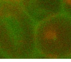


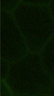
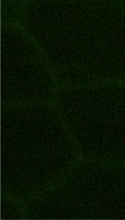
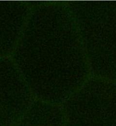
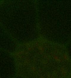
1 Electronic Supplementary Material (ESI) for Lab on a Chip. This journal is The Royal Society of Chemistry 2014 Supplementary Informatio on - A Paper Microfluidic Cartridge for Automated Staining of Malaria Parasites with an Optically Transparent Microscopy Window Matthew P. Horning, Charles Delahunt, Ryan Singh, Spencer H.. Garing, Kevin P. Nichols* Typical thick and thin smear, as manually prepared The cartridge is inted to duplicate the utility provided by manually prepared thick and thin smears. An example image of such a manually prepared smear on a microscope slide is shown. blood Figure S1 A typical thick and thin blood smear as prepared on a microscope slide by an expert microscopist. The thick smear is the circle on the left, and the thin smear iss the gradient on the right. Optimization of stain concentration Concentration of stain greatly affects the final image quality. Example non-optimal images (above 0.06 X) are shown. Figure S2 Stain concentration is varied to reduce background signal to a level that permits clear visualization of both RNA and DNA within parasites, while still leaving the outlines of the RBC intact to permit cell counting. Stain concentration is reported as a fraction of the saturated solution described in the methods section.


")


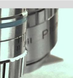


2 Cutting pattern for paper The SI accessible SVG file cutting_guide.svg, can be usedd directly to duplicate the paper pattern cut using an ecraft Electronic Die Cutter (Craftwell, USA). The width of the file (the length of the long, horizontal cutting line) should be verified to be cm. In the ecraftt die cutter, paper towels were placed between two sheets of card stock, and cut with a depthh of 7 (arbitrary units). Microscope slide adapter. An adapter was useful to prevent the clips utilized to compress the slide from nterfering with the microscope stage, and to allow the stage clamps to grip the cartridge. Figure S2 shows the adapter, as 3D printed and utilized. An STL file is included with SI to aid in duplication of this piece via external 3D printing foundries. Figure S3 The 3D adapter. printed slide adapter, as utilized. An STL file is available in SI to duplicate the
3 MATLAB code for detection of malaria parasites in microscopy images after staining in cartridge To duplicate the image processing algorithm utilized in this report, a new.m filed titled countparasitesandrbcsinaoimages.m should be created, and the following code copied and pasted into it. There may be slight adjustments required if line breaks do not match up correctly after pasting. This script was tested on MATLAB 2013a with the image processing toolbox. Two images are also included in SI, one of which is an example image that can be directly extracted from the PDF and utilized as a test image for future code development, the other of which is the output of the script below on that image. Run the code by calling the function in MATLAB as: countparasitesandrbcsinaoimages( filename.jpg,1) Additionally, a more detailed technical description of the algorithm than that provided in the methods section is provided: The image-processing algorithm has two goals: To detect parasites, and to count red blood cells (RBCs). Parasites: The targeted parasite signature is a bright orange blob (stained DNA) in close proximity to a bright green blob (stained RNA). Given channels R, G, and B, and pixels p, a bright orange binary image is created by masking the red channel R with: {p:r(p) > Rthresh && R(p) > G(p)} = 1, where Rthresh is an adaptive threshold Rthresh = median(r) + 2*stdDev(R). Similarly, a bright green binary image is created by masking the green channel G with: {p: G(p) > Gthresh && G(p) > R(p)} = 1, where Gthresh = median(g) + 2*stdDev(G). Both masked images are filtered to remove overly large blobs and single pixel noise. To find orange and green regions that are close together, the orange masked image is dilated. Dilation is an operation on binary images in which a disc is placed on each 1-valued pixel in turn, and all pixels within the disc are converted to 1 s. The effect is to increase the size of the 1-valued regions by adding a margin around them. Parasite locations are represented by the overlap of the two images: {dilated masked orange image = 1} && {masked green image = 1}. Red blood cells: The cell walls of RBCs stain pale green, while their interiors are typically dark. The cell walls are captured in a binary green image by first histogram-equalizing the green channel G, then masking G with: {p: G(p) > localgthresh} = 1, where localgthresh = 1.1*median(G) is calculated in smaller squares that partition the image. The purpose of the partition is to minimize the effect of large green artifacts. Median filters eliminate scattered pixel noise. The image is then reversed, so that cell walls are black and cell interiors are white. The cell interiors are separated by eroding the image. Erosion is an operation on binary images whereby a disc is placed on each {0} pixel in turn; all pixels within the disc are converted to 0 s. The effect is to decrease the size of the 1-valued regions by removing their border areas; in this case cell interiors connected by noise are effectively separated. The cell interiors are then solidified (so that each cell interior is a single connected region) by dilation using a smaller disc. The number of distinct connected regions is taken to be the number of RBCs. % This function counts the number of parasites and number of RBCs in an % Acridine Orange image. It assumes that the cell walls are brighter green % than the cell interiors. It does not distinguish between empty space and % cell interior. % inputs:
4 % filename = image filename (including path if needed) % showimagesboolean: if True, 2 images will be shown: the original, % and a greyscale with the estimated locations of % parasites and RBCs. Yellow circles are parasites. % A double circle implies 2 parasites close % together. % Red crosses are RBCs. % outputs: % number of parasites, and number of RBCs. % % This function requires the image processing toolbox, and was tested % with MATLAB 2013a % % Written by Charles Delahunt, 26 August 2013 function [numparasites, numrbcs] = countparasitesandrbcsinaoimages(filename, showimagesboolean) % load image: [~,~,imtype] = fileparts(filename); imtype = imtype(2:); im = imread(filename,imtype); % parasites: % method: get greenish-yellow blobs and orange blobs, then see if any are % the right size and close to each other % green-yellow = 150,250,0. (image is basically 2 color) % orange = 240,170,0 % basic processing: imr = im(:,:,1); imr = double(imr); imr = imr/max(imr(:)); img = im(:,:,2); img = double(img); img = img/ max(img(:)); thrparam = 0.3; % used to set thresholds for R and G channel images % isolate orange: % threshold image: thr = median (imr(:)) + thrparam*(max(imr(:)) - median(imr(:))); imthr = imr; imthr(imthr < thr imr < img) = 0; % ie require that red > green to keep % isolate green-yellow: thg = median (img(:)) + thrparam*(max(img(:)) - median(img(:))); imthg = img; imthg(imthg < thg img < imr) = 0; % ie require that green > red to keep % We want to find green pixels (ie from imthg) that are close to orange % pixels (ie from imthr). Do this by taking a binary image of imthr, then % dilating it with a large disc to give an image of pixels in the % neighborhood of orange pixels. temp = ones(size(imthr));
5 temp (imthr == 0) = 0; temp = imerode(temp, strel('disk',2)); % kill single pixels sec = strel('disk',25); imdilater = imdilate(temp, sec); % get the overlap of the green channel and the dilated orange channel: imgoodg = imthg; imgoodg(imthg == 0 imdilater == 0) = 0; % threshold the results in order to separate candidates: imgoodthresh = 0.45; imgoodg(imgoodg < imgoodthresh) = 0; % use a binary image to count blobs. Reject small blobs imgoodbin = zeros(size(imgoodg)); imgoodbin(imgoodg > 0) = 1; cc = bwconncomp(imgoodbin); parastats = regionprops(cc,'area', 'Centroid'); parasitethresh = 60; % reject blobs below this size. 60 to 80 works. paraareas = [parastats.area]; numparasites = sum(paraareas > parasitethresh); parasitesizethresh = 800; numdoubles = sum(paraareas > parasitesizethresh); % ie this blob is too big to be just one parasite. % detect pairs of blobs corresponding to just one parasite: % is distance between centroids is less than 25: parakeep = parastats(paraareas > parasitethresh); count = 0; for i = 1:size(paraKeep,1), temp = parakeep(i).centroid; for j = 1:size(paraKeep,1), temp2 = parakeep(j).centroid; if norm(temp - temp2) > 0 && norm(temp - temp2) < 25, count = count + 1; numduplicates = count/2; % divide by two since each neighboring pair is counted twice. numparasites = numparasites + numdoubles - numduplicates; % this is output % % RBCs: % detect cell walls: % remove bright areas: img1 = img; thg2 = median (img(:)) + 0.6*(max(imG(:)) - median(img(:))); img1(img > thg*0.9) = median(img(:)); img2 = adapthisteq(img1);
6 % do local median thresholding on a partition into squares of size 2*'border': border = 20; wallim = zeros(size(img2)); for i = [1+border:2*border:size(imG,1)-border, size(wallim,1)-border] for j = [1+border:2*border:size(imG,2)-border, size(wallim,2)-border] temp = img2(i-border:i+border,j-border:j+border); temp(temp < median(temp(:))*1.1) = 0; wallim(i-border:i+border,j-border:j+border) = temp; % do right hand edge: temp = img2(i-border:i+border,j-border:j+border); temp(temp < median(temp(:))*1.1) = 0; wallim(i-border:i+border,j-border:j+border) = temp; % do bottom edge: % do repeated median filters to clarify image: wallim2 = medfilt2(wallim); wallim2 = medfilt2(wallim2); wallim2 = medfilt2(wallim2); % open the image to separate cells wallim3 = zeros(size(wallim)); wallim3(wallim2 == 0) = 1; sed = strel('disk',5); wallim3 = imopen(wallim3, sed); see = strel('disk',2); wallim3 = imclose(wallim3,see); % count the number of blobs (RBCs): cc = bwconncomp(wallim3); rbcstats = regionprops(cc,'area','filledarea', 'FilledImage', 'Centroid'); rbcareas = [rbcstats.area]; rbckeep = rbcstats(rbcareas > 2000); % kill small spots numrbcs = size(rbckeep,1); % this is an output % % plot results % plot estimated cell centers: if showimagesboolean, figure, hold on, imshow (im), title(filename) figure, imshow(img) % mark parasites: for i = 1:size(paraKeep), if parakeep(i).area > parasitethresh, figure(2), hold on, xy = parakeep(i).centroid; a = parakeep(i).area; viscircles([xy(1), xy(2)], 25, 'EdgeColor', 'y');
7 if a > parasitesizethresh, viscircles([xy(1), xy(2)], 35, 'EdgeColor', 'y');, % mark RBCs: for i = 1:size(rbcKeep,1), xy = rbckeep(i).centroid; x(i) = xy(1); y(i) = xy(2); hold on, plot(x,y,'r+') title(['estimated numparasites = ' num2str(numparasites) '. Estimated numrbcs = ' num2str(numrbcs)]) %


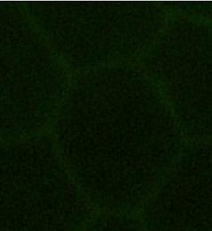


8 Example image of parasites: Figure S4 - This image can be extracted from the PDF file (either by taking a screen shot, or more directly using software such as Adobe Illustrator) and used to create a new.jpg file for testing the countparasitesandrbcsinaoimages script with.


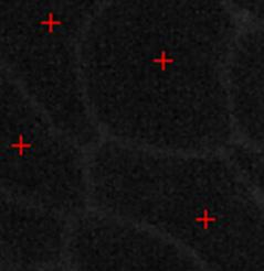


9 Output results from countparasitesandrbcsinaoimages script Figure S5 - The expected output from running the countparasitesandrbcsinaoimages script on the example image in Figure S2.
AUTOMATED MALARIA PARASITE DETECTION BASED ON IMAGE PROCESSING PROJECT REFERENCE NO.: 38S1511
 AUTOMATED MALARIA PARASITE DETECTION BASED ON IMAGE PROCESSING PROJECT REFERENCE NO.: 38S1511 COLLEGE : BANGALORE INSTITUTE OF TECHNOLOGY, BENGALURU BRANCH : COMPUTER SCIENCE AND ENGINEERING GUIDE : DR.
Traffic Sign Recognition Senior Project Final Report
 Traffic Sign Recognition Senior Project Final Report Jacob Carlson and Sean St. Onge Advisor: Dr. Thomas L. Stewart Bradley University May 12th, 2008 Abstract - Image processing has a wide range of real-world
Estimating malaria parasitaemia in images of thin smear of human blood
 CSIT (March 2014) 2(1):43 48 DOI 10.1007/s40012-014-0043-7 Estimating malaria parasitaemia in images of thin smear of human blood Somen Ghosh Ajay Ghosh Sudip Kundu Received: 3 April 2014 / Accepted: 4
8.2 IMAGE PROCESSING VERSUS IMAGE ANALYSIS Image processing: The collection of routines and
 8.1 INTRODUCTION In this chapter, we will study and discuss some fundamental techniques for image processing and image analysis, with a few examples of routines developed for certain purposes. 8.2 IMAGE
Adobe Studio on Adobe Photoshop CS2 Enhance scientific and medical images. 2 Hide the original layer.
 1 Adobe Studio on Adobe Photoshop CS2 Light, shadow and detail interact in wild and mysterious ways in microscopic photography, posing special challenges for the researcher and educator. With Adobe Photoshop
Finger print Recognization. By M R Rahul Raj K Muralidhar A Papi Reddy
 Finger print Recognization By M R Rahul Raj K Muralidhar A Papi Reddy Introduction Finger print recognization system is under biometric application used to increase the user security. Generally the biometric
NON UNIFORM BACKGROUND REMOVAL FOR PARTICLE ANALYSIS BASED ON MORPHOLOGICAL STRUCTURING ELEMENT:
 IJCE January-June 2012, Volume 4, Number 1 pp. 59 67 NON UNIFORM BACKGROUND REMOVAL FOR PARTICLE ANALYSIS BASED ON MORPHOLOGICAL STRUCTURING ELEMENT: A COMPARATIVE STUDY Prabhdeep Singh1 & A. K. Garg2
COMPUTERIZED HEMATOLOGY COUNTER
 , pp.-190-194. Available online at http://www.bioinfo.in/contents.php?id=39 COMPUTERIZED HEMATOLOGY COUNTER KHOT S.T.* AND PRASAD R.K. Bharati Vidyapeeth (Deemed Univ.) Pune- 411 030, MS, India. *Corresponding
CHAPTER 4 LOCATING THE CENTER OF THE OPTIC DISC AND MACULA
 90 CHAPTER 4 LOCATING THE CENTER OF THE OPTIC DISC AND MACULA The objective in this chapter is to locate the centre and boundary of OD and macula in retinal images. In Diabetic Retinopathy, location of
Scrabble Board Automatic Detector for Third Party Applications
 Scrabble Board Automatic Detector for Third Party Applications David Hirschberg Computer Science Department University of California, Irvine hirschbd@uci.edu Abstract Abstract Scrabble is a well-known
Vehicle Number Plate Recognition with Bilinear Interpolation and Plotting Horizontal and Vertical Edge Processing Histogram with Sound Signals
 Vehicle Number Plate Recognition with Bilinear Interpolation and Plotting Horizontal and Vertical Edge Processing Histogram with Sound Signals Aarti 1, Dr. Neetu Sharma 2 1 DEPArtment Of Computer Science
Version 6. User Manual OBJECT
 Version 6 User Manual OBJECT 2006 BRUKER OPTIK GmbH, Rudolf-Plank-Str. 27, D-76275 Ettlingen, www.brukeroptics.com All rights reserved. No part of this publication may be reproduced or transmitted in any
Enhanced Identification of Malarial Infected Objects using Otsu Algorithm from Thin Smear Digital Images
 International Journal of Latest Research in Science and Technology Vol.1,Issue 2 :Page No159-163,July-August(2012) http://www.mnkjournals.com/ijlrst.htm ISSN (Online):2278-5299 Enhanced Identification
Digital Image Processing 3/e
 Laboratory Projects for Digital Image Processing 3/e by Gonzalez and Woods 2008 Prentice Hall Upper Saddle River, NJ 07458 USA www.imageprocessingplace.com The following sample laboratory projects are
Digital Image Processing. Lecture # 3 Image Enhancement
 Digital Image Processing Lecture # 3 Image Enhancement 1 Image Enhancement Image Enhancement 3 Image Enhancement 4 Image Enhancement Process an image so that the result is more suitable than the original
CSE 564: Scientific Visualization
 CSE 564: Scientific Visualization Lecture 5: Image Processing Klaus Mueller Stony Brook University Computer Science Department Klaus Mueller, Stony Brook 2003 Image Processing Definitions Purpose: - enhance
TECHNICAL REPORT VSG IMAGE PROCESSING AND ANALYSIS (VSG IPA) TOOLBOX
 TOOLBOX") TECHNICAL REPORT VSG IMAGE PROCESSING AND ANALYSIS (VSG IPA) TOOLBOX Version 3.1 VSG IPA: Application Programming Interface May 2013 Paul F Whelan 1 Function Summary: This report outlines the mechanism
TECHNICAL REPORT VSG IMAGE PROCESSING AND ANALYSIS (VSG IPA) TOOLBOX Version 3.1 VSG IPA: Application Programming Interface May 2013 Paul F Whelan 1 Function Summary: This report outlines the mechanism
Image Processing for feature extraction
 Image Processing for feature extraction 1 Outline Rationale for image pre-processing Gray-scale transformations Geometric transformations Local preprocessing Reading: Sonka et al 5.1, 5.2, 5.3 2 Image
International Journal of Advance Engineering and Research Development
 Scientific Journal of Impact Factor (SJIF): 4.72 International Journal of Advance Engineering and Research Development Volume 4, Issue 10, October -2017 e-issn (O): 2348-4470 p-issn (P): 2348-6406 REVIEW
Checkerboard Tracker for Camera Calibration. Andrew DeKelaita EE368
 Checkerboard Tracker for Camera Calibration Abstract Andrew DeKelaita EE368 The checkerboard extraction process is an important pre-preprocessing step in camera calibration. This project attempts to implement
An Image Processing Approach for Screening of Malaria
 An Image Processing Approach for Screening of Malaria Nagaraj R. Shet 1 and Dr.Niranjana Sampathila 2 1 M.Tech Student, Department of Biomedical Engineering, Manipal Institute of Technology, Manipal University,
Automated workflow for Core Saturation experiment
 Automated workflow for Core Saturation experiment 1. Introduction This tutorial will detail how to develop and use an automated workflow for a core flooding experiment. The workflow consists of a recipe
IMAGE PROCESSING: AREA OPERATIONS (FILTERING)
") IMAGE PROCESSING: AREA OPERATIONS (FILTERING) N. C. State University CSC557 Multimedia Computing and Networking Fall 2001 Lecture # 13 IMAGE PROCESSING: AREA OPERATIONS (FILTERING) N. C. State University
IMAGE PROCESSING: AREA OPERATIONS (FILTERING) N. C. State University CSC557 Multimedia Computing and Networking Fall 2001 Lecture # 13 IMAGE PROCESSING: AREA OPERATIONS (FILTERING) N. C. State University
L2. Image processing in MATLAB
 L2. Image processing in MATLAB 1. Introduction MATLAB environment offers an easy way to prototype applications that are based on complex mathematical computations. This annex presents some basic image
Image Enhancement in the Spatial Domain Low and High Pass Filtering
 Image Enhancement in the Spatial Domain Low and High Pass Filtering Topics Low Pass Filtering Averaging Median Filter High Pass Filtering Edge Detection Line Detection Low Pass Filtering Low pass filters
Computer Graphics (CS/ECE 545) Lecture 7: Morphology (Part 2) & Regions in Binary Images (Part 1)
 Lecture 7: Morphology (Part 2) & Regions in Binary Images (Part 1)") Computer Graphics (CS/ECE 545) Lecture 7: Morphology (Part 2) & Regions in Binary Images (Part 1) Prof Emmanuel Agu Computer Science Dept. Worcester Polytechnic Institute (WPI) Recall: Dilation Example
Computer Graphics (CS/ECE 545) Lecture 7: Morphology (Part 2) & Regions in Binary Images (Part 1) Prof Emmanuel Agu Computer Science Dept. Worcester Polytechnic Institute (WPI) Recall: Dilation Example
][ R G [ Q] Y =[ a b c. d e f. g h I
![][ R G [ Q] Y =[ a b c. d e f. g h I](/thumbs/89/99401536.jpg "][ R G [ Q] Y =[ a b c. d e f. g h I") Abstract Unsupervised Thresholding and Morphological Processing for Automatic Fin-outline Extraction in DARWIN (Digital Analysis and Recognition of Whale Images on a Network) Scott Hale Eckerd College
Abstract Unsupervised Thresholding and Morphological Processing for Automatic Fin-outline Extraction in DARWIN (Digital Analysis and Recognition of Whale Images on a Network) Scott Hale Eckerd College
Chapter 6. [6]Preprocessing
![Chapter 6. [6]Preprocessing](/thumbs/88/115179409.jpg "Chapter 6. [6]Preprocessing") Chapter 6 [6]Preprocessing As mentioned in chapter 4, the first stage in the HCR pipeline is preprocessing of the image. We have seen in earlier chapters why this is very important and at the same time
Chapter 6 [6]Preprocessing As mentioned in chapter 4, the first stage in the HCR pipeline is preprocessing of the image. We have seen in earlier chapters why this is very important and at the same time
ECE 619: Computer Vision Lab 1: Basics of Image Processing (Using Matlab image processing toolbox Issued Thursday 1/10 Due 1/24)
") ECE 619: Computer Vision Lab 1: Basics of Image Processing (Using Matlab image processing toolbox Issued Thursday 1/10 Due 1/24) Task 1: Execute the steps outlined below to get familiar with basics of
ECE 619: Computer Vision Lab 1: Basics of Image Processing (Using Matlab image processing toolbox Issued Thursday 1/10 Due 1/24) Task 1: Execute the steps outlined below to get familiar with basics of
Color and More. Color basics
 Color and More In this lesson, you'll evaluate an image in terms of its overall tonal range (lightness, darkness, and contrast), its overall balance of color, and its overall appearance for areas that
Point Spread Function Estimation Tool, Alpha Version. A Plugin for ImageJ
 Tutorial Point Spread Function Estimation Tool, Alpha Version A Plugin for ImageJ Benedikt Baumgartner Jo Helmuth jo.helmuth@inf.ethz.ch MOSAIC Lab, ETH Zurich www.mosaic.ethz.ch This tutorial explains
Image processing for gesture recognition: from theory to practice. Michela Goffredo University Roma TRE
 Image processing for gesture recognition: from theory to practice 2 Michela Goffredo University Roma TRE goffredo@uniroma3.it Image processing At this point we have all of the basics at our disposal. We
Light Microscopy. Upon completion of this lecture, the student should be able to:
 Light Light microscopy is based on the interaction of light and tissue components and can be used to study tissue features. Upon completion of this lecture, the student should be able to: 1- Explain the
IMAGE ENHANCEMENT - POINT PROCESSING
 1 IMAGE ENHANCEMENT - POINT PROCESSING KOM3212 Image Processing in Industrial Systems Some of the contents are adopted from R. C. Gonzalez, R. E. Woods, Digital Image Processing, 2nd edition, Prentice
Preprocessing and Segregating Offline Gujarati Handwritten Datasheet for Character Recognition
 Preprocessing and Segregating Offline Gujarati Handwritten Datasheet for Character Recognition Hetal R. Thaker Atmiya Institute of Technology & science, Kalawad Road, Rajkot Gujarat, India C. K. Kumbharana,
The Unique Role of Lucis Differential Hysteresis Processing (DHP) in Digital Image Enhancement
 in Digital Image Enhancement") The Unique Role of Lucis Differential Hysteresis Processing (DHP) in Digital Image Enhancement Brian Matsumoto, Ph.D. Irene L. Hale, Ph.D. Imaging Resource Consultants and Research Biologists, University
The Unique Role of Lucis Differential Hysteresis Processing (DHP) in Digital Image Enhancement Brian Matsumoto, Ph.D. Irene L. Hale, Ph.D. Imaging Resource Consultants and Research Biologists, University
Automatic Morphological Segmentation and Region Growing Method of Diagnosing Medical Images
 International Journal of Information & Computation Technology. ISSN 0974-2239 Volume 2, Number 3 (2012), pp. 173-180 International Research Publications House http://www. irphouse.com Automatic Morphological
Using Adobe Photoshop
 Using Adobe Photoshop 4 Colour is important in most art forms. For example, a painter needs to know how to select and mix colours to produce the right tones in a picture. A Photographer needs to understand
Using Image Processing to Enhance Vehicle Safety
 Cedarville University DigitalCommons@Cedarville The Research and Scholarship Symposium The 2013 Symposium Apr 10th, 2:40 PM - 3:00 PM Using Image Processing to Enhance Vehicle Safety Malia Amling Cedarville
CoE4TN4 Image Processing. Chapter 3: Intensity Transformation and Spatial Filtering
 CoE4TN4 Image Processing Chapter 3: Intensity Transformation and Spatial Filtering Image Enhancement Enhancement techniques: to process an image so that the result is more suitable than the original image
Table of Contents 1. Image processing Measurements System Tools...10
 Introduction Table of Contents 1 An Overview of ScopeImage Advanced...2 Features:...2 Function introduction...3 1. Image processing...3 1.1 Image Import and Export...3 1.1.1 Open image file...3 1.1.2 Import
MATLAB 6.5 Image Processing Toolbox Tutorial
 MATLAB 6.5 Image Processing Toolbox Tutorial The purpose of this tutorial is to gain familiarity with MATLAB s Image Processing Toolbox. This tutorial does not contain all of the functions available in
KEYWORDS Cell Segmentation, Image Segmentation, Axons, Image Processing, Adaptive Thresholding, Watershed, Matlab, Morphological
 Automated Axon Counting via Digital Image Processing Techniques in Matlab Joshua Aylsworth Department of Electrical Engineering and Computer Science, Case Western Reserve University, Cleveland, OH Email:
Week IX: INTERFEROMETER EXPERIMENTS
 Week IX: INTERFEROMETER EXPERIMENTS Notes on Adjusting the Michelson Interference Caution: Do not touch the mirrors or beam splitters they are front surface and difficult to clean without damaging them.
IDENTIFICATION OF FISSION GAS VOIDS. Ryan Collette
 IDENTIFICATION OF FISSION GAS VOIDS Ryan Collette Introduction The Reduced Enrichment of Research and Test Reactor (RERTR) program aims to convert fuels from high to low enrichment in order to meet non-proliferation
BCC Glow Filter Glow Channels menu RGB Channels, Luminance, Lightness, Brightness, Red Green Blue Alpha RGB Channels
 BCC Glow Filter The Glow filter uses a blur to create a glowing effect, highlighting the edges in the chosen channel. This filter is different from the Glow filter included in earlier versions of BCC;
Color Space 1: RGB Color Space. Color Space 2: HSV. RGB Cube Easy for devices But not perceptual Where do the grays live? Where is hue and saturation?
 Color Space : RGB Color Space Color Space 2: HSV RGB Cube Easy for devices But not perceptual Where do the grays live? Where is hue and saturation? Hue, Saturation, Value (Intensity) RBG cube on its vertex
DOING PHYSICS WITH MATLAB COMPUTATIONAL OPTICS. GUI Simulation Diffraction: Focused Beams and Resolution for a lens system
 DOING PHYSICS WITH MATLAB COMPUTATIONAL OPTICS GUI Simulation Diffraction: Focused Beams and Resolution for a lens system Ian Cooper School of Physics University of Sydney ian.cooper@sydney.edu.au DOWNLOAD
Basics of Quantitative Imaging and Image Processing Using ImageJ / Fiji. Dan White Nov 2008
 MPI-CBG LMF / IPF Basics of Quantitative Imaging and Image Processing Using ImageJ / Fiji Dan White Nov 2008 Before you start writing... Presentations soon available at: http://tu-dresden.de/med/ifn Light
Detection and Counting of Blood Cells in Blood Smear Image
 Asian Journal of Engineering and Applied Technology ISSN: 2249-068X Vol. 5 No. 2, 2016, pp.1-5 The Research Publication, www.trp.org.in Detection and Counting of Blood Cells in Blood Smear Image K.Pradeep
INDIAN VEHICLE LICENSE PLATE EXTRACTION AND SEGMENTATION
 International Journal of Computer Science and Communication Vol. 2, No. 2, July-December 2011, pp. 593-599 INDIAN VEHICLE LICENSE PLATE EXTRACTION AND SEGMENTATION Chetan Sharma 1 and Amandeep Kaur 2 1
Detection of Malaria Parasite Using K-Mean Clustering
 Detection of Malaria Parasite Using K-Mean Clustering Avani Patel, Zalak Dobariya Electronics and Communication Department Silver Oak College of Engineering and Technology, Ahmedabad I. INTRODUCTION Malaria
Computational approach for diagnosis of malaria through classification of malaria parasite from microscopic image of blood smear.
 Biomedical Research 2018; 29 (18): 3464-3468 ISSN 0970-938X www.biomedres.info Computational approach for diagnosis of malaria through classification of malaria parasite from microscopic image of blood
Table of contents. Vision industrielle 2002/2003. Local and semi-local smoothing. Linear noise filtering: example. Convolution: introduction
 Table of contents Vision industrielle 2002/2003 Session - Image Processing Département Génie Productique INSA de Lyon Christian Wolf wolf@rfv.insa-lyon.fr Introduction Motivation, human vision, history,
Digital Image Processing Lec.(3) 4 th class
 4 th class") Digital Image Processing Lec.(3) 4 th class Image Types The image types we will consider are: 1. Binary Images Binary images are the simplest type of images and can take on two values, typically black
Digital Image Processing Lec.(3) 4 th class Image Types The image types we will consider are: 1. Binary Images Binary images are the simplest type of images and can take on two values, typically black
Automated inspection of microlens arrays
 Automated inspection of microlens arrays James Mure-Dubois and Heinz Hügli University of Neuchâtel - Institute of Microtechnology, 2 Neuchâtel, Switzerland ABSTRACT Industrial inspection of micro-devices
Image Processing. Adam Finkelstein Princeton University COS 426, Spring 2019
 Image Processing Adam Finkelstein Princeton University COS 426, Spring 2019 Image Processing Operations Luminance Brightness Contrast Gamma Histogram equalization Color Grayscale Saturation White balance
IMAGE PROCESSING: POINT PROCESSES
 IMAGE PROCESSING: POINT PROCESSES N. C. State University CSC557 Multimedia Computing and Networking Fall 2001 Lecture # 11 IMAGE PROCESSING: POINT PROCESSES N. C. State University CSC557 Multimedia Computing
Microvasculature on a chip: study of the Endothelial Surface Layer and the flow structure of Red Blood Cells
 Supplementary Information Microvasculature on a chip: study of the Endothelial Surface Layer and the flow structure of Red Blood Cells Daria Tsvirkun 1,2,5, Alexei Grichine 3,4, Alain Duperray 3,4, Chaouqi
Typical Uses of Erosion
 Erosion: Erosion is used for shrinking of element A by using element B One of the simplest uses of erosion is for eliminating irrelevant details from a binary image. Erosion: Erosion Typical Uses of Erosion
Image Manipulation: Filters and Convolutions
 Dr. Sarah Abraham University of Texas at Austin Computer Science Department Image Manipulation: Filters and Convolutions Elements of Graphics CS324e Fall 2017 Student Presentation Per-Pixel Manipulation
Digital Image Processing
 Digital Image Processing Part 2: Image Enhancement Digital Image Processing Course Introduction in the Spatial Domain Lecture AASS Learning Systems Lab, Teknik Room T26 achim.lilienthal@tech.oru.se Course
Correction of Clipped Pixels in Color Images
 Correction of Clipped Pixels in Color Images IEEE Transaction on Visualization and Computer Graphics, Vol. 17, No. 3, 2011 Di Xu, Colin Doutre, and Panos Nasiopoulos Presented by In-Yong Song School of
Computer Vision. Non linear filters. 25 August Copyright by NHL Hogeschool and Van de Loosdrecht Machine Vision BV All rights reserved
 Computer Vision Non linear filters 25 August 2014 Copyright 2001 2014 by NHL Hogeschool and Van de Loosdrecht Machine Vision BV All rights reserved j.van.de.loosdrecht@nhl.nl, jaap@vdlmv.nl Non linear
Mech 296: Vision for Robotic Applications. Vision for Robotic Applications
 Mech 296: Vision for Robotic Applications Lecture 1: Monochrome Images 1.1 Vision for Robotic Applications Instructors, jrife@engr.scu.edu Jeff Ota, jota@scu.edu Class Goal Design and implement a vision-based,
AUTOMATIC QUANTIFICATION OF CELL VIABILITY IN HIPPOCAMPAL ORGANOTYPIC CULTURES APARNA KANNAN. A thesis submitted to the. Graduate School-New Brunswick
 AUTOMATIC QUANTIFICATION OF CELL VIABILITY IN HIPPOCAMPAL ORGANOTYPIC CULTURES By APARNA KANNAN A thesis submitted to the Graduate School-New Brunswick Rutgers, The State University of New Jersey In partial
LESSON 09: THE STYLISH SCRAPPER FOR PS & PSE USERS COMPANION BOOK. Digital Scrapbook Academy
 Digital Scrapbook Academy September 2018: Lesson 09 LESSON 09: THE STYLISH SCRAPPER FOR PS & PSE USERS COMPANION BOOK Page 1 of 12 Table of Contents Table of Contents 2 Welcome to Lesson 09 for Photoshop
Image Recognition for PCB Soldering Platform Controlled by Embedded Microchip Based on Hopfield Neural Network
 436 JOURNAL OF COMPUTERS, VOL. 5, NO. 9, SEPTEMBER Image Recognition for PCB Soldering Platform Controlled by Embedded Microchip Based on Hopfield Neural Network Chung-Chi Wu Department of Electrical Engineering,
COLOR LASER PRINTER IDENTIFICATION USING PHOTOGRAPHED HALFTONE IMAGES. Do-Guk Kim, Heung-Kyu Lee
 COLOR LASER PRINTER IDENTIFICATION USING PHOTOGRAPHED HALFTONE IMAGES Do-Guk Kim, Heung-Kyu Lee Graduate School of Information Security, KAIST Department of Computer Science, KAIST ABSTRACT Due to the
Method to acquire regions of fruit, branch and leaf from image of red apple in orchard
 Modern Physics Letters B Vol. 31, Nos. 19 21 (2017) 1740039 (7 pages) c World Scientific Publishing Company DOI: 10.1142/S0217984917400395 Method to acquire regions of fruit, branch and leaf from image
Positive Pixel Count Algorithm. User s Guide
 Positive Pixel Count Algorithm User s Guide Copyright 2004, 2006 2008 Aperio Technologies, Inc. Part Number/Revision: MAN 0024, Revision B Date: December 9, 2008 This document applies to software versions
Introduction to MATLAB and the DIPimage toolbox 1
 15th Special Course on Image Introduction to MATLAB and the DIPimage toolbox 1 Contents 1 Introduction...1 2 MATLAB...1 3 DIPimage...2 3.1 Edit a MATLAB command file under Windows...2 3.2 Edit a MATLAB
Carmen Alonso Montes 23rd-27th November 2015
 Practical Computer Vision: Theory & Applications calonso@bcamath.org 23rd-27th November 2015 Alternative Software Alternative software to matlab Octave Available for Linux, Mac and windows For Mac and
Chapter 17. Shape-Based Operations
 Chapter 17 Shape-Based Operations An shape-based operation identifies or acts on groups of pixels that belong to the same object or image component. We have already seen how components may be identified
Supplementary Figures and Videos for
 Electronic Supplementary Material (ESI) for Lab on a Chip. This journal is The Royal Society of Chemistry 2016 Supplementary Figures and Videos for Motorized actuation system to perform droplet operations
Morphological Image Processing
 Morphological Image Processing Examples 1 Example 1 Estimate the number of balls (the number won t be exact) You will have to shrink the balls so that they don t touch 2 Example 1 (continued) clear all
IMAGE PROCESSING AS A POSSIBILITY OF AUTOMATIC QUALITY CONTROL
 1 2 IMAGE PROCESSING AS A POSSIBILITY OF AUTOMATIC QUALITY CONTROL 1 Elemér NAGY 2 Margaret NAGY 1 UNIVERSITY OF SZEGED 2 UNIVERSITY OF SZEGED Abstract: This poster displays the definition of the age of
MECOS-C2 microscopy systems
 MECOS-C2 microscopy systems Microscopy systems of the MECOS-C2 family production LLC "Medical computer Systems (MECOS)" belong to a class of scanning microscopes-analyzers and are intended for: Increase
Image Filtering Josef Pelikán & Alexander Wilkie CGG MFF UK Praha
 Image Filtering 1995-216 Josef Pelikán & Alexander Wilkie CGG MFF UK Praha pepca@cgg.mff.cuni.cz http://cgg.mff.cuni.cz/~pepca/ 1 / 32 Image Histograms Frequency table of individual brightness (and sometimes
Digital Image Processing. Digital Image Fundamentals II 12 th June, 2017
 Digital Image Processing Digital Image Fundamentals II 12 th June, 2017 Image Enhancement Image Enhancement Types of Image Enhancement Operations Neighborhood Operations on Images Spatial Filtering Filtering
What is image enhancement? Point operation
 IMAGE ENHANCEMENT 1 What is image enhancement? Image enhancement techniques Point operation 2 What is Image Enhancement? Image enhancement is to process an image so that the result is more suitable than
Supplementary Materials for
 advances.sciencemag.org/cgi/content/full/1/11/e1501057/dc1 Supplementary Materials for Earthquake detection through computationally efficient similarity search The PDF file includes: Clara E. Yoon, Ossian
Image representation, sampling and quantization
 Image representation, sampling and quantization António R. C. Paiva ECE 6962 Fall 2010 Lecture outline Image representation Digitalization of images Changes in resolution Matlab tutorial Lecture outline
Development of Image Processing Tools for Analysis of Laser Deposition Experiments
 Development of Image Processing Tools for Analysis of Laser Deposition Experiments Todd Sparks Department of Mechanical and Aerospace Engineering University of Missouri, Rolla Abstract Microscopical metallography
OPTOFLUIDIC ULTRAHIGH-THROUGHPUT DETECTION OF FLUORESCENT DROPS. Electronic Supplementary Information
 Electronic Supplementary Material (ESI) for Lab on a Chip. This journal is The Royal Society of Chemistry 2015 OPTOFLUIDIC ULTRAHIGH-THROUGHPUT DETECTION OF FLUORESCENT DROPS Minkyu Kim 1, Ming Pan 2,
Introduction Visible light is an electromagnetic wave, characterized by a wavelength, an amplitude
 Thin Film Interferences of SiO2 and TiO2 : Thickness and Iridescence Eman Mousa Alhajji North Carolina State University Department of Materials Science and Engineering MSE 355 Lab Report 201 A Matthew
Segmentation of Microscopic Bone Images
 International Journal of Electronics Engineering, 2(1), 2010, pp. 11-15 Segmentation of Microscopic Bone Images Anand Jatti Research Scholar, Vishveshvaraiah Technological University, Belgaum, Karnataka
Fundamentals of Multimedia
 Fundamentals of Multimedia Lecture 2 Graphics & Image Data Representation Mahmoud El-Gayyar elgayyar@ci.suez.edu.eg Outline Black & white imags 1 bit images 8-bit gray-level images Image histogram Dithering
Standardized Micro-Scale Mixing Evaluation
 Standardized Micro-Scale Mixing Evaluation Abstract Guillermo González-Fernández, Benjamin Yang Department of Electrical and Computer Engineering, University of Wisconsin-Madison 1415 Engineering Drive,
Analyst. Supplementary Data
 Electronic Supplementary Material (ESI) for Analyst. This journal is The Royal Society of Chemistry 2015 Analyst RSCPublishing Cite this: DOI: 10.1039/x0xx00000x Received 00th January 2015, Accepted 00th
Mahdi Amiri. March Sharif University of Technology
 Course Presentation Multimedia Systems Image II (Image Enhancement) Mahdi Amiri March 2014 Sharif University of Technology Image Enhancement Definition Image enhancement deals with the improvement of visual
ImageJ: Introduction to Image Analysis 3 May 2012 Jacqui Ross
 Biomedical Imaging Research Unit School of Medical Sciences Faculty of Medical and Health Sciences The University of Auckland Private Bag 92019 Auckland 1142, NZ Ph: 373 7599 ext. 87438 http://www.fmhs.auckland.ac.nz/sms/biru/.
Illumination Correction tutorial
 Illumination Correction tutorial I. Introduction The Correct Illumination Calculate and Correct Illumination Apply modules are intended to compensate for the non uniformities in illumination often present
User Reference Manual
 User Reference Manual IN SITU HYBRIDIZATION QUANTIFICATION (for research purposes only) Contents Overview... 3 System Requirements... 3 Compatible File Formats... 3 Input Parameters... 4 Output Parameters...
Application of Machine Vision Technology in the Diagnosis of Maize Disease
 Application of Machine Vision Technology in the Diagnosis of Maize Disease Liying Cao, Xiaohui San, Yueling Zhao, and Guifen Chen * College of Information and Technology Science, Jilin Agricultural University,
3) Start ImageJ, install CM Engine as a macro (instructions here:
 Start ImageJ, install CM Engine as a macro (instructions here:") Instructions for CM Engine use 1) Download CM Engine from SourceForge (http://cm- engine.sourceforge.net/) or from the Rothstein Lab website (http://www.rothsteinlab.com/cm- engine.zip ). 2) Download ImageJ
Instructions for CM Engine use 1) Download CM Engine from SourceForge (http://cm- engine.sourceforge.net/) or from the Rothstein Lab website (http://www.rothsteinlab.com/cm- engine.zip ). 2) Download ImageJ
Tutorial document written by Vincent Pelletier and Maria Kilfoil 2007.
 Tutorial document written by Vincent Pelletier and Maria Kilfoil 2007. Overview This code finds and tracks round features (usually microscopic beads as viewed in microscopy) and outputs the results in
Computer Assisted Image Analysis 1 GW 1, Filip Malmberg Centre for Image Analysis Deptartment of Information Technology Uppsala University
 Computer Assisted Image Analysis 1 GW 1, 2.1-2.4 Filip Malmberg Centre for Image Analysis Deptartment of Information Technology Uppsala University 2 Course Overview 9+1 lectures (Filip, Damian) 5 computer
MRI Grid. The MRI Grid is a tool in MRI Cell Image Analyzer, that can be used to associate measurements with labeled positions on a board.
 Abstract The is a tool in MRI Cell Image Analyzer, that can be used to associate measurements with labeled positions on a board. Illustration 2: A grid on a binary image. Illustration 1: The interface
Dip-and-read paper-based analytical devices using distance-based detection with color screening
 Electronic Supplementary Material (ESI) for Lab on a Chip. This journal is The Royal Society of Chemistry 2018 Supplementary Information for Dip-and-read paper-based analytical devices using distance-based