Proximal Humerus Plate 3.5 Surgical Technique
|
|
|
- Lionel Evans
- 5 years ago
- Views:
Transcription
1 Locking Compression Technology by aap
2
3 1
4 Disclaimer This surgical technique is exclusively intended for medical professionals, especially physicians, and therefore may not be regarded as a source of information for non-medical persons. The description of this surgical technique does not constitute medical advice or medical recommendations nor does it convey any diagnostic or therapeutic information on individual cases. Therefore, the attending physician is fully responsible for providing medical advice to the patient and obtaining the informed consent of the patient which this surgical technique does not supersede. The description of this surgical technique has been compiled by medical experts and trained staff of aap mplantate AG with utmost diligence and to the best of their knowledge. However, aap Implantate AG excludes any liability for the completeness, accuracy, currentness, and quality of the information as well as for material or immaterial damages arising from the use of this information. 2
5 Content Introduction Material Indications / Contraindications Processing (Sterilization & Cleaning) Features & Benefits Preoperative planning Patient positioning Approach Preparing the plate Reduction and primary fixation Insertion of cortical screws (gold) Insertion of locking screws (green) Insertion of locking compression screws (red) without compression with compression Reattachment of the tuberosities Explantation Assembly instructions compression drill guide Implants Instruments Case Study
6 Introduction The LOQTEQ is part of the LOQTEQ plate system and unifies angular stability with anatomical plate design. Material The LOQTEQ implants and instruments are manufactured using high-quality materials, which have been proven to be successful in medical technology for decades. The anatomical plates and bone screws are made of titanium alloy. All materials employed comply with national and international standards. They are characterized by good biocompatibility, a high degree of safety against allergic reactions and good mechanical properties. LOQTEQ implants show an excellent highly polished surface. Indications/Contraindications Indications Fractures and fracture dislocations Osteotomies Non-unions of the proximal humerus, particularly in osteopenic bone Contraindications Infection or inflammation (localized or systemic) Allergies against the implant material High anesthesia risk patients Severe soft tissue swelling impacting normal wound healing Insufficient soft tissue coverage Fractures in children and adolescents with epiphyseal plates not yet ossified Processing (Sterilization & Cleaning) The implants are supplied sterile and non-sterile. Implants and instruments that are supplied in non-sterile condition must be sterilized before use. For this purpose, please refer to the Instructions for Use that are enclosed with the plates, instruments, and trays. Do not use (sterile) implants from damaged or open inner packaging. Implant components that have come or might have come into contact with infectious fluids (e.g. blood) must not be resterilized and reused in another surgery. They must be returned to the manufacturer. Resterilization is prohibited under any circumstances (see Instructions for Use). 4
 Fitted, radiolucent aiming devices")


7 Introduction Features & Benefits The anatomical plate design minimizes the need for intraoperative plate contouring All plate holes, with the exception of the oblong hole, are compatible with locking as well as cortical screws (gold) Fitted, radiolucent aiming devices designed for the safe placement of drill guides at a preset angle Minor contact undercuts may help to preserve the blood supply to the periosteum Suture holes enable additional fixation of the tubercle fragments Holes for K-wires for temporary fixation of bone fragments or of the plate to the bone Locking screws are positioned in a diverging manner in order to ensure a high degree of stability in normal and osteoporotic bones as well as for multifragment fractures Two angled calcar support screws running in a cranial direction may help The oblong hole allows for easy adjustment of the plate to increase the stability of the humeral head Gliding locking holes in the shaft area allow compression and angular stability with LOQTEQ technology The flattened end of the plate is designed for tissue-conserving, submuscular insertion 5


8 Preoperative planning 4 holes 5 holes 6 holes 8 holes 10 holes 12 holes Evaluation of the fracture situation on the basis of an X-ray/CT scan and selection of the appropriate plate length. 92 mm 105 mm 118 mm 143mm Patient positioning 169mm The patient is positioned in the beach chair position. This facilitates AP and 195mm axial fluoroscopic imaging. Prepare the patients arm so that it can be moved intraoperatively. Approach The deltoidopectoral or lateral transdeltoid approach is recommended. 6
9 Preparing the plate INSTRUMENTS ART.-NO. Aiming device LOQTEQ IU Fixing screw aiming device LOQTEQ SFI T15 IU Screwdriver Duo, T15, quick coupling IU Mount the aiming device on the plate using the fixing screw. NOTE: A thread holds the fixing screw in the aiming device. For cleaning purposes, the screw must be screwed out of the aiming device. For this purpose, apply slight pressure onto the screw from the underside of the aiming device and remove the screw. 7


10 Reduction and primary fixation 10 mm INSTRUMENTS ART.-NO. K-wire with trocar point, ø1.6. L 150 NK Aiming device LOQTEQ IU Fixing screw aiming device LOQTEQ SFI T15 IU Screwdriver Duo, T15, quick coupling IU Large handle, cannulated, quick coupling IU Drill guide for round hole LOQTEQ 3.5, I-ø 2.4, green IU Reduction sleeve for K-wire ø1.6, green IU Reposition the head fragments and check the repositioning using fluoroscopy. Fixate the repositioned fragments temporarily with K-wires or suture material. Ensure that K-wires do not interfere with subsequent plate placement. NOTE: Reposition as gently as possible to prevent additional iatrogenic injury to the blood supply. Repositioning of the head fragments must be completed before fixing the plate in place. Depending on the access, carefully insert the plate, and position it at the lateral humeral head and shaft, or place it directly at the bone CAUTION: To minimize the risk of subacromial impingement, the plate must be placed approximately 10-15mm distal to the greater tuberosity. Fixate the plate temporarily with K-wires either through the appropriate holes in the plate or alternatively through a drill guide (green) with a reduction sleeve for K-wire (green). 8

11 Insertion of cortical screws (gold) INSTRUMENTS ART.-NO. Double drill guide, ø2.7/3.5, with spring aided centering IU Twist Drill ø2.7, L 150, coil 50, quick coupling IU Depth gauge for screws ø , up to L 90 IS Screw forceps, self-holding IU Screwdriver Duo, T15, quick coupling IU Large handle, cannulated, quick coupling IU For the primary fixation of the plate shaft, a non-locking cortical screw 3.5 mm (gold) can be inserted into the oblong hole. For this purpose use a double drill guide and a twist drill ø2.7 and drill to the desired depth. Then determine the length of the screw using the depth gauge and insert a screw of appropriate length by using the screwdriver T15. The plate can be pulled against the bone using this screw. NOTE: Securing the oblong hole before inserting screws in other plate holes can facilitate the positioning of the plate on the bone. NOTE: If a combination of non-locking cortical screws (gold) and locking compression screws (red) is used, non-locking cortical screws (gold) must be inserted first. For inserting a non-locking cortical screw 3.5 mm (gold) in a locking hole, use the double drill guide ø2.7/3.5 in a neutral position, i.e. center in the plate hole by applying slight pressure on the adjustable part. Drill using a twist drill ø2.7, determine the length of the screw using the depth gauge, and insert a nonlocking cortical screw 3.5 mm (gold) of the appropriate length. Check plate position using fluoroscopy and adjust if required. 9

12 Insertion of locking screws (green) INSTRUMENTS ART.-NO. K-wire with trocar point, ø1.6. L 150 NK Drill guide for round hole LOQTEQ 3.5, I-ø 2.4, green IU Reduction sleeve for K-wire ø1.6, green IU Screwdriver Duo, T15, quick coupling IU The six proximal plate holes are secured with LOQTEQ cancellous screws 3.8 mm (green). Before inserting the screws, check the subsequent position of the screws using K-wires. For this purpose, insert one drill guide for round hole (green) each in both the proximal and the distal head area in a plate hole. Insert the reduction sleeve for K-wire (green). Insert the K-wire through the reduction sleeve up to the far cortical bone. Check the position of the K-wires using fluoroscopy. NOTE: Using the screwdriver duo, T15 can facilitate screwing in, or later unscrewing, the drill guide (green). 10

, remove the K-wire and the reduction sleeve for K-wire (green) and pilot drill with a twist drill ø2.3 (green) to the required depth, up to the subchondral zone.")
13 INSTRUMENTS ART.-NO. K-wire with trocar point, ø1.6. L 150 NK Reduction sleeve for K-wire ø1.6, green IU Twist Drill ø2.3, L 180, coil 50, quick coupling IU Drill guide for round hole LOQTEQ 3.5, I-ø 2.4, green IU Direct measuring device LOQTEQ, green, for K-wire L 150 IU To insert a LOQTEQ cancellous screw 3.8 mm (green), remove the K-wire and the reduction sleeve for K-wire (green) and pilot drill with a twist drill ø2.3 (green) to the required depth, up to the subchondral zone. The penetration depth of the drill in the bone can be read off from the drill guide (green) to determine the required screw length. CAUTION: When determining the screw length, the probability of bone resorption and sintering of screws at the fracture site must be taken into account. Ensure that the screw tip is an adequate distance away from the subchondral zone. NOTE: Measuring of the screw length via the K-wire is possible prior to drilling. Slide the direct measuring device (green) on the reduction sleeve for K-wire (green), and determine the length of the required screw. It is recommended to check the position of the K-wire by fluoroscopy before measuring, so that the determined screw length can be adjusted, if necessary. Before measuring the screw length, the total length of the K-wire should be checked using the scale on the screw rack. 11

14 INSTRUMENTS ART.-NO. Screwdriver Duo, T15, quick coupling IU Handle with quick coupling, with torque limiter, 2.0 Nm IU Handle for quick coupling large, cannulated IU Aiming device LOQTEQ IU K-wire with trocar point, ø1.6, L 150 NK Select a LOQTEQ cancellous screw 3.8 mm (green) of the appropriate length and loosely insert with screwdriver T15. Finally, tighten the screw with the torque limiter 2.0 Nm. Optimal fixation is achieved when an audible click is heard. Secure all proximal plate holes in this way. Then remove the aiming device and any remaining K-wires. NOTE: As soon as the head of the screw reaches the plate hole it is compulsory to switch to the torque limiter. 12

15 Insertion of locking compression screws (red) without compression INSTRUMENTS ART.-NO. Drill guide for gliding hole LOQTEQ 3.5, I-ø 2.8, red IU Twist Drill ø2.7, L 150, coil 50, quick coupling IU Depth gauge for screws ø , up to L 90 IS Screwdriver Duo, T15, quick coupling IU Handle with quick coupling, with torque limiter, 2.0 Nm IU Large handle, cannulated, quick coupling IU Screw the drill guide for gliding hole (red) into the desired plate hole and pilot drill to the desired depth using the twist drill ø2.7 (blue-red). Remove the drill guide for gliding hole (red) and determine the required screw length using the depth gauge. Loosely insert a LOQTEQ cortical screw 3.5 mm (red) of the appropriate length using screwdriver T15 and tighten the screw with the torque limiter 2.0 Nm. Optimal fixation is achieved when an audible click is heard. NOTE: As soon as the head of the screw reaches the plate hole it is compulsory to switch to the torque limiter. In cases of very hard bone in the diaphysis it is necessary to make sure that the screw head is flush to the plate. Therefore, it is permissible in exceptionally hard bone of the diaphysis to finish the screw without the torque limiter. For an optimal plate-to-screw connection, it is necessary to use the drill guide for gliding hole LOQTEQ for the insertion of locking screws. If the locking screw is inserted obliquely, the angular stability may be reduced. 13


 or LOQTEQ locking compression screw 3.5 mm (red) into the compression position.")
16 Insertion of locking compression screws (red) with compression INSTRUMENTS ART.-NO. Basic Insert for load drill guide LOQTEQ 3.5 IU Load Drill guide LOQTEQ 3.5, compression 1 mm IU Load Drill guide LOQTEQ 3.5, compression 2 mm IU Twist Drill ø2.7, L 150, coil 50, quick coupling IU Depth gauge for screws ø , up to L 90 IS Screwdriver Duo, T15, quick coupling IU Handle with quick coupling, with torque limiter, 2.0 Nm IU Large handle, cannulated, quick coupling IU OPTIONAL Load drill guide LOQTEQ 3.5, adjustable up to 2 mm IU For combined shaft fractures, the required fracture compression can be achieved by inserting a non-locking cortical screw 3.5 mm (gold) or LOQTEQ locking compression screw 3.5 mm (red) into the compression position. Screw the basic insert for load drill guide (IU ) into a shaft hole near the fracture line or, if necessary, above the fracture line. Choose a load drill guide in accordance with the compression distance (1mm or 2mm) and position on the basic insert away from the fracture gap. Alternatively, the adjustable load drill guide (IU ) can be used. The fracture gap serves as orientation in setting the compression distance (max. 2mm). For this purpose, turn the wheel of the load drill guide until an appropriate gap forms in the upper part of the instrument and position the drill guide on the basic insert for load drill guide away from the fracture gap. 14
 of the appropriate length with screwdriver T15 and finally tighten the screw with the torque limiter 2.0 Nm. Optimal fixation is achieved when an audible click is heard.")


17 Drill to the desired depth using a twist drill ø2.7 (blue/red) and determine the depth with the depth gauge. Loosely insert a LOQTEQ locking compression screw 3.5 mm (red) of the appropriate length with screwdriver T15 and finally tighten the screw with the torque limiter 2.0 Nm. Optimal fixation is achieved when an audible click is heard. NOTE: Care should be taken to select the proper compression distance (1 or 2mm). If the fracture gap is too small and the bone very hard, excessive compression may prevent full locking of the angle stable screw. NOTE: As soon as the head of the screw reaches the plate hole it is compulsory to switch to the torque limiter. In cases of very hard bone in the diaphysis it is necessary to make sure that the screw head is flush to the plate. Therefore, it is permissible in exceptionally hard bone of the diaphysis to finish the screw without the torque limiter. Alternatively, a non-locking cortical screw (gold) can be placed as a compression screw. For this purpose, use the double drill guide in offset position (do not apply pressure on the drill guide) and drill using a twist drill ø2.7 (see page 10). When all required screws have been inserted, perform final check using fluoroscopy, AP and lateral, and close the wound. 15

18 Reattachment of the tuberosities If required, sutures can be sewn through the suture holes available at the plate periphery to facilitate reattachment of the tuberosities. The oblique suture holes are specially aligned to the direction of tension. Before closing the wound, make a final check on the repositioning result, the plate position and the screw lengths using fluoroscopy in all planes. Ensure that the screws do not penetrate the articular surface. 16

19 Explantation INSTRUMENTS ART.-NO. Explantation screwdriver, T15, round handle IU NOTE: Use the appropriate explantation screwdriver T15 (IU ) for a safe screw removal. The explantation screwdrivers are not self-holding and allow for higher torque transmission during screw removal. They should be ordered separately. The screwdrivers T15 in the set (IU ) are self-holding and should not be used for screw explantation. Place an incision on the old scar. Manually undo all screws and sequentially remove them. NOTE: After manually unlocking all screws, the removal can be performed using a power tool. 17
20 Assembly instructions, compression drill guide The load drill guide facilitates setting a variable compression path. It can be disassembled and reassembled in only a few steps. NOTE: When ordering the adjustable load drill guide LOQTEQ 3.5 (IU ), please add a screwdriver hexagonal 2.5 (IU ) together with your order. Disassembly 2 1 Remove screws (item 4) using a hexagonal srewdriver 2.5 Unscrew the set screw (item 3) Pull the compression block apart (items 1 and 2) 5 Assembly 2 1 Fit together the compression block (items 1 and 2) Insert the set screw (item 3) into the compression block, middle hole Insert the retaining screws (items 4.1 and 4.2) using a hexagonal srewdriver
21 Implants & Instruments 19

22 Implants Plates LOQTEQ HOLES LENGTH (mm) 4 92 PH PH PH PH PH PH For ordering of the sterile plates please add S to the article number, e.g. PH S Aiming device LOQTEQ IU Fixing screw aiming device LOQTEQ SFI T15 IU
23 Implants Screws LOQTEQ Cortical Screw 3.5, T15, self-tapping LOQTEQ Cancellous Screw 3.8, T15 Cortical Screw 3.5, T15, self-tapping L 12 SK L 14 SK L 16 SK L 18 SK L 20 SK L 22 SK L 24 SK L 26 SK L 28 SK L 30 SK L 32 SK L 34 SK L 36 SK L 38 SK L 40 SK L 42 SK L 45 SK L 50 SK L 55 SK L 60 SK L 65 SK L 70 SK L 28 SP L 30 SP L 32 SP L 34 SP L 36 SP L 38 SP L 40 SP L 42 SP L 44 SP L 46 SP L 48 SP L 50 SP L 52 SP L 54 SP L 56 SP L 58 SP L 60 SP L 12 SK L 14 SK L 16 SK L 18 SK L 20 SK L 22 SK L 24 SK L 26 SK L 28 SK L 30 SK L 32 SK L 34 SK L 36 SK L 38 SK L 40 SK L 42 SK L 45 SK L 50 SK L 55 SK L 60 SK L 65 SK L 70 SK For ordering of the sterile screws please add S to the article number, e.g. SK S 21
24 Instruments Small Fragment 3.5 Depth gauge for screws ø , up to L 90 IS Twist drill ø2.3, L 180, coil 50, quick coupling IU * Twist drill ø2.7, L 150, coil 50, quick coupling IU * Twist drill ø3.5, L 110, coil 50, quick coupling IU * * For ordering single use drills please add -1U to the article number (e.g. IU U) Large handle, cannulated, quick coupling IU Handle with quick coupling, with torque limiter 2.0Nm IU Screwdriver Duo, T15, quick coupling IU Direct measuring device LOQTEQ, green IU Screw forceps, self-holding IU
25 Instruments Small Fragment 3.5 Double drill guide ø2.7/3.5, with spring aided centering IU Load drill guide LOQTEQ 3.5, compression 1mm IU Load drill guide LOQTEQ 3.5, compression 2mm IU Load drill guide LOQTEQ 3.5, adjustable up to 2mm IU Basic insert for load drill guide LOQTEQ 3.5 IU Drill guide for gliding hole LOQTEQ 3.5, I-ø 2.8, red IU Reduction sleeve for K-wire ø1.6, green IU Drill guide for round hole LOQTEQ 3.5, I-ø 2.4, green IU K-wire with trocar point, ø1.6, L 150 NK


")
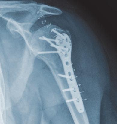
26 Case Study Fracture of the Proximal Humerus (AO 11 C2) Preoperative Postoperative 24
27 Subject to technical modifications, errors and misprints. WP 4OP020 EN / 1503 Layout, typesetting: design graphic - Wolfram Passlack Illustrations: Karen Hilberg Lorenzweg Berlin Germany Phone Fax customer.service@aap.de
28 Lorenzweg Berlin Germany Phone Fax customer.service@aap.de WP 4OP020 EN / 1503
Distal Medial Tibia Plate Surgical Technique
 Locking Compression Technology by aap 1 Disclaimer This surgical technique is exclusively intended for medical professionals, especially physicians, and therefore may not be regarded as a source of information
Instructions for Use. LCP Locking Compression Plate. Combine without Compromise.
 Instructions for Use LCP Locking Compression Plate. Combine without Compromise. Table of Contents LCP: Combine without Compromise 2 AO ASIF Principles of Osteosynthesis 4 Indications and Contraindications
Proximal Humerus System 3.5
 SURGICAL TECHNIQUE STEP BY STEP Proximal Humerus System 3.5 APTUS Shoulder 2 Proximal Humerus System 3.5 Contents 3 Introduction 3 Product Materials 3 Indications 3 Contraindications 3 Color Coding 3 Symbols
Integra. Stainless Headed Compression Screw System SURGICAL TECHNIQUE
 Integra Stainless Headed Compression Screw System SURGICAL TECHNIQUE Table of Contents Design Rationale...2 Indications...2 Contraindications...2 Surgical Technique Step 1: Inserting Guide Wire... 3 Step
DISTAL RADIUS PLATES 3.5 mm / ANGULARLY STABLE. Distal radius plates 3,5 mm / angularly stable. Locking bone screws. Cortical bone screw
 SURGICAL NÁSTROJE TECHNIQUE PRO ARTROSKOPII DISTAL INSTRUMENTS RADIUS PLATES FOR ARTHROSCOPY 3.5 mm / ANGULARLY STABLE Distal radius plates 3.5 mm / angularly stable Indication The plates are used for
Technique Guide. 4.5 mm Cannulated Screws. Part of the Synthes Cannulated Screw System.
 Technique Guide 4.5 mm Cannulated Screws. Part of the Synthes Cannulated Screw System. TableofContents Introduction 4.5 mm Cannulated Screws 2 AO Principles 3 Indications 4 Surgical Technique Surgical
Integra. Capture Screw System SURGICAL TECHNIQUE
 Integra Capture Screw System SURGICAL TECHNIQUE Table of Contents Indications... 2 Contraindications... 2 System Description... 2 System Features... 2 Cannulated Low-Profile Screws (AC-Series) Overview...
Fibula Plating System
 ANATOMIC LOCKED PLATING SYSTEM Fibula Plating System Securing optimal fixation through versatile locked and compression plating technology Contents Surgeon Design Team 2 Introduction 3 Anatomic Fibula
WRIST SYSTEM. ARIX Volar Distal Radius Locking Plate System
 WRIST SYSTEM A Contents 3 4 7 14 15 16 19 21 Indications Product Overview Features & Benefits Ordering Information - Screws -2.5mm Self-Tapping Cortical Screws (Non-Locking) -2.5mm Self-Tapping Locking
DLS Dynamic Locking Screw. Combined with LCP Locking Compression Plate.
 DLS Dynamic Locking Screw. Combined with LCP Locking Compression Plate. Instructions for Use Discontinued June 2016 DSEM/TRM/0517/0844(1) Table of Contents Introduction DLS Dynamic Locking Screw 2 Indications
LCP Pilon Plate 2.7/3.5
 Surgical Technique LCP Locking Compression Plate Original Instruments and Implants of the Association for the Study of Internal Fixation AO/ASIF Table of contents Indications 3 Implants 4 Instruments 5
Small Fragment Locking Compression Plate (LCP ) System Stainless Steel and Titanium TECHNIQUE GUIDE
 System Stainless Steel and Titanium TECHNIQUE GUIDE") Small Fragment Locking Compression Plate (LCP ) System Stainless Steel and Titanium TECHNIQUE GUIDE R Original Instruments and Implants of the Association for the Study of Internal Fixation AO ASIF Introduction
Small Fragment Locking Compression Plate (LCP ) System Stainless Steel and Titanium TECHNIQUE GUIDE R Original Instruments and Implants of the Association for the Study of Internal Fixation AO ASIF Introduction
The Percutaneous Reduction Forceps Technique Guide
 The Percutaneous Reduction Forceps Technique Guide Indications + Product Overview Introduction The Percutaneous Reduction Forceps The Percutaneous Reduction Forceps facilitate standard technique for fixation
3.5 mm Cannulated Screw Technique Guide
 3.5 mm Cannulated Screw Technique Guide An Integral Part of the SYNTHES Cannulated Screw System Original Instruments and Implants of the Association for the Study of Internal Fixation AO ASIF The 3.5 mm
Technique Guide. 7.0 mm Cannulated Screws. Part of the Synthes Cannulated Screw System.
 Technique Guide 7.0 mm Cannulated Screws. Part of the Synthes Cannulated Screw System. Table of Contents Introduction 7.0 mm Cannulated Screws 2 AO Principles 3 Indications 4 Surgical Technique Surgical
Universal Humeral Nail
 990210009 INDEX Indications Preoperative Planning Patient Position Surgical Technique - Step 1 Open Humerus - Step 2 Calibrate The Nail - Step 3 Insert Nail - Step 4 Proximal Locking - Step 5 Assemble
1.5 MM LCP SYSTEM. For treatment of fractures and arthrodeses of canines and felines SURGICAL TECHNIQUE
 1.5 MM LCP SYSTEM For treatment of fractures and arthrodeses of canines and felines SURGICAL TECHNIQUE TABLE OF CONTENTS INTRODUCTION 1.5 mm LCP System 2 AO Principles 4 SURGICAL TECHNIQUE Reduce Fracture
Technique Guide. LCP Pilon Plate 2.7/3.5
 Technique Guide LCP Pilon Plate 2.7/3.5 LCP Pilon Plate 2.7/3.5 Table of contents Indications 3 Implants 4 Instruments 5 Surgical technique 6 Implant removal 12 Image intensifier control Warning This
6.5 mm and 7.3 mm Cannulated Screws Technique Guide
 6.5 mm and 7.3 mm Cannulated Screws Technique Guide An Integral Part of the SYNTHES Cannulated Screw System Original Instruments and Implants of the Association for the Study of Internal Fixation AO ASIF
Technique Guide. 2.4/2.7 mm Locking Tarsal Plates. Talus Plate, Navicular Plate and Cuboid Plate.
 Technique Guide 2.4/2.7 mm Locking Tarsal Plates. Talus Plate, Navicular Plate and Cuboid Plate. Table of Contents Introduction 2.4/2.7 mm Locking Tarsal Plates 2 AO Principles 4 Indications 5 Clinical
Mecron Cannulated Screws
 Surgical Technique and Ordering Information 2 Table of contents Description... 4 Indications for use... 4 Contraindications... 4 State-of-the-art design features... 5 Surgical Technique... 6 Surgery Steps
OPERATIVE TECHNIQUE RIVAL VIEW PLATING SYSTEM. foot & ankle reconstruction procedures
 OPERATIVE TECHNIQUE RIVAL VIEW PLATING SYSTEM foot & ankle reconstruction procedures INTRODUCTION 3 SYSTEM DESCRIPTION 3 TECHNICAL DETAILS 4 SALES AND MARKETING CONFIGURATION 5 OPERATIVE TECHNIQUE 7 OPERATIVE
OPERATIVE TECHNIQUE RIVAL REDUCE FRACTURE PLATING SYSTEM. foot & ankle trauma procedures
 OPERATIVE TECHNIQUE RIVAL REDUCE FRACTURE PLATING SYSTEM foot & ankle trauma procedures INTRODUCTION 3 SYSTEM DESCRIPTION 3 TECHNICAL DETAILS 4 SALES AND MARKETING CONFIGURATION 5 OPERATIVE TECHNIQUE 7
Technique Guide. Quadrilateral Surface Plates 3.5. Part of the Low Profile Pelvic System 3.5.
 Technique Guide Quadrilateral Surface Plates 3.5. Part of the Low Profile Pelvic System 3.5. Table of Contents Introduction Quadrilateral Surface Plates 3.5 2 AO Principles 4 Indications 5 Surgical Technique
Ulna Shortening System 2.5
 SURGICAL TECHNIQUE STEP BY STEP Ulna Shortening System 2.5 APTUS Wrist 2 Ulna Shortening System 2.5 Contents 3 Introduction Product Materials Indications Contraindications Color Coding 4 General Instrument
SpeedTip CCS 5.0, 7.0
 SURGICAL TECHNIQUE STEP BY STEP SpeedTip CCS 5.0, 7.0 Cannulated Compression Screws APTUS 2 SpeedTip CCS 5.0, 7.0 Cannulated Compression Screws Contents 3 Introduction Product Materials Indications Contraindications
LCP Pilon Plate 2.7/3.5
 LCP Pilon Plate 2.7/3.5 Surgical Technique This publication is not intended for distribution in the USA. Instruments and implants approved by the AO Foundation. Table of contents Indications 2 Implants
Distal Radius System 2.5
 SURGICAL TECHNIQUE STEP BY STEP Distal Radius System 2.5 APTUS Wrist 2 Distal Radius System 2.5 Contents 3 Introduction 3 Product Materials 3 Indications 3 Contraindications 3 Color Coding 3 Possible Combination
HCS 1.5. The countersinkable compression screw.
 HCS 1.5. The countersinkable compression screw. Surgical Technique This publication is not intended for distribution in the USA. Instruments and implants approved by the AO Foundation. Table of Contents
MaxTorque. surgical technique. Cannulated Screw System. Foot & Ankle. OrthoHelix Technology
 MaxTorque Cannulated Screw System OrthoHelix Technology surgical technique Foot & Ankle 2 M A X T O R Q U E C A N N U L A T E D S C R E W S Y S T E M Table of Contents Advantages 3 Indications 4 Contraindications
Part of the DePuy Synthes Cannulated Screw System. 3.5 mm Cannulated Screws
 Part of the DePuy Synthes Cannulated Screw System 3.5 mm Cannulated Screws Surgical Technique Table of Contents Introduction 3.5 mm Cannulated Screws 2 AO Principles 3 Indications 4 Surgical Technique
7.0 mm Cannulated Screws
 Part of the DePuy Synthes Cannulated Screw System 7.0 mm Cannulated Screws Surgical Technique Table of Contents Introduction 7.0 mm Cannulated Screws 2 AO Principles 3 Indications 4 Surgical Technique
SpeedTip CCS 5.0, 7.0
 SURGICAL TECHNIQUE STEP BY STEP SpeedTip CCS 5.0, 7.0 Cannulated Compression Screws APTUS 2 SpeedTip CCS 5.0, 7.0 Cannulated Compression Screws SpeedTip CCS 5.0, 7.0 Cannulated Compression Screw 3 SpeedTip
5th Metatarsal Fracture System Surgical Technique
 5th Metatarsal Fracture System Surgical Technique 5th Metatarsal Fracture System 5th Metatarsal Fracture System The 5th Metatarsal Fracture System (AR-8956S) is a uniquely designed screw and plate system
OPERATIVE TECHNIQUE RIVAL BITE HEADED CANNULATED AND HEADLESS COMPRESSION SCREWS. foot & ankle applications
 OPERATIVE TECHNIQUE RIVAL BITE HEADED CANNULATED AND HEADLESS COMPRESSION SCREWS foot & ankle applications INTRODUCTION 3 SYSTEM DESCRIPTION 3 TECHNICAL DETAILS 4 SALES AND MARKETING CONFIGURATION 6 OPERATIVE
Technique Guide. Modular Sternal Cable System. Flexibility and strength in sternal closure and repair.
 Technique Guide Modular Sternal Cable System. Flexibility and strength in sternal closure and repair. Table of Contents Introduction Overview 2 Indications and Contraindications 3 Surgical Technique A.
LCP Pilon Plate 2.7/3.5. Surgical Technique
 LCP Pilon Plate 2.7/3.5 Surgical Technique Image intensifier control This description alone does not provide sufficient background for direct use of DePuy Synthes products. Instruction by a surgeon experienced
Humeral Nail System Procedural Steps.
 Humeral Nail System Procedural Steps www.carbo-fix.com Table of Contents Introduction..3 Instrumentation Set... 8 Procedural Steps: Humeral Nail.........10 Procedural Steps: Proximal Humeral Nail.....13
3.5 mm and 4.5 mm Curved Locking Compression Plates (LCP )
") For Minimally Invasive Osteosynthesis 3.5 mm and 4.5 mm Curved Locking Compression Plates (LCP ) Surgical Technique Table of Contents Introduction 3.5 mm and 4.5 mm Curved Locking Compression 2 Plates
For Minimally Invasive Osteosynthesis 3.5 mm and 4.5 mm Curved Locking Compression Plates (LCP ) Surgical Technique Table of Contents Introduction 3.5 mm and 4.5 mm Curved Locking Compression 2 Plates
Table of Contents 2-6. Introduction. Indications Surgical Technique. Ordering Information 15-24
 Table of Contents Introduction Product information ExtremiFix Midsize Large Screw Offering Headless Screw Characteristics Design Features & Benefits Instrumentation Technical Details Calibrated Drill Bits
SURGICAL TECHNIQUE GUIDE
 SURGICAL TECHNIQUE GUIDE DANGER indicates an imminently hazardous situation which, if not avoided, will result in death or serious injury. WARNING indicates a potentially hazardous situation which, if
Locking Small Fragment
 Locking Small Fragment Page 1 Locking Small Fragment Table of contents Implants 5 Operation technique 7 Inserting drill guides 7 Drilling 7 Length measurement 7 Inserting the measured screw 8 Different
TORNIER MAXLOCK EXTREME. Clavicle Plating System SURGICAL TECHNIQUE
 TORNIER MAXLOCK EXTREME Clavicle Plating System SURGICAL TECHNIQUE 2 Table of Contents: Key Design Features...4 Surgical Technique...5 Implants & Instruments...9 3 Key Design Features There are 7 anatomically
Variable Angle LCP Mesh Plate 2.4/2.7. Part of the Variable Angle LCP Forefoot/Midfoot System 2.4/2.7.
 Variable Angle LCP Mesh Plate 2.4/2.7. Part of the Variable Angle LCP Forefoot/Midfoot System 2.4/2.7. Surgical Technique This publication is not intended for distribution in the USA. Instruments and implants
2.4 mm and 3.0 mm Headless Compression Screws
 For Fixation of Small Bones and Small Bone Fragments 2.4 mm and 3.0 mm Headless s Surgical Technique Table of Contents Introduction 2.4 mm and 3.0 mm Headless 2 Technique Overview 4 AO Principles 5 Indications
VariAx DistalFibula. Foot & Ankle. Locking Plate System. Operative Technique
 VariAx DistalFibula Locking Plate System Operative Technique Foot & Ankle Distal Fibula Fracture Repair Polyaxial Locking Technology Low Profile Design VariAx 2 Color Coded Screws and Instruments VariAx
Integra. Tibio Talo Calcaneus Plate SURGICAL TECHNIQUE
 Integra Tibio Talo Calcaneus Plate SURGICAL TECHNIQUE Table of Contents Indications...02 Contraindications...02 Description...02 Surgical Technique...03 Step 1: Articular Surfaces Preparation...03 Step
DART-FIRE. Small Screw System SURGICAL TECHNIQUE
 DART-FIRE Small Screw System SURGICAL TECHNIQUE DART-FIRE Small Screw System Surgical Technique Contents Chapter 1 4 Chapter 2 6 Chapter 3 7 Appendix 1 10 Appendix 2 12 Introduction Intended Use DART-FIRE
MEDIALMAX System SURGICAL TECHNIQUE
 MAXLOCK EXTREME MEDIALMAX System SURGICAL TECHNIQUE Contents The MEDIALMAX Advantage Design Feature Various flex options POCKETLOCK Technology Anatomic design Advantage Provides the surgeon with multiple
Zimmer Natural Nail System. Cephalomedullary Nail Surgical Technique SMALL
 Zimmer Natural Nail System Cephalomedullary Nail Surgical Technique SMALL Zimmer Natural Nail System Cephalomedullary Nail Technique - Small 1 Zimmer Natural Nail System Cephalomedullary Nail Surgical
VARIABLE ANGLE LOCKING HAND SYSTEM
 VARIABLE ANGLE LOCKING HAND SYSTEM For fragment-specific fracture fixation with variable angle locking and locking technology Instruments and implants approved by the AO Foundation. This publication is
VLP FOOT Variable Angle Locked Plating System
 Surgical Technique VLP FOOT Variable Angle Locked Plating System Surgical Technique Table of Contents Product overview Implant selection...2 Introduction...3 System overview...4 Indications...4 Design
Zimmer Natural Nail System
 Zimmer Natural Nail System Cephalomedullary Small Nail Surgical Technique Table of Contents Product Overview... 2 Implant Overview... 2 Indications... 3 Contraindications... 3 Surgical Technique... 4 Preoperative
VARIABLE ANGLE LOCKING HAND SYSTEM
 VARIABLE ANGLE LOCKING HAND SYSTEM For fragment-specific fracture fixation with variable angle locking and locking technology SURGICAL TECHNIQUE TABLE OF CONTENTS INTRODUCTION Variable Angle Locking Hand
VariAx Foot Locking Plate System
 VariAx Foot Locking Plate System Operative Technique Trauma and Deformity Correction Polyaxial Locking Technology Comprehensive Calcaneal Fracture Plates Table of Contents 1. Introduction 3 2. Overview
DART-FIRE. Small Screw System SURGIC A L T ECHNIQUE
 DART-FIRE Small Screw System SURGIC A L T ECHNIQUE Contents Headline Headline PREFACE Chapter 1 4 Chapter 2 6 Chapter 3 7 Appendix A 10 Appendix B 12 Introduction Intended Use DART-FIRE Small Screw System
Orthopedic Bone Nail System Universal Humeral Nail
 Orthopedic Bone Nail System Universal Humeral Nail Surgical Technique Manual Note: The surgical procedures should be performed under the guidance of qualified skilled orthopedic surgeons, and this surgical
MatrixMANDIBLE Preformed Reconstruction Plates. Preshaped to the mandibular anatomy.
 MatrixMANDIBLE Preformed Reconstruction Plates. Preshaped to the mandibular anatomy. Technique Guide CMF Matrix Table of Contents Introduction MatrixMANDIBLE Preformed Reconstruction Plates 2 AO Principles
ACLP Anterior Cervical Locking Plate System TECHNIQUE GUIDE
 ACLP Anterior Cervical Locking Plate System TECHNIQUE GUIDE Instruments and implants approved by the AO Foundation ACLP Anterior Cervical Locking Plate System The ACLP System is designed to reduce the
HCS 2.4/3.0. The countersinkable compression screw.
 Technique Guide HCS 2.4/3.0. The countersinkable compression screw. Table of Contents Introduction Features and Benefits 2 Functional Principle 3 Indications 4 Surgical Technique Hand Scaphoid 5 Foot
Vortex TRAUMATOLOGY. Vortex Distal Femur
 Vortex TRAUMATOLOGY Vortex Distal Femur 1 Content 1. Introduction 4 4. Implant list 16-17 The following surgical description contains general outlines for Vortex Distal Femur plating. However, the operating
Ankle Fracture System. Surgical Technique STRENGTH FROM WITHIN
 Ankle Fracture System Surgical Technique STRENGTH FROM WITHIN Ankle Fracture System The Sonoma FibuLock nail is the first intramedullary device that has the same indications as plates and delivers anatomic
DART-FIRE. Small Screw System SURGICAL TECHNIQUE
 DART-FIRE Small Screw System SURGICAL TECHNIQUE DART-FIRE Small Screw System SURGICAL TECHNIQUE Contents Chapter 1 4 Chapter 2 6 Appendix 1 9 Appendix 2 11 Introduction DART-FIRE Small Screw System Surgical
Technique Guide. Cable System. For Orthopaedic Trauma Surgery.
 Technique Guide Cable System. For Orthopaedic Trauma Surgery. Table of Contents Introduction Overview 2 AO Principles 4 Indications and Contraindications 5 Surgical Technique Standard Cerclage Technique
Instruments for Removing DePuy Synthes Screws. Screw Removal Set
 Instruments for Removing DePuy Synthes Screws Screw Removal Set Surgical Technique Table of Contents Introduction Screw Removal Set 2 Surgical Technique Preoperative Planning and Preparation 6 Removal
BioDrive Micro Screw System
 At Biomet, engineering excellence is our heritage and our passion. For over 25 years, through various divisions worldwide, we have applied the most advanced engineering and manufacturing technology to
The Titanium Cannulated Lateral Entry Femoral Recon Nail. Expert nailing system with radiolucent instrumentation.
 The Titanium Cannulated Lateral Entry Femoral Recon Nail. Expert nailing system with radiolucent instrumentation. Technique Guide EXPERT Nailing System Table of Contents Introduction Titanium Cannulated
Zimmer Natural Nail System
 Zimmer Natural Nail System Cephalomedullary Nail Surgical Technique Compact Case- Short Nails Only SMALL Zimmer Natural Nail System Cephalomedullary Nail Technique - Small 1 Zimmer Natural Nail System
Variable Angle LCP Forefoot/Midfoot System 2.4/2.7. Procedure specific plates for osteotomies, arthrodeses and fractures of the foot.
 Instruction for Use Variable Angle LCP Forefoot/Midfoot System 2.4/2.7. Procedure specific plates for osteotomies, arthrodeses and fractures of the foot. Table of Contents Introduction VA-LCP Forefoot/Midfoot
Technique Guide. LCP Dynamic Helical Hip System (DHHS). Part of the Synthes Large Fragment LCP System.
. Part of the Synthes Large Fragment LCP System.") Technique Guide LCP Dynamic Helical Hip System (DHHS). Part of the Synthes Large Fragment LCP System. Table of Contents Introduction LCP Dynamic Helical Hip System (DHHS) 2 AO Principles 4 Indications
Technique Guide LCP Dynamic Helical Hip System (DHHS). Part of the Synthes Large Fragment LCP System. Table of Contents Introduction LCP Dynamic Helical Hip System (DHHS) 2 AO Principles 4 Indications
Variable Angle LCP Tarsal Plates 2.4/2.7. Navicular Plate and Cuboid Plates.
 Variable Angle LCP Tarsal Plates 2.4/2.7. Navicular Plate and Cuboid Plates. Surgical Technique This publication is not intended for distribution in the USA. Instruments and implants approved by the AO
Zimmer Natural Nail System
 Zimmer Natural Nail System Cephalomedullary Nail Surgical Technique Compact Case - Short Nails Only STANDARD Zimmer Natural Nail System Cephalomedullary Nail Surgical Technique - Standard 1 Zimmer Natural
Cannulated Screws Ø 3.5 mm / 4.5 mm
 Cannulated Screws Ø / 4.5 mm Table of contents Implants Ø 4 Implants Ø 4.5 mm 6 Operation technique - Ø Cannulated Screws 9 Reduce fracture and insert guide wire 9 Determine the screw length 9 Drilling
Digital Compression Screw
 Digital Compression Screw Surgical Technique Contents Product The BioPro Digital Compression Screw is a stainless steel lag screw designed for digital fusions. Table of contents Indications & Contraindications
Technique Guide MTP Arthrodesis System
 Technique Guide MTP Arthrodesis System The CheckMATE MTP Arthrodesis System features low-profile, anatomically pre-contoured Bone Plates with either a combination of locking and non-locking holes or all-locking
Small Plate and Screw System SURGICAL TECHNIQUE
 MINI MAXLOCK EXTREME Small Plate and Screw System SURGICAL TECHNIQUE Contents Key Design Features 2 Surgical Technique 3 Implants and Instruments 9 Proper surgical procedures and techniques are the responsibility
Technique Guide Supplement. Standard DHS Lag Screw with LCP DHHS Sideplate.
 Technique Guide Supplement Standard DHS Lag Screw with LCP DHHS Sideplate. Table of Contents Surgical Technique Standard DHS Lag Screw with LCP DHHS 2 Sideplate Technique DHS One-step Lag Screw with DHHS
For Minimally Invasive Application of Cerclage Wires. Cerclage Passer. Surgical Technique
 For Minimally Invasive Application of Cerclage Wires Cerclage Passer Surgical Technique Table of Contents Introduction Cerclage Passer 2 Surgical Technique Preparation 4 Insert Cerclage Passer 5 Connect
ACCS Anterior Cervical Compression System TECHNIQUE GUIDE
 ACCS Anterior Cervical Compression System TECHNIQUE GUIDE Original Instruments and Implants of the Association for the Study of Internal Fixation AO ASIF ACCS Anterior Cervical Compression System The Anterior
Omega 3 System Compression Hip Screw
 Omega 3 System Compression Hip Screw Hip Fracture Axially Stable Locking Option Contents Omega3 Compression Hip Screw Introduction 4 Potential Features & Benefits 5 Relative Indications & Contraindications
OsteoBridge IKA Intramedullary Knee Arthrodesis Fixation System. From the «BioBall Company» OsteoBridge Family
 From the «BioBall Company» OsteoBridge Family OsteoBridge IKA Intramedullary Knee Arthrodesis Fixation System The modular system for the fixation of the knee joint 01. OsteoBridge IKA The OsteoBridge IKA
Operative Technique Distal Fibula Fracture Repair Polyaxial Locking Technology Low Profile Design
 VariAx Fibula Locking Plate System Operative Technique Distal Fibula Fracture Repair Polyaxial Locking Technology Low Profile Design Contributing Surgeon: Bradley R. Merk, MD Associate Professor of Orthopaedic
2.4 mm Cannulated Screw. An integral part of the Synthes Cannulated Screw System (CSS).
.") 2.4 mm Cannulated Screw. An integral part of the Synthes Cannulated Screw System (CSS). Surgical Technique This publication is not intended f distribution in the USA. and implants approved by the AO Foundation.
2.4 mm Cannulated Screw. An integral part of the Synthes Cannulated Screw System (CSS). Surgical Technique This publication is not intended f distribution in the USA. and implants approved by the AO Foundation.
ISO Plate SURGICAL TECHNIQUE
 MINI MAXLOCK EXTREME ISO Plate SURGICAL TECHNIQUE Contents Table of Contents Key Design Features 2 Surgical Technique 3 Implants and Instruments 8 Key Design Features The MINI MAXLOCK EXTREME ISO (Intraosseous
Technique Guide. Modular Sternal Cable System. Flexibility and strength in sternal closure and repair.
 Technique Guide Modular Sternal Cable System. Flexibility and strength in sternal closure and repair. Table of Contents Introduction Modular Sternal Cable System 2 Indications 4 Modular Sternal Closure
Surgical Technique. Customer Service:
 Patent Pending CAUTION: Federal Law (USA) restricts this device to sale by or on the order of a physician. Notes This page is blank INDICATIONS FOR USE The Extremity Medical Hallu X Intramedullary Fusion
Technique Chart. DHS/DCS One-Step Insertion Wrench. For use with DHS/DCS One-Step Lag Screws.
 Technique Chart DHS/DCS One-Step Insertion Wrench. For use with DHS/DCS One-Step Lag Screws. DHS/DCS One-Step Insertion Wrench DHS Triple Reamer (338.13) Assembly 338.10 8.0 mm Drill Bit 338.11 DHS Reaming
VariAx. Foot & Ankle. Foot Locking Plate System
 VariAx Foot Locking Plate System Operative Technique Foot & Ankle Trauma and Deformity Correction Polyaxial Locking Technology Comprehensive Calcaneal Fracture Plates VariAx 2 Plates VariAx Foot Locking
TwinFix Surgical Protocol. 3.2mm Cannulated Compression Screw System
 TwinFix Surgical Protocol Compression Screw System TwinFix Compression Screw System Indications for Use: The Stryker TwinFix Interfragmentary Compression Screw System is intended to be used for fractures
Lag Screw Device TECHNIQUE GUIDE. Indicated for symphyseal fracture fixation of the mandible. Instruments and implants approved by the AO Foundation
 Lag Screw Device TECHNIQUE GUIDE Indicated for symphyseal fracture fixation of the mandible Instruments and implants approved by the AO Foundation Lag Screw Device Indicated for symphyseal fracture fixation
Technique Guide. Occipito-Cervical Fusion System. Implants and instruments designed to optimize fixation to the occiput.
 Technique Guide Occipito-Cervical Fusion System. Implants and instruments designed to optimize fixation to the occiput. Table of Contents Introduction Overview 2 AO ASIF Principles 4 Indications and Contraindications
Distal Fibula Plate SURGICAL TECHNIQUE
 MAXLOCK EXTREME Distal Fibula Plate SURGICAL TECHNIQUE Contents Key Design Features 3 Surgical Technique 4 Implants and Instruments 8 Proper surgical procedures and techniques are the responsibility of
VariAx Fibula Locking Plate System
 VariAx Fibula Locking Plate System Operative Technique Distal Fibula Fracture Repair Polyaxial Locking Technology Low Profile Design 1 Contributing Surgeon: Bradley R. Merk, MD Associate Professor of Orthopaedic
MTP Fusion Surgical Technique
 MTP Fusion Surgical Technique Patent and Patent Pending CAUTION: Federal Law (USA) restricts this device to sale by or on the order of a physician. INDICATIONS FOR USE The Omni Foot Plating System is intended
Peanut Growth Control Plating System. Surgical Technique
 Peanut Growth Control Plating System Surgical Technique 1 Peanut Growth Control Plating System Table of Contents System Design Features... 2 Instrument Tray... 5 Implant Caddy... 6 Surgical Technique...
Cerclage Passer. For minimally invasive application of cerclage cables.
 Cerclage Passer. For minimally invasive application of cerclage cables. Handling Technique Cable application This publication is not intended for distribution in the USA. Instruments and implants approved
Integra. HALLU -Lock M.T.P. Arthrodesis System SURGICAL TECHNIQUE
 Integra HALLU -Lock M.T.P. Arthrodesis System SURGICAL TECHNIQUE Table of Contents... 02 Indications... 02 Contraindications... 02 Surgical Technique... 03 Step 1: Incision... 03 Step 2: Preparation of
URS Degen. Top loading pedicle screw system for posterior stabilization.
 URS Degen. Top loading pedicle screw system for posterior stabilization. Technique Guide This publication is not intended for distribution in the USA. Table of Contents Introduction URS Degen 2 AO Principles
S U R G I C A L T E C H N I Q U E TRAUMA & EXTREMITIES GROUP
 S U R G I C A L T E C H N I Q U E TRAUMA & EXTREMITIES GROUP TABLE OF CONTENTS ATN NAIL SYSTEM DESIGN RATIONALE INDICATIONS/CONTRAINDICATIONS PREOPERATIVE PLANNING AND PATIENT POSITIONING NAIL INSERTION
Integra SURGICAL TECHNIQUE. HALLU -Fix and HALLU -Lock MTP Arthrodesis Systems. HALLU Lock C plate. HALLU -C plate. HALLU -S plate. HALLU Lock S plate
 EN Integra HALLU -Fix and HALLU -Lock MTP Arthrodesis Systems SURGICAL TECHNIQUE HALLU -C plate HALLU Lock C plate HALLU -S plate HALLU Lock S plate Products for sale in Europe, Middle-East and Africa