INTRODUCTION We believe that every laboratory working in the field of nanotechnology needs an SEM, therefore we would like to introduce to you our IEM
|
|
|
- Bethany Webb
- 5 years ago
- Views:
Transcription
1

. The SEM can be used in many areas and there is no restriction to analyses on samples.")
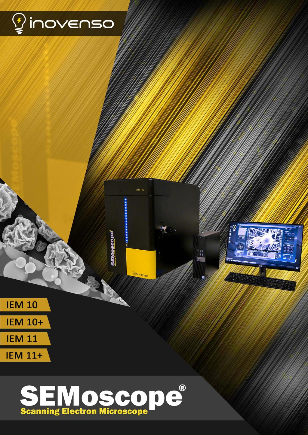
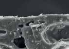
2 INTRODUCTION We believe that every laboratory working in the field of nanotechnology needs an SEM, therefore we would like to introduce to you our IEM series of SEM. In short space of time, our device that is the size of an old computer will prove its effectiveness, and will help you achieve a significant step in your research. The advantage of the images obtained by SEM lies in the high magnification factor: you will be able to magnify an image by up to 150,000 times which will enable you to conduct analyses up to a nanoscale using different detectors such as BSE, SE and EDS (X-ray detector). The SEM can be used in many areas and there is no restriction to analyses on samples. Another important factor to purchase our SEM is to avoid waiting the annoying time for obtaining images from a nearby facility, instead of that your images can be available in an unbelievable short time. Our SEMs are easy to operate and your employees or students can use it without any waste of time in a special training. lnovenso Team 2


3 SEM Why? Quick and simple installation and no maintenance needed 1 3 Times More affordable than other SEMs 2 Obtaining results in less than 4 minutes 3 Magnification range: x20 ~ x150, nanometer resolution 5 Easy Navigation with the User-Friendly Software 6 3
 Acc Voltage 1 ~ 30 kv (1kV increments) Electron Gun Tungsten Filament (W) Detector SE Detector Stage Auto Stage (X: 35mm,Y: 35mm,T: 0")
 - 187 (lbs) Stage Position Users can easily locate the sample on the stage Auto Focus & Fine Focus Auto focus function makes the operation easier and clearer images can be obtained even at")
4 IEM 10 Magnification Up To x 100,000 Auto Stage: X, Y, T Axis Click & Move Stage Control Auto Function: Filament Memory, Focus, Contrast, Brightness High Definition Image : 5120 X 3840 Pixel Specifications Magnification x20 ~ x100,000 (Efficient: ~x50,000) Acc Voltage 1 ~ 30 kv (1kV increments) Electron Gun Tungsten Filament (W) Detector SE Detector Stage Auto Stage (X: 35mm,Y: 35mm,T: 0 to 45o) Manual Stage (Z: 5 to 50mm) Image Shift X, Y, R(Rotation) Operating System Microsoft Windows 7 Dimensions (mm) 400(W) x 600(L) x 550(H) Dimensions (inch) 15,74(W) x 23,62(L) x 21,65(H) Weight 85(kgs) (lbs) Stage Position Users can easily locate the sample on the stage Auto Focus & Fine Focus Auto focus function makes the operation easier and clearer images can be obtained even at the higher magnifications Click & Move By applying the auto stage, stage is controlled simply with a mouse click Filament Memory Filament s saturation point is automatically stored and activated 4
5 IEM 10+ Magnification Up To x 100,000 Auto Stage: X, Y, T Axis Click & Move Stage Control Auto Function: Filament Memory, Focus, Contrast, Brightness High Definition Image: 5120 X 3840 Pixel Thermo EDS Detector: 133 ev at Mn, B(5) ~ U(92) Specifications Magnification x20 ~ x100,000 (Efficient: ~x50,000) Acc Voltage 1 ~ 30 kv (1kV increments) Electron Gun Tungsten Filament (W) Detector SE and EDS Detectors Stage Auto Stage (X: 35mm,Y: 35mm,T: 0 to 45o) Manual Stage (Z: 5 to 50mm) Image Shift X, Y, R(Rotation) Operating System Microsoft Windows 7 Dimensions (mm) 400(W) x 600(L) x 550(H) Dimensions (inch) 15,74(W) x 23,62(L) x 21,65(H) Weight 95(kgs) - 210(lbs) Rapid EDS Analysis Point and shoot an alysis with NORAN System 7 software uses standardless quantitative analysis to enable rapid identification of various regions within a material. Extracted Linescans Extracted Area Analysis EDS Analysis with NORAN System 7 Spectral Imaging The UltraDry Compact EDS detector provides outstanding elemental mapping within few minutes. With Spectral Imaging, where a full EDS spectrum is stored at every pixel, samples can be analyzed after they have been removed from the microscope. NORAN System 7 tools provide several analytical methods for the best results. 5


 Detector SE Detector Stage Auto Stage (X: 35mmY:")
 Operating System Microsoft Windows 7 Dimensions (mm) 400(W) x")
 - 187(lbs) SE: Composition Material: Alloy Metal BSE: Topography")

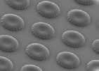
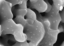
6 IEM 11 High Quality Images (5nm resolution) Magnification Up To 150,000x Easy Navigation with the Navigation Mode Precise Control with a Joystick and the Driving Mode Combined SE and BSE Images Low Energy Consumption Intuitive User Interface Specifications Magnification x20 ~ x100,000 (Efficient: ~x50,000) Acc Voltage 1 ~ 30 kv (1kV increments) Electron Gun Tungsten Filament (W) Detector SE Detector Stage Auto Stage (X: 35mmY: 35mm,T: 0 to 45o) Manual Stage (Z: 5 to 50mm) Image Shift X, Y, R(Rotation) Operating System Microsoft Windows 7 Dimensions (mm) 400(W) x 600(L) x 550(H) Dimensions (inch) 15,74(W) x 23,62(L) x 21,65(H) Weight 85(kgs) - 187(lbs) SE: Composition Material: Alloy Metal BSE: Topography Material: Alloy Metal Removable BSE Detector By applying 4-channel BSE detector, composition and topography functions are available. These functions are optional and can be opted out. 6


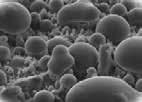
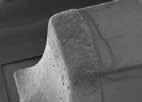
7 IEM 11 Low Voltage Analysis Information on the morphology of a sample can be obtained with voltage ranging from 1 to 30kV. ACC.Voltage: 20kV / SE Image / Mag: x50,000 ACC.Voltage : 5 kv / SE Image / Mag: x50,000 Move "Left" Focus "-" Focus "+" Move "Up" Move "Right" Low Magnification High Magnification Driving Mode 2-axes stage motion, magnification, and focus can be easily motorized and controlled with a joystick. Move "Down" Combined Signaling SE and BSE can be detected separately and together. ACC.Voltage: 20kV Material: Alloy Metal SE + BSE Image Highest Resolution Provides high quality images (5nm resolution, Max accelerating voltage: 30kV) ACC. Voltage : 30kV / SE Image / Mag : x100,000 Driving Mode By loading seven samples simultaneously into a multi holder, samples can be easily located with the help of a mini map, sample area, and a live display. 7
8 IEM 11+ High Quality Images (5nm resolution) Magnification Up To 150,000x Easy Navigation with the Navigation Mode Precise Control with a Joystick and the Driving Mode Combined SE and BSE Images Low Energy Consumption Intuitive User Interface Oxford:130eV at Mnk, C(6) ~ U(92) / EDAX:133eV at Mnk, Be(4) ~ U(92) Specifications Magnification Acc Voltage Electron Gun Detector EDS EDS x20 ~ x150,000 (Efficient: ~x80,000) 1 ~ 30kV (1kV increments) Tungsten Filament (W) SE and BSE Detectors Oxford:130eV at Mnk, C(6) ~ U(92) EDAX:133eV at Mnk, Be(4) ~ U(92) Image Shift Auto Stage (X: 35mm, Y: 35mm, T: 0 to 45o) Manual Stage (Z: 5 to 50mm) Operating System Microsoft Windows 7 Dimensions (mm) 400(W) x 600(L) x 550(H) Dimensions (inch) 15,74(W) x 23,62(L) x 21,65(H) Weight 95(kgs)- 210(lbs) Brand: Oxford Tru-Q Analysis Engine: Accurate Result By Multi Algorithm Multilingual Operation Fast & Easy Analysis With Flow Chart Style Menu Diverse Element Analysis Functions: Point & ID, Line Scan, Mapping, Line Overlap & Background Correction Quantitative Mapping (QuantLineScan, QuantMap) Brand: EDAX Highest Performance SDD Sensor Size: 30mm2 High Quality Light Element Analysis: Be(4) ~ Am(95) Diverse Element Analysis Functions: Point&ID Line Scan, Mapping 8


9 IEM 11+ Oxford - Report & Data Save Various report templates are provided. Users can generate a report with the Report Generate function. Data can be saved, copied, printed, and ed directly. - Available in Various Languages The program is offered in various languages including English, Japanese, Chinese simplified, Russian, French, Korean and Portuguese. - -LineScan Linescan determines elemental concentration variations along a line defined by the users. Linescans are corrected for peak overlaps and any false variations due to X-ray background. - Point & ID Spectrum that shows elemental composition can be acquired from multiple points and areas. EDAX Outstanding Light Element Performance Silicon nitride window improves performance for light elements at low energies. EDS Reporting The TEAMTM EDS Smart Data Management system for SEM achieves new levels of ease and flexibility with simplified file management and dynamic reporting. Fast, Efficient Results for Industrial Needs All results can be reviewed anytime. Spectrum, microelement detection, quantitative mapping are construed accurately. 9
 Target Target Size Power")
![Ionization Current Au (Standard) 50mm[Dia] AC110-240V, 50 / 60Hz,](/docs-images/94/121756971/images/10-2.jpg "50W (Except Rotary Pump) Oxford:130eV at Mnk, C(6) ~ U(92)")
 100(mm)[Dia] 420(W)x220(D)x230(H) 15,74(W)x")

[Dia]-")
10 Options Ion Coater Simple Operation Compact Rotary Pump Suitable for Metal Targets (Au,Pt,Pd,Cr,Pt-Pd,Cu,Ni) Target Target Size Power Ionization Current Au (Standard) 50mm[Dia] AC V, 50 / 60Hz, 50W (Except Rotary Pump) Oxford:130eV at Mnk, C(6) ~ U(92) EDAX:133eV at Mnk, Be(4) ~ U(92) Chamber Size Dimension (mm) Dimension (inch) 100(mm)[Dia] 420(W)x220(D)x230(H) 15,74(W)x 8,66(D)x 9,05(H) Cool Stage Suitable for Liquid or Biological Samples Cool Stage Temperature: -25oC to 50oC Temperature Resolution: ±0.1oC Temperature Accuracy: ±1oC Specimen Holder Size: 18(mm)[Dia]- 0,70(inch) [Dia] Vacuum Feed Through Flange Onion Skin without Cool Stage Onion Skin with Cool Stage 10


11 Options Low Vacuum System -High Vacuum SE Image -Low Vacuum BSE Image Suitable for Non-Conductive Samples BSE Required Coating Not Required Vacuum Condition: 100pa to 1pa BSE Detector BSE Detector -SE Image of Film Surface 4 Channel Solid State Type: 4 Segment Excellent Composition and Topography Functions Combined SE and BSE Images Synergistic Use of BSE Detector and LV Mode -BSE Image of Film Surface 11
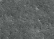
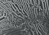
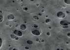


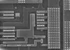


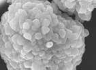
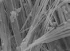
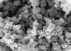
12 Applications Automotive Iron Plate Glass Filter Tire Semiconductor Chip Layer Package Material PCB Si Wafer Chemical ZrO2 Package Material Fiber Silver 12
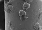
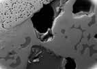
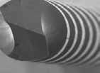
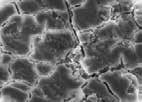


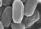
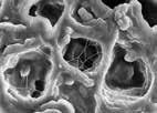
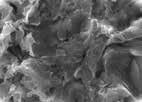

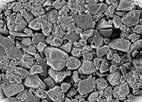
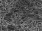
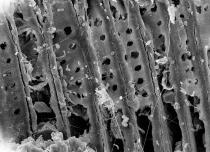
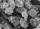

13 Applications Materials Wire Alloy Metal Drill AI Deposition Bioengineering Bacteria Fungus Coffee Implant Construction Paint Water Filter Wood Stone Wood 13
14 Comparison Table Items/Model IEM - 11 IEM IEM - 10 IEM Resolution SE 5.0nm at 30kV 20nm at 30kV Magnification x20 ~ x150,000 x20 ~ x100,000 Efficient Magnification Acceleration Voltage Vacuum Mode ~ x80,000 ~ x50,000 1 ~ 30 kv High Vacuum Low Vacuum (Optional) - EDS Maximum Specimen Size External (Optional) Integrated (Standard) External (Optional) 60 (mm) - 2,36 (inch) in diameter Integrated (Standard) Stage 3-Axes Motorized X 0~35(mm) - 1,37(inch) Y 0~35(mm) - 1,37(inch) T 0~45 (mm) - 1,77(inch) Z 5~50(mm) (Manual) R 360o (Raster) Observation Area Maximum Height 40(mm) - 1,57(inch) in diameter 45(mm) - 1,77(inch) Electron Gun Pre-centered Cartridge Source Tungsten Detector BSE(Standard) SE BSE(Optional) 14
15 Comparison Table Mouse Control Keyboard Joystick - Auto Focus Auto lmage Adjustment Auto Brightness & Contrast Auto Filament Auto Start Navigation View Special Multi Holder Measurement Tool Remote Control Special Features Signal Mixing (SE+BSE) Dual Display(SE/BSE) BSE(Compo,Topo) Line Profile Image Process Measurement Tool - Remote Control EDS - EDS - - Options - BSED Low Vacuum Cool Stage 15

16
Quick and simple installation and no maintenance needed. 3 Times More affordable Than a normal SEM. Obtaining results in less than 4 minutes
 INTRODUCTION We believe that every laboratory working in the field of nanotechnology needs an SEM, therefore we would like to introduce to you our IEM series of SEM. In short space of time, our device
ALTURA EDS. Rev. 0915
 ALTURA EDS Rev. 0915 Enable the Oxford PC Enable the Altura-EDS under Dual Beam Tools in Coral. Or enter your NETID and password directly into the Oxford PC. Warning: Ion-milling, GISs/microprobe, and
Introduction of New Products
 Field Emission Electron Microscope JEM-3100F For evaluation of materials in the fields of nanoscience and nanomaterials science, TEM is required to provide resolution and analytical capabilities that can
NeoScope. Simple Operation to 40,000. Table Top SEM. Serving Advanced Technology
 Table Top SEM Simple Operation to 40,000 Serving Advanced Technology From 10 to 40,000 Table Top SEM Notebook PC version Just plug it to a wall outlet after placing it on a table Desktop PC version Option
SCIENTIFIC INSTRUMENT NEWS. Introduction. Design of the FlexSEM 1000
 SCIENTIFIC INSTRUMENT NEWS 2017 Vol. 9 SEPTEMBER Technical magazine of Electron Microscope and Analytical Instruments. Technical Explanation The FlexSEM 1000: A Scanning Electron Microscope Specializing
Functions of the SEM subsystems
 Functions of the SEM subsystems Electronic column It consists of an electron gun and two or more electron lenses, which influence the path of electrons traveling down an evacuated tube. The base of the
Model SU3500 Scanning Electron Microscope
 Model SU3500 Scanning Electron Microscope Modified and Parts taken from Hitachi Easy Operation Guide. Before using the Model SU3500 SEM, be sure to read the [GENERAL SAFETY GUIDELINES] in the instruction
Leading in Desktop SEM Imaging and Analysis
 Leading in Desktop SEM Imaging and Analysis Fast. Outstanding. Reliable SEM imaging and analysis. The Phenom: World s Fastest Scanning Electron Microscope With its market-leading Phenom desktop Scanning
JEM-F200. Multi-purpose Electron Microscope. Scientific / Metrology Instruments Multi-purpose Electron Microscope
 Scientific / Metrology Instruments Multi-purpose Electron Microscope JEM-F200 Multi-purpose Electron Microscope JEM-F200/F2 is a multi-purpose electron microscope of the new generation to meet today's
LVEM 25. Low Voltage Electron Microscope Fast Compact Powerful.... your way to electron microscopy
 LVEM 25 Low Voltage Electron Microscope Fast Compact Powerful... your way to electron microscopy INTRODUCING THE LVEM 25 High Contrast & High Resolution Unmatched contrast of biologic and light material
Scanning Electron MICROSCOPES. SEM-20/30, TableTop SEM-20. The Wise SEM for Your Application
 SEM-20/30, TableTop SEM-20 The Wise SEM for Your Application Navigation to Nanoworld With SEM How do you keep the forefront from competition in researching nanometer small world. MRC provides convenient
Huvitz Digital Microscope HDS-5800
 Huvitz Digital Microscope HDS-5800 Dimensions unit : mm Huvitz Digital Microscope HDS-5800 HDS-MC HDS-SS50 The world s first, convert the magnification from 50x to 5,800x with a zoom lens HDS-TS50 Huvitz
Scanning Electron Microscope FEI INSPECT F50. Step by step operation manual
 Scanning Electron Microscope FEI INSPECT F50 Step by step operation manual Scanning Electron Microscope, FEI Inspect F50 FE-SEM-F Observation Flow Saving Data And Analysis Specimen preparation Error check
Schottky Emission VP FE-SEM
 Schottky Emission VP FE-SEM Variable Pressure The Scanning Electron Microscope (SEM) has played an important role for many years for research and development of advanced materials in the leading edge of
Thermo Scientific SPECTRONIC 200 Visible Spectrophotometer. The perfect tool. for routine measurements
 Thermo Scientific SPECTRONIC 200 Visible Spectrophotometer The perfect tool for routine measurements The Standard for Routine Measurements Thermo Scientific SPECTRONIC spectrophotometers have served as
OPERATION OF THE HITACHI S-450 SCANNING ELECTRON MICROSCOPE. by Doug Bray Department of Biological Sciences University of Lethbridge
 OPERATION OF THE HITACHI S-450 SCANNING ELECTRON MICROSCOPE by Doug Bray Department of Biological Sciences University of Lethbridge Revised September, 2000 Note: The terms in bold in this document represent
Development of JEM-2800 High Throughput Electron Microscope
 Development of JEM-2800 High Throughput Electron Microscope Mitsuhide Matsushita, Shuji Kawai, Takeshi Iwama, Katsuhiro Tanaka, Toshiko Kuba and Noriaki Endo EM Business Unit, JEOL Ltd. Electron Optics
Thermo Scientific SPECTRONIC 200 Education
 molecular spectroscopy Thermo Scientific SPECTRONIC 200 Education Part of Thermo Fisher Scientific Designed for the Teaching Laboratory Classroom Friendly Sample Compartment Whether you measure in 10 mm
Thermo Scientific SPECTRONIC 200
 molecular spectroscopy Thermo Scientific SPECTRONIC 200 Part of Thermo Fisher Scientific The New Standard for Routine Measurements Robust, Multifunction Sample Compartment Whether you measure in 10 mm
Scanning Electron Microscope in Our Facility
 SEM Training Scanning Electron Microscope in Our Facility Specifications Table SEM ESEM FE-SEM-F FE-SEM-J FE-SEM-H FE-SEM-CZ Device name TM3030 Inspect S50 Inspect F50 JSM-7600 S-4700 Marlin compact Company
Scanning electron microscope
 Scanning electron microscope 6 th CEMM workshop Maja Koblar, Sc. Eng. Physics Outline The basic principle? What is an electron? Parts of the SEM Electron gun Electromagnetic lenses Apertures Chamber and
Thermo Scientific SPECTRONIC 200 Visible Spectrophotometer. The perfect. teaching instrument
 Thermo Scientific SPECTRONIC 200 Visible Spectrophotometer The perfect teaching instrument Designed for the Teaching Laboratory Thermo Scientific SPECTRONIC spectrophotometers have served as core analytical
SEM OPERATION IN LOW VACUUM MODE
 SEM OPERATION IN LOW VACUUM MODE Instructions for JEOL 5800 LV The EVAC light of the SEM specimen chamber should be already lit when you approach the SEM & the SEM will have been left in the high vacuum
MODULE I SCANNING ELECTRON MICROSCOPE (SEM)
") MODULE I SCANNING ELECTRON MICROSCOPE (SEM) Scanning Electron Microscope (SEM) Initially, the plan of SEM was offered by H. Stintzing in 1927 (a German patent application). His suggested procedure was
MODULE I SCANNING ELECTRON MICROSCOPE (SEM) Scanning Electron Microscope (SEM) Initially, the plan of SEM was offered by H. Stintzing in 1927 (a German patent application). His suggested procedure was
Scanning electron microscope
 Scanning electron microscope 5 th CEMM workshop Maja Koblar, Sc. Eng. Physics Outline The basic principle? What is an electron? Parts of the SEM Electron gun Electromagnetic lenses Apertures Detectors
CLEMEX intelligent microscopy
 CLEMEX intelligent microscopy Vision PE 5.0 Advanced Image Analysis Experience in Image Analysis Research and Quality Control Solutions With Vision PE, Clemex provides a powerful image analysis solution
Dickinson College Department of Geology
 Dickinson College Department of Geology Title: Equipment: BASIC OPERATION OF THE SCANNING ELECTRON MICROSCOPE (SEM) JEOL JSM-5900 SCANNING ELECTRON MICROSCOPE Revision: 2.2 Effective Date: 1/29/2003 Author(s):
Standard Operating Procedure for the Amray 1810 Scanning Electron Microscope Version: 29 NOVEMBER 2014
 Standard Operating Procedure for the Amray 1810 Scanning Electron Microscope Version: 29 NOVEMBER 2014 1. Utility Requirements a. System power is supplied by two 120 VAC/20 A circuits. When doing maintenance
S200 Course LECTURE 1 TEM
 S200 Course LECTURE 1 TEM Development of Electron Microscopy 1897 Discovery of the electron (J.J. Thompson) 1924 Particle and wave theory (L. de Broglie) 1926 Electromagnetic Lens (H. Busch) 1932 Construction
Using the Hitachi 3400-N VP-SEM
 Using the Hitachi 3400-N VP-SEM Opening the Chamber to Load Specimens (This may also be done later using the software) 1. Click the AIR button on the front of the machine: 2. Wait a few minutes until you
Nanovie. Scanning Tunnelling Microscope
 Nanovie Scanning Tunnelling Microscope Nanovie STM Always at Hand Nanovie STM Lepto for Research Nanovie STM Educa for Education Nanovie Auto Tip Maker Nanovie STM Lepto Portable 3D nanoscale microscope
AZtecEnergy. EDS Acquisition and Analysis....the ultimate EDS System
 AZtecEnergy EDS EDS Acquisition and Analysis...the ultimate EDS System AZtecEnergy EDS The most powerful, the most flexible EDS system you ll ever see AZtec integrates AZtecEnergy acquisition and analysis
Low Voltage Electron Microscope
 LVEM5 Low Voltage Electron Microscope Nanoscale from your benchtop LVEM5 Delong America DELONG INSTRUMENTS COMPACT BUT POWERFUL The LVEM5 is designed to excel across a broad range of applications in material
ZEISS EVO SOP. May 2017 ELECTRON OPTICS
 ZEISS EVO SOP May 2017 ELECTRON OPTICS The patented EVO column is the area of the SEM, where electrons are emitted, accelerated, deflected, focused, and scanned. Main characteristics of the EVO optics
SEM Training Notebook
 SEM Training Notebook Lab Manager: Dr. Perry Cheung MSE Fee-For-Service Facility Materials Science and Engineering University of California, Riverside December 21, 2017 (rev. 3.4) 1 Before you begin Complete
SOP for Hitachi S-2150 Scanning Electron Microscope For review purposes only
 SOP for Hitachi S-2150 Scanning Electron Microscope For review purposes only Version 1.0 Prepared by D. Turnbull February 21, 2007. Please submit any omissions to the Author Note: This SEM is a recent
PrismaPlus. The New Mass Spectrometer with the Added Plus! Modular Design. Powerful Software. Wide Range of Applications.
 PrismaPlus The New Mass Spectrometer with the Added Plus! Modular Design. Powerful Software. Wide Range of Applications. PrismaPlus The PrismaPlus innovation The optimum combination of high sensitivity,
Scanning Electron Microscopy SEM. Warren Straszheim, PhD MARL, 23 Town Engineering
 Scanning Electron Microscopy SEM Warren Straszheim, PhD MARL, 23 Town Engineering wesaia@iastate.edu 515-294-8187 How it works Create a focused electron beam Accelerate it Scan it across the sample Map
Full-screen mode Popup controls. Overview of the microscope user interface, TEM User Interface and TIA on the left and EDS on the right
 Quick Guide to Operating FEI Titan Themis G2 200 (S)TEM: TEM mode Susheng Tan Nanoscale Fabrication and Characterization Facility, University of Pittsburgh Office: M104/B01 Benedum Hall, 412-383-5978,
 07 Setting Place a specimen, and select a fluorescence dye. The FV10i automatically selects the most suitable imaging conditions based on the fluorescence dye selection. Set Image mapping menu Just click
A DIVISION OF FORENSIC TECHNOLOGY. UCM pia The Universal Comparison Macroscope for Forensic Investigations
 A DIVISION OF FORENSIC TECHNOLOGY UCM pia-7000 The Universal Comparison Macroscope for Forensic Investigations PROJECTINA UCM - Outstanding optical performance combined with excellent ergonomics Innovations
Low Voltage Electron Microscope. Nanoscale from your benchtop LVEM5. Delong America
 LVEM5 Low Voltage Electron Microscope Nanoscale from your benchtop LVEM5 Delong America DELONG INSTRUMENTS COMPACT BUT POWERFUL The LVEM5 is designed to excel across a broad range of applications in material
PRECISION CUTTING MICRACUT 202
 PRECISION CUTTING MICRACUT 202 MICRACUT 202 MICRACUT 202 precision cutters are used for precise and deformation-free cutting of Metals, Ceramics, Electronic Components, Crystals, Composites, Biomaterials,
JSM 6060 LV SCANNING ELECTRON MICROSCOPE STANDARD OPERATING PROCEDURES
 JSM 6060 LV SCANNING ELECTRON MICROSCOPE STANDARD OPERATING PROCEDURES RULES All users must go through a series of standard operation procedure training. For more information contact: Longlong Liao Teaching
A DIVISION OF FORENSIC TECHNOLOGY. UCM pia The Universal Comparison Macroscope for Forensic Investigations
 A DIVISION OF FORENSIC TECHNOLOGY UCM pia-7000 The Universal Comparison Macroscope for Forensic Investigations PROJECTINA UCM - Outstanding optical performance combined with excellent ergonomics Innovations
PRISMAPLUS. The precise solution for mass spectrometry. Modular design. Powerful software. Wide range of applications.
 PRISMAPLUS The precise solution for mass spectrometry. Modular design. Powerful software. Wide range of applications. PRISMAPLUS The precise solution for mass spectrometry. The combination of high sensi
Low Voltage Electron Microscope
 LVEM 25 Low Voltage Electron Microscope fast compact powerful Delong America FAST, COMPACT AND POWERFUL The LVEM 25 offers a high-contrast, high-throughput, and compact solution with nanometer resolutions.
SMX-1000 Plus SMX-1000L Plus
 Microfocus X-Ray Inspection Systems SMX-1000 Plus SMX-1000L Plus C251-E023A Taking Innovation to New Heights with Shimadzu X-Ray Inspection Systems Microfocus X-Ray Inspection Systems SMX-1000 Plus SMX-1000L
LVEM 25. Low Voltage Electron Mictoscope. fast compact powerful
 LVEM 25 Low Voltage Electron Mictoscope fast compact powerful FAST, COMPACT AND POWERFUL The LVEM 25 offers a high-contrast, high-throughput, and compact solution with nanometer resolutions. All the benefits
Material analysis by infrared mapping: A case study using a multilayer
 Material analysis by infrared mapping: A case study using a multilayer paint sample Application Note Author Dr. Jonah Kirkwood, Dr. John Wilson and Dr. Mustafa Kansiz Agilent Technologies, Inc. Introduction
Check that the pneumatic hose is disconnected!!!! (unless your using the BSE detector, of course)
") JEOL 7000F BASIC OPERATING INSTRUCTIONS-Ver.-2.0 Note: This is minimal operation checklist and does not replace the other reference manuals. Read the manual for Specimen Exchange (JEOL 7000 Specimen Exchange
JEOL 7000F BASIC OPERATING INSTRUCTIONS-Ver.-2.0 Note: This is minimal operation checklist and does not replace the other reference manuals. Read the manual for Specimen Exchange (JEOL 7000 Specimen Exchange
SCANNING ELECTRON MICROSCOPY AND X-RAY MICROANALYSIS
 SCANNING ELECTRON MICROSCOPY AND X-RAY MICROANALYSIS Robert Edward Lee Electron Microscopy Center Department of Anatomy and Neurobiology Colorado State University P T R Prentice Hall, Englewood Cliffs,
JEOL 6500 User Manual
 LOG IN to your session on the computer to the left of the microscope. Starting Conditions 1. Press Ctrl-Alt-Del and log on to the microscope computer. Click on JEOL PC SEM 6500 icon. Click yes if message
SOP: EDAX Eagle III Microspot XRF
 SOP: EDAX Eagle III Microspot XRF Page 1 of 6 SOP: EDAX Eagle III Microspot XRF 1. Scope 1.1 This document describes the standard operating procedure (SOP) for the EDAX Eagle III Microspot XRF. This X-ray
University of Washington Molecular Analysis Facility
 University of Washington Molecular Analysis Facility Apreo-S (Variable Pressure) is a Schottky Field Emission Scanning Electron Microscope (FESEM) that combines high- and low-voltage ultra-high resolution
Systematic Workflow via Intuitive GUI. Easy operation accomplishes your goals faster than ever.
 Systematic Workflow via Intuitive GUI Easy operation accomplishes your goals faster than ever. 16 With the LEXT OLS4100, observation or measurement begins immediately once the sample is placed on the stage.
(Refer Slide Time: 00:10)
") Fundamentals of optical and scanning electron microscopy Dr. S. Sankaran Department of Metallurgical and Materials Engineering Indian Institute of Technology, Madras Module 03 Unit-6 Instrumental details
Fundamentals of optical and scanning electron microscopy Dr. S. Sankaran Department of Metallurgical and Materials Engineering Indian Institute of Technology, Madras Module 03 Unit-6 Instrumental details
This procedure assumes the user is already familiar with basic operation of the SEM and the MiraTC interface.
 Tescan MIRA3 SEM: EDS using EDAX TEAM Nicholas G. Rudawski ngr@ufl.edu Cell: (805) 252-4916 Office: (352) 392-3077 Last updated: 12/04/17 This procedure assumes the user is already familiar with basic
Philip Sperling. Sales Science and New Materials, YXLON International GmbH, Essener Bogen 15, Hamburg, Germany.
 A new generation of x-ray computed tomography devices for quality inspection and metrology inspection in the field of additive manufacturing and other sciences Philip Sperling Sales Science and New Materials,
Basic Operating Instructions for Strata Dual Beam 235 FIB/SEM
 Basic Operating Instructions for Strata Dual Beam 235 FIB/SEM Warning Always adjust your specimen height before closing the chamber door to make sure your specimen will not hit the bottom of the lens;
Quantax 100 Low-Cost EDS System. Innovation with Integrity
 Quantax 100 Low-Cost EDS System Innovation with Integrity XFlash 410 Low Cost Silicon Drift Detector Quantax 100 EDS system includes the XFlash 410 SDD Proven Bruker SDD technology, since 1997 Maintenance-free,
Scanning Electron Microscopy. EMSE-515 F. Ernst
 Scanning Electron Microscopy EMSE-515 F. Ernst 1 2 Scanning Electron Microscopy Max Knoll Manfred von Ardenne Manfred von Ardenne Principle of Scanning Electron Microscopy 3 Principle of Scanning Electron
M4 TORNADO PLUS. Innovation with Integrity. Super Light Element Micro-XRF Spectrometer. Micro-XRF
 M4 TORNADO PLUS Super Light Element Micro-XRF Spectrometer Innovation with Integrity Micro-XRF M4 TORNADO PLUS - A New Era in Micro-XRF M4 TORNADO PLUS is the world's first Micro-XRF spectrometer that
Operating the Hitachi 7100 Transmission Electron Microscope Electron Microscopy Core, University of Utah
 Operating the Hitachi 7100 Transmission Electron Microscope Electron Microscopy Core, University of Utah Follow the procedures below when you use the Hitachi 7100 TEM. Starting Session 1. Turn on the cold
Fast Laser Raman Microscope RAMAN
 Fast Laser Raman Microscope RAMAN - 11 www.nanophoton.jp Fast Raman Imaging A New Generation of Raman Microscope RAMAN-11 developed by Nanophoton was created by combining confocal laser microscope technology
Residual Gas Analyzers XT Series
 Residual Gas Analyzers XT Series Products from ExTorr Inc. - Pirani, Ion Gauge, Quadrupole - All Included The Extorr XT residual gas analyzer is a quadrupole mass spectrometer complete with a built-in
SECONDARY ELECTRON DETECTION
 SECONDARY ELECTRON DETECTION CAMTEC Workshop Presentation Haitian Xu June 14 th 2010 Introduction SEM Raster scan specimen surface with focused high energy e- beam Signal produced by beam interaction with
JEOL 6700 User Manual 05/18/2009
 JEOL 6700 User Manual 05/18/2009 LOG IN to your session on the computer to the right of the microscope. Starting Conditions 1. Click the button and read the Penning Gauge to ensure that the microscope
Fast Laser Raman Microscope RAMAN
 Fast Laser Raman Microscope RAMAN - 11 www.nanophoton.jp Fast Raman Imaging A New Generation of Raman Microscope RAMAN-11 developed by Nanophoton was created by combining confocal laser microscope technology
Introduction to Scanning Electron Microscopy
 Introduction to Scanning Electron Microscopy By: Brandon Cheney Ant s Leg Integrated Circuit Nano-composite This document was created as part of a Senior Project in the Materials Engineering Department
Operating Checklist for using the Scanning Electron Microscope, JEOL JSM 6400.
 Smith College August 2005 Operating Checklist for using the Scanning Electron Microscope, JEOL JSM 6400. CONTENT, page no. Pre-Check, 1 Specimen Insertion, 1 Startup, 2 Filament Saturation, 2 Beam Alignment,
Opternus GmbH optische Spleiss- und Messtechnik
 Spec. No. B-11M3001A Date of Issue : March 3, 2011 FSM-100M+ and FSM-100P+ ARC FUSION SPLICER 1. GENERAL These specifications cover Sophisticated Specialty fiber fusion splicer FSM-100M+ and Sophisticated
Measurement of Microscopic Three-dimensional Profiles with High Accuracy and Simple Operation
 238 Hitachi Review Vol. 65 (2016), No. 7 Featured Articles Measurement of Microscopic Three-dimensional Profiles with High Accuracy and Simple Operation AFM5500M Scanning Probe Microscope Satoshi Hasumura
Advancing EDS Analysis in the SEM Quantitative XRF. International Microscopy Congress, September 5 th, Outline
 Advancing EDS Analysis in the SEM with in-situ Quantitative XRF Brian J. Cross (1) & Kenny C. Witherspoon (2) 1) CrossRoads Scientific, El Granada, CA 94018, USA 2) ixrf Systems, Inc., Houston, TX 77059,
SAMPLE PREPARATION SPECTRAL MM. metkon.com
 SAMPLE PREPARATION SPECTRAL MM metkon.com AUTOMATIC,FINE SURFACE MILLING MACHINE SPECTRAL MM SPECTRAL MM is designed for automatic sample preparation of OES and XRF analysis. It is fast and efficient in
Operating Checklist for using the Scanning Electron. Microscope, JEOL JSM 6400.
 Smith College August 2009 Operating Checklist for using the Scanning Electron Microscope, JEOL JSM 6400. CONTENT, page no. Pre-Check 1 Startup 1 Specimen Insertion 2 Filament Saturation 2 Beam Alignment
-_.-~ Sample. HIGH SENSITIVITY TYPE TOTAL REFLECTION X-RAY SPECTROMETER SYSTEM Wafer Surface Analysis System --
 THE RIGAKU JOURNAL VOl. 8 / NO. 1 / 1991 HIGH SENSITIVITY TYPE TOTAL REFLECTION X-RAY SPECTROMETER SYSTEM 3726 --Wafer Surface Analysis System -- 1. Introduction System 3726 utilizes the total reflection
Add CLUE to your SEM. High-efficiency CL signal-collection. Designed for your SEM and application. Maintains original SEM functionality
 Add CLUE to your SEM Designed for your SEM and application The CLUE family offers dedicated CL systems for imaging and spectroscopic analysis suitable for most SEMs. In addition, when combined with other
ELECTRON MICROSCOPY AN OVERVIEW
 ELECTRON MICROSCOPY AN OVERVIEW Anjali Priya 1, Abhishek Singh 2, Nikhil Anand Srivastava 3 1,2,3 Department of Electrical & Instrumentation, Sant Longowal Institute of Engg. & Technology, Sangrur, India.
The SS6000 Gold Mate Series For analyzing all precious metals and other elements from Mg to U
 The SS6000 Gold Mate Series For analyzing all precious metals and other elements from Mg to U Portable desk top EDXRF analyzers Responsive, bright, color touch screen display Uses Silicon Drift or Silicon
Series. Shaping The Future of Electron Microscopy. Hitachi Tabletop Microscope TM3030 / TM3030 Plus. Hitachi Electron Microscope
 Hitachi Tabletop Microscope TM3030 / TM3030 Plus Shaping The Future of Electron Microscopy Series Hitachi Electron Microscope https://www.facebook.com/ HitachiElectronMicroscope/ Public Facebook Page for
Appreciating the very little things: Status and future prospects of TEM at NUANCE
 Appreciating the very little things: Status and future prospects of TEM at NUANCE Dr. Roberto dos Reis roberto.reis@northwestern.edu 11/28/2018 Nature 542, pages75 79 (2017) TEM Facility Manager: Dr. Xiaobing
NSS Spectral Imaging System Handbook Version 3.0
 NSS Spectral Imaging System Handbook Version 3.0 The information in this publication is provided for reference only. All information contained in this publication is believed to be correct and complete.
Miniflex. Rigaku/ Miniflex X-ray Diffractometer System. Rigaku Corporation
 Miniflex Rigaku/ Miniflex X-ray Diffractometer System Rigaku Corporation Rigaku/ Miniflex X-ray Diffractometer System 1. Introduction Rigaku s general purpose X-ray diffractometer systems are broadly classified
SPECTRAL SCANNER. Recycling
 SPECTRAL SCANNER The Spectral Scanner, produced on an original project of DV s.r.l., is an instrument to acquire with extreme simplicity the spectral distribution of the different wavelengths (spectral
Scanning Electron Microscopy Basics and Applications
 Scanning Electron Microscopy Basics and Applications Dr. Julia Deuschle Stuttgart Center for Electron Microscopy MPI for Solid State Research Room: 1E15, phone: 0711/ 689-1193 email: j.deuschle@fkf.mpg.de
SEM Training Notebook
 SEM Training Notebook Lab Manager: Dr. Perry Cheung MSE Fee-For-Service Facility Materials Science and Engineering University of California, Riverside March 8, 2018 (rev. 3.5) 1 Before you begin Complete
Scanning Electron Microscopy
 Scanning Electron Microscopy For the semiconductor industry A tutorial Titel Vorname Nachname Titel Jobtitle, Bereich/Abteilung Overview Scanning Electron microscopy Scanning Electron Microscopy (SEM)
1. Preliminary sample preparation
 FEI Helios NanoLab 600 standard operating procedure Nicholas G. Rudawski ngr@ufl.edu (352) 392 3077 (office) (805) 252-4916 (cell) Last updated: 03/02/18 What this document provides: an overview of basic
Cincom Evolution Line
 Efficient Production Impressive Value Cincom Evolution Line Sliding Headstock Type Automatic CNC Lathe Cincom Evolution line from Citizen Introducing the K16E faster processing with outstanding ease-of-use.
Scanning Electron Microscope. Instructions for Use
 Scanning Electron Microscope Instructions for Use The reproduction, transmission or use of this document or its contents is not permitted without express written authority. Offenders are liable for damages.
UCM: The Universal Comparison Macroscope for Forensic Investigations
 UCM: The Universal Comparison Macroscope for Forensic Investigations New: Motorized objective and magnification changer New: Motorized optics adjustment New: LCD display for operating modes New: Live digital
Operation Guide. Hitachi S-3400N. Variable Pressure Scanning Electron Microscope. with. Deben Peltier Coolstage
 Operation Guide Hitachi S-3400N Variable Pressure Scanning Electron Microscope with Deben Peltier Coolstage www.deben.co.uk www.taltos.stanford.edu www.hitachi-hta.com Index Main Unit 3 Electron Optical
Bringing Answers to the Surface
 3D Bringing Answers to the Surface 1 Expanding the Boundaries of Laser Microscopy Measurements and images you can count on. Every time. LEXT OLS4100 Widely used in quality control, research, and development
In-Vivo IMAGING SYSTEMS. A complete line of high resolution optical & X-ray systems for pre-clinical imaging
 In-Vivo IMAGING SYSTEMS A complete line of high resolution optical & X-ray systems for pre-clinical imaging In-Vivo Imaging Systems Carestream is a strong, successful, multi-billion dollar, international
Agilent 8700 LDIR Chemical Imaging System. Bringing Clarity and Unprecedented Speed to Chemical Imaging.
 Agilent 8700 LDIR Chemical Imaging System Bringing Clarity and Unprecedented Speed to Chemical Imaging. What if you could save time and achieve better results? The Agilent 8700 Laser Direct Infrared (LDIR)
Fastest high definition Raman imaging. Fastest Laser Raman Microscope RAMAN
 Fastest high definition Raman imaging Fastest Laser Raman Microscope RAMAN - 11 www.nanophoton.jp Observation A New Generation in Raman Observation RAMAN-11 developed by Nanophoton was newly created by
JEOL 5800LV SCANNING ELECTRON MICROSCOPE OPERATOR'S MANUAL
 JEOL 5800LV SCANNING ELECTRON MICROSCOPE OPERATOR'S MANUAL Michael N. Spilde Christopher Adcock Original version: October 1999 Revised: January 2006 Department of Earth and Planetary Sciences and Institute
Title: Amray 1830 SEM#2 Semiconductor & Microsystems Fabrication Laboratory Revision: D Rev Date: 03/18/2016
 Approved by: Process Engineer / / / / Equipment Engineer 1 SCOPE The purpose of this document is to detail the use of the Amray 1830 SEM. All users are expected to have read and understood this document.