SPOT PathSuite Solutions
|
|
|
- Barnaby Arnold
- 5 years ago
- Views:
Transcription
1

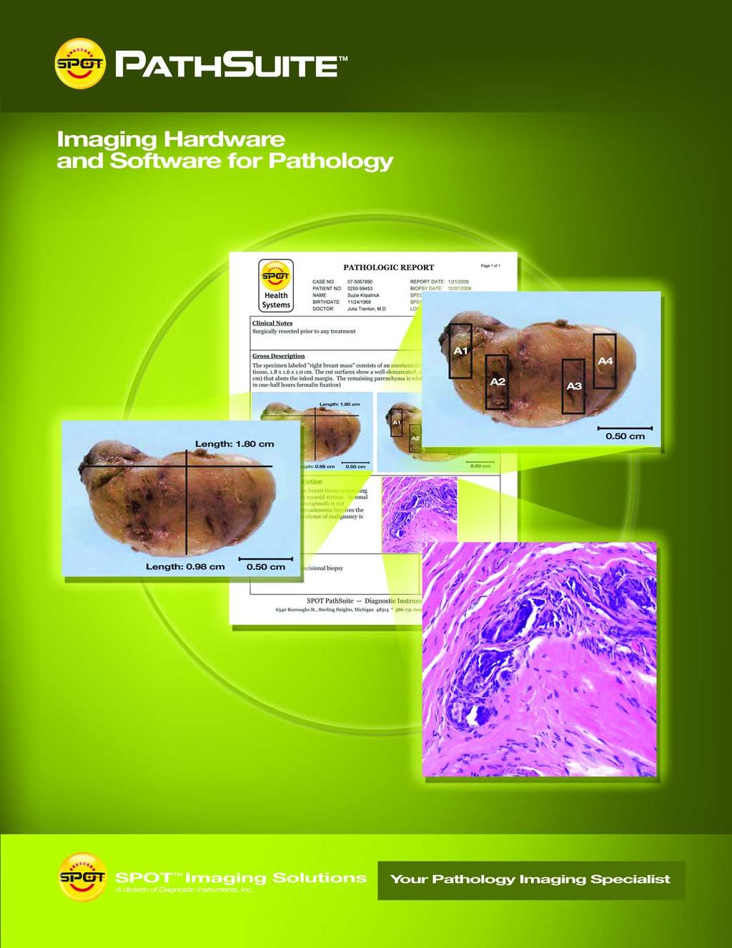
2 SPOT PathSuite Solutions The Perfect Fit PathStation TM Imaging system for the gross dissection in hood PathStand TM 40 imaging station for medium to large specimens PathSuite Is Easy To Use A turnkey solution for each of your needs Configurable user interface to meet your labs work practices Barcode data input speeds clerical work Simple on-screen image preview and image editing Computer controlled lens and camera provides wide field and magnified images PathSuite Enhances Traceability Eliminate clerical errors with barcode entry of case and session data Documentation of sample as received Documentation of labels as received Documentation of section and block location Documentation of cassettes used Touch-screen annotation, sample and section labeling Calibrated image capture allows quick measurement of length, area and more via touch screen interface Touch-screen keyboard for gloved-up work areas Foot switch control of software functions such as: Preview, Zoom, Focus, Capture, Dump, Save Auto-incrementing of sample, section and block labels PathSuite Is A World Class Solution Premium grade optics, cameras, computers and accessories Purpose designed - not a consumer product adaptation Outstanding images result from: > High resolution - 5Mp, 3Mp, 1.3 Mp options > CCT Correct Color Technology for "SPOT ON COLOR! Washable screens, stands and accessories to fit your lab environment

3 SPOT PathSuite Provides Systems to meet both your Macro and Micro imaging requirements: PathStand TM 24 imaging station for small to medium samples PathStereo TM small tissue dissections and needle biopsies PathScope TM microscopy slides PathSuite Gives You A Professional Image All-In-One computer implementation eliminates clutter Digital documents present a progressive impression to associates and patients Hosting web conferences tells your referral doctors and associates that you value their time PathSuite Provides The Digital Imaging Advantage Ability to save images to individual or multiple locations: > OS directories > SPOT database > Lab or hospital database Immediate access to images for other colleagues within your lab or at remote sites Interactive image sharing via internet web conference interface

4 PathSuite Provides A Full Set Of Easy To Use Tools Log In Case and sample login validates the sample-case relationship to ensure samples are not misfiled with the wrong case. Logging can be made via barcode scan or keystroke entry. Preview & Capture Easily start a preview session with a click of the foot pedal. Then compose your image with the zoom and focus controls and when satisfied, a single click captures the image. Measure Precalibrated captured images allow for immediate measurement of lengths, areas, angles or the simple addition of a calibration mark. Automatic Label Annotations Reduce your labeling work with auto-sequenced sample, block, section and feature counters. Formatting is configurable for site preferences (A1, A2 or 1A, 1B ). Freehand Drawing In a hurry or need the freedom associated with freehand drawing? PathSuite provides you with the tool and saves it digitally. Formal Drawing Formal presentation is supported with the full set of structured drawing tools: Line, Arrow, Text, Ellipse, Rectangle, Image/Logo Imprint. Formatting All annotation and measurement tools are supported by a full set of formatting options that can be set globally and edited. See the Details, Show the Details Digitally zoom and pan to see the details of your specimen. Need to present to a group? Use the full screen mode to provide the maximum resolution. Image Composite Tile images side by side or stack with the Merge function. LIMS Interface Laboratory Information Systems connect directly to PathSuite via the SPOT Extended LIMS Interface. Performing imaging with PathSuite hardware is as simple as: 1) clicking the Acquire button in your LIMS software, 2) Previewing, capturing and annotating with the PathSuite interface 3) Clicking the Transfer button in PathSuite to have the LIMS interface resume control with the newly acquired image ready for use. It couldn t be easier. Auto-Filing Saves Your Images Logically PathSuite automatically files your images based on Case #, Sample #, and Image #. Additionally, annotated and un-annotated versions of the image are saved during the same operation. What previously took minutes is done in seconds. Print Function Quickly select images for your printout, then select Auto Arrange or manually layout a page, it s simple. Consult Need to communicate with colleagues down the hall or across the country? GoToMeeting provides PathSuite systems with a cost effective, secure tele-pathology interface. Ask for a demo. Reporting PathSuite s standard reports provide you with a Pathology Diagnostic Report, a Gross Dissection Process Tracking Report, and Case Question Report. The reporting interface supports input during the three distinctly separate case tasks: registration, gross dissection and microscopy/diagnosis.


5 SPOT PathSuite Systems Supports Your Imaging and Data Collection from Grossing to Microscopy Grossing Process PathSuite Systems allow you to easily and routinely document your grossing process, providing the necessary images for tracking audits, informational reference images, report fulfillment and publishing support. Sample log in validates the Case-Sample relationship avoiding mismatches (via keystroke or barcode). Detailed close ups of lesions provide reference images of biopsy prior to dissection essential for telepathology discussion, follow up diagnostic reference, reporting and publication support. Case log in establishes the case session (via keystroke or barcode). Imaging of the sample container As Received provides visual verification of the containers received. Annotation of dissection sectioning and blocking concretely answers the pathologist questions of where tissue blocks originated. Imaging of the samples As Received with numerical count and measurements provides visual verification of the sample pieces received. Imaging of the loaded cassettes provides visual confirmation of what is being supplied to Histology. HISTOLOGY Slide Preparation Microscopy Process PathSuite Systems support your microscopy work by providing the pathologist tracking of the case and slide index, review of the gross dissection via images that provide details of the biopsy As Received, As Sectioned and As Blocked and then a convenient and easy to use imaging interface to document the microscopy samples of interest. Additionally, dictation and reporting are supported if needed. Case log in establishes the case session (via keystroke or barcode). Retrieve reference gross images and read gross description notes to answer any questions with regard to the biopsy dissection. Slide index log in validates the Case-Sample relationship, avoiding mismatches (via keystroke or barcode). View slides and capture microscopy images of diagnostic significance. Dictate description and diagnosis for use in report Populate, save and print a clean, professional diagnostic report. Dictation of gross description to be added to the diagnostic report.


6 SPOT TM PathSuite TM Software provides a common interface for... PathStation TM the gross dissection in hood PathStereo TM small tissue dissections and needle biopsies PathStand 24 TM imaging station for small to medium samples Certifications: FCC Part 15 Class A device CE: EN Product Safety CE: EN61326:2005 Emissions and Immunity testing PathStand 40 TM imaging station for medium to large specimens PathScope TM microscopy slides SPOT TM Imaging Solutions A division of Diagnostic Instruments, Inc Burroughs Avenue Sterling Heights, Michigan USA Toll Free USA: SPOT Phone: Fax: sales@spotimaging.com Web address:
PathStation TM Fits Your Lab
 PathStation TM Fits Your Lab PathStation hood Compact In-hood Imaging system. Supports in-process documentation PathStation Key Features 20 MP Image Capture Great HD Video 8.33 X Optical zoom 4X Digital
34X ZOOM. PathStand TM Routine. Superior Image Quality. PathStand Key Features. 1X Zoom. 8.33X Zoom
 PathStand TM Routine Complete stand-alone Imaging system. Supports in-process documentation. PathStand Key Features 20 MP Image Capture 1080p Live Video 34X zoom Metal Construction All-in-one Computer
34X ZOOM. PathStand TM Routine. Superior Image Quality. PathStand Key Features. 1X Zoom. 8.33X Zoom
 PathStand TM Routine Complete stand-alone Imaging system. Supports in-process documentation. PathStand Key Features 20 MP Image Capture 720p Live Video 34X zoom Metal Construction All-in-one Computer LED
MacroPATH The new line of Digital Imaging Systems for Grossing
 MILESTONE H E L P I N G P A T I E N T S MacroPATH The new line of Digital Imaging Systems for Grossing If the dimensions of the specimen are not recorded, the key section not taken, and the proper special
egross Ergonomic, mobile grossing workcenter with digital documentation of surgical specimens MILESTONE
 egross Ergonomic, mobile grossing workcenter with digital documentation of surgical specimens MILESTONE H E L P I N G P A T I E N T S The egross This all-in-one grossing workstation has been specifically
FRAUNHOFER INSTITUTE FOR INTEGRATED CIRCUITS IIS. MANUAL PANORAMIC MICROSCOPY WITH istix
 FRAUNHOFER INSTITUTE FOR INTEGRATED CIRCUITS IIS MANUAL PANORAMIC MICROSCOPY WITH istix CLINICAL DIAGNOSTICS AND MATERIAL SCIENCES IMPROVED BY DIGITAL MICROSCOPY B A C K G R O U N D Due to a high grade
DIGITAL-MICROSCOPY CAMERA SOLUTIONS USB 3.0
 DIGITAL-MICROSCOPY CAMERA SOLUTIONS USB 3.0 PixeLINK for Microscopy Applications PixeLINK will work with you to choose and integrate the optimal USB 3.0 camera for your microscopy project. Ideal for use
User Manual. Copyright 2010 Lumos. All rights reserved
 User Manual The contents of this document may not be copied nor duplicated in any form, in whole or in part, without prior written consent from Lumos. Lumos makes no warranties as to the accuracy of the
Teaching Digital Histology
 Teaching Digital Histology Carlos R. Morales Department of Anatomy and Cell Biology, McGill University, Montreal, Quebec, Canada The light microscope is one of the most widely used scientific instruments
Automated Digitization of Gram Stains. Centralized Reading. Decentralized Assessment. Improved Quality Management.
 Automated Digitization of Gram Stains Centralized Reading. Decentralized Assessment. Improved Quality Management. A GROWING DEMAND Gram staining is the rapid, easy, and inexpensive method for the assessment
Using the Microscope for a NANSLO Remote Web-based Science Lab Activity
 Using the Microscope for a NANSLO Remote Web-based Science Lab Activity MICROSCOPE RWSL LAB INTERFACE INSTRUCTIONS The Remote Web-based Science Lab (RWSL) microscope is a high quality digital microscope
Second Announcement Call for Participation. (Evaluation Criteria added)
") Second Announcement Call for Participation 2 nd International Scanner Contest (ISC) (Evaluation Criteria added) P. Hufnagl 1, T. Schrader 1, 2, M.G. Rojo 3, A. Laurinavicius 4, G. Kayser 5, Y. Yagi 6 1
Second Announcement Call for Participation 2 nd International Scanner Contest (ISC) (Evaluation Criteria added) P. Hufnagl 1, T. Schrader 1, 2, M.G. Rojo 3, A. Laurinavicius 4, G. Kayser 5, Y. Yagi 6 1
μscope Microscopy Software
 μscope Microscopy Software Pixelink μscope Essentials (ES) Software is an easy-to-use robust image capture tool optimized for productivity. Pixelink μscope Standard (SE) Software had added features, making
Positive Pixel Count Algorithm. User s Guide
 Positive Pixel Count Algorithm User s Guide Copyright 2004, 2006 2008 Aperio Technologies, Inc. Part Number/Revision: MAN 0024, Revision B Date: December 9, 2008 This document applies to software versions
MIRAX SCAN The new way of looking at pathology
 Microscopy from Carl Zeiss MIRAX SCAN The new way of looking at pathology Greater reliability. Greater efficiency. Plus points for your diagnostics Better. More efficient. Quality as a factor for success
Color aspects and Color Standardization in Digital Microscopy
 Color aspects and Color Standardization in Digital Microscopy Yukako Yagi, PhD yyagi@partners.org Director of the MGH Pathology Imaging & Communication Technology Center Assistant Professor of Pathology,
InScape: Making Virtual Pathology a Reality
 InScape: Making Virtual Pathology a Reality Sally S. Agersborg, M.D., Ph.D. Quest Diagnostics, Nichols Institute San Juan Capistrano, CA Company Overview Quest Diagnostics, Nichols Institute the world
CHROMACAL User Guide (v 1.1) User Guide
 User Guide") CHROMACAL User Guide (v 1.1) User Guide User Guide Notice Hello and welcome to the User Guide for the Datacolor CHROMACAL Color Calibration System for Optical Microscopy, a cross-platform solution that
CHROMACAL User Guide (v 1.1) User Guide User Guide Notice Hello and welcome to the User Guide for the Datacolor CHROMACAL Color Calibration System for Optical Microscopy, a cross-platform solution that
Image Viewing. with ImageScope
 Image Viewing with ImageScope ImageScope Components Use ImageScope to View These File Types: ScanScope Virtual Slides.SVS files created when the ScanScope scanner scans glass microscope slides. JPEG files
High-sensitivity. optical molecular imaging and high-resolution digital X-ray. In-Vivo Imaging Systems
 High-sensitivity optical molecular imaging and high-resolution digital X-ray In-Vivo Imaging Systems In vivo imaging solutions available in several packages Carestream Molecular Imaging offers a selection
Training Guide for Carl Zeiss AxioZoom V16 Stereo Microscope
 Training Guide for Carl Zeiss AxioZoom V16 Stereo Microscope ZEN 2012 Optical Imaging & Vital Microscopy Core Baylor College of Medicine (2017) Power ON Routine 1 2 If you require fluorescence imaging,
Axioscan - Startup. 1. Turn on the Axioscan on (button to the left) and turn on the computer
 and turn on the computer") Axioscan - Startup 1. Turn on the Axioscan on (button to the left) and turn on the computer 2. Log on and start the ZEN Blue software from the desktop 3. Press ZEN slidescan and Start System 4. Start by
Axioscan - Startup 1. Turn on the Axioscan on (button to the left) and turn on the computer 2. Log on and start the ZEN Blue software from the desktop 3. Press ZEN slidescan and Start System 4. Start by
ScanArray Overview. Principle of Operation. Instrument Components
 ScanArray Overview The GSI Lumonics ScanArrayÒ Microarray Analysis System is a scanning laser confocal fluorescence microscope that is used to determine the fluorescence intensity of a two-dimensional
This document contains work instructions related to utilizing the dental imaging application, XrayVision version 4.0.
 Apteryx Inc. 313 S. High St. Suite 200 Akron, OH 44308 330-376-0889 voice 330-376-0788 fax sales@apteryx.com www.apteryx.com XrayVision Quick Start User Manual Abstract Abstract Abstract This document
Bowen Hills Histopathology
 Bowen Hills Histopathology Histology s Bowen Hills Challenges Relocate and amalgamate Taringa and Indooroopilly Labs into a single complex histology laboratory Change workflow, staff structure, install
Installation & User Manual Micro-Image Capture 7
 Installation & User Manual Micro-Image Capture 7 Ver1.2016 Product Warranty Quality Assurance Every Micro-Image Capture system passes quality assurance tests including focus, resolution quality and mechanical
IHE Anatomic Pathology Redesign. Sardinia, Italy Nov , 2017
 IHE Anatomic Pathology Redesign Sardinia, Italy Nov. 13-15, 2017 Specimen Workflow in a Nutshell (variations likely depending on context, e.g. collection location) Create Encounter Generate Results Order
Tissue-Tek TEC 6 Embedding Console System
 The most trusted name in reliable, comfortable and easy-to-use embedding Tissue-Tek TEC 6 Embedding Console System 2 The embedding system you trust with all of the enhancements you need The Tissue-Tek
User Operation of JEOL 1200 EX II
 **Log onto Computer** Open item program Start Up Procedure User Operation of JEOL 1200 EX II 1. If scope is not running, locate an electron microscopy technician (EMT) to find out why not. 2. Turn up brightness
FCR XL-2 and FCR XC-2. High-quality digital x-ray that's perfect for your private practice.
 F U J I F I L M D I G I T A L X - R A Y FCR XL-2 and FCR XC-2. High-quality digital x-ray that's perfect for your private practice. So small, yet so powerful. With a compact footprint of less than 2.5
The ideal K-12 science microscope solution. User Guide. for use with the Nova5000
 The ideal K-12 science microscope solution User Guide for use with the Nova5000 NovaScope User Guide Information in this document is subject to change without notice. 2009 Fourier Systems Ltd. All rights
BioSpectrum Imaging System
 BioSpectrum Imaging System Imaging Made Easy for Chemiluminescence Bioluminescence Colorimetric Fluorescence MegaCam 810 Camera OptiChemi 610 Camera BioChemi 510 Camera GelCam 310 Camera 8.1 megapixel
User Manual Veterinary
 Veterinary Acquisition and diagnostic software Doc No.: Rev 1.0.1 Aug 2013 Part No.: CR-FPM-04-022-EN-S 3DISC, FireCR, Quantor and the 3D Cube are trademarks of 3D Imaging & Simulations Corp, South Korea,
Digital Imaging in Anatomic Pathology
 your lab focus CE update [generalist histology] Digital Imaging in Anatomic Pathology Ted F. Beals, MD From the Department of Veteran Affairs, Veterans Health Administration, and Department of Pathology,
This document contains work instructions related to utilizing the dental imaging application, XVLite version 3.12 and above.
 XVLITE Apteryx Inc. 313 S. High St. Suite 200 Akron, OH 44308 330-376-0889 voice 330-376-0788 fax sales@apteryx.com www.apteryx.com XVLITE User Manual Abstract Abstract This document contains work instructions
Quick Guide. LSM 5 MP, LSM 510 and LSM 510 META. Laser Scanning Microscopes. We make it visible. M i c r o s c o p y f r o m C a r l Z e i s s
 LSM 5 MP, LSM 510 and LSM 510 META M i c r o s c o p y f r o m C a r l Z e i s s Quick Guide Laser Scanning Microscopes LSM Software ZEN 2007 August 2007 We make it visible. Contents Page Contents... 1
Cast Iron Analysis. Nodular cast iron analysis
 Cast Iron Analysis This dhs Image Data Base module analyzes the microstructure of cast iron. The graphite particles actual make-up is crucial for material properties such as elasticity and fracture behaviour.
10.2. Scanning Document Camera Scoring. Page 1 of 5. How do I score answer sheets using a document camera? STEP 1
 Step by Step How do I score answer sheets using a document camera? STEP 1 Click on the Assessment icon in the top navigation bar. STEP 2 To locate your assessment in an assessment list, first select the
EXC500p-- PATHOLOGY MICROSCOPE. EXC500hd -- HD DIGITAL PATHOLOGY MICROSCOPE. EXC500r -- RESEARCH MICROSCOPE EXC500-LABORATORY SCOPE
 EXC500p-- PATHOLOGY MICROSCOPE EXC500hd -- HD DIGITAL PATHOLOGY MICROSCOPE EXC500r -- RESEARCH MICROSCOPE EXC500-LABORATORY SCOPE The EXC500 Pathology and Laboratory Microscope is the most optically advanced
Users Guide. Meditory CORPORATION. Version 2015C v1.0 7/20/2015
 Patent No.: US 8,823,770 B2 & other patents pending Users Guide Version 2015C v1.0 7/20/2015 Meditory CORPORATION 7600 Grand River Road, Suite 230 Brighton, MI 48114 T (810) 225-9720 F (810) 225-9726 Toll
User Manual for PROGRES GRYPHAX software
 User Manual for PROGRES GRYPHAX software PROGRES GRYPHAX software JENOPTIK l Healthcare & Industry JENOPTIK Optical Systems GmbH Göschwitzer Straße 25 07745 Jena, Germany Dear user, Please read the instructions
Innovation and Quality Made in Germany
 EN Headquarters Canfield Scientific, Inc. Parsippany New Jersey Canfield Scientific GmbH Bielefeld Germany Innovation and Quality Made in Germany VISIOMED GmbH has been one of the most innovative companies
Quality-Management. Portable micrograph laboratory Microlab ML 3002
 Quality-Management Portable micrograph laboratory Microlab ML 3002 Fully functional micrograph laboratory for creating crimp micrographs in a handy case Portable laboratory to prepare crimp cross sections
ImageScope. User s Guide
 ImageScope User s Guide Copyright 2006 2011, Aperio Technologies, Inc. Part Number/Revision: MAN 0001, Revision I Date: 5 January 2011 This document applies to software versions Release 11 and later. All
Which equipment is necessary? How is the panorama created?
 Congratulations! By purchasing your Panorama-VR-System you have acquired a tool, which enables you - together with a digital or analog camera, a tripod and a personal computer - to generate high quality
Buxton & District U3A Digital Photography Beginners Group Lesson 5: Simple Editing. 5 November 2013
 U3A Group Lesson 5: Simple Editing 5 November 2013 Programme Buxton & District 19 September Exploring your camera 1 October You ve taken some pictures now what? (Viewing pictures; filing on your computer)
4.0 How to Turn On the Selenia Dimensions
 Chapter 2 System Controls and Indicators How to Turn On the Selenia Dimensions 4.0 How to Turn On the Selenia Dimensions 4.1 Preparation 1. Reset all three Emergency Off switches. Emergency Off Switches
In-Vivo IMAGING SYSTEMS. A complete line of high resolution optical & X-ray systems for pre-clinical imaging
 In-Vivo IMAGING SYSTEMS A complete line of high resolution optical & X-ray systems for pre-clinical imaging In-Vivo Imaging Systems Carestream is a strong, successful, multi-billion dollar, international
Copyright 2014 SOTA Imaging. All rights reserved. The CLIOSOFT software includes the following parts copyrighted by other parties:
 2.0 User Manual Copyright 2014 SOTA Imaging. All rights reserved. This manual and the software described herein are protected by copyright laws and international copyright treaties, as well as other intellectual
AF Area Mode. Face Priority
 Chapter 4: The Shooting Menu 71 AF Area Mode This next option on the second screen of the Shooting menu gives you several options for controlling how the autofocus frame is set up when the camera is in
Technical Aspects in Digital Pathology
 Technical Aspects in Digital Pathology Yukako Yagi, PhD yyagi@mgh.harvard.edu Director of the MGH Pathology Imaging & Communication Technology Center Assistant Professor of Pathology, Harvard Medical School
Solutions Page Content ImagePilot. Primary keywords: Digital radiography Computed radiography Image viewing and storage
 Solutions Page Content Primary keywords: Digital radiography Computed radiography Image viewing and storage Solution Description For facilities with medium volume imaging, Konica Minolta s original all-in-one
GlobiScope Analysis Software for the Globisens QX7 Digital Microscope. Quick Start Guide
 GlobiScope Analysis Software for the Globisens QX7 Digital Microscope Quick Start Guide Contents GlobiScope Overview... 1 Overview of home screen... 2 General Settings... 2 Measurements... 3 Movie capture...
Relationship to theory: This activity involves the motion of bodies under constant velocity.
 UNIFORM MOTION Lab format: this lab is a remote lab activity Relationship to theory: This activity involves the motion of bodies under constant velocity. LEARNING OBJECTIVES Read and understand these instructions
GlassSpection User Guide
 i GlassSpection User Guide GlassSpection User Guide v1.1a January2011 ii Support: Support for GlassSpection is available from Pyramid Imaging. Send any questions or test images you want us to evaluate
Recitation 2 Introduction to Photoshop
 Recitation 2 Introduction to Photoshop What is Adobe Photoshop? Adobe Photoshop is a tool for creating digital graphics either by starting with a scanned photograph or artwork or by creating the graphics
China Resources Wandong Medical Equipment Co., Ltd. High Frequency 50kW Digital RF System - HF51-5
 China Resources Wandong Medical Equipment Co., Ltd. High Frequency 50kW Digital RF System - HF51-5 #3, No.9, Jiuxianqiaodong Road, Chaoyang District, Beijing 100015, P.R. China E-mail: international@wandong.com.cn
Huvitz Digital Microscope HDS-5800
 Huvitz Digital Microscope HDS-5800 Dimensions unit : mm Huvitz Digital Microscope HDS-5800 HDS-MC HDS-SS50 The world s first, convert the magnification from 50x to 5,800x with a zoom lens HDS-TS50 Huvitz
Figure 1. Oil-immersion objectives available for use with the Lionheart FX.
 Tech Note Oil Objective Introduction The Lionheart FX automated imager is compatible with high numerical aperture oil immersion objectives. These objectives offer magnification up to 100X and significantly
PLD5600A High Frequency Digital Gastrointestinal &DR System(630mA)
") PLD5600A High Frequency Digital Gastrointestinal &DR System(630mA) Application: Full support perspective, gastrointestinal spot film, GI (barium meal, barium enema), orthopedic photography, pediatrics
PLD5600A High Frequency Digital Gastrointestinal &DR System(630mA) Application: Full support perspective, gastrointestinal spot film, GI (barium meal, barium enema), orthopedic photography, pediatrics
GEA Omni TM control panel. The intuitive touch for refrigeration and gas compression control technology. Evaporator. Compressor. OmniView.
 Compressor Evaporator OmniView Condenser Sequencer Energy Saver GEA Omni TM control panel The intuitive touch for refrigeration and gas compression control technology engineering for a better world GEA
ImagesPlus Basic Interface Operation
 ImagesPlus Basic Interface Operation The basic interface operation menu options are located on the File, View, Open Images, Open Operators, and Help main menus. File Menu New The New command creates a
The Flash IIP Console is the heart of every FCR system. It s designed to maximize productivity in the busiest environments.
 Choose the FCR system that best fits your practice. The FCR XL-2. Perfect for higher-volume environments. It can process up to 94 images per hour yet it fits right into small exam rooms or offices where
Reliably embed with comfort and speed
 Reliably embed with comfort and speed Tissue-Tek TEC 5 Tissue Embedding Console System Satisfy all of your tissue embedding needs quickly, reliably and ergonomically The proven reliability of the Tissue-Tek
Optika ISview. Image acquisition and processing software. Instruction Manual
 Optika ISview Image acquisition and processing software Instruction Manual Key to the Instruction Manual IS is shortened name used for OptikaISview Square brackets are used to indicate items such as menu
jfo^u=sfbtbo= oéäé~ëé=nkno= lééê~íáåö=j~åì~ä=
 jfo^u=sfbtbo= oéäé~ëé=nkno= lééê~íáåö=j~åì~ä= Carl Zeiss COPYRIGHT / TRADEMARK MIRAX VIEWER Prior knowledge of this manual is required for proper Viewer operation. You are therefore advised to familiarize
Progeny Imaging. User Guide V x and Higher. Part Number: ECN: P1808 REV. F
 Progeny Imaging User Guide V. 1.6.0.x and Higher Part Number: 00-02-1598 ECN: P1808 REV. F Contents 1 About This Manual... 5 How to Use this Guide... 5 Text Conventions... 5 Getting Assistance... 6 2 Overview...
University of California, Santa Barbara. CS189 Fall 17 Capstone. VR Telemedicine. Product Requirement Documentation
 University of California, Santa Barbara CS189 Fall 17 Capstone VR Telemedicine Product Requirement Documentation Jinfa Zhu Kenneth Chan Shouzhi Wan Xiaohe He Yuanqi Li Supervised by Ole Eichhorn Helen
Network Scanner Guide for Fiery S300 50C-KM
 Network Scanner Guide for Fiery S300 50C-KM Read this manual before printing. Keep readily available for reference. User's Guide Introduction Thank you very much for purchasing the Fiery S300 50C-KM. This
ChemiDoc-It Imaging System
 ChemiDoc-It Imaging System Ultra dark chamber and highly sensitive, scientific-grade CCD camera for chemiluminescence imaging ChemiDoc-It darkroom is light tight creating optimum imaging conditions for
SHORT INSTRUCTIONS FOR OPERATING LSM1/2 (Zeiss LSM510) AT CIAN Version 1.4, September 2014
 AT CIAN Version 1.4, September 2014") CIAN LSM1 or LSM2 short instructions, version 1.4, September 2014 page 1 of 6 SHORT INSTRUCTIONS FOR OPERATING LSM1/2 (Zeiss LSM510) AT CIAN Version 1.4, September 2014 Before starting To work with LSM1
CIAN LSM1 or LSM2 short instructions, version 1.4, September 2014 page 1 of 6 SHORT INSTRUCTIONS FOR OPERATING LSM1/2 (Zeiss LSM510) AT CIAN Version 1.4, September 2014 Before starting To work with LSM1
WELCOME WHAT S IN THE BOX
 WELCOME Congratulations on purchasing your Visioneer PaperPort flatbed scanner. With your scanner, you can quickly scan paper documents and color photos to place their electronic images on your computer.
User Manual. cellsens 1.16 LIFE SCIENCE IMAGING SOFTWARE
 User Manual cellsens 1.16 LIFE SCIENCE IMAGING SOFTWARE Any copyrights relating to this manual shall belong to OLYMPUS CORPORATION. We at OLYMPUS CORPORATION have tried to make the information contained
2D, 3D CT Intervention, and CT Fluoroscopy
 2D, 3D CT Intervention, and CT Fluoroscopy SOMATOM Definition, Definition AS, Definition Flash Answers for life. Siemens CT Vision Siemens CT Vision The justification for the existence of the entire medical
CR30-ORACLE CR. with Office Viewer. A Revelation in Computed Radiography. Innovations in Digital Imaging. Solution for Quantum Medical Imaging
 CR30-ORACLE CR with Office Viewer A Revelation in Computed Radiography Innovations in Digital Imaging Solution for Quantum Medical Imaging CR30-ORACLE CR with Office Viewer The CR30-ORACLE CR System Package
House Design Tutorial
 House Design Tutorial This House Design Tutorial shows you how to get started on a design project. The tutorials that follow continue with the same plan. When you are finished, you will have created a
Zeiss LSM 780 Protocol
 Zeiss LSM 780 Protocol 1) System Startup F Please note the sign-up policy. You must inform the facility at least 24 hours beforehand if you can t come; otherwise, you will receive a charge for unused time.
Basic Users Manual for Tecnai-F20 TEM
 Basic Users Manual for Tecnai-F20 TEM NB: This document contains my personal notes on the operating procedure of the Tecnai F20 and may be used as a rough guide for those new to the microscope. It may
Operating Rausch ScanCam within POSM.
 Operating Rausch ScanCam within POSM. POSM (Pipeline Observation System Management) // posmsoftware.com // info@posmsoftware.com // 859-274-0041 RAUSCH USA // www.rauschusa.com // reusa@rauschusa.com //
Axioscan - Startup. 1. Turn on the Axioscan (button to the left) and turn on the computer. 2. Log on and start the ZEN Blue software from the desktop
 and turn on the computer. 2. Log on and start the ZEN Blue software from the desktop") Axioscan - Startup 1. Turn on the Axioscan (button to the left) and turn on the computer 2. Log on and start the ZEN Blue software from the desktop 3. Press ZEN slidescan and Start System 4. Start by changing
Axioscan - Startup 1. Turn on the Axioscan (button to the left) and turn on the computer 2. Log on and start the ZEN Blue software from the desktop 3. Press ZEN slidescan and Start System 4. Start by changing
In our previous lecture, we understood the vital parameters to be taken into consideration before data acquisition and scanning.
 Interactomics: Protein Arrays & Label Free Biosensors Professor Sanjeeva Srivastava MOOC NPTEL Course Indian Institute of Technology Bombay Module 7 Lecture No 34 Software for Image scanning and data processing
Brief Operation Manual for Imaging on BX61W1
 DBS CONFOCAL LAB Brief Operation Manual for Imaging on BX61W1 Olympus cellsens Dimension Tong Yan 9/19/2011 This briefing manual is for quick setup of imaging experiment. It includes Acquiring a single
JSM 6060 LV SCANNING ELECTRON MICROSCOPE STANDARD OPERATING PROCEDURES
 JSM 6060 LV SCANNING ELECTRON MICROSCOPE STANDARD OPERATING PROCEDURES RULES All users must go through a series of standard operation procedure training. For more information contact: Longlong Liao Teaching
Label Studio Quick Start Guide
 Label Studio Quick Start Guide Overview The goal of the LabelStudio program is to help you layout and manage bulk jobs containing a mix of different logos, that all require a specified quanity. LabelStudio
Catholic Education Center, LLC User Manual. If you need further assistance, please
 1 Catholic Education Center, LLC User Manual If you need further assistance, please email: catholiceducationcenter@yahoo.com or, telephone: 703-785-2319 (M-F: 9-5) Getting Started. Using your internet
SPECTRALIS Training Guide
 SPECTRALIS Training Guide SPECTRALIS Diagram 1 SPECTRALIS Training Guide Table of Contents 1. Entering Patient Information & Aligning the Patient a. Start Up/Shut Down the System... 4 b. Examine a New
Mira M-3 Handheld Raman Spectrometer. On-site verification of materials in seconds
 Mira M-3 Handheld Raman Spectrometer On-site verification of materials in seconds Mira M-3 compact, fast, and highly accurate 02 The Mira M-3 is one of the fastest and most compact handheld Raman spectrometers
Zeiss 880 Training Notes Zen 2.3
 Zeiss 880 Training Notes Zen 2.3 1 Turn on the HXP 120V Lamp 2 Turn on Main Power Switch Turn on the Systems PC Switch Turn on the Components Switch. 3 4 5 Turn on the PC and log into your account. Start
Supplement. ScanMaker s450/s350 features, scenarios, and information. Getting to Know Your ScanMaker s450/s350
 Supplement ScanMaker s450/s350 features, scenarios, and information Getting to Know Your ScanMaker s450/s350 The ScanMaker s450/s350 high-performance scanner with the versatility to scan photos and film.
Camera settings for photographic documentation with the HEINE DELTA 20 PLUS / DELTA 20 Dermatoscope and HEINE SLR Photo adaptor
 Camera settings for photographic documentation with the HEINE DELTA 20 PLUS / DELTA 20 Dermatoscope and HEINE SLR Photo adaptor Digital high-resolution photography is the method of choice for optimum image
Zeiss 780 Training Notes
 Zeiss 780 Training Notes Turn on Main Switch, System PC and Components Switches 780 Start up sequence Do you need the argon laser (458, 488, 514 nm lines)? Yes Turn on the laser s main power switch and
CircumSpect TM 360 Degree Label Verification and Inspection Technology
 CircumSpect TM 360 Degree Label Verification and Inspection Technology Written by: 7 Old Towne Way Sturbridge, MA 01518 Contact: Joe Gugliotti Cell: 978-551-4160 Fax: 508-347-1355 jgugliotti@machinevc.com
Automated Imaging Technology to Simplify Your Workflow!
 Automated Imaging Technology to Simplify Your Workflow! BioSpectrum Imaging System Imaging Made Easy for Chemiluminescence Bioluminescence Colorimetric Fluorescence MegaCam 810 Camera OptiChemi 600 Camera
Y N C R O S C O P Y A DIVISION OF THE SYNOPTICS GROUP
 S Y N C R O S C O P Y A DIVISION OF THE SYNOPTICS GROUP THE PROBLEM: As a microscopist you often have to work with samples that are difficult to focus. When viewing a 3-D sample using an optical microscope
XF Feature Update #4 Firmware Release Note
 XF Feature Update #4 Firmware Release Note This release note describes the new features of Feature Update #4 for the XF Camera System. Downloading and installing Feature Update #4 (Camera package file
ThermaViz. Operating Manual. The Innovative Two-Wavelength Imaging Pyrometer
 ThermaViz The Innovative Two-Wavelength Imaging Pyrometer Operating Manual The integration of advanced optical diagnostics and intelligent materials processing for temperature measurement and process control.
Cutwork With Generations Automatic Digitizing Software By Bernadette Griffith, Director of Educational Services, Notcina Corp
 In this lesson we are going to create a cutwork pattern using our scanner, an old pattern, a black felt tip marker (if necessary) and the editing tools in Generations. You will need to understand the basics
Supplement. ScanMaker s480/s380 features, scenarios, and information. Getting to Know Your ScanMaker s480/s380
 Supplement ScanMaker s480/s380 features, scenarios, and information Getting to Know Your ScanMaker s480/s380 The ScanMaker s480/s380 is a high-performance scanner with the versatility to scan photos and
THEORY AND APPROACHES TO AUTOMATED IMAGE ANALYSIS IN DIGITAL PATHOLOGY
 THEORY AND APPROACHES TO AUTOMATED IMAGE ANALYSIS IN DIGITAL PATHOLOGY Kyle Takayama, MS Charles River Laboratories EVERY STEP OF THE WAY EVERY STEP OF THE WAY MORPHOMETRY Measurements or counts performed
Quick Guide for Zeiss 710 Laser Scanning Confocal MGH Cancer Center
 Quick Guide for Zeiss 710 Laser Scanning Confocal MGH Cancer Center For any questions or concerns, please contact: Linda Nieman lnieman@mgh.harvard.edu Office: (617) 643-9684 Cell: (512) 565-8076 Chenyue