4D dynamic imaging of the eye using ultrahigh speed SS- OCT
|
|
|
- Marlene Bryant
- 6 years ago
- Views:
Transcription
1 4D dynamic imaging of the eye using ultrahigh speed SS- OCT The MT Faculty has made this article openly available. Please share how this access benefits you. Your story matters. Citation As Published Publisher Liu, Jonathan J., reneusz Grulkowski, Benjamin Potsaid, Vijaysekhar Jayaraman, Alex E. Cable, Martin F. Kraus, Joachim Hornegger, Jay S. Duker, and James G. Fujimoto. 4D Dynamic maging of the Eye Using Ultrahigh Speed SS-OCT. Edited by Fabrice Manns, Per G. Söderberg, and Arthur Ho. Ophthalmic Technologies XX (March 26, 2013). (2013) COPYRGHT Society of Photo-Optical nstrumentation Engineers (SPE) SPE Version Final published version Accessed Thu Jun 07 13:06:43 EDT 2018 Citable Link Terms of Use Detailed Terms Article is made available in accordance with the publisher's policy and may be subject to US copyright law. Please refer to the publisher's site for terms of use.
2 4-D dynamic imaging of the eye using ultrahigh speed SS-OCT Jonathan J. Liu 1, reneusz Grulkowski 1, Benjamin Potsaid 1,2, Vijaysekhar Jayaraman 3, Alex E. Cable 2, Martin F. Kraus 4, Joachim Hornegger 4, Jay S. Duker 5, and James G. Fujimoto 1 1 Department of Electrical Engineering and Computer Science & Research Laboratory of Electronics, Massachusetts nstitute of Technology, Cambridge, MA, USA 02139; 2 Advanced maging Group, Thorlabs nc., Newton, NJ, USA 07860; 3 Praevium Research nc., Santa Barbara, CA, USA 93111; 4 Pattern Recognition Lab & School of Advanced Optical Technologies, University Erlangen-Nuremberg, Erlangen, Germany 91054; 5 New England Eye Center & Tufts Medical Center, Tufts University, Boston, MA, USA 02116; ABSTRACT Recent advances in swept-source / Fourier domain optical coherence tomography (SS-OCT) technology enable in vivo ultrahigh speed imaging, offering a promising technique for four-dimensional (4-D) imaging of the eye. Using an ultrahigh speed tunable vertical cavity surface emitting laser (VCSEL) light source based SS-OCT prototype system, we performed imaging of human eye dynamics in four different imaging modes: 1) Pupillary reaction to light at 200,000 axial scans per second and 9 μm resolution in tissue. 2) Anterior eye focusing dynamics at 100,000 axial scans per second and 9 μm resolution in tissue. 3) Tear film break up at 50,000 axial scans per second and 19 μm resolution in tissue. 4) Retinal blood flow at 800,000 axial scans per second and 12 μm resolution in tissue. The combination of tunable ultrahigh speeds and long coherence length of the VCSEL along with the outstanding roll-off performance of SS-OCT makes this technology an ideal tool for time-resolved volumetric imaging of the eye. Visualization and quantitative analysis of 4-D OCT data can potentially provide insight to functional and structural changes in the eye during disease progression. Ultrahigh speed imaging using SS-OCT promises to enable novel 4-D visualization of realtime dynamic processes of the human eye. Furthermore, this non-invasive imaging technology is a promising tool for research to characterize and understand a variety of visual functions. Keywords: 4D, SS-OCT, VCSEL, ophthalmology, pupil, accommodation, tear film, retinal blood flow 1. NTRODUCTON Recent developments in swept-source and spectral domain optical coherence tomography (OCT) technologies have enabled ultrahigh speed OCT imaging at over 100,000 axial scans (A-scans) per second [1-4]. Novel ultralong range and ultrahigh speed imaging of the eye has been demonstrated using SS-OCT with vertical cavity surface emitting laser (VCSEL) technology [5]. n this paper, we demonstrate time resolved volumetric data using four-dimensional (4-D) OCT with speeds up to forty volumes per second. We imaged pupillary reactions, anterior eye focusing dynamics, tear film break up, and retinal blood flow. The real-time dynamics of the human eye is captured and analyzed for characterizing visual functions. 2. MATERALS AND METHODS We developed a VCSEL light source based prototype SS-OCT system (Figure 1) [5]. The laser was operated in four different modes with center wavelengths at around 1060 nm (Table 1). To image the pupillary reaction, the VCSEL light source was operated at an axial scan rate of 200 khz with 85 nm tuning range yielding 9 μm resolution and 5.6 mm Nyquist-limited imaging range in tissue. To image anterior eye accommodation dynamics, the axial scan rate was reduced to 100 khz to achieve a 10.3 mm Nyquist-limited imaging range in tissue. To image tear film break up, the VCSEL light source was operated at a reduced axial scan rate of 50 khz and reduced 30 nm tuning range for 19 μm Ophthalmic Technologies XX, edited by Fabrice Manns, Per G. Söderberg, Arthur Ho, Proc. of SPE Vol. 8567, 85670X 2013 SPE CCC code: /13/$18 doi: / Proc. of SPE Vol X-1

 at either GSPS or a fixed 500 MSPS sampling rate with dual channel")
 safety limits.")
 Referernce MZ (B) VCSEL j Module r 80:20 'k \.")
 VCSEL based SS-OCT prototype setup. (B) Different interface configurations.")


3 resolution and 60 mm Nyquist-limited imaging range in tissue. Finally, to image retinal blood flow, the VCSEL light source was operated at an axial scan rate of 800 khz and 50 nm tuning range for 12 μm resolution and 2.5mm Nyquista fixed 1 limited imaging range in tissue. A/D data acquisition cards (ATS9870 & ATS9350; AlazarTech) at either GSPS or a fixed 500 MSPS sampling rate with dual channel acquisition were used. The incident power on the cornea for all imaging modes was 1.9 mw, within the American National Standard nstitute (ANS) safety limits. Studies were performed under protocols approved by the MT Committee on n the Use of Humans as Experimental Subjects (COUHES) and written consent from participants was obtained. (A) Referernce MZ (B) VCSEL j Module r 80:20 'k \._ 8c Sample Arm Patient nterface SC 1 Anterior - Segment Full -Eye- Length nterface FT J Sc ox T i L _2 1 \ U S,H VTM RR 1 1 Reference Figure 1. (A) VCSEL based SS-OCT prototype setup. (B) Different interface configurations. Full-eye-length imaging was performed by using a longer focal length lens in the anterior segment configuration. Retinal imaging was performed by adding ocular lens to the anterior segment configuration and adjusting thee fixation target path. SC galvanometric scanners, FT fixation target, DM dichroic mirror, DC dispersion compensation glass, RR retroreflector, PDB balanced photodetectors, MZ Mach-Zehnder interferometer. Table 1. Configuration of VCSEL SS-OCT system. Parameter Effective imaging speed Wavelength tuning range 85 nm Axial resolution (tissue) Sample arm interface Transversee resolution Acquisition card Pupillary response 200,000 A-scans/s 100,000 A-scans/s 50,0000 A-scans/s 9 µm Anteriorr segment 30 µm ATS9870 (1 GSPS) Accommodat tion 85 nm 9 µm Anterior segment 30 µm ATS9870 (1 GSPS) Tear film break up 30 nm 19 µm Full eye length 73 µm ATS9870 (1 GSPS) Retinal blood flow 800,000 A-scans/s 50 nm 12 µm Retinal 25 µm ATS9350 (500 MSPS) Proc. of SPE Vol X-2


![closure glaucoma [6-8].](/docs-images/79/79563778/images/4-11.jpg "We demonstrated 4-D imaging")


.")


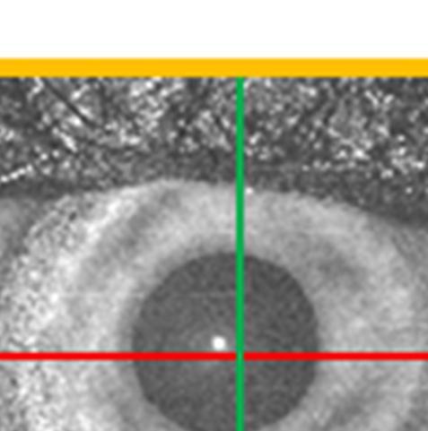
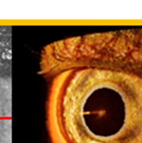
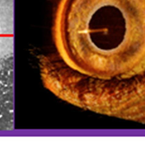

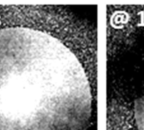
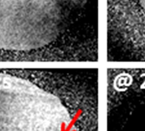
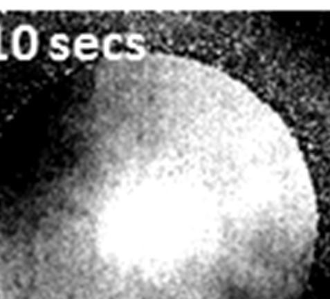
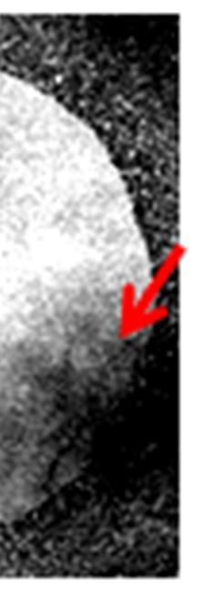
4 3. RESULTSS AND DSCUSSON 3.1 Pupillary response The iris is a dynamic structure whose configuration regularly changes in response to light and during accommodation. Dynamic changes in intraocular structures caused by illumination or dilation are suggested to be risk factors for glaucoma development. Studying the dynamic response of the pupil to dark-light stimulus may provide a more comprehensive assessment of risks to primary-angle closure development and may help understand the pathophysiology of angle closure glaucoma [6-8]. We demonstrated 4-D imaging of the pupil response to light stimulus from an LED positioned adjacent to the eye, where sequential 150 x 150 axial-scan volumes over 17 mm x 17 mm were acquired at ~8 volumes per second for 5 seconds with an axial scan rate of 200 khzz enabling the visualization of changes in the iris in time and in three dimensions (Video 1). The pupillary response is shown in Figuree 2 where two time points in the 4-D data are displayed. The iris response to light stimulus can be quantitatively analyzed by measuring pupil size/area changes in time. As shown in Figure 2(A), the pupil area decreased drastically when light stimulus was applied. n contrast, the time constant of the pupil diameter recovery was longer than when stimulus was applied. Video 1. 4-D OCT imaging of the pupillary reflex. Light stimulus ; sec M! JO' J 1 Figure 2. 4-D OCT imaging of the pupillary reflex. (A) Plot of pupil areaa vs time. (B) Volume#1 at 0.13 seconds. (C) Volume# #19 at 2.38 seconds. (D) Volume#38 at 4.5 seconds. The constriction of the iriss and the decrease in pupil area is apparent when comparing volume#1 and volume#19. The dilation of thee iris and increase in pupil area is visible when comparing volume#19 and volume #38. The cross-sectional OCT images show the iris structure while the enface OCT image depicts the pupil area. 1 Proc. of SPE Vol X-3


 Volume #30 at 3 seconds.")


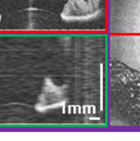
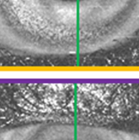
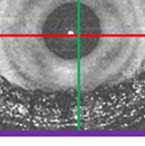
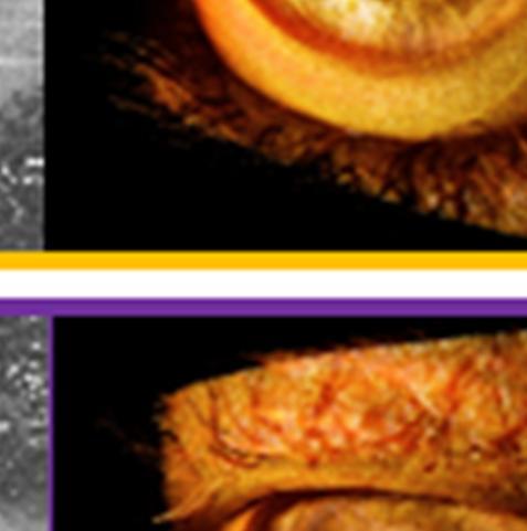
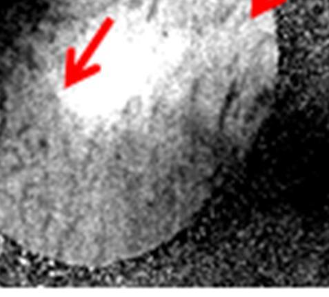
5 3.2 Accommodation dynamics n the anterior eye, accommodation is the process by which the eye focuses on near objects. This occurs mainly through the deformation of the crystalline lens geometry. Studies of the dynamics of far to near focusing processes may enable understanding several aspects like the age-dependent loss of accommodative amplitude in presbyopia, the development of myopia, and the ageing of the crystalline lens as well as the mechanism of accommodation [9]. We performed 4-D imaging of the focusing process in the anterior eye acquiring 100 x 100 axial-scan volumes over 15mm x 15mmm at ~10 volumes per second for 5 seconds at an axial scan rate of 100 khz (Video 2). The target was moved in position from far to near and the subject was asked to focus on the target and accommodate the target s position change. Two time points are shown from the 4-D data in Figure 3. Pupil diameter decreasess when the eyee focuses at near positions. The iris becomes smaller while the crystalline lens becomes thicker with surfaces curvature changes. r L Video 2. 4-D OCT imaging of the anterior eye focusing on a near object. Figure 3. 4-D OCT imaging of the anterior eye focusing on a near object. A) Volume#55 at 0.5 seconds. B) Volume #30 at 3 seconds. The decrease in pupil size and increase in lens thickness is observed and measured. 3.3 Tear film break up The tear film is the first refractive surface for light incident on the eye and plays an important role in the optical quality of the human eye. Tear film dynamics has been used to evaluate the tear system and diagnose dry eye. Tear film break up time (TBUT) is defined as the time interval between a complete blink and the appearancee of the first randomly distributed dry spot [10]. A TBUT of less than ten seconds is considered abnormal [11]. Fluorescein dye is typically Proc. of SPE Vol X-4


.")


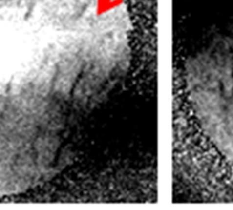
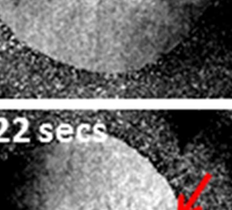
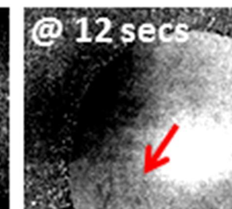
6 used for clinical TBUT measurements. OCT allows for non-invasive tear film dynamics imaging without the need of fluorescein staining. We implemented a 4-D scan protocol with 300 x 300 axial-scan volumes over 8.5 mm x 8..5 mm at ~0.5 volumes per second for 20 seconds at an axial scan rate of 50 khz. n order to best visualize tear film break up, we examine the shadowing effect by summing the signal between thee anterior surface to the posterior surface of the crystalline lens (Video 3). Tear film break up can be clearly observedd in the frame-by-frame breakdown (Figure 4). a ii he region of enfa Video 3. llustration of th ace summation in a full-eye-length OCT dataset at a single time point for tear film visualization. Figure 4. Tear film break up observed in enface images extracted from 4-D OCT data. beginningg at ~12 secondss where vertical streaking patterns appear. Tear film break up is observed 3.4 Retinal blood flow Retinal blood flow may play a role in the pathogenesis of diseases such as diabetic retinopathy, age-related macular degeneration, and glaucoma. The ability to monitor retinal blood flow may be important not only for treatment, but also for understanding the pathophysiology of these diseases [12-14]. We acquired 4-D data in the retina with 200 x 100 axial-scan volumes over 1 mm x 0.5 mm at ~40 volumes per second for 1 second at an axial scan rate of 800 khz (Video 4). Since the galvanometer scanners are driven at very high speeds, a conservative flyback pattern is chosen resulting in only a 0.5 mm x 0.5 mm evenly sampled region. mages from a single time point is extracted and shown in Figure 5(B)(C)(D). By taking the variance of maximum intensity projectionn images at alll time points from multiple volumes, we are able to visualize capillaries from the inner to the outer plexiform layers (Figure 5(E)). Proc. of SPE Vol X-5


 Cross-sectional OCT image.")

 Maximum")
 Variance image generated")


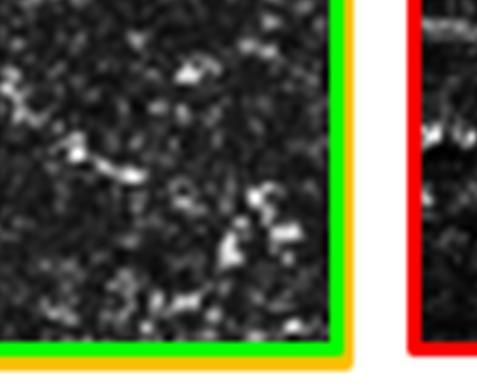
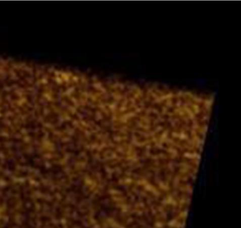
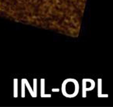
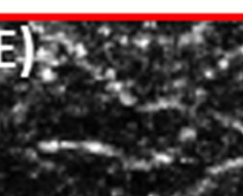
7 NL-OPL Video 4. 4-D OCT imaging of the retina [15]. The video is slowed downn 5x with 0.2x playback speed. GCL: ganglion cell layer. PL-NL: inner plexiform layer to inner nuclear layer. NL-OPL: inner nuclearlayer to outer plexiform layer. Figure 5. 4-D OCT imaging of retinal blood flow. A) Cross-sectional OCT image. B) Enface summation image in the axial direction of a single volume. C) Enface summation image of a region of interest in the axial direction of a single volume. D) Maximum projection image of a region of interest in the axial direction of a single volume. E) Variance image generated from maximum projection images (like E) of multiple volumes in the 4-DD data. The maximum projection image from a single volume shows moving highly reflective scatters. The variance image generated from multiple volumes highlight retinal capillaries. 4. CONCLUSONS n conclusion, 4-D OCT imaging was demonstrated in the human eyee using ultrahigh speed SS-OCT. Visualization and quantitative analysis of 4-D OCT imaging can potentially provide insight to functional and structural changes in the eye with disease. The tunable VCSEL light source enables different imaging modes for variable imaging ranges, speeds, and resolutions. Ultrahigh speed imaging using SS-OCT promises to enable novel 4-D visualization of real-time dynamics of the human eye and may benefit studiess by visualizing and characterizing a variety of visual functions. ACKNOWLEDGMENTS The authors want to thank Dr. Al-Hafeez Dhalla, WooJhon Choi, Chen D. Lu, Kathrin J. Molher, and Tsung-Hann Tsai at MT for technical discussions. Financial support from the National nstitutes of Health (NH R01-EY , R01- EY , R01-EY , R01-EY , R01-EY , R01-CA , R01-HL , R01-NS ), Air Force Office of Scientific Research (AFOSRR FA ), Medical Free Electron Laser Program (FA ) and Deutsche Forschungsgesellschaft (DFG-GSC80-SAOT, DFG-HO-1791/ /11-1) is gratefully acknowledged. Dr. reneusz Grulkowski acknowledges partial support from KOLUMB Fellowship, Foundation for Polish Science (KOL/3/2010-). Dr. Jay S. Duker is supported by an unrestricted grant from Research to Prevent Blindness as well as the Massachusetts Lions Clubs. Dr. reneusz Grulkowski is a visiting scientist from the nstitute of Physics, Nicolaus Copernicus University, Torun, Poland. Proc. of SPE Vol X-6
8 REFERENCES [1] B. Potsaid,. Gorczynska, V. J. Srinivasan, Y. Chen, J. Jiang, A. Cable and J. G. Fujimoto, "Ultrahigh speed spectral / Fourier domain OCT ophthalmic imaging at 70,000 to 312,500 axial scans per second," Opt Express 16(19), (2008) [2] M. Gora, K. Karnowski, M. Szkulmowski, B. J. Kaluzny, R. Huber, A. Kowalczyk and M. Wojtkowski, "Ultra high-speed swept source OCT imaging of the anterior segment of human eye at 200 khz with adjustable imaging range," Opt Express 17(17), (2009) [3] B. Potsaid, B. Baumann, D. Huang, S. Barry, A. E. Cable, J. S. Schuman, J. S. Duker and J. G. Fujimoto, "Ultrahigh speed 1050nm swept source/fourier domain OCT retinal and anterior segment imaging at 100,000 to 400,000 axial scans per second," Opt Express 18(19), (2010) [4] T. Klein, W. Wieser, C. M. Eigenwillig, B. R. Biedermann and R. Huber, "Megahertz OCT for ultrawide-field retinal imaging with a 1050 nm Fourier domain mode-locked laser," Opt Express 19(4), (2011) [5]. Grulkowski, J. J. Liu, B. Potsaid, V. Jayaraman, C. D. Lu, J. Jiang, A. E. Cable, J. S. Duker and J. G. Fujimoto, "Retinal, anterior segment and full eye imaging using ultrahigh speed swept source OCT with vertical-cavity surface emitting lasers," Biomed Opt Express 3(11), (2012) [6] C. K. Leung, C. Y. Cheung, H. Li, S. Dorairaj, C. K. Yiu, A. L. Wong, J. Liebmann, R. Ritch, R. Weinreb and D. S. Lam, "Dynamic analysis of dark-light changes of the anterior chamber angle with anterior segment OCT," nvest Ophthalmol Vis Sci 48(9), (2007) [7] C. Y. Cheung, S. Liu, R. N. Weinreb, J. Liu, H. Li, D. Y. Leung, S. Dorairaj, J. Liebmann, R. Ritch, D. S. Lam and C. K. Leung, "Dynamic analysis of iris configuration with anterior segment optical coherence tomography," nvest Ophthalmol Vis Sci 51(8), (2010) [8] Y. Lee, K. R. Sung, J. H. Na and J. H. Sun, "Dynamic changes in anterior segment (AS) parameters in eyes with primary angle closure (PAC) and PAC glaucoma and open-angle eyes assessed using AS optical coherence tomography," nvest Ophthalmol Vis Sci 53(2), (2012) [9] M. Ruggeri, S. R. Uhlhorn, C. De Freitas, A. Ho, F. Manns and J. M. Parel, "maging and full-length biometry of the eye during accommodation using spectral domain OCT with an optical switch," Biomed Opt Express 3(7), (2012) [10] M. A. Lemp and J. R. Hamill, Jr., "Factors affecting tear film breakup in normal eyes," Arch Ophthalmol 89(2), (1973) [11] M. A. Lemp, C. H. Dohlman, T. Kuwabara, F. J. Holly and J. M. Carroll, "Dry eye secondary to mucus deficiency," Trans Am Acad Ophthalmol Otolaryngol 75(6), (1971) [12] B. Baumann, B. Potsaid, M. F. Kraus, J. J. Liu, D. Huang, J. Hornegger, A. E. Cable, J. S. Duker and J. G. Fujimoto, "Total retinal blood flow measurement with ultrahigh speed swept source/fourier domain OCT," Biomed Opt Express 2(6), (2011) [13] Y. Jia, O. Tan, J. Tokayer, B. Potsaid, Y. Wang, J. J. Liu, M. F. Kraus, H. Subhash, J. G. Fujimoto, J. Hornegger and D. Huang, "Split-spectrum amplitude-decorrelation angiography with optical coherence tomography," Opt Express 20(4), (2012) [14] W. Choi, B. Baumann, J. J. Liu, A. C. Clermont, E. P. Feener, J. S. Duker and J. G. Fujimoto, "Measurement of pulsatile total blood flow in the human and rat retina with ultrahigh speed spectral/fourier domain OCT," Biomed Opt Express 3(5), (2012) [15] T. Schmoll, C. Kolbitsch and R. A. Leitgeb, "Ultra-high-speed volumetric tomography of human retinal blood flow," Opt Express 17(5), (2009) Proc. of SPE Vol X-7
Ultrahigh speed volumetric ophthalmic OCT imaging at 850nm and 1050nm
 Ultrahigh speed volumetric ophthalmic OCT imaging at 850nm and 1050nm The MIT Faculty has made this article openly available. Please share how this access benefits you. Your story matters. Citation As
MEMS tunable VCSEL light source for ultrahigh speed 60kHz - 1MHz axial scan rate and long range centimeter class OCT imaging
 MEMS tunable VCSEL light source for ultrahigh speed 60kHz - 1MHz axial scan rate and long range centimeter class OCT imaging The MIT Faculty has made this article openly available. Please share how this
Axsun OCT Swept Laser and System
 Axsun OCT Swept Laser and System Seungbum Woo, Applications Engineer Karen Scammell, Global Sales Director Bill Ahern, Director of Marketing, April. Outline 1. Optical Coherence Tomography (OCT) 2. Axsun
Visualization of human retinal micro-capillaries with phase contrast high-speed optical coherence tomography
 Visualization of human retinal micro-capillaries with phase contrast high-speed optical coherence tomography Dae Yu Kim 1,2, Jeff Fingler 3, John S. Werner 1,2, Daniel M. Schwartz 4, Scott E. Fraser 3,
Volumetric microvascular imaging of human retina using optical coherence tomography with a novel motion contrast technique
 Volumetric microvascular imaging of human retina using optical coherence tomography with a novel motion contrast technique Jeff Fingler 1,*, Robert J. Zawadzki 2, John S. Werner 2, Dan Schwartz 3, Scott
Handheld ultrahigh speed swept source optical coherence tomography instrument using a MEMS scanning mirror
 Handheld ultrahigh speed swept source optical coherence tomography instrument using a MEMS scanning mirror Chen D. Lu, 1 Martin F. Kraus, 1,2 Benjamin Potsaid, 1,3 Jonathan J. Liu, 1 WooJhon Choi, 1 Vijaysekhar
Megahertz FDML Laser with up to 143nm Sweep Range for Ultrahigh Resolution OCT at 1050nm
 Megahertz FDML Laser with up to 143nm Sweep Range for Ultrahigh Resolution OCT at 1050nm Jan Philip Kolb 1,2, Thomas Klein 2,3, Mattias Eibl 1,2, Tom Pfeiffer 1,2, Wolfgang Wieser 2,3 and Robert Huber
Piezoelectric transducer based miniature catheter for ultrahigh speed endoscopic optical coherence tomography
 Piezoelectric transducer based miniature catheter for ultrahigh speed endoscopic optical coherence tomography The MIT Faculty has made this article openly available. Please share how this access benefits
Axsun Technologies Inc. Swept Laser based OCT Subsystems. May 2012
 Axsun Technologies Inc. Swept Laser based OCT Subsystems May 2012 Outline Axsun Overview Axsun Technology and Manufacturing Axsun Swept Laser Engine products Product Roadmap Images Summary Axsun Technologies
Extended coherence length megahertz FDML and its application for anterior segment imaging
 Extended coherence length megahertz FDML and its application for anterior segment imaging Wolfgang Wieser, 1 Thomas Klein, 1 Desmond C. Adler, 2 Francois Trépanier, 3 Christoph M. Eigenwillig, 1 Sebastian
Ultrahigh speed endoscopic optical coherence tomography using micro-motor imaging catheter and VCSEL technology
 Ultrahigh speed endoscopic optical coherence tomography using micro-motor imaging catheter and VCSEL technology The MIT Faculty has made this article openly available. Please share how this access benefits
Ultrahigh Speed Spectral / Fourier Domain Ophthalmic OCT Imaging
 Ultrahigh Speed Spectral / Fourier Domain Ophthalmic OCT Imaging Benjamin Potsaid 1,3, Iwona Gorczynska 1,2, Vivek J. Srinivasan 1, Yueli Chen 1,2, Jonathan Liu 1, James Jiang 3, Alex Cable 3, Jay S. Duker
High-speed imaging of human retina in vivo with swept-source optical coherence tomography
 High-speed imaging of human retina in vivo with swept-source optical coherence tomography H. Lim, M. Mujat, C. Kerbage, E. C. W. Lee, and Y. Chen Harvard Medical School and Wellman Center for Photomedicine,
The TRC-NW8F Plus: As a multi-function retinal camera, the TRC- NW8F Plus captures color, red free, fluorescein
 The TRC-NW8F Plus: By Dr. Beth Carlock, OD Medical Writer Color Retinal Imaging, Fundus Auto-Fluorescence with exclusive Spaide* Filters and Optional Fluorescein Angiography in One Single Instrument W
Optical coherence tomography
 Optical coherence tomography Peter E. Andersen Optics and Plasma Research Department Risø National Laboratory E-mail peter.andersen@risoe.dk Outline Part I: Introduction to optical coherence tomography
Spectral domain optical coherence tomography with balanced detection using single line-scan camera and optical delay line
 Spectral domain optical coherence tomography with balanced detection using single line-scan camera and optical delay line Min Gyu Hyeon, 1 Hyung-Jin Kim, 2 Beop-Min Kim, 1,2,4 and Tae Joong Eom 3,5 1 Department
Ultra High Speed Space Division Multiplexing OCT
 Lehigh University Lehigh Preserve Theses and Dissertations 5-1-2018 Ultra High Speed Space Division Multiplexing OCT Guo-Jhe Syu Lehigh University, s0987599709@gmail.com Follow this and additional works
some aspects of Optical Coherence Tomography
 some aspects of Optical Coherence Tomography SSOM Lectures, Engelberg 17.3.2009 Ch. Meier 1 / 34 Contents 1. OCT - basic principles (Time Domain Frequency Domain) 2. Performance and limiting factors 3.
Simultaneously measuring ocular aberration and anterior segment biometry during accommodation
 Journal of Innovative Optical Health Sciences Vol. 8, No. 2 (2015) 1550005 (6 pages) #.c The Authors DOI: 10.1142/S1793545815500054 Simultaneously measuring ocular aberration and anterior segment biometry
Swept source / Fourier domain polarization sensitive optical coherence tomography with a passive polarization delay unit
 Swept source / Fourier domain polarization sensitive optical coherence tomography with a passive polarization delay unit The MIT Faculty has made this article openly available. Please share how this access
Fourier Domain (Spectral) OCT OCT: HISTORY. Could OCT be a Game Maker OCT in Optometric Practice: A THE TECHNOLOGY BEHIND OCT
 OCT OCT: HISTORY. Could OCT be a Game Maker OCT in Optometric Practice: A THE TECHNOLOGY BEHIND OCT") Could OCT be a Game Maker OCT in Optometric Practice: A Hands On Guide Murray Fingeret, OD Nick Rumney, MSCOptom Fourier Domain (Spectral) OCT New imaging method greatly improves resolution and speed of
Could OCT be a Game Maker OCT in Optometric Practice: A Hands On Guide Murray Fingeret, OD Nick Rumney, MSCOptom Fourier Domain (Spectral) OCT New imaging method greatly improves resolution and speed of
Medical imaging has long played a critical role in diagnosing
 Three-Dimensional Optical Coherence Tomography (3D-OCT) Image Enhancement with Segmentation-Free Contour Modeling C-Mode Hiroshi Ishikawa, 1,2 Jongsick Kim, 1,2 Thomas R. Friberg, 1,2 Gadi Wollstein, 1
Biology 70 Slides for Lecture 1 Fall 2007
 Biology 70 Part II Sensory Systems www.biology.ucsc.edu 1 2 intensity vs spatial position (image formation) color 3 4 motion depth (monocular) 5 6 1 depth (binocular) 1. In the lectures on perception we
A miniature all-optical photoacoustic imaging probe
 A miniature all-optical photoacoustic imaging probe Edward Z. Zhang * and Paul C. Beard Department of Medical Physics and Bioengineering, University College London, Gower Street, London WC1E 6BT, UK http://www.medphys.ucl.ac.uk/research/mle/index.htm
Going beyond the surface of your retina OCT-HS100 OPTICAL COHERENCE TOMOGRAPHY
 Going beyond the surface of your retina OCT-HS100 OPTICAL COHERENCE TOMOGRAPHY Automatic functions make examinations short and simple. Perform the examination with only two simple mouse clicks! 1. START
Going beyond the surface of your retina OCT-HS100 OPTICAL COHERENCE TOMOGRAPHY
 Going beyond the surface of your retina OCT-HS100 OPTICAL COHERENCE TOMOGRAPHY Full Auto OCT High specifications in a very compact design Automatic functions make examinations short and simple. Perform
Ultrahigh-resolution high-speed retinal imaging using spectral-domain optical coherence tomography
 Ultrahigh-resolution high-speed retinal imaging using spectral-domain optical coherence tomography Barry Cense, Nader A. Nassif Harvard Medical School and Wellman Center for Photomedicine, Massachusetts
Static and dynamic crystalline lens accommodation evaluated using quantitative 3-D OCT
 Static and dynamic crystalline lens accommodation evaluated using quantitative 3-D OCT Enrique Gambra, 1,* Sergio Ortiz, 1 Pablo Perez-Merino, 1 Michalina Gora, 2,3 Maciej Wojtkowski, 2 and Susana Marcos
Going beyond the surface of your retina
 Going beyond the surface of your retina OCT-HS100 Optical Coherence Tomography Canon s expertise in optics and innovative technology have resulted in a fantastic 3 μm optical axial resolution for amazing
Lecture 2 Slit lamp Biomicroscope
 Lecture 2 Slit lamp Biomicroscope 1 Slit lamp is an instrument which allows magnified inspection of interior aspect of patient s eyes Features Illumination system Magnification via binocular microscope
Phase-stabilized optical frequency domain imaging at 1-µm for the measurement of blood flow in the human choroid
 Phase-stabilized optical frequency domain imaging at 1-µm for the measurement of blood flow in the human choroid Boy Braaf, 1,* Koenraad A. Vermeer, 1 Victor Arni D.P. Sicam, 1 Elsbeth van Zeeburg, 1,2
Frequency comb swept lasers for optical coherence tomography
 Frequency comb swept lasers for optical coherence tomography The MIT Faculty has made this article openly available. Please share how this access benefits you. Your story matters. Citation As Published
PHGY Physiology. SENSORY PHYSIOLOGY Vision. Martin Paré
 PHGY 212 - Physiology SENSORY PHYSIOLOGY Vision Martin Paré Assistant Professor of Physiology & Psychology pare@biomed.queensu.ca http://brain.phgy.queensu.ca/pare The Process of Vision Vision is the process
OCT. Optical Coherence Tomography
 OCT Optical Coherence Tomography Optical Coherence Tomography (OCT) is a non-invasive, non-destructive imaging technique that enables high-resolution, cross-sectional imaging of a wide range of highly
Nature Neuroscience: doi: /nn Supplementary Figure 1. Optimized Bessel foci for in vivo volume imaging.
 Supplementary Figure 1 Optimized Bessel foci for in vivo volume imaging. (a) Images taken by scanning Bessel foci of various NAs, lateral and axial FWHMs: (Left panels) in vivo volume images of YFP + neurites
Cheng, KHY; Standish, BA; Yang, VXD; Cheung, KKY; Gu, X; Lam, EY; Wong, KKY
 Title Hybrid Fourier domain modelocked laser utilizing a fiber optical parametric amplifier and an erbium doped fiber amplifier Author(s) Citation Cheng, KHY; Standish, BA; Yang, VXD; Cheung, KKY; Gu,
Kent Academic Repository
 Kent Academic Repository Full text document (pdf) Citation for published version Toadere, Florin and Stancu, Radu.-F. and Poon, Wallace and Schultz, David and Podoleanu, Adrian G.H. (2017) 1 MHz Akinetic
Image Modeling of the Human Eye
 Image Modeling of the Human Eye Rajendra Acharya U Eddie Y. K. Ng Jasjit S. Suri Editors ARTECH H O U S E BOSTON LONDON artechhouse.com Contents Preface xiiii CHAPTER1 The Human Eye 1.1 1.2 1. 1.4 1.5
Real-Time Experimental Measurement of Swept Source VCSEL Properties Relevant to OCT Imaging
 Open Access Real-Time Experimental Measurement of Swept Source VCSEL Properties Relevant to OCT Imaging Volume 9, Number 5, October 2017 T. P. Butler S. Slepneva P. M. McNamara K. Neuhaus D. Goulding M.
High-Coherence Wavelength Swept Light Source
 Kenichi Nakamura, Masaru Koshihara, Takanori Saitoh, Koji Kawakita [Summary] Optical technologies that have so far been restricted to the field of optical communications are now starting to be applied
3550 Aberdeen Ave SE, Kirtland AFB, NM 87117, USA ABSTRACT 1. INTRODUCTION
 Beam Combination of Multiple Vertical External Cavity Surface Emitting Lasers via Volume Bragg Gratings Chunte A. Lu* a, William P. Roach a, Genesh Balakrishnan b, Alexander R. Albrecht b, Jerome V. Moloney
Transferring wavefront measurements to ablation profiles. Michael Mrochen PhD Swiss Federal Institut of Technology, Zurich IROC Zurich
 Transferring wavefront measurements to ablation profiles Michael Mrochen PhD Swiss Federal Institut of Technology, Zurich IROC Zurich corneal ablation Calculation laser spot positions Centration Calculation
Short Ring Cavity Swept Source Based on a Highly Reflective Chirped FBG
 PHOTONIC SENSORS / Vol. 5, No. 3, 215: 251 256 Short Ring Cavity Swept Source Based on a Highly Reflective Chirped FBG Radu-Florin STANCU * and Adrian PODOLEANU Applied Optics Group, School of Physical
Frequency comb swept lasers
 Frequency comb swept lasers The MIT Faculty has made this article openly available. Please share how this access benefits you. Your story matters. Citation As Published Publisher Tsai, Tsung-Han et al.
Frequency comb swept lasers
 Frequency comb swept lasers Tsung-Han Tsai 1, Chao Zhou 1, Desmond C. Adler 1, and James G. Fujimoto 1* 1 Department of Electrical Engineering and Computer Science and Research Laboratory of Electronics,
Design of a Swept-Source, Anatomical OCT System for Pediatric Bronchoscopy
 Design of a Swept-Source, Anatomical OCT System for Pediatric Bronchoscopy Kushal C. Wijesundara a, Nicusor V. Iftimia c, and Amy L. Oldenburg a,b a Department of Physics and Astronomy and the b Biomedical
60 MHz A-line rate ultra-high speed Fourier-domain optical coherence tomography
 60 MHz Aline rate ultrahigh speed Fourierdomain optical coherence tomography K. Ohbayashi a,b), D. Choi b), H. HiroOka b), H. Furukawa b), R. Yoshimura b), M. Nakanishi c), and K. Shimizu c) a Graduate
PHGY Physiology. The Process of Vision. SENSORY PHYSIOLOGY Vision. Martin Paré. Visible Light. Ocular Anatomy. Ocular Anatomy.
 PHGY 212 - Physiology SENSORY PHYSIOLOGY Vision Martin Paré Assistant Professor of Physiology & Psychology pare@biomed.queensu.ca http://brain.phgy.queensu.ca/pare The Process of Vision Vision is the process
Temporal coherence characteristics of a superluminescent diode system with an optical feedback mechanism
 VI Temporal coherence characteristics of a superluminescent diode system with an optical feedback mechanism Fang-Wen Sheu and Pei-Ling Luo Department of Applied Physics, National Chiayi University, Chiayi
Vision. The eye. Image formation. Eye defects & corrective lenses. Visual acuity. Colour vision. Lecture 3.5
 Lecture 3.5 Vision The eye Image formation Eye defects & corrective lenses Visual acuity Colour vision Vision http://www.wired.com/wiredscience/2009/04/schizoillusion/ Perception of light--- eye-brain
Photoacoustic ophthalmoscopy for in vivo retinal imaging
 Photoacoustic ophthalmoscopy for in vivo retinal imaging Shuliang Jiao, 1,5 Minshan Jiang, 1 Jianming Hu, 1 Amani Fawzi, 1 Qifa Zhou, 2 K. Kirk Shung, 2 Carmen A. Puliafito, 1 and Hao F. Zhang 3,4 1 Department
PHY 431 Homework Set #5 Due Nov. 20 at the start of class
 PHY 431 Homework Set #5 Due Nov. 0 at the start of class 1) Newton s rings (10%) The radius of curvature of the convex surface of a plano-convex lens is 30 cm. The lens is placed with its convex side down
Split-spectrum amplitude-decorrelation angiography with optical coherence tomography
 Split-spectrum amplitude-decorrelation angiography with optical coherence tomography Yali Jia, 1 Ou Tan, 1 Jason Tokayer, 2 Benjamin Potsaid, 3,4 Yimin Wang, 1 Jonathan J. Liu, 3 Martin F. Kraus, 3,5 Hrebesh
Optical Coherence Tomography Retina Scan Duo
 Optical Coherence Tomography Retina Scan Duo High Definition OCT & Fundus Imaging in One Compact System The Retina Scan Duo is a combined OCT and fundus camera system that is a user friendly and versatile
Multi-Megahertz OCT: High quality 3D imaging at 20 million A-scans and 4.5 GVoxels per second
 Multi-Megahertz OCT: High quality 3D imaging at 20 million A-scans and 4.5 GVoxels per second Wolfgang Wieser, Benjamin R. Biedermann, Thomas Klein, Christoph M. Eigenwillig and Robert Huber* Lehrstuhl
High Resolution Ocular Surface OCT to Directly Measure Tear Film Thickness in Human Eyes
 High Resolution Ocular Surface OCT to Directly Measure Tear Film Thickness in Human Eyes Rahul Yadav, B.Tech., Kyesung Lee, Ph.D., Jannick Rolland, Ph.D., James Zavislan, Ph.D., James Aquavella, M.D. and
AP PSYCH Unit 4.2 Vision 1. How does the eye transform light energy into neural messages? 2. How does the brain process visual information? 3.
 AP PSYCH Unit 4.2 Vision 1. How does the eye transform light energy into neural messages? 2. How does the brain process visual information? 3. What theories help us understand color vision? 4. Is your
High-speed ultra-broad tuning MEMS-VCSELs for imaging and spectroscopy
 High-speed ultra-broad tuning MEMS-VCSELs for imaging and spectroscopy The MIT Faculty has made this article openly available. Please share how this access benefits you. Your story matters. Citation As
Customized Lasers for Specific Swept Source OCT Applications. Bill Ahern Axsun Technologies, Inc. June 20, 2013
 Customized Lasers for Specific Swept Source OCT Applications Bill Ahern Axsun Technologies, Inc. June 20, 2013 Outline Axsun Overview Axsun Technology and Manufacturing Axsun OCT Laser Platform Laser Operation
Integre Pro Scan combines pattern scanning and multi-color photocoagulation in our unique all-in-one laser/slit lamp design.
 Integre Pro Scan combines pattern scanning and multi-color photocoagulation in our unique all-in-one laser/slit lamp design. Multi-color scanning photocoagulation takes on a new look. Integre Pro Scan
Photoacoustic imaging using an 8-beam Fabry-Perot scanner
 Photoacoustic imaging using an 8-beam Fabry-Perot scanner Nam Huynh, Olumide Ogunlade, Edward Zhang, Ben Cox, Paul Beard Department of Medical Physics and Biomedical Engineering, University College London,
EnFocus Your Upgrade Path to High Performance Intrasurgical OCT
 Your Upgrade Path to High Performance Intrasurgical OCT is FDA 510(k) Cleared > Ultra HD OCT extends your microscope s potential with intrasurgical OCT BRILLIANT IMAGES, SUB-SURFACE KNOWLEDGE is an intrasurgical
Introduction. Chapter Aim of the Thesis
 Chapter 1 Introduction 1.1 Aim of the Thesis The main aim of this investigation was to develop a new instrument for measurement of light reflected from the retina in a living human eye. At the start of
Customized Correction of Wavefront Aberrations in Abnormal Human Eyes by Using a Phase Plate and a Customized Contact Lens
 Journal of the Korean Physical Society, Vol. 49, No. 1, July 2006, pp. 121 125 Customized Correction of Wavefront Aberrations in Abnormal Human Eyes by Using a Phase Plate and a Customized Contact Lens
OPTICAL COHERENCE TOMOGRAPHY: OCT supports industrial nondestructive depth analysis
 OPTICAL COHERENCE TOMOGRAPHY: OCT supports industrial nondestructive depth analysis PATRICK MERKEN, RAF VANDERSMISSEN, and GUNAY YURTSEVER Abstract Optical coherence tomography (OCT) has evolved to a standard
Wide-field high-speed space-division multiplexing optical coherence tomography using an integrated photonic device
 Vol. 8, No. 8 1 Aug 2017 BIOMEDICAL OPTICS EXPRESS 3856 Wide-field high-speed space-division multiplexing optical coherence tomography using an integrated photonic device YONGYANG HUANG,1,2 MUDABBIR BADAR,1,2
OCULAR MEDIA* PHOTOGRAPHIC RECORDING OF OPACITIES OF THE. development by the control of diabetes, the supply of a deficient hormone
 Brit. J. Ophthal. (1955) 39, 85. PHOTOGRAPHIC RECORDING OF OPACITIES OF THE OCULAR MEDIA* BY E. F. FINCHAM Institute of Ophthalmology, University of London THE value of photography for recording pathological
Ron Liu OPTI521-Introductory Optomechanical Engineering December 7, 2009
 Synopsis of METHOD AND APPARATUS FOR IMPROVING VISION AND THE RESOLUTION OF RETINAL IMAGES by David R. Williams and Junzhong Liang from the US Patent Number: 5,777,719 issued in July 7, 1998 Ron Liu OPTI521-Introductory
Pulsed illumination spectral-domain optical coherence tomography for human retinal imaging
 Pulsed illumination spectral-domain optical coherence tomography for human retinal imaging Jang-Woo You 1, 1) Department of Mechanical Engineering, Korea Advanced Institute of Science and Technology 373-1,
Eye. Eye Major structural layer of the wall of the eye is a thick layer of dense C.T.; that layer has two parts:
 General aspects Sensory receptors ; External or internal environment. A stimulus is a change in the environmental condition which is detectable by a sensory receptor 1 Major structural layer of the wall
Primary angle-closure glaucoma is a major cause of visual
 Glaucoma Dynamic Analysis of Iris Configuration with Anterior Segment Optical Coherence Tomography Carol Yim-lui Cheung, 1 Shu Liu, 1 Robert N. Weinreb, 2 Jing Liu, 1 Haitao Li, 1 Dexter Yu-lung Leung,
Imaging the Subcellular Structure of Human Coronary Atherosclerosis Using 1-µm Resolution
 Imaging the Subcellular Structure of Human Coronary Atherosclerosis Using 1-µm Resolution Optical Coherence Tomography (µoct) Linbo Liu, Joseph A. Gardecki, Seemantini K. Nadkarni, Jimmy D. Toussaint,
III: Vision. Objectives:
 III: Vision Objectives: Describe the characteristics of visible light, and explain the process by which the eye transforms light energy into neural. Describe how the eye and the brain process visual information.
Light has some interesting properties, many of which are used in medicine:
 LIGHT IN MEDICINE Light has some interesting properties, many of which are used in medicine: 1- The speed of light changes when it goes from one material into another. The ratio of the speed of light in
Diabetic Retinopathy Clinical Research Network (DRCR.net) UWF Optos 200Tx Imaging Protocol. Version 3.0 9/19/16
 UWF Optos 200Tx Imaging Protocol. Version 3.0 9/19/16") Diabetic Retinopathy Clinical Research Network (DRCR.net) UWF Optos 200Tx Imaging Protocol Version 3.0 9/19/16 DRCR.net UWF 200 Tx Imaging Protocol V3.0 9-19-15 Final Page 1 of 14 Table of Contents Background...
Diabetic Retinopathy Clinical Research Network (DRCR.net) UWF Optos 200Tx Imaging Protocol Version 3.0 9/19/16 DRCR.net UWF 200 Tx Imaging Protocol V3.0 9-19-15 Final Page 1 of 14 Table of Contents Background...
Optical Coherence Tomography. RS-3000 Advance
 Optical Coherence Tomography RS-3000 Advance See it in Advance See it in high resolution with the AngioScan* image. SLO Superficial capillary OCT-Angiography (3 x 3 mm) Deep capillary OCT-Angiography (3
Diabetic Retinopathy Clinical Research Network (DRCR.net) UWF Optos Imaging Protocol. Version /14/14
 UWF Optos Imaging Protocol. Version /14/14") Diabetic Retinopathy Clinical Research Network (DRCR.net) UWF Optos Imaging Protocol Version 1.0 10/14/14 DRCR.net UWF Imaging Protocol FINAL 10-14-14 Page 1 of 14 Table of Contents Background... 3 P200Tx
Diabetic Retinopathy Clinical Research Network (DRCR.net) UWF Optos Imaging Protocol Version 1.0 10/14/14 DRCR.net UWF Imaging Protocol FINAL 10-14-14 Page 1 of 14 Table of Contents Background... 3 P200Tx
2 The First Steps in Vision
 2 The First Steps in Vision 2 The First Steps in Vision A Little Light Physics Eyes That See light Retinal Information Processing Whistling in the Dark: Dark and Light Adaptation The Man Who Could Not
Swept Wavelength Testing:
 Application Note 13 Swept Wavelength Testing: Characterizing the Tuning Linearity of Tunable Laser Sources In a swept-wavelength measurement system, the wavelength of a tunable laser source (TLS) is swept
1272. Phase-controlled vibrational laser percussion drilling
 1272. Phase-controlled vibrational laser percussion drilling Chao-Ching Ho 1, Chih-Mu Chiu 2, Yuan-Jen Chang 3, Jin-Chen Hsu 4, Chia-Lung Kuo 5 National Yunlin University of Science and Technology, Douliou,
High-speed spectral-domain optical coherence tomography at 1.3 µm wavelength
 High-speed spectral-domain optical coherence tomography at 1.3 µm wavelength S. H. Yun, G. J. Tearney, B. E. Bouma, B. H. Park, and J. F. de Boer Harvard Medical School and Wellman Center of Photomedicine,
Understanding Optical Specifications
 Understanding Optical Specifications Optics can be found virtually everywhere, from fiber optic couplings to machine vision imaging devices to cutting-edge biometric iris identification systems. Despite
UC Davis UC Davis Previously Published Works
 UC Davis UC Davis Previously Published Works Title Progress on developing adaptive optics-optical coherence tomography for in vivo retinal imaging: Monitoring and correction of eye motion artifacts Permalink
Single camera spectral domain polarizationsensitive optical coherence tomography using offset B-scan modulation
 Single camera spectral domain polarizationsensitive optical coherence tomography using offset B-scan modulation Chuanmao Fan 1,2 and Gang Yao 1,3 1 Department of Biological Engineering, University of Missouri,
What s Fundus photography s purpose? Why do we take them? Why do we do it? Why do we do it? Why do we do it? 11/3/2014. To document the retina
 What s Fundus photography s purpose? To document the retina Photographers role to show the retina Document other ocular structures Why do we take them? Why do we do it? We as photographers help the MD
The First True Color Confocal Scanner on the Market
 The First True Color Confocal Scanner on the Market White color and infrared confocal images: the advantages of white color and confocality together for better fundus images. The infrared to see what our
Choices and Vision. Jeffrey Koziol M.D. Thursday, December 6, 12
 Choices and Vision Jeffrey Koziol M.D. How does the eye work? What is myopia? What is hyperopia? What is astigmatism? What is presbyopia? How the eye works How the Eye Works 3 How the eye works Light rays
Goldmann Visual Field. Humphrey Visual Field 4/25/2017. What s So Special About Special Testing?! Houston, we have a problem.
 What s So Special About Special Testing?! Why can t they get the schedule right? looneytunes.com Houston, we have a problem. Communication is the key. We all assume that the other people in the office
Akinori Mitani and Geoff Weiner BGGN 266 Spring 2013 Non-linear optics final report. Introduction and Background
 Akinori Mitani and Geoff Weiner BGGN 266 Spring 2013 Non-linear optics final report Introduction and Background Two-photon microscopy is a type of fluorescence microscopy using two-photon excitation. It
Optical Coherence Tomography Systems and signal processing in SD-OCT
 Optical Coherence Tomography Systems and signal processing in SD-OCT Chandan S.Rawat 1, Vishal S.Gaikwad 2 1 Associate Professor V.E.S.I.T., Mumbai chandansrawat@gmail.com 2 P.G.Student, V.E.S.I.T., Mumbai
Off-axis full-field swept-source optical coherence tomography using holographic refocusing
 Off-axis full-field swept-source optical coherence tomography using holographic refocusing Dierck Hillmann *,a, Gesa Franke b,c, Laura Hinkel b, Tim Bonin b, Peter Koch a, Gereon Hüttmann b,c a Thorlabs
Visual System I Eye and Retina
 Visual System I Eye and Retina Reading: BCP Chapter 9 www.webvision.edu The Visual System The visual system is the part of the NS which enables organisms to process visual details, as well as to perform
Swept source optical coherence tomography Gabor fusion splicing technique for microscopy of thick samples using a deformable mirror
 Swept source optical coherence tomography Gabor fusion splicing technique for microscopy of thick samples using a deformable mirror Christopher Costa, a Adrian Bradu, a John Rogers, a Pauline Phelan, b
Choices and Vision. Jeffrey Koziol M.D. Friday, December 7, 12
 Choices and Vision Jeffrey Koziol M.D. How does the eye work? What is myopia? What is hyperopia? What is astigmatism? What is presbyopia? How the eye works Light rays enter the eye through the clear cornea,
LMT F14. Cut in Three Dimensions. The Rowiak Laser Microtome: 3-D Cutting and Imaging
 LMT F14 Cut in Three Dimensions The Rowiak Laser Microtome: 3-D Cutting and Imaging The Next Generation of Microtomes LMT F14 - Non-contact laser microtomy The Rowiak laser microtome LMT F14 is a multi-purpose
Optical Coherence Tomography. RS-3000 Advance 2
 Optical Coherence Tomography RS-3000 Advance 2 -Providing a comprehensive solution for retina and glaucom Retina Analysis Retinal mode Glaucoma Analysis Choroidal mode Image courtesy of Hokkaido University
Dynamic Phase-Shifting Microscopy Tracks Living Cells
 from photonics.com: 04/01/2012 http://www.photonics.com/article.aspx?aid=50654 Dynamic Phase-Shifting Microscopy Tracks Living Cells Dr. Katherine Creath, Goldie Goldstein and Mike Zecchino, 4D Technology
SUPPLEMENTARY INFORMATION
 Computational high-resolution optical imaging of the living human retina Nathan D. Shemonski 1,2, Fredrick A. South 1,2, Yuan-Zhi Liu 1,2, Steven G. Adie 3, P. Scott Carney 1,2, Stephen A. Boppart 1,2,4,5,*
Version 1.0. th March 2011
 Optical Coherence Tomography Scan and Retinal Imagingg Version 1.0 http://www.ukbiobank.ac.uk/ 5 th March 2011 This manual details the procedure for Scan and Retinal Imagingg at an Assessment Centre of
Integrated photonic circuit in silicon on insulator for Fourier domain optical coherence tomography
 Integrated photonic circuit in silicon on insulator for Fourier domain optical coherence tomography Günay Yurtsever *,a, Pieter Dumon a, Wim Bogaerts a, Roel Baets a a Ghent University IMEC, Photonics