Quantitative, spectrally- resolved intraoperative
|
|
|
- Kathleen Rich
- 6 years ago
- Views:
Transcription
1 1 Quantitative, spectrally- resolved intraoperative fluorescence imaging Pablo A. Valdés 1,2,3, Frederic Leblond 1, Valerie L. Jacobs 2, Brian C. Wilson 5, Keith D. Paulsen 1,2,4, & David W. Roberts 2,3,4 1 Thayer School of Engineering, and 2 Dartmouth Medical School, Dartmouth College, Hanover, NH 03755, USA; 3 Section of Neurosurgery; 4 Norris Cotton Cancer Center, Dartmouth Hitchcock Medical Center, Lebanon, New Hampshire 03756, USA; 5 University of Toronto, Ontario Cancer Institute, 610 University Avenue, Toronto, Ontario M5G 2M9, Canada. Correspondence should be addressed to P.A.V. (Pablo.A.Valdes@Dartmouth.edu) or D.W.R. (David.W.Roberts@Dartmouth.edu)
2 2 SUPPLEMENTARY METHODS AND RESULTS Spatial resolution of optical components. A USAF 1951 (Edmund Optics) contrast resolution target was used to determine the spatial resolution of the qfi imaging system under white light exposure (Suppl. Fig. 3a-c). The target was composed of n = [0, 1, 2, 3] groups, each with i =[1, 2, 3, 4, 5, 6] horizontal and vertical elements consisting of black and white bar patterns (Suppl. Fig. 3c). The crosssectional intensity profiles of each element were extracted to calculate the element-specific contrast transfer function (CTF) from CTF(i) = I max (i) " I min (i) I max (i) + I min (i) *100 where I max and I min are the maximum and minimum intensities of the bar pattern s cross-sectional profile. The calculated element specific resolution was determined from Re solution(i) = 1 2 n + i"1 6 *1000 where n is the group number, i is the element number, and resolution is given in µm. We computed the Rayleigh and Sparrow horizontal (Suppl. Fig. 3a) and vertical (Suppl. Fig. 3b) spatial resolutions by determining the point on the graph were the CTF = 26.4% or 0%, respectively. The optical components of the qfi system demonstrated submillimeter resolutions both in the vertical (214 μm and 125 μm) and horizontal directions (217 μm and 125 μm), which are approximately 5 to 10 times smaller, respectively, than the smallest length scale relevant to the execution of surgical procedures.
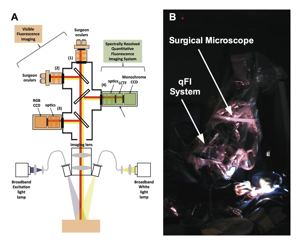
3 3 SUPPLEMENTARY FIGURE LEGENDS Supplementary Figure 1. Quantitative fluorescence imaging (qfi) system. (A) Schematic of the qfi design integrated with a conventional surgical microscope enabled for fluorescence imaging. The microscope consists of two optical ports (1 and 2) with associated oculars for direct viewing by two surgeons and a third (3) optical port that transmits light onto a RGB CCD camera for video rate and snapshot image acquisition. A fourth (4) free optical port integrates with the portable qfi system. The microscope functions in violet-blue light and white light modes - one for fluorescence imaging and one for white light reflectance imaging, respectively. The qfi system consists of an optical adapter which connects to the free optical port of the surgical microscope, a liquid crystal tunable filter for fast (ms range) wavelength selection and light filtration, and a CCD camera for detection. (B) Illustration of intraoperative use of qfi in a human surgery with fluorescence guidance. Supplementary Figure 2. Scatter plot of qfi-derived C PpIX estimates vs. probe C PpIX values (in triplicate) showing a strong linear relationship with a highly significant Pearson s correlation coefficient, R = 0.79 (p-value<0.0001). Data produced from 41 locations in 4 rats with CNS-1 tumors. Both normal and tumor tissue was sampled. Error bars denote +/- standard deviation. Supplementary Figure 3. Spatial resolution of the optical components comprising the qfi system. Contrast transfer function analysis demonstrates submillimeter vertical spatial resolutions (in A) of 214 μm and 125 μm, and horizontal spatial resolutions (in B) of 217 μm and 125 μm (Rayleigh and Sparrow criteria, respectively) using a standard contrast resolution target (in C).
4 4 Supplementary Figure 1
*+$#',$4! \" # &\"\" %\" $\" #\"!\" \" '45+(.67080!$9#:0;0!&A!2 <=4>>*?080\":0;0&!@0!2!\"\" #\"\" $\"\" %\"\" &\"\"\" '()*+,-.*/01!23!\"#$%&'( )*+$#',$-.#'+,/\"#-01+&$%*+- 23-)*+$#',$4 &\"\" %\" $\" #\"!")
5 5 Supplementary Figure 2! $%&' ()!*+,-.(/(45& #!"#!"!# $ ( :1003-6;<18 8((?(E# %/B6-C3(D!"!!!#!"!!#!"!!#!"!#!"# #! $%&' ()!*+,-.(/($0123 Supplementary Figure 3 5*#%6*+$'( )*+$#',$-.#'+,/"#-01+&$%*+- 23-)*+$#',$4! " # &"" %" $" #"!" " '45+(.67080!$9#:0;0!&A!2 <=4>>*?080":0;0&!@0!2!"" #"" $"" %"" &""" '()*+,-.*/01!23!"#$%&'( )*+$#',$-.#'+,/"#-01+&$%*+- 23-)*+$#',$4 &"" %" $" #"!" " '45+(.67080!$9#:0;0!&#!2 <=4>>*?080":0;0&!@0!2!"" #"" $"" %"" &""" '()*+,-.*/01!23
ABOUT RESOLUTION. pco.knowledge base
 The resolution of an image sensor describes the total number of pixel which can be used to detect an image. From the standpoint of the image sensor it is sufficient to count the number and describe it
Supplementary Figure S1. Schematic representation of different functionalities that could be
 Supplementary Figure S1. Schematic representation of different functionalities that could be obtained using the fiber-bundle approach This schematic representation shows some example of the possible functions
Improving the Collection Efficiency of Raman Scattering
 PERFORMANCE Unparalleled signal-to-noise ratio with diffraction-limited spectral and imaging resolution Deep-cooled CCD with excelon sensor technology Aberration-free optical design for uniform high resolution
Microstructured Air Cavities as High-Index-Contrast Substrates with
 Supporting Information for: Microstructured Air Cavities as High-Index-Contrast Substrates with Strong Diffraction for Light-Emitting Diodes Yoon-Jong Moon, Daeyoung Moon, Jeonghwan Jang, Jin-Young Na,
Fast, high-contrast imaging of animal development with scanned light sheet based structured-illumination microscopy
 nature methods Fast, high-contrast imaging of animal development with scanned light sheet based structured-illumination microscopy Philipp J Keller, Annette D Schmidt, Anthony Santella, Khaled Khairy,
Material analysis by infrared mapping: A case study using a multilayer
 Material analysis by infrared mapping: A case study using a multilayer paint sample Application Note Author Dr. Jonah Kirkwood, Dr. John Wilson and Dr. Mustafa Kansiz Agilent Technologies, Inc. Introduction
EnFocus Your Upgrade Path to High Performance Intrasurgical OCT
 Your Upgrade Path to High Performance Intrasurgical OCT is FDA 510(k) Cleared > Ultra HD OCT extends your microscope s potential with intrasurgical OCT BRILLIANT IMAGES, SUB-SURFACE KNOWLEDGE is an intrasurgical
PGx11 series. Transform Limited Broadly Tunable Picosecond OPA APPLICATIONS. Available models
 PGx1 PGx3 PGx11 PT2 Transform Limited Broadly Tunable Picosecond OPA optical parametric devices employ advanced design concepts in order to produce broadly tunable picosecond pulses with nearly Fourier-transform
BIG PIXELS VS. SMALL PIXELS THE OPTICAL BOTTLENECK. Gregory Hollows Edmund Optics
 BIG PIXELS VS. SMALL PIXELS THE OPTICAL BOTTLENECK Gregory Hollows Edmund Optics 1 IT ALL STARTS WITH THE SENSOR We have to begin with sensor technology to understand the road map Resolution will continue
Hyperspectral Imaging Basics for Forensic Applications
 Hyperspectral Imaging Basics for Forensic Applications Sara Nedley, ChemImage Corp. June 14, 2011 1 ChemImage Corporation Pioneers in Hyperspectral Imaging industry Headquartered in Pittsburgh, PA In operation
Confocal Imaging Through Scattering Media with a Volume Holographic Filter
 Confocal Imaging Through Scattering Media with a Volume Holographic Filter Michal Balberg +, George Barbastathis*, Sergio Fantini % and David J. Brady University of Illinois at Urbana-Champaign, Urbana,
Practical work no. 3: Confocal Live Cell Microscopy
 Practical work no. 3: Confocal Live Cell Microscopy Course Instructor: Mikko Liljeström (MIU) 1 Background Confocal microscopy: The main idea behind confocality is that it suppresses the signal outside
Circular Dichroism Microscopy Free from Commingling Linear Dichroism via Discretely Modulated Circular Polarization
 Supplementary information Circular Dichroism Microscopy Free from Commingling Linear Dichroism via Discretely Modulated Circular Polarization Tetsuya Narushima AB and Hiromi Okamoto A* A Institute for
Chapter 1. Basic Electron Optics (Lecture 2)
") Chapter 1. Basic Electron Optics (Lecture 2) Basic concepts of microscope (Cont ) Fundamental properties of electrons Electron Scattering Instrumentation Basic conceptions of microscope (Cont ) Ray diagram
Chapter 1. Basic Electron Optics (Lecture 2) Basic concepts of microscope (Cont ) Fundamental properties of electrons Electron Scattering Instrumentation Basic conceptions of microscope (Cont ) Ray diagram
Fast Laser Raman Microscope RAMAN
 Fast Laser Raman Microscope RAMAN - 11 www.nanophoton.jp Fast Raman Imaging A New Generation of Raman Microscope RAMAN-11 developed by Nanophoton was created by combining confocal laser microscope technology
BaySpec SuperGamut OEM
 BaySpec SuperGamut OEM Spectrographs & Spectrometers RUGGED SOLID STATE HIGH RESOLUTION OPTIMIZED COOLING COST EFFECTIVE HIGH THROUGHPUT www.bayspec.com Specifications Model UV-NIR VIS-NIR NIR 900-1700nm
Development of a new multi-wavelength confocal surface profilometer for in-situ automatic optical inspection (AOI)
") Development of a new multi-wavelength confocal surface profilometer for in-situ automatic optical inspection (AOI) Liang-Chia Chen 1#, Chao-Nan Chen 1 and Yi-Wei Chang 1 1. Institute of Automation Technology,
Development of a new multi-wavelength confocal surface profilometer for in-situ automatic optical inspection (AOI) Liang-Chia Chen 1#, Chao-Nan Chen 1 and Yi-Wei Chang 1 1. Institute of Automation Technology,
Instructions for the Experiment
 Instructions for the Experiment Excitonic States in Atomically Thin Semiconductors 1. Introduction Alongside with electrical measurements, optical measurements are an indispensable tool for the study of
Fast Laser Raman Microscope RAMAN
 Fast Laser Raman Microscope RAMAN - 11 www.nanophoton.jp Fast Raman Imaging A New Generation of Raman Microscope RAMAN-11 developed by Nanophoton was created by combining confocal laser microscope technology
Chemical Imaging. Whiskbroom Imaging. Staring Imaging. Pushbroom Imaging. Whiskbroom. Staring. Pushbroom
 Chemical Imaging Whiskbroom Chemical Imaging (CI) combines different technologies like optical microscopy, digital imaging and molecular spectroscopy in combination with multivariate data analysis methods.
High Speed Hyperspectral Chemical Imaging
 High Speed Hyperspectral Chemical Imaging Timo Hyvärinen, Esko Herrala and Jouni Jussila SPECIM, Spectral Imaging Ltd 90570 Oulu, Finland www.specim.fi Hyperspectral imaging (HSI) is emerging from scientific
Rapid and inexpensive fabrication of polymeric microfluidic devices via toner transfer masking
 Easley et al. Toner Transfer Masking Page -1- B816575K_supplementary_revd.doc December 3, 2008 Supplementary Information for Rapid and inexpensive fabrication of polymeric microfluidic devices via toner
Deep learning enhanced mobile-phone microscopy
 Supplementary Information Deep learning enhanced mobile-phone microscopy Yair Rivenson 1,2,3, Hatice Ceylan Koydemir 1,2,3, Hongda Wang 1,2,3, Zhensong Wei 1, Zhengshuang Ren 1, Harun Günaydın 1, Yibo
Supplementary Information
 Supplementary Information Supplementary Figure 1. Modal simulation and frequency response of a high- frequency (75- khz) MEMS. a, Modal frequency of the device was simulated using Coventorware and shows
Optical Imaging of Intrinsic Signals with Blue Light
 Optical Imaging of Intrinsic Signals with Blue Light Andrei Cimponeriu and Ehud Kaplan The Mount Sinai School of Medicine, New York, NY E-mail: andrei@camelot.mssm.edu Phone: (212) 241-0843 Poster NE 03-18
Nature Neuroscience: doi: /nn Supplementary Figure 1. Optimized Bessel foci for in vivo volume imaging.
 Supplementary Figure 1 Optimized Bessel foci for in vivo volume imaging. (a) Images taken by scanning Bessel foci of various NAs, lateral and axial FWHMs: (Left panels) in vivo volume images of YFP + neurites
Modulation Transfer Function
 Modulation Transfer Function The resolution and performance of an optical microscope can be characterized by a quantity known as the modulation transfer function (MTF), which is a measurement of the microscope's
Migration from Contrast Transfer Function to ISO Spatial Frequency Response
 IS&T's 22 PICS Conference Migration from Contrast Transfer Function to ISO 667- Spatial Frequency Response Troy D. Strausbaugh and Robert G. Gann Hewlett Packard Company Greeley, Colorado Abstract With
Fastest high definition Raman imaging. Fastest Laser Raman Microscope RAMAN
 Fastest high definition Raman imaging Fastest Laser Raman Microscope RAMAN - 11 www.nanophoton.jp Observation A New Generation in Raman Observation RAMAN-11 developed by Nanophoton was newly created by
Lecture 2. Electromagnetic radiation principles. Units, image resolutions.
 NRMT 2270, Photogrammetry/Remote Sensing Lecture 2 Electromagnetic radiation principles. Units, image resolutions. Tomislav Sapic GIS Technologist Faculty of Natural Resources Management Lakehead University
DESIGN AND CHARACTERIZATION OF A HYPERSPECTRAL CAMERA FOR LOW LIGHT IMAGING WITH EXAMPLE RESULTS FROM FIELD AND LABORATORY APPLICATIONS
 DESIGN AND CHARACTERIZATION OF A HYPERSPECTRAL CAMERA FOR LOW LIGHT IMAGING WITH EXAMPLE RESULTS FROM FIELD AND LABORATORY APPLICATIONS J. Hernandez-Palacios a,*, I. Baarstad a, T. Løke a, L. L. Randeberg
SUPPLEMENTARY INFORMATION
 Supplementary Information S1. Theory of TPQI in a lossy directional coupler Following Barnett, et al. [24], we start with the probability of detecting one photon in each output of a lossy, symmetric beam
Endoscopic laser speckle contrast imaging system using a fibre image guide
 Endoscopic laser speckle contrast imaging system using a fibre image guide Lipei Song* and Daniel Elson Hamlyn Centre for Robotic Surgery; Institute of Global Health Innovation and Department of Surgery
NanoSpective, Inc Progress Drive Suite 137 Orlando, Florida
 TEM Techniques Summary The TEM is an analytical instrument in which a thin membrane (typically < 100nm) is placed in the path of an energetic and highly coherent beam of electrons. Typical operating voltages
3D light microscopy techniques
 3D light microscopy techniques The image of a point is a 3D feature In-focus image Out-of-focus image The image of a point is not a point Point Spread Function (PSF) 1D imaging 1 1 2! NA = 0.5! NA 2D imaging
Pop In Monitor Test at European XFEL GmbH. Nouman Zia, University of Eastern Finland Supervisors: Andreas Koch, Jan Gruenert, European XFEL GmbH
 Pop In Monitor Test at European XFEL GmbH Nouman Zia, University of Eastern Finland Supervisors: Andreas Koch, Jan Gruenert, European XFEL GmbH DESY Summer Student, Hamburg 2014 Abstract This report presents
High Power and Energy Femtosecond Lasers
 High Power and Energy Femtosecond Lasers PHAROS is a single-unit integrated femtosecond laser system combining millijoule pulse energies and high average powers. PHAROS features a mechanical and optical
Module 6: Liquid Crystal Thermography Lecture 37: Calibration of LCT. Calibration. Calibration Details. Objectives_template
 Calibration Calibration Details file:///g /optical_measurement/lecture37/37_1.htm[5/7/2012 12:41:50 PM] Calibration The color-temperature response of the surface coated with a liquid crystal sheet or painted
IMAGE SENSOR SOLUTIONS. KAC-96-1/5" Lens Kit. KODAK KAC-96-1/5" Lens Kit. for use with the KODAK CMOS Image Sensors. November 2004 Revision 2
 KODAK for use with the KODAK CMOS Image Sensors November 2004 Revision 2 1.1 Introduction Choosing the right lens is a critical aspect of designing an imaging system. Typically the trade off between image
Light Microscopy. Upon completion of this lecture, the student should be able to:
 Light Light microscopy is based on the interaction of light and tissue components and can be used to study tissue features. Upon completion of this lecture, the student should be able to: 1- Explain the
High resolution extended depth of field microscopy using wavefront coding
 High resolution extended depth of field microscopy using wavefront coding Matthew R. Arnison *, Peter Török #, Colin J. R. Sheppard *, W. T. Cathey +, Edward R. Dowski, Jr. +, Carol J. Cogswell *+ * Physical
Imaging Retreat - UMASS Customized real-time confocal and 2-photon imaging
 Imaging Retreat - UMASS 2012 Customized real-time confocal and 2-photon imaging Mike Sanderson Department of Microbiology and Physiological Systems University of Massachusetts Medical School Thanks for
Beam Analysis BeamWatch Non-contact, Focus Spot Size and Position monitor for high power YAG, Diode and Fiber lasers. Disruptive Technology
 3.8 BeamWatch Non-contact, Focus Spot Size and Position monitor for high power YAG, Diode and Fiber lasers Instantly measure focus spot size Dynamically measure focal plane location during start-up From
Reprint (R37) DLP Products DMD-Based Hyperspectral Imager Makes Surgery Easier
 DLP Products DMD-Based Hyperspectral Imager Makes Surgery Easier") Reprint (R37) DLP Products DMD-Based Hyperspectral Imager Makes Surgery Easier Reprinted with permission by Dr. Karel J. Zuzak University of Texas/Arlington October 2008 Gooch & Housego 4632 36 th Street,
Reprint (R37) DLP Products DMD-Based Hyperspectral Imager Makes Surgery Easier Reprinted with permission by Dr. Karel J. Zuzak University of Texas/Arlington October 2008 Gooch & Housego 4632 36 th Street,
Horiba LabRAM ARAMIS Raman Spectrometer Revision /28/2016 Page 1 of 11. Horiba Jobin-Yvon LabRAM Aramis - Raman Spectrometer
 Page 1 of 11 Horiba Jobin-Yvon LabRAM Aramis - Raman Spectrometer The Aramis Raman system is a software selectable multi-wavelength Raman system with mapping capabilities with a 400mm monochromator and
Nature Methods: doi: /nmeth Supplementary Figure 1
 . Supplementary Figure 1 Schematics and characterization of our AO two-photon fluorescence microscope. (a) Essential components of our AO two-photon fluorescence microscope: Ti:Sapphire laser; optional
Computer analysis of optic disc images. Comparison with HRT data
 Computer analysis of optic disc images. Comparison with HRT data Mihai Bîscă, Liliana Voinea, Radu Burcin, Mădălina Voicu University Hospital Bucureşti, Ophthalmology Clinic, Oftalux Medical Center 1.
Supplementary Information for. Surface Waves. Angelo Angelini, Elsie Barakat, Peter Munzert, Luca Boarino, Natascia De Leo,
 Supplementary Information for Focusing and Extraction of Light mediated by Bloch Surface Waves Angelo Angelini, Elsie Barakat, Peter Munzert, Luca Boarino, Natascia De Leo, Emanuele Enrico, Fabrizio Giorgis,
Three-dimensional quantitative phase measurement by Commonpath Digital Holographic Microscopy
 Available online at www.sciencedirect.com Physics Procedia 19 (2011) 291 295 International Conference on Optics in Precision Engineering and Nanotechnology Three-dimensional quantitative phase measurement
Linear arrays used in ultrasonic evaluation
 Annals of the University of Craiova, Mathematics and Computer Science Series Volume 38(1), 2011, Pages 54 61 ISSN: 1223-6934 Linear arrays used in ultrasonic evaluation Laura-Angelica Onose and Luminita
High-speed Micro-crack Detection of Solar Wafers with Variable Thickness
 High-speed Micro-crack Detection of Solar Wafers with Variable Thickness T. W. Teo, Z. Mahdavipour, M. Z. Abdullah School of Electrical and Electronic Engineering Engineering Campus Universiti Sains Malaysia
Adventures of a Laserchick Sr. Physics Educator ExplOratorium
 Adventures of a Laserchick Sr. Physics Educator ExplOratorium @DarthScience @Dr.Laserchick Conference for Undergraduate Women in Physics Dr. Desiré Whitmore, PhD January 18th, 2019 Who am I? - I am the
X-ray light valve (XLV): a novel detectors technology for digital mammography*
: a novel detectors technology for digital mammography*") X-ray light valve (XLV): a novel detectors technology for digital mammography* Sorin Marcovici, Vlad Sukhovatkin, Peter Oakham XLV Diagnostics Inc., Thunder Bay, ON P7A 7T1, Canada ABSTRACT A novel method,
X-ray light valve (XLV): a novel detectors technology for digital mammography* Sorin Marcovici, Vlad Sukhovatkin, Peter Oakham XLV Diagnostics Inc., Thunder Bay, ON P7A 7T1, Canada ABSTRACT A novel method,
A Pin-Hole Projection System: Status
 Spot-o-Matic A Pin-Hole Projection System: Status Wolfgang Lorenzon Work performed by: Michael Borysow Nate Barron SNAP Detector Design We need to test: Intra-pixel response Lateral Charge Diffusion Must
Spectral Analysis of the LUND/DMI Earthshine Telescope and Filters
 Spectral Analysis of the LUND/DMI Earthshine Telescope and Filters 12 August 2011-08-12 Ahmad Darudi & Rodrigo Badínez A1 1. Spectral Analysis of the telescope and Filters This section reports the characterization
Imaging the Subcellular Structure of Human Coronary Atherosclerosis Using 1-µm Resolution
 Imaging the Subcellular Structure of Human Coronary Atherosclerosis Using 1-µm Resolution Optical Coherence Tomography (µoct) Linbo Liu, Joseph A. Gardecki, Seemantini K. Nadkarni, Jimmy D. Toussaint,
Multispectral. imaging device. ADVANCED LIGHT ANALYSIS by. Most accurate homogeneity MeasureMent of spectral radiance. UMasterMS1 & UMasterMS2
 Multispectral imaging device Most accurate homogeneity MeasureMent of spectral radiance UMasterMS1 & UMasterMS2 ADVANCED LIGHT ANALYSIS by UMaster Ms Multispectral Imaging Device UMaster MS Description
Spectral and Polarization Configuration Guide for MS Series 3-CCD Cameras
 Spectral and Polarization Configuration Guide for MS Series 3-CCD Cameras Geospatial Systems, Inc (GSI) MS 3100/4100 Series 3-CCD cameras utilize a color-separating prism to split broadband light entering
Lecture 15. Lecture 15
 Lecture 15 Charge coupled device (CCD) The basic CCD is composed of a linear array of MOS capacitors. It functions as an analog memory and shift register. The operation is indicated in the diagram below:
CANTY PROCESS TECHNOLOGY
 CANTY PROCESS TECHNOLOGY Ballycoolin Business Park Blanchardstown. Dublin 15 Phone: +353 1 8829621 Fax +353 1 8829622 Portable InFlow Lab Test Aluminum Industry Particle Sizing Analysis Colin Dalton Applications
Dynamic Phase-Shifting Microscopy Tracks Living Cells
 from photonics.com: 04/01/2012 http://www.photonics.com/article.aspx?aid=50654 Dynamic Phase-Shifting Microscopy Tracks Living Cells Dr. Katherine Creath, Goldie Goldstein and Mike Zecchino, 4D Technology
UV AQUAtracka. In-situ PMT Fluorimeter
 Fact Sheet UV AQUAtracka In-situ PMT Fluorimeter The UV AQUAtracka is a highly sensitive in-situ fluorimeter designed to monitor concentrations of hydrocarbons (360nm) & Gelbstoff (440). The UV AQUAtracka
LSM 780 Confocal Microscope Standard Operation Protocol
 LSM 780 Confocal Microscope Standard Operation Protocol Basic Operation Turning on the system 1. Sign on log sheet according to Actual start time 2. Check Compressed Air supply for the air table 3. Switch
An Implantable Microfluidic Device for Self Monitoring of Intraocular Pressure
 An Implantable Microfluidic Device for Self Monitoring of Intraocular Pressure 1,2 Ismail E. Araci, 1 Baolong Su, 1-3 Stephen R. Quake *, 4-6 Yossi Mandel * 1 Department of Bioengineering, Stanford University,
Phased Array Velocity Sensor Operational Advantages and Data Analysis
 Phased Array Velocity Sensor Operational Advantages and Data Analysis Matt Burdyny, Omer Poroy and Dr. Peter Spain Abstract - In recent years the underwater navigation industry has expanded into more diverse
GeoSAR P-band and X-band Performance In Southern California and Colombia, South America
 GeoSAR P-band and X-band Performance In Southern California and Colombia, South America ISPRS International WG 1/2 Workshop 2005 James J Reis, EarthData Technologies Dr. Scott Hensley, Jet Propulsion Laboratory
CTF Correction with IMOD
 CTF Correction with IMOD CTF Correction When microscope is operated in underfocus to produce phase contrast, the contrast is inverted in some spatial frequency ranges 1 We See Only Amplitudes, Not Phases,
Exercise questions for Machine vision
 Exercise questions for Machine vision This is a collection of exercise questions. These questions are all examination alike which means that similar questions may appear at the written exam. I ve divided
Nature Methods: doi: /nmeth Supplementary Figure 1. Schematic of 2P-ISIM AO optical setup.
 Supplementary Figure 1 Schematic of 2P-ISIM AO optical setup. Excitation from a femtosecond laser is passed through intensity control and shuttering optics (1/2 λ wave plate, polarizing beam splitting
Single-photon excitation of morphology dependent resonance
 Single-photon excitation of morphology dependent resonance 3.1 Introduction The examination of morphology dependent resonance (MDR) has been of considerable importance to many fields in optical science.
Seishi IKAMI* Takashi KOBAYASHI** Yasutake TANAKA* and Akira YAMAGUCHI* Abstract. 2. System configuration. 1. Introduction
 Development of a Next-generation CCD Imager for Life Sciences Research Seishi IKAMI* Takashi KOBAYASHI** Yasutake TANAKA* and Akira YAMAGUCHI* Abstract We have developed a next-generation CCD-based imager
:... resolution is about 1.4 μm, assumed an excitation wavelength of 633 nm and a numerical aperture of 0.65 at 633 nm.
 PAGE 30 & 2008 2007 PRODUCT CATALOG Confocal Microscopy - CFM fundamentals :... Over the years, confocal microscopy has become the method of choice for obtaining clear, three-dimensional optical images
Optimal Pupil Design for Confocal Microscopy
 Optimal Pupil Design for Confocal Microscopy Yogesh G. Patel 1, Milind Rajadhyaksha 3, and Charles A. DiMarzio 1,2 1 Department of Electrical and Computer Engineering, 2 Department of Mechanical and Industrial
Tunable Color Filters Based on Metal-Insulator-Metal Resonators
 Chapter 6 Tunable Color Filters Based on Metal-Insulator-Metal Resonators 6.1 Introduction In this chapter, we discuss the culmination of Chapters 3, 4, and 5. We report a method for filtering white light
FRESNEL LENS TOPOGRAPHY WITH 3D METROLOGY
 FRESNEL LENS TOPOGRAPHY WITH 3D METROLOGY INTRO: Prepared by Benjamin Mell 6 Morgan, Ste156, Irvine CA 92618 P: 949.461.9292 F: 949.461.9232 nanovea.com Today's standard for tomorrow's materials. 2010
Vision. The eye. Image formation. Eye defects & corrective lenses. Visual acuity. Colour vision. Lecture 3.5
 Lecture 3.5 Vision The eye Image formation Eye defects & corrective lenses Visual acuity Colour vision Vision http://www.wired.com/wiredscience/2009/04/schizoillusion/ Perception of light--- eye-brain
Confocal Microscopy and Related Techniques
 Confocal Microscopy and Related Techniques Chau-Hwang Lee Associate Research Fellow Research Center for Applied Sciences, Academia Sinica 128 Sec. 2, Academia Rd., Nankang, Taipei 11529, Taiwan E-mail:
Reikan FoCal Aperture Sharpness Test Report
 Focus Calibration and Analysis Software Test run on: 26/01/2016 17:02:00 with FoCal 2.0.6.2416W Report created on: 26/01/2016 17:03:39 with FoCal 2.0.6W Overview Test Information Property Description Data
We attempted to separate the two dyes by acquiring images using a single excitation wavelength and just two emission wavelengths.
 TN437: Spectral Separation of monochrome images using Volocity 4.0 Introduction Spectral Separation is a technique that allows the user to separate images containing data from more than one fluorochrome
A simulation tool for evaluating digital camera image quality
 A simulation tool for evaluating digital camera image quality Joyce Farrell ab, Feng Xiao b, Peter Catrysse b, Brian Wandell b a ImagEval Consulting LLC, P.O. Box 1648, Palo Alto, CA 94302-1648 b Stanford
Swept Wavelength Testing:
 Application Note 13 Swept Wavelength Testing: Characterizing the Tuning Linearity of Tunable Laser Sources In a swept-wavelength measurement system, the wavelength of a tunable laser source (TLS) is swept
Aurora II Integra OPO Integrated Nd:YAG Pumped Type II BBO OPO
 L i t r o n T o t a l L a s e r C a p a b i l i t y Aurora II Integra OPO Integrated Nd:YAG Pumped Type II BBO OPO The Litron Aurora II Integra is an innovative, fully motorised, type II BBO OPO and Nd:YAG
Reikan FoCal Aperture Sharpness Test Report
 Focus Calibration and Analysis Software Reikan FoCal Sharpness Test Report Test run on: 26/01/2016 17:14:35 with FoCal 2.0.6.2416W Report created on: 26/01/2016 17:16:16 with FoCal 2.0.6W Overview Test
Low Voltage Electron Microscope
 LVEM5 Low Voltage Electron Microscope Nanoscale from your benchtop LVEM5 Delong America DELONG INSTRUMENTS COMPACT BUT POWERFUL The LVEM5 is designed to excel across a broad range of applications in material
Nikon D2x Simple Spectral Model for HDR Images
 Nikon D2x Simple Spectral Model for HDR Images The D2x was used for simple spectral imaging by capturing 3 sets of images (Clear, Tiffen Fluorescent Compensating Filter, FLD, and Tiffen Enhancing Filter,
ADAPTIVE CORRECTION FOR ACOUSTIC IMAGING IN DIFFICULT MATERIALS
 ADAPTIVE CORRECTION FOR ACOUSTIC IMAGING IN DIFFICULT MATERIALS I. J. Collison, S. D. Sharples, M. Clark and M. G. Somekh Applied Optics, Electrical and Electronic Engineering, University of Nottingham,
Εισαγωγική στην Οπτική Απεικόνιση
 Εισαγωγική στην Οπτική Απεικόνιση Δημήτριος Τζεράνης, Ph.D. Εμβιομηχανική και Βιοϊατρική Τεχνολογία Τμήμα Μηχανολόγων Μηχανικών Ε.Μ.Π. Χειμερινό Εξάμηνο 2015 Light: A type of EM Radiation EM radiation:
Copyright 2000 Society of Photo Instrumentation Engineers.
 Copyright 2000 Society of Photo Instrumentation Engineers. This paper was published in SPIE Proceedings, Volume 4043 and is made available as an electronic reprint with permission of SPIE. One print or
Image Capture TOTALLAB
 1 Introduction In order for image analysis to be performed on a gel or Western blot, it must first be converted into digital data. Good image capture is critical to guarantee optimal performance of automated
Microscopic Structures
 Microscopic Structures Image Analysis Metal, 3D Image (Red-Green) The microscopic methods range from dark field / bright field microscopy through polarisation- and inverse microscopy to techniques like
Multi-spectral acoustical imaging
 Multi-spectral acoustical imaging Kentaro NAKAMURA 1 ; Xinhua GUO 2 1 Tokyo Institute of Technology, Japan 2 University of Technology, China ABSTRACT Visualization of object through acoustic waves is generally
attocfm I for Surface Quality Inspection NANOSCOPY APPLICATION NOTE M01 RELATED PRODUCTS G
 APPLICATION NOTE M01 attocfm I for Surface Quality Inspection Confocal microscopes work by scanning a tiny light spot on a sample and by measuring the scattered light in the illuminated volume. First,
Operating longitudinal mode Several Polarization ratio > 100:1. Power. Warranty. 30 <1.5 <5% Near TEM ~4.0 one year
 DL CW Blue Violet Laser, 405nm 405 nm Operating longitudinal mode Several Applications: DNA Sequencing Spectrum analysis Optical Instrument Flow Cytometry Interference Measurements Laser lighting show
Examination, TEN1, in courses SK2500/SK2501, Physics of Biomedical Microscopy,
 KTH Applied Physics Examination, TEN1, in courses SK2500/SK2501, Physics of Biomedical Microscopy, 2009-06-05, 8-13, FB51 Allowed aids: Compendium Imaging Physics (handed out) Compendium Light Microscopy
Measurement of channel depth by using a general microscope based on depth of focus
 Eurasian Journal of Analytical Chemistry Volume, Number 1, 007 Measurement of channel depth by using a general microscope based on depth of focus Jiangjiang Liu a, Chao Tian b, Zhihua Wang c and Jin-Ming
GafChromic EBT2 and EBT3 Films for Ball Cube II Phantom
 GafChromic EBT2 and EBT3 Films for Ball Cube II Phantom Introduction: These EBT2/EBT3 films, shown in Figure 1a-c, are specially sized and formatted to uniquely fit the Accuray Ball Cube II Phantom. Each
CAMAG TLC VISUALIZER 2
 CAMAG TLC VISUALIZER 2 Professional Imaging and Documentation System for TLC/HPTLC Chromatograms with a new Digital CCD Camera, connected by USB 3.0 WORLD LEADER IN PLANAR CHROMATOGRAPHY Visualization,
OPTICAL BACKSCATTER REFLECTOMETER TM (Model OBR 5T-50)
") OPTICAL BACKSCATTER REFLECTOMETER TM (Model OBR 5T-50) The Luna OBR 5T-50 delivers fast, accurate return loss, insertion loss, and length measurements with 20 micron spatial resolution. PERFORMANCE HIGHLIGHTS
OPTICAL BACKSCATTER REFLECTOMETER TM (Model OBR 5T-50) The Luna OBR 5T-50 delivers fast, accurate return loss, insertion loss, and length measurements with 20 micron spatial resolution. PERFORMANCE HIGHLIGHTS
Low Voltage Electron Microscope
 LVEM 25 Low Voltage Electron Microscope fast compact powerful Delong America FAST, COMPACT AND POWERFUL The LVEM 25 offers a high-contrast, high-throughput, and compact solution with nanometer resolutions.
Lecture 23 MNS 102: Techniques for Materials and Nano Sciences
 Lecture 23 MNS 102: Techniques for Materials and Nano Sciences Reference: #1 C. R. Brundle, C. A. Evans, S. Wilson, "Encyclopedia of Materials Characterization", Butterworth-Heinemann, Toronto (1992),