Special Senses: The Eye
|
|
|
- Bryan Harvey
- 6 years ago
- Views:
Transcription
 that separate the palpebral fissue Eyelashes Tarsal glands or Meibomian glands in the eyelids Lacrimal apparatus Lacrimal")
1 Collin County Community College BIOL 2401: Week 9 Special Senses: The Eye 1 VISION As humans, we rely on Vision more than any other special sense. The eye itself is surrounded by accessory structures Eyelids (palpebrae) that separate the palpebral fissue Eyelashes Tarsal glands or Meibomian glands in the eyelids Lacrimal apparatus Lacrimal gland Lacrimal canuncle with lcrimal canaliculi Lacrimal sac and naso lacrimal duct 2 1

2 VISION Conjuctiva : Epithelium covering the inner eyelid and outer surface of the eye Extends to the edges of the cornea. The cornea itself is transparent and contains no blood-vessels. Lacrimal apparatus : Produces, distributes and removes tears. 3 VISION: The EYE The eye is a more or less spherical structure, almost the size of a ping pong ball and about 8 grams in weight. It is located within the eye socket and cushioned by orbital fat. The eye-ball is hollow and it can be divided into two chambers according to the location with respect to the lens. Posterior cavity or vitreous chamber : filled with gelatinous liquid called vitreous humor or vitreous body Anterior cavity located in front of the lens and divided into two chambers ( anterior and posterior chamber). It is filled with aqueous humor. 4 2

 muscles that allows us to move the eye")
3 VISION: The EYE The eye is a more or less spherical structure, almost the size of a ping pong ball and about 8 grams in weight. It is located within the eye socket and cushioned by orbital fat. Excessive fat behind the eye causes the eyes the bulge forward ( such as may occur due to hyperthyroidism). Exophthalmos 5 VISION: The EYE The eye-ball has 6 extra-occular (external) muscles that allows us to move the eye in quick fashion in almost all directions. 6 3

 7 VISION:")
4 VISION: The EYE The medial rectus ( not visible) Cranial nerves that activate these muscles : A : Abducens nerve (VI) E : Trochlear nerve (IV) B,C,D and the medial rectus : Occulomotor nerve (III) 7 VISION: The EYE 8 4

5 VISION: The EYE There are basically three main layers (tunics) to the wall of eye organ Outer fibrous tunic Sclera, cornea, limbus Middle vascular tunic Iris, ciliary body, choroid Inner nervous tunic Retina 9 VISION: The EYE 10 5
6 The EYE: Internal Structures Fibrous Tunic The outer layer Composed out of the SCLERA and CORNEA SCLERA is white part of the eye (the majority ) while the cornea is transparent anterior portion. The limbus is the border between these two 11 The EYE: Internal Structures Vascular Tunic or UVEA The middle layer Composed out of the CHOROID, CILIARY BODY and IRIS CHOROID is follows the majority of the sclera and is very vascular. It also contains many melanocytes near the border with the sclera. 12 6

7 The EYE: Internal Structures Near the anterior portion of the eye, the choroid develops into the CILIARY BODY It is composed out of the ciliary smooth muscles that extend inwards towards the lens. The lens attaches to the ciliary body via suspensory ligaments. It keeps the lens in front 13 of the iris and centered. The EYE: Internal Structures Vascular Tunic or UVEA The IRIS extend as a flap of tissue beyond the Cilairy body and provide a central opening for light to enter the eyeball ( the pupil) It is composed out of two layers of smooth muscles : radial dilator muscles and circular constrictor muscles The iris also contains pigments and blood vessels. 14 7
8 The EYE: Internal Structures Vascular Tunic or UVEA 1 : 2 : 3 : 4 : 5 : Sclera Cornea Iris Pupil Lens 6 : Limbus 7 : Ciliary Body 15 The EYE: Internal Structures Vascular Tunic or UVEA Contraction of the two layers of muscles results in pupil dilation or pupil constriction. It thus functions just like a camera diaphragm, letting more or less light inside. Circular constrictor contraction is under para-sympathetic influence (part of pupillary reflex; makes pupil smaller) Radial dilator contraction is under sympathetic influence (makes pupil larger). 16 8

9 The EYE: Internal Structures 17 The EYE: Internal Structures Retina or Neural Tunic It is the innermost layer of the eye wall Consist out of a pigmented part and a neural part Pigmented part is also called the pigmented epithelial layer It has an important function in preventing light from bouncing back It also is important in providing biochemical feedback to the light receptors in the retina 18 9
10 The EYE: Internal Structures Retina or Neural Tunic The neural part is the actual retina with the light receptors Rods : do not discriminate color - good for gray shades - highly sensitive to light - good for dim light vision Cones : discriminate color - require higher light intensities It extends anteriorly only as far as what is called the ora serrata. (light does not hit the inside of the eye anterior to the ora serrata. Thus no need for a retina there). The retina contains several layers of cells that are important in relaying captured light energy to the brain. 19 The EYE: Internal Structures Retina or Neural Tunic 8 : Ora Serrata 20 10
11 The EYE: Internal Structures 21 The EYE: Internal Structures Rods and Cones Synapse with Bipolar Cells Synapse with Ganglion Cells Their Axons form the Optic Nerve Axons of the ganglion cells leave the eye at the optic disc ( contains no light receptors ; reason for the blind spot) 22 11
 cavity and anterior cavity Anterior cavity further divided anterior chamber in front of eye")
12 The EYE: Internal Structures Macula lutea is a depression in the retina where no rods occur. Fovea centralis is the center and has highest levels of cones ; provides the sharpest vision. 23 The EYE Chambers Ciliary body and lens divide the anterior cavity of the eye into posterior (vitreous) cavity and anterior cavity Anterior cavity further divided anterior chamber in front of eye posterior chamber between the iris and the lens Aqueous humor circulates within the anterior eye cavity Made by ciliary body and diffuses through the walls of the anterior chamber passes through canal of Schlemm and re-enters circulation Vitreous humor fills the posterior cavity. Not recycled permanent fluid 24 12
 it as it")
13 The EYE: Internal Structures Blockage of this drainage pathway may result in an increase in ocular pressure, resulting in glaucoma! 25 The EYE: The Lens The lens is located Posterior to the cornea and forms the anterior boundary of the posterior cavity The Lens helps to focus light on the retina by refracting (bending) it as it passes through lens. The lens is made from slender, elongated cells filled with transparant proteins called crystallins. Loss of transparency = cataract ( cloudy lens) 26 13


14 The EYE: The Lens Hyper-mature cataract 27 The EYE: The Lens Refraction Light changes direction when it passes from one medium to another medium with different density. Most of the refraction occurs when light passes through the cornea. The lens provides the extra refraction adjustments needed to focus the light onto the retina. If light is not centered on the retina, we end up with blurry vision. Why do things look blurry under water when we open our eyes? 28 14
15 The EYE: The Lens Refraction Accommodation is the process where the shape (thickness) of the lens is adjusted to keep the focal distance constant During Accommodation the lens becomes fatter when we try to focus on a near-by object and thinner when the object is distant. 29 The EYE: The Lens Accommodation Accommodation is executed via CN III by action on the ciliary body Contraction of the cilary muscles causes relaxation of the ligaments and bulging of the lens Relaxation of the ciliary muscles results in a pull on the suspensory ligaments, which in turn flattens the lens 30 15

16 The EYE: The Lens Accommodation Problems 31 VISUAL PHYSIOLOGY VISUAL physiology relates to understanding how we actually see images. First of all, the image created on the retina is almost the same as when we look through a microscope : it is upside down and backwards The brain compensates for this image reversal and we are never aware of this

17 VISUAL PHYSIOLOGY The rods and the cones are responsible for picking up the information in visible light ( the photons) Our rods and comes are sensitive to visible light only, a portion of the electromagnetic spectrum with wavelenghts between 400 nm to 700 nm. 33 VISUAL PHYSIOLOGY Anatomy of Rods and Cones Rods and cones are elongated specialized nerve receptor cells There is an Outer segment embedded into the pigmented epithelial layer contains membranous discs with visual pigments Narrow stalk connecting outer segment to inner segment Inner segments synapse with bipolar cells 34 17
18 VISUAL PHYSIOLOGY RODS Functional characteristics Very sensitive to dim light Best suited for night vision and peripheral vision Perceived input is in gray tones only Pathways converge, resulting in fuzzy and indistinct images CONES Functional characteristics Need bright light for activation (have low sensitivity) Have one of three pigments that furnish a vividly colored view Nonconverging pathways result in detailed, high-resolution vision 35 VISUAL PHYSIOLOGY Visual Pigments Visual pigments are located in the membranes of the membraneous discs pigments The visual pigment is called Rhodpsin Rhodopsin is a molecule made from Opsin ( a larger protein) Retinal ( a smaller visual pigment) Retinal is a derivative of Vitamin A In cones, the Opsin protein is slightly different, accounting for the color sensitivity of the cones 36 18

19 VISUAL PHYSIOLOGY PhotoReception Rhodopsin, the visual pigment in rods, is embedded in the membrane that forms discs in the outer segment. Rod discs It is made from retinal, the Vitamin A derivative, and a larger protein part, called Opsin Visual pigment consists of Retinal Opsin 37 VISUAL PHYSIOLOGY PhotoReception Retinal is derivative from Vitamin A (Retinol) Retinal has two different configurations Trans form : molecule has a straight tail Cis form : molecule tail has a bend in it Light energy converts Retinal from the Cis to Trans form. The conversion from the Cis to Trans state is at the basis of photoreception 38 19

 and eventually releases from opsin. All-trans-retinal 39 VISUAL PHYSIOLOGY In vision, cgmp is important in that it opens up a chemically regulated Na + channel.")
20 11-cis-retinal VISUAL PHYSIOLOGY Vitamin A 2H + Oxidation Reduction 2H + 2 Regeneration of the pigment: Enzymes slowly convert all-trans retinal to its 11-cis form in the pigmented epithelium; requires ATP. 11-cis-retinal Rhodopsin Dark Light Opsin and All-trans-retinal 1 Bleaching of the pigment: Light absorption by rhodopsin triggers a rapid series of steps in which retinal changes shape (11-cis to all-trans) and eventually releases from opsin. All-trans-retinal 39 VISUAL PHYSIOLOGY In vision, cgmp is important in that it opens up a chemically regulated Na + channel. This is similar like camp opening up Na + channels in olfaction. In photoreceptors, under resting conditions (dark conditions), cgmp is present in high concentrations. GTP Guanyl Cyclase cgmp 40 20
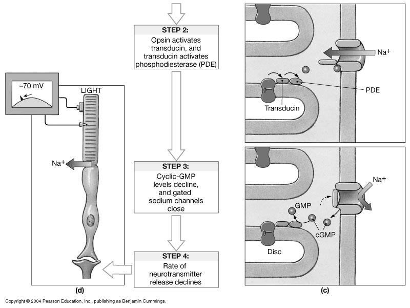
21 VISUAL PHYSIOLOGY THUS, when not stimulated by light, the photoreceptors cells are always depolarized (around - 40 mv) due to a Na + ion current ( called the dark current)! The cells continuously release neurotransmitters at the bipolar synapse! Steps in PhotoReception STEP 1 : Light energy converts Retinal from the CIS form to the TRANS form. This now activates the OPSIN part of Rhodopsin STEP 2 : OPSIN activates the enzyme TRANSDUCIN (G-protein complex) TRANSDUCIN in turn activates a phosphodiesterase. Phosphodiesterase breaks down cyclic GMP (cgmp) levels 41 VISUAL PHYSIOLOGY PhotoReception STEP 3 : Phosphodiesterase thus reduces cgmp levels ; this reduces the numbers of Na+ channels that are open. It results in a hyper-polarization! STEP 4 : The membrane potential drifts back to around - 70 mv. This reduces the release of neurotransmitters at the synapse with the bipolar cells. Thus, in contrast with what we seen so far, stimulation of the receptor results in a decrease in released number of N.T. This is a graded response; the higher the intensity of light, the greater the hyperpolarization and the less the amount of N.T. released! 42 21

22 VISUAL PHYSIOLOGY 43 VISUAL PHYSIOLOGY 44 22
 11-cis-retinal Transducin (a G protein) Open cgmp-gated cation channel Closed cgmp-gated cation channel 2 Visual pigment activates")
23 VISUAL PHYSIOLOGY 1 Light (photons) activates visual pigment. Visual pigment Light All-trans-retinal Phosphodiesterase (PDE) 11-cis-retinal Transducin (a G protein) Open cgmp-gated cation channel Closed cgmp-gated cation channel 2 Visual pigment activates transducin (G protein). 3 Transducin activates phosphodiester ase (PDE). 4 PDE converts cgmp into GMP, causing cgmp levels to fall. 5 As cgmp levels fall, cgmp-gated cation channels close, resulting in hyperpolarization. 45 VISUAL PHYSIOLOGY So, how does the brain receive the information of the light stimuli? The N.T. released by the photoreceptors are inhibitory neurotransmitters (glutamate). They thus cause IPSP s in the bipolar cells ( resulting in nyperpolarixzation) The bipolar cells in turn reduce their frequency of stimulation to the ganglion cells. The reduction in N.T. release induced by the light stimuli thus reduces the amount of IPSP s. The removal of inhibition equates with stimulation. It is similar like having your foot on the brake and slowly releasing the action of your foot! 46 23

 Bipolar cell Ganglion cell mv - 40 mv - 70 mv Dark Due to the open Na+ channel, the rod experiences a dark current that keeps the cell depolarized 47 In the")
24 VISUAL PHYSIOLOGY In the dark 1 cgmp-gated channels open, allowing cation influx; the photoreceptor depolarizes. 2 Voltage-gated Ca 2+ channels open in synaptic terminals. 3 Neurotransmitter is released continuously. 4 Neurotransmitter causes IPSPs in bipolar cell; hyperpolarization results. 5 Hyperpolarization closes voltage-gated Ca 2+ channels, inhibiting neurotransmitter release. 6 No EPSPs occur in ganglion cell. 7 No action potentials occur along the optic nerve. Ca 2+ Na + Ca 2+ Photoreceptor cell (rod) Bipolar cell Ganglion cell mv - 40 mv - 70 mv Dark Due to the open Na+ channel, the rod experiences a dark current that keeps the cell depolarized 47 In the light VISUAL PHYSIOLOGY Photoreceptor cell (rod) Bipolar cell Ganglion cell Light Ca 2+ 1 cgmp-gated channels are closed, so cation influx stops; the photoreceptor hyperpolarizes. 2 Voltage-gated Ca 2+ channels close in synaptic terminals. 3 No neurotransmitter is released. 4 Lack of IPSPs in bipolar cell results in depolarization. 5 Depolarization opens voltage-gated Ca 2+ channels; neurotransmitter is released. 6 EPSPs occur in ganglion cell. 7 Action potentials propagate along the optic nerve. mv - 40 mv - 70 mv Dark Light Light response closes Na+ channel due to the cgmp breakdown and the rod experiences a hyperpolarization
. Photoreceptors cannot function with damaged rhodopsin.")
25 VISUAL PHYSIOLOGY Recovery after stimulation After light stimulation, retinal is in the TRANS form and does not spontaneously convert back to the CIS form Shortly after light stimulation, rhodopsin actually breaks down into retinal and opsin. (called bleaching effect). Photoreceptors cannot function with damaged rhodopsin. Thus, before it can become an active molecule again, it needs to be glued back together. This can only occur if retinal is in the CIS position. The occurs in the dark and requires enzymes and ATP. 49 VISUAL PHYSIOLOGY Recovery after stimulation 50 25

26 VISUAL PHYSIOLOGY Dark Adapted State When exposed to the dark long enough, all photoreceptors are loaded and ready. Our visual system is then in a highly sensitive state and receptive to small amounts of light. Light Adapted State When moving from a dark area to a bright area, all photoreceptors become bleached and thus reduce the immediate sensitivity to a series of additional light stimuli. 51 VISUAL PHYSIOLOGY Color Vision White light is a spectrum of all different colors. If an object absorbs all color, it appears black. The color of an object is determined by the wavelength it reflects ( and thus does not absorb )! A red apple looks red because it absorbs all wavelengths of the visible spectrum except the wavelengths between 620 and 700 nm (red colors)
27 VISUAL PHYSIOLOGY The cones are sensitive to colored light. There are three different kinds of cones : Blue cones have highest sensitivity in blue region Green cones are most sensitive in green region Red cones are most sensitive in red region In a person with normal vision 16 % Blue cones 10 % Green cones 74 % Red cones Perception of color is due to the relative integration of information arriving from each cone type. 53 VISUAL PHYSIOLOGY ColorBlindness is the inability to perceive certain colors. It occurs when one or more of the classes of cones become non functional The most common type of color blindness ids red-green color blindness; the red cones are missing and the person cannot differentiate between red or green. The genes for the cones are located on the X chromosome. Thus, color blindness is more common in males ( 10% of all males) than females (0.67% of al females). Total colorblindness is rare! 54 27
28 VISUAL PHYSIOLOGY Visual Pathway The retina has about 130 million rods/cones 6 million bipolar cells 1 million ganglion cells Thus considerable convergence occurs with respect to signal processing Most convergence occurs with the rods! About thousand rods converge on a single ganglion cell. This ganglion cell ( and thus the rods) monitor a certain portion of the visual field. Such a cell is called an M cell! Since rods are more effective in dim light, M cells provide information about the fact that light has arrived in a certain area. 55 VISUAL PHYSIOLOGY Visual Pathway The cones shows little convergence The ratio of cones to bipolar cells is almost 1 : 1 in the fovea Ganglion cells that monitor cones are called P cells They are active in bright light and provide information about detail, color from a very specific area About thousand rods converge on a single ganglion cell. Difference between M and P cells can be explained in terms of a computer screen or photography M cells (rods) provide grainy, fuzzy pictures with low resolution, blurry details P cells (cones) provide high resolution, fine grained, sharp and clear detail 56 28


29 VISUAL PHYSIOLOGY Visual Pathway in CNS Axons from the ganglion cells meet up at the optic disc and exit the eyeball They proceed as the OPTIC nerve towards the diencephalon The optic nerves cross over at the optic chiasm and become the optic tracts The optic tract proceeds to the Laterate Geniculate Nucleus in the Thalamus Here information is passed on via the projection fibers, called the optic radiation, to the visual cortex of the occipital lobe. Collateral fibers at the Geniculate Nucleus connect with the Superior collicluli, pineal gland, RAS, and other nuclei 57 VISUAL PHYSIOLOGY The Binocular zone is the overlapping zone seen by both eyes, which is important for depth perception. Not all axons cross over at the optic chiasm. The axons that come from the left side of each eye proceed to the left hemisphere, while those from the right proceed to the right hemisphere. This partial cross over thus brings together that information that comes from the same area of the visual field 58 29

30 VISUAL PHYSIOLOGY This partial cross over has some interesting effects
Coarse hairs that overlie the supraorbital margins Functions include: Shading the eye Preventing perspiration from reaching the eye
 SPECIAL SENSES (INDERA KHUSUS) Dr.Milahayati Daulay Departemen Fisiologi FK USU Eye and Associated Structures 70% of all sensory receptors are in the eye Most of the eye is protected by a cushion of fat
10/8/ dpt. n 21 = n n' r D = The electromagnetic spectrum. A few words about light. BÓDIS Emőke 02 October Optical Imaging in the Eye
 A few words about light BÓDIS Emőke 02 October 2012 Optical Imaging in the Eye Healthy eye: 25 cm, v1 v2 Let s determine the change in the refractive power between the two extremes during accommodation!
11/23/11. A few words about light nm The electromagnetic spectrum. BÓDIS Emőke 22 November Schematic structure of the eye
 11/23/11 A few words about light 300-850nm 400-800 nm BÓDIS Emőke 22 November 2011 The electromagnetic spectrum see only 1/70 of the electromagnetic spectrum The External Structure: The Immediate Structure:
The Special Senses: Vision
 OLLI Lecture 5 The Special Senses: Vision Vision The eyes are the sensory organs for vision. They collect light waves through their photoreceptors (located in the retina) and transmit them as nerve impulses
Visual System I Eye and Retina
 Visual System I Eye and Retina Reading: BCP Chapter 9 www.webvision.edu The Visual System The visual system is the part of the NS which enables organisms to process visual details, as well as to perform
PHGY Physiology. SENSORY PHYSIOLOGY Vision. Martin Paré
 PHGY 212 - Physiology SENSORY PHYSIOLOGY Vision Martin Paré Assistant Professor of Physiology & Psychology pare@biomed.queensu.ca http://brain.phgy.queensu.ca/pare The Process of Vision Vision is the process
Vision. By. Leanora Thompson, Karen Vega, and Abby Brainerd
 Vision By. Leanora Thompson, Karen Vega, and Abby Brainerd Anatomy Outermost part of the eye is the Sclera. Cornea transparent part of outer layer Two cavities by the lens. Anterior cavity = Aqueous humor
Sensory receptors External internal stimulus change detectable energy transduce action potential different strengths different frequencies
 General aspects Sensory receptors ; respond to changes in the environment. External or internal environment. A stimulus is a change in the environmental condition which is detectable by a sensory receptor
PHGY Physiology. The Process of Vision. SENSORY PHYSIOLOGY Vision. Martin Paré. Visible Light. Ocular Anatomy. Ocular Anatomy.
 PHGY 212 - Physiology SENSORY PHYSIOLOGY Vision Martin Paré Assistant Professor of Physiology & Psychology pare@biomed.queensu.ca http://brain.phgy.queensu.ca/pare The Process of Vision Vision is the process
Special Senses. Important Concepts. Anatomy of the Eye. Anatomy of the Eye. Biol 219 Lecture 17 Vision Fall The Eye and Vision
 Special Senses The Eye and Vision Important Concepts Describe the structures of the eye and the role of each structure in vision. Trace the pathway for vis ion from the retina to the visual cortex. Explain
The Eye. Nakhleh Abu-Yaghi, M.B.B.S Ophthalmology Division
 The Eye Nakhleh Abu-Yaghi, M.B.B.S Ophthalmology Division Coats of the Eyeball 1- OUTER FIBROUS COAT is made up of : Posterior opaque part 2-THE SCLERA the dense white part 1- THE CORNEA the anterior
Vision. By: Karen, Jaqui, and Jen
 Vision By: Karen, Jaqui, and Jen Activity: Directions: Stare at the black dot in the center of the picture don't look at anything else but the black dot. When we switch the picture you can look around
THE EYE. People of Asian descent have an EPICANTHIC FOLD in the upper eyelid; no functional difference.
 THE EYE The eye is in the orbit of the skull for protection. Within the orbit are 6 extrinsic eye muscles, which move the eye. There are 4 cranial nerves: Optic (II), Occulomotor (III), Trochlear (IV),
Chapter Six Chapter Six
 Chapter Six Chapter Six Vision Sight begins with Light The advantages of electromagnetic radiation (Light) as a stimulus are Electromagnetic energy is abundant, travels VERY quickly and in fairly straight
November 14, 2017 Vision: photoreceptor cells in eye 3 grps of accessory organs 1-eyebrows, eyelids, & eyelashes 2- lacrimal apparatus:
 Vision: photoreceptor cells in eye 3 grps of accessory organs 1-eyebrows, eyelids, & eyelashes eyebrows: protection from debris & sun eyelids: continuation of skin, protection & lubrication eyelashes:
-eyelashes are richly innervated and triggers reflex blinking
 The Eye and Vision -vision is the dominant sense -70% of all sensory receptors in the body are in the eyes -half of the cerebral cortex is involved in some aspect of visual processing -accessory structures
EYE ANATOMY. Multimedia Health Education. Disclaimer
 Disclaimer This movie is an educational resource only and should not be used to manage your health. The information in this presentation has been intended to help consumers understand the structure and
EYE STRUCTURE AND FUNCTION
 Name: Class: Date: EYE STRUCTURE AND FUNCTION The eye is the body s organ of sight. It gathers light from the environment and forms an image on specialized nerve cells on the retina. Vision occurs when
Eye. Eye Major structural layer of the wall of the eye is a thick layer of dense C.T.; that layer has two parts:
 General aspects Sensory receptors ; External or internal environment. A stimulus is a change in the environmental condition which is detectable by a sensory receptor 1 Major structural layer of the wall
4Basic anatomy and physiology
 Hene_Ch09.qxd 8/30/04 6:51 AM Page 348 348 4Basic anatomy and physiology The eye is a highly specialized organ with an average axial length of 24 mm and a volume of 6.5 ml. Except for its anterior aspect,
1. Introduction to Anatomy of the Eye and its Adnexa
 1. Introduction to Anatomy of the Eye and its Adnexa Fig 1: A Cross section of the human eye. Let us imagine we are traveling with a ray of light into the eye. The first structure we will encounter is
Special Senses- THE EYE. Pages
 Special Senses- THE EYE Pages 548-569 Accessory Structures Eyebrows Eyelids Conjunctiva Lacrimal Apparatus Extrinsic Eye Muscles EYEBROWS Deflect debris to side of face Facial recognition Nonverbal communication
The Eye. Morphology of the eye (continued) Morphology of the eye. Sensation & Perception PSYC Thomas E. Van Cantfort, Ph.D
 Morphology of the eye. Sensation & Perception PSYC Thomas E. Van Cantfort, Ph.D") Sensation & Perception PSYC420-01 Thomas E. Van Cantfort, Ph.D The Eye The Eye The function of the eyeball is to protect the photoreceptors The role of the eye is to capture an image of objects that we
Sensation & Perception PSYC420-01 Thomas E. Van Cantfort, Ph.D The Eye The Eye The function of the eyeball is to protect the photoreceptors The role of the eye is to capture an image of objects that we
Objectives. 3. Visual acuity. Layers of the. eye ball. 1. Conjunctiva : is. three quarters. posteriorly and
 OCULAR PHYSIOLOGY (I) Dr.Ahmed Al Shaibani Lab.2 Oct.2013 Objectives 1. Review of ocular anatomy (Ex. after image) 2. Visual pathway & field (Ex. Crossed & uncrossed diplopia, mechanical stimulation of
III: Vision. Objectives:
 III: Vision Objectives: Describe the characteristics of visible light, and explain the process by which the eye transforms light energy into neural. Describe how the eye and the brain process visual information.
Visual Optics. Visual Optics - Introduction
 Visual Optics Jim Schwiegerling, PhD Ophthalmology & Optical Sciences University of Arizona Visual Optics - Introduction In this course, the optical principals behind the workings of the eye and visual
25 Things To Know. Vision
 25 Things To Know Vision Magnetism Electromagnetic Energy Electricity Magnetism Electromagnetic Energy Electricity Light Frequency Amplitude Light Frequency How often it comes Wave length Peak to peak
ABO Certification Training. Part I: Anatomy and Physiology
 ABO Certification Training Part I: Anatomy and Physiology Major Ocular Structures Centralis Nerve Major Ocular Structures The Cornea Cornea Layers Epithelium Highly regenerative: Cells reproduce so rapidly
BIOPHYSICS OF VISION GEOMETRIC OPTICS OF HUMAN EYE. Refraction media of the human eye. D eye = 63 diopter, D cornea =40, D lens = 15+
 BIOPHYSICS OF VISION THEORY OF COLOR VISION ELECTRORETINOGRAM Two problems: All cows are black in dark! Playing tennis in dark with illuminated lines, rackets, net, and ball! Refraction media of the human
Slide 4 Now we have the same components that we find in our eye. The analogy is made clear in this slide. Slide 5 Important structures in the eye
 Vision 1 Slide 2 The obvious analogy for the eye is a camera, and the simplest camera is a pinhole camera: a dark box with light-sensitive film on one side and a pinhole on the other. The image is made
iris pupil cornea ciliary muscles accommodation Retina Fovea blind spot
 Chapter 6 Vision Exam 1 Anatomy of vision Primary visual cortex (striate cortex, V1) Prestriate cortex, Extrastriate cortex (Visual association coretx ) Second level association areas in the temporal and
Early Visual Processing: Receptive Fields & Retinal Processing (Chapter 2, part 2)
") Early Visual Processing: Receptive Fields & Retinal Processing (Chapter 2, part 2) Lecture 5 Jonathan Pillow Sensation & Perception (PSY 345 / NEU 325) Princeton University, Spring 2015 1 Summary of last
Early Visual Processing: Receptive Fields & Retinal Processing (Chapter 2, part 2) Lecture 5 Jonathan Pillow Sensation & Perception (PSY 345 / NEU 325) Princeton University, Spring 2015 1 Summary of last
2 The First Steps in Vision
 2 The First Steps in Vision 2 The First Steps in Vision A Little Light Physics Eyes That See light Retinal Information Processing Whistling in the Dark: Dark and Light Adaptation The Man Who Could Not
Handout G: The Eye and How We See
 Handout G: The Eye and How We See Prevent Blindness America. (2003c). The eye and how we see. Retrieved July 31, 2003, from http://www.preventblindness.org/resources/howwesee.html Your eyes are wonderful
EYE. The eye is an extension of the brain
 I SEE YOU EYE The eye is an extension of the brain Eye brain proxomity Can you see : the optic nerve bundle? Spinal cord? The human Eye The eye is the sense organ for light. Receptors for light are found
Chapter 6 Human Vision
 Chapter 6 Notes: Human Vision Name: Block: Human Vision The Humane Eye: 8) 1) 2) 9) 10) 4) 5) 11) 12) 3) 13) 6) 7) Functions of the Eye: 1) Cornea a transparent tissue the iris and pupil; provides most
AP PSYCH Unit 4.2 Vision 1. How does the eye transform light energy into neural messages? 2. How does the brain process visual information? 3.
 AP PSYCH Unit 4.2 Vision 1. How does the eye transform light energy into neural messages? 2. How does the brain process visual information? 3. What theories help us understand color vision? 4. Is your
The Human Eye and a Camera 12.1
 The Human Eye and a Camera 12.1 The human eye is an amazing optical device that allows us to see objects near and far, in bright light and dim light. Although the details of how we see are complex, the
Materials Cow eye, dissecting pan, dissecting kit, safety glasses, lab apron, and gloves
 Cow Eye Dissection Guide Introduction How do we see? The eye processes the light through photoreceptors located in the eye that send signals to the brain and tells us what we are seeing. There are two
Sheep Eye Dissection
 Sheep Eye Dissection Question: How do the various parts of the eye function together to make an image appear on the retina? Materials and Equipment: Preserved sheep eye Scissors Dissection tray Tweezers
Vision. The eye. Image formation. Eye defects & corrective lenses. Visual acuity. Colour vision. Lecture 3.5
 Lecture 3.5 Vision The eye Image formation Eye defects & corrective lenses Visual acuity Colour vision Vision http://www.wired.com/wiredscience/2009/04/schizoillusion/ Perception of light--- eye-brain
Seeing and Perception. External features of the Eye
 Seeing and Perception Deceives the Eye This is Madness D R Campbell School of Computing University of Paisley 1 External features of the Eye The circular opening of the iris muscles forms the pupil, which
The Eye. (We ll leave the Lord Sauron jokes to you.)
") The Eye (We ll leave the Lord Sauron jokes to you.) When you look in the mirror, you only see a very small part of your eyes. In reality, they are incredibly complex organs with a pretty big job: enabling
The Eye (We ll leave the Lord Sauron jokes to you.) When you look in the mirror, you only see a very small part of your eyes. In reality, they are incredibly complex organs with a pretty big job: enabling
Biology 70 Slides for Lecture 1 Fall 2007
 Biology 70 Part II Sensory Systems www.biology.ucsc.edu 1 2 intensity vs spatial position (image formation) color 3 4 motion depth (monocular) 5 6 1 depth (binocular) 1. In the lectures on perception we
PSY 214 Lecture # (09/14/2011) (Introduction to Vision) Dr. Achtman PSY 214. Lecture 4 Topic: Introduction to Vision Chapter 3, pages 44-54
 (Introduction to Vision) Dr. Achtman PSY 214. Lecture 4 Topic: Introduction to Vision Chapter 3, pages 44-54") Corrections: A correction needs to be made to NTCO3 on page 3 under excitatory transmitters. It is possible to excite a neuron without sending information to another neuron. For example, in figure 2.12
Corrections: A correction needs to be made to NTCO3 on page 3 under excitatory transmitters. It is possible to excite a neuron without sending information to another neuron. For example, in figure 2.12
Zoology. Lesson: Physiology of Vision. Lesson Developer: Dr. Mahtab Zarin. College/Dept: Zoology, University of Delhi
 Zoology Lesson: Physiology of Vision Lesson Developer: Dr. Mahtab Zarin College/Dept: Zoology, University of Delhi Institute of Life Long Learning, University of Delhi 1 Table of Contents Introduction
Visual Perception of Images
 Visual Perception of Images A processed image is usually intended to be viewed by a human observer. An understanding of how humans perceive visual stimuli the human visual system (HVS) is crucial to the
HW- Finish your vision book!
 March 1 Table of Contents: 77. March 1 & 2 78. Vision Book Agenda: 1. Daily Sheet 2. Vision Notes and Discussion 3. Work on vision book! EQ- How does vision work? Do Now 1.Find your Vision Sensation fill-in-theblanks
Chapter Human Vision
 Chapter 6 6.1 Human Vision How Light Enters the Eye Light enters the eye through the pupil. The pupil appears dark because light passes through it without reflecting back Pupil Iris = Coloured circle of
Sensation. What is Sensation, Perception, and Cognition. All sensory systems operate the same, they only use different mechanisms
 Sensation All sensory systems operate the same, they only use different mechanisms 1. Have a physical stimulus (e.g., light) 2. The stimulus emits some sort of energy 3. Energy activates some sort of receptor
Sensation. Sensation. Perception. What is Sensation, Perception, and Cognition
 All sensory systems operate the same, they only use different mechanisms Sensation 1. Have a physical stimulus (e.g., light) 2. The stimulus emits some sort of energy 3. Energy activates some sort of receptor
Retina. Convergence. Early visual processing: retina & LGN. Visual Photoreptors: rods and cones. Visual Photoreptors: rods and cones.
 Announcements 1 st exam (next Thursday): Multiple choice (about 22), short answer and short essay don t list everything you know for the essay questions Book vs. lectures know bold terms for things that
Vision Science I Exam 1 23 September ) The plot to the right shows the spectrum of a light source. Which of the following sources is this
 The plot to the right shows the spectrum of a light source. Which of the following sources is this") Vision Science I Exam 1 23 September 2016 1) The plot to the right shows the spectrum of a light source. Which of the following sources is this spectrum most likely to be taken from? A) The direct sunlight
Vision Science I Exam 1 23 September 2016 1) The plot to the right shows the spectrum of a light source. Which of the following sources is this spectrum most likely to be taken from? A) The direct sunlight
The eye* The eye is a slightly asymmetrical globe, about an inch in diameter. The front part of the eye (the part you see in the mirror) includes:
 includes:") The eye* The eye is a slightly asymmetrical globe, about an inch in diameter. The front part of the eye (the part you see in the mirror) includes: The iris (the pigmented part) The cornea (a clear dome
The eye* The eye is a slightly asymmetrical globe, about an inch in diameter. The front part of the eye (the part you see in the mirror) includes: The iris (the pigmented part) The cornea (a clear dome
ensory System III Eye Reflexes
 ensory System III Eye Reflexes Quick Review from Last Week Eye Anatomy Inside of the Eye choroid Eye Reflexes Eye Reflexes A healthy person has a number of eye reflexes: Pupillary light reflex Vestibulo-ocular
Topic 4: Lenses and Vision. Lens a curved transparent material through which light passes (transmit) Ex) glass, plastic
 Ex) glass, plastic") Topic 4: Lenses and Vision Lens a curved transparent material through which light passes (transmit) Ex) glass, plastic Double Concave Lenses Are thinner and flatter in the middle than around the edges.
Topic 4: Lenses and Vision Lens a curved transparent material through which light passes (transmit) Ex) glass, plastic Double Concave Lenses Are thinner and flatter in the middle than around the edges.
Chapter 2: The Beginnings of Perception
 Chapter 2: The Beginnings of Perception We ll see the first three steps of the perceptual process for vision https:// 49.media.tumblr.co m/ 87423d97f3fbba8fa4 91f2f1bfbb6893/ tumblr_o1jdiqp4tc1 qabbyto1_500.gif
Introduction. Chapter Aim of the Thesis
 Chapter 1 Introduction 1.1 Aim of the Thesis The main aim of this investigation was to develop a new instrument for measurement of light reflected from the retina in a living human eye. At the start of
Digital Image Processing
 Digital Image Processing Lecture # 3 Digital Image Fundamentals ALI JAVED Lecturer SOFTWARE ENGINEERING DEPARTMENT U.E.T TAXILA Email:: ali.javed@uettaxila.edu.pk Office Room #:: 7 Presentation Outline
EYE: THE PHOTORECEPTOR SYSTEM. Prof. Dr. Huda Al Khateeb
 EYE: THE PHOTORECEPTOR SYSTEM Prof. Dr. Huda Al Khateeb Lecture 1 The eye ball Objectives By the end of this lecture the student should: 1. List the layers and chambers of the eye ball 2. Describe the
Spatial Vision: Primary Visual Cortex (Chapter 3, part 1)
") Spatial Vision: Primary Visual Cortex (Chapter 3, part 1) Lecture 6 Jonathan Pillow Sensation & Perception (PSY 345 / NEU 325) Princeton University, Fall 2017 Eye growth regulation KL Schmid, CF Wildsoet
Spatial Vision: Primary Visual Cortex (Chapter 3, part 1) Lecture 6 Jonathan Pillow Sensation & Perception (PSY 345 / NEU 325) Princeton University, Fall 2017 Eye growth regulation KL Schmid, CF Wildsoet
Psych 333, Winter 2008, Instructor Boynton, Exam 1
 Name: Class: Date: Psych 333, Winter 2008, Instructor Boynton, Exam 1 Multiple Choice There are 35 multiple choice questions worth one point each. Identify the letter of the choice that best completes
SCIENCE 8 WORKBOOK Chapter 6 Human Vision Ms. Jamieson 2018 This workbook belongs to:
 SCIENCE 8 WORKBOOK Chapter 6 Human Vision Ms. Jamieson 2018 This workbook belongs to: Eric Hamber Secondary 5025 Willow Street Vancouver, BC Table of Contents A. Chapter 6.1 Parts of the eye.. Parts of
Photography (cont d)
") Lecture 13 Ch. 4 Photography continued Ch. 5 The Eye Feb. 23, 2010 Exams will be back on Feb. 25 Homework 5 is due Feb. 25 Read all of Ch. 5. on The Eye. 1 Photography (cont d) Polarizing and haze filters
Lecture 13 Ch. 4 Photography continued Ch. 5 The Eye Feb. 23, 2010 Exams will be back on Feb. 25 Homework 5 is due Feb. 25 Read all of Ch. 5. on The Eye. 1 Photography (cont d) Polarizing and haze filters
By Dr. Abdelaziz Hussein
 By Dr. Abdelaziz Hussein Light is a form of radiant energy, consisting of electromagnetic waves a. Velocity of light: In air it is 300,000 km/second. b. Wave length: The wave-length of visible light to
A&P 1 Eye & Vision Lab Vision Concepts
 A&P 1 Eye & Vision Lab Vision Concepts In this "Lab Exercise Guide", we will be looking at the basics of vision. NOTE: these notes do not follow the order of the videos. You should be able to read this
Ocular Jeopardy. The major refractive portion of the eye 5/12/2015. Presented by Jill J Luebbert, CPOT, ABOC. Watch This Refractive optios
 Ocular Jeopardy Presented by Jill J Luebbert, CPOT, ABOC In the beginning anterior Way back Visual Pathway Say What? terminolog y Watch This Refractive optios Posterior Segment 10 10 10 10 10 20 20 20
Sense Organs (Eye) The eye is the sense organ of sight. The eye is shaped like a ball and is located in bony
 The eye is the sense organ of sight. The eye is shaped like a ball and is located in bony") Sense Organs (Eye) The eye is the sense organ of sight. The eye is shaped like a ball and is located in bony sockets in the skull. It is held in place by six muscles which are joined to the outside of
Sense Organs (Eye) The eye is the sense organ of sight. The eye is shaped like a ball and is located in bony sockets in the skull. It is held in place by six muscles which are joined to the outside of
Biophysics of the senses: vision
 Medical Physics I. Biophysics of the senses: vision Ferenc Bari Professor & chairman Department of Medical Physics & Informatics Szeged, December 3, 2015. Basic properties of light Visible electromagnetic
Unit 1 DIGITAL IMAGE FUNDAMENTALS
 Unit 1 DIGITAL IMAGE FUNDAMENTALS What Is Digital Image? An image may be defined as a two-dimensional function, f(x, y), where x and y are spatial (plane) coordinates, and the amplitude of f at any pair
SCIENCE 8 WORKBOOK Chapter 6 Human Vision Ms. Jamieson 2018 This workbook belongs to:
 SCIENCE 8 WORKBOOK Chapter 6 Human Vision Ms. Jamieson 2018 This workbook belongs to: Eric Hamber Secondary 5025 Willow Street Vancouver, BC Table of Contents A. Chapter 6.1 Parts of the eye.. Parts of
Vision. Definition. Sensing of objects by the light reflected off the objects into our eyes
 Vision Vision Definition Sensing of objects by the light reflected off the objects into our eyes Only occurs when there is the interaction of the eyes and the brain (Perception) What is light? Visible
[Chapter 2] Ocular Geometry and Topography. Elements of Ocular Structure
![[Chapter 2] Ocular Geometry and Topography. Elements of Ocular Structure](/thumbs/95/124174711.jpg "[Chapter 2] Ocular Geometry and Topography. Elements of Ocular Structure") [Chapter 2] Ocular Geometry and Topography Before Sam Clemens became Mark Twain, he had been, among other things, a riverboat pilot, a placer miner, and a newspaper reporter, occupations in which success
[Chapter 2] Ocular Geometry and Topography Before Sam Clemens became Mark Twain, he had been, among other things, a riverboat pilot, a placer miner, and a newspaper reporter, occupations in which success
Sensation, Part 4 Gleitman et al. (2011), Chapter 4
, Chapter 4") Sensation, Part 4 Gleitman et al. (2011), Chapter 4 Mike D Zmura Department of Cognitive Sciences, UCI Psych 9A / Psy Beh 11A February 20, 2014 T. M. D'Zmura 1 From last time T. M. D'Zmura 2 Rod Transduction
Sensation, Part 4 Gleitman et al. (2011), Chapter 4 Mike D Zmura Department of Cognitive Sciences, UCI Psych 9A / Psy Beh 11A February 20, 2014 T. M. D'Zmura 1 From last time T. M. D'Zmura 2 Rod Transduction
The Human Eye Looking at your own eye with an Eye Scope
 The Human Eye Looking at your own eye with an Eye Scope Rochelle Payne Ondracek Edited by Anne Starace Abstract The human ability to see is the result of an intricate interconnection of muscles, receptors
Lectures on Medical Biophysics Department of Biophysics, Medical Faculty, Masaryk University, Brno. Biophysics of visual perception
 Lectures on Medical Biophysics Department of Biophysics, Medical Faculty, Masaryk University, Brno 1 Lecture outline Basic properties of light Anatomy of eye Optical properties of eye Retina biological
Eyeball Model Lab Date Block
 Science 8 Name Eyeball Model Lab Date Block Problem: Identify the twelve key parts of the eye and describe their function. Materials: dissecting scissors ping pong ball transparent plastic ordinary scissors
This question addresses OPTICAL factors in image formation, not issues involving retinal or other brain structures.
 Bonds 1. Cite three practical challenges in forming a clear image on the retina and describe briefly how each is met by the biological structure of the eye. Note that by challenges I do not refer to optical
A piece of white paper can be 1,000,000,000 times brighter in outdoor sunlight than in a moonless night.
 Light intensities range across 9 orders of magnitude. A piece of white paper can be 1,000,000,000 times brighter in outdoor sunlight than in a moonless night. But in a given lighting condition, light ranges
Sensation and Perception
 Sensation and Perception PSY 100: Foundations of Contemporary Psychology Basic Terms Sensation: the activation of receptors in the various sense organs Perception: the method by which the brain takes all
Chapter 22: Illumination and Vision
 Chapter 22: Illumination and Vision Learning Outcomes After successful studying this chapter, You should be able to Explain how we see objects? Discus the anatomical structure of the eye, Describe the
Psychology in Your Life
 Sarah Grison Todd Heatherton Michael Gazzaniga Psychology in Your Life FIRST EDITION Chapter 5 Sensation and Perception 2014 W. W. Norton & Company, Inc. Section 5.1 How Do Sensation and Perception Affect
VISUAL SYSTEM PHYSIOLOGY. Discipline of Physiology and Neuroscience, Carol Davila University of Medicine and Pharmacy, Bucharest, Romania
 VISUAL SYSTEM PHYSIOLOGY Discipline of Physiology and Neuroscience, Carol Davila University of Medicine and Pharmacy, Bucharest, Romania Outer layer of the eye Cornea - No bood vessels - Most powerful
Retinal stray light originating from intraocular lenses and its effect on visual performance van der Mooren, Marie Huibert
 University of Groningen Retinal stray light originating from intraocular lenses and its effect on visual performance van der Mooren, Marie Huibert IMPORTANT NOTE: You are advised to consult the publisher's
12.1. Human Perception of Light. Perceiving Light
 12.1 Human Perception of Light Here is a summary of what you will learn in this section: Focussing of light in your eye is accomplished by the cornea, the lens, and the fluids contained in your eye. Light
In the following diagram the parts of the eye are visualized and labeled for you.
 Investigation 3.12B: The Eye In the preceding case study marker of the problem of greatest concern to you lay in finding the pupils fixed in a dilated position. But what is the pupil and what makes it
DIGITAL IMAGE PROCESSING LECTURE # 4 DIGITAL IMAGE FUNDAMENTALS-I
 DIGITAL IMAGE PROCESSING LECTURE # 4 DIGITAL IMAGE FUNDAMENTALS-I 4 Topics to Cover Light and EM Spectrum Visual Perception Structure Of Human Eyes Image Formation on the Eye Brightness Adaptation and
Yokohama City University lecture INTRODUCTION TO HUMAN VISION Presentation notes 7/10/14
 Yokohama City University lecture INTRODUCTION TO HUMAN VISION Presentation notes 7/10/14 1. INTRODUCTION TO HUMAN VISION Self introduction Dr. Salmon Northeastern State University, Oklahoma. USA Teach
Cow Eye Dissection. Online dissection, for kids abstaining:
 Cow Eye Dissection Introductory Discussion: Tell the students that we will be learning about what eyes are made of and how they work by dissecting a cow eye. Talk about where the eye comes from, and how
LECTURE 2. Vision Accomodation& pupillary light reflex By Prof/Faten zakareia
 LECTURE 2 Vision Accomodation& pupillary light reflex By Prof/Faten zakareia Objectives: At the end of this lecture,the student should be able to;- -Describe visual acuity & depth perception -Contrast
Lab #8: The Special Senses: Hearing, Vision, and Orientation
 Lab #8: The Special Senses: Hearing, Vision, and Orientation Background The special senses (vision, hearing, equilibrium, gustation, and olfaction) differ from the somatesthetic senses in two fundamental
Vision. Sensation & Perception. Functional Organization of the Eye. Functional Organization of the Eye. Functional Organization of the Eye
 Vision Sensation & Perception Part 3 - Vision Visible light is the form of electromagnetic radiation our eyes are designed to detect. However, this is only a narrow band of the range of energy at different
HOW THE EYE EVOLVED By Adrea R. Benkoff, M.D.
 HOW THE EYE EVOLVED By Adrea R. Benkoff, M.D. HOW THE EYE EVOLVED BY ADREA R. BENKOFF, M.D. CREATIONISM vs. NATURAL SELECTION The complex structure of the eye has been used as evidence to support the theory
1. What are the components of your nervous system? 2. How do telescopes and human eyes work?
 Chapter 18 Vision and Hearing Although small, your eyes and ears are amazingly important and complex organs. Do you know how your eyes and ears work? Scientists have learned enough about these organs to
INTRODUCING OPTICS CONCEPTS TO STUDENTS THROUGH THE OX EYE EXPERIMENT
 INTRODUCING OPTICS CONCEPTS TO STUDENTS THROUGH THE OX EYE EXPERIMENT Marcela L. Redígolo redigolo@univap.br Leandro P. Alves leandro@univap.br Egberto Munin munin@univap.br IP&D Univap Av. Shishima Hifumi,
HSC Biology. Published Feb 9, 2017 HSC BIOLOGY OPTION: COMMUNICATION. By Sahar (99.1 ATAR)
") HSC Biology Year 2014 Mark 92.00 Pages 11 Published Feb 9, 2017 HSC BIOLOGY OPTION: COMMUNICATION By Sahar (99.1 ATAR) Your notes author, Sahar. Sahar achieved an ATAR of 99.1 in 2014 while attending Carlingford
HSC Biology Year 2014 Mark 92.00 Pages 11 Published Feb 9, 2017 HSC BIOLOGY OPTION: COMMUNICATION By Sahar (99.1 ATAR) Your notes author, Sahar. Sahar achieved an ATAR of 99.1 in 2014 while attending Carlingford
11.5 The Senses Tuesday January 7, Wednesday, 8 January, 14
 11.5 The Senses Tuesday January 7, 2014. TEST ON ALL OF HOMEOSTASIS (FOCUS ON REPRODUCTIVE AND NERVOUS SYSTEM) ON FRIDAY. Structure of the Eye Eye Anatomy and Function http://www.youtube.com/watch? v=0hzwmldldhi&feature=related
Lecture 4 Foundations and Cognitive Processes in Visual Perception From the Retina to the Visual Cortex
 Lecture 4 Foundations and Cognitive Processes in Visual Perception From the Retina to the Visual Cortex 1.Vision Science 2.Visual Performance 3.The Human Visual System 4.The Retina 5.The Visual Field and
OPTICAL DEMONSTRATIONS ENTOPTIC PHENOMENA, VISION AND EYE ANATOMY
 OPTICAL DEMONSTRATIONS ENTOPTIC PHENOMENA, VISION AND EYE ANATOMY The pupil as a first line of defence against excessive light. DEMONSTRATION 1. PUPIL SHAPE; SIZE CHANGE Make a triangular shape with the
Class 10 Science NCERT Exemplar Solutions Human Eye and Colourful World
 Class 10 Science NCERT Exemplar Solutions Human Eye and Colourful World Short Answer Questions Question 1. A student sitting at the back of the classroom cannot read clearly the letters written on the